




Congress of Neurological Surgeons Systematic Review and Evidence-Based Guidelines for Chiari Malformation: Diagnosis
Sponsored by: Congress of Neurological Surgeons (CNS) and the Section on Pediatrics
Endorsed by: The Congress of Neurological Surgeons (CNS), American Association of Neurological Surgeons (AANS), and the Bobby Jones Chiari and Syringomyelia Foundation (Bobby Jones CSF)
Authors:
David F. Bauer, MD, MPH1, Toba Niazi, MD2, Rabia Qaiser, MD3, Libby Kosnik Infinger, MD, MPH4, Shobhan Vachhrajani MD, PhD, FRCSC5, Laurie L Ackerman, MD6, Eric M. Jackson, MD7, Sarah Jernigan, MD, MPH8, Cormac O. Maher, MD, FAAP, FACS, FAANS9, Jogi V. Pattisapu MD FAAP FACS FAANS10, Carolyn Quinsey, MD11, Jeffrey S. Raskin MS MD12, Brandon G. Rocque, MD, MS13, Howard Silberstein, MD14
Departmental and institutional affiliations:
- Department of Neurosurgery, Baylor College of Medicine, Division of Pediatric Neurosurgery, Texas Children’s Hospital, Houston, TX
- Department of Neurological Surgery, Nicklaus Children’s Hospital, Miami, FL
- Department of Neurological Surgery, Indiana University School of Medicine, Indianapolis, IN
- Department of Neurosurgery, Medical University of South Carolina (MUSC), Charleston, SC
- Department of Pediatrics, Wright State University Boonshoft School of Medicine, Dayton, OH
- Department of Neurological Surgery, Indiana University Health, Indianapolis, IN
- Department of Neurosurgery, Johns Hopkins University School of Medicine, Baltimore, MD
- Carolina Neurosurgery & Spine Associates, Charlotte, NC
- Department of Neurosurgery, Stanford Medicine, Palo Alto, CA
- Pediatric Neurosurgery, University of Central Florida College of Medicine, Orlando FL
- Department of Neurosurgery, University of North Carolina Chapel Hill, Chapel Hill, NC
- Department of Neurological Surgery, Northwestern University Feinberg School of Medicine, Chicago, IL
- Division of Pediatric Neurosurgery, Department of Neurosurgery, University of Alabama at Birmingham, Birmingham, AL
- Department of Neurosurgery, University of Rochester School of Medicine and Dentistry, Rochester, NY
Corresponding Author contact information:
David F. Bauer, MD, MPH
Department of Neurosurgery, Baylor College of Medicine
Division of Pediatric Neurosurgery, Texas Children’s Hospital
Houston, TX
No part of this article has been published or submitted for publication elsewhere.
Keywords:Chiari, diagnosis, guideline, systematic review, treatment
Abbreviations: CIM (Chiari I Malformation), MRI (Magnetic Resonance Imaging), CSF (Cerebral spinal fluid)
ABSTRACT
Background: Chiari I malformation (CIM) is characterized by descent of the cerebellar tonsils through the foramen magnum, potentially causing symptoms from compression or obstruction of the flow of cerebrospinal fluid (CSF). Diagnosis and treatment of CIM is varied, and guidelines produced through systematic review may be helpful for clinicians.
Objective: We performed a systematic review of the medical literature to answer specific questions on the diagnosis and treatment of CIM.
Methods: PubMed and Embase were queried between 1946 and January 23, 2021 using the search strategies provided in Appendix I.
Results: The literature search yielded 567 abstracts, of which 151 were selected for full-text review, 109 were then rejected for not meeting the inclusion criteria or for being off-topic, and 42 were included in this systematic review.
Conclusion: Three Grade C recommendations were made based on Level III evidence.
RECOMMENDATIONS
1-1. In patients with CIM diagnosed only with brain or cervical spine MRI, is complete imaging needed to evaluate for clinically relevant pathology such as brain tumor, hydrocephalus, spine syrinx, or tethered spinal cord?
Recommendation: In patients with CIM diagnosed only with brain or cervical spine MRI, further imaging of the brain and spine may be helpful to evaluate for clinically relevant pathology such as hydrocephalus or spine syrinx.
Strength of recommendation: Grade C
Level III evidence
1-2. In patients with CIM, are advanced imaging modalities such as Cine MRI helpful to predict benefit from surgical decompression?
Recommendation: In patients with CIM, advanced imaging modalities such as Cine MRI may or may not predict benefit from surgical decompression.
Strength of recommendation: Grade C
Level III evidence
1-3. In patients with CIM, should flexion and extension radiographs be routinely performed to evaluate for cervical instability? Should preoperative evaluation include any specific evaluation for cranial cervical instability or ventral compression (clivoaxial angle, pB-C2, etc)?
Recommendation: In patients with CIM, measurement of the clivoaxial angle, pB-C2, or C- C2 sagittal vertebral alignment (C-C2SVA) may predict future craniocervical instability and the need for surgical stabilization.
Strength of recommendation: Grade C
Level III evidence
There is insufficient evidence to support the use of flexion-extension films to predict future craniocervical instability in this population.
Strength of recommendation: Grade insufficient
Insufficient evidence
INTRODUCTION
Goals and Rationale
This clinical guideline has been created to improve patient care by outlining the appropriate diagnostic and decision-making processes involved in the treatment of patients with Chiari I malformation (CIM). Diagnosis and treatment of CIM can be challenging because not all patients are symptomatic and many patients do not require surgery. In addition, variations in surgical treatment can make surgical decision-making challenging. This guideline was created as an educational tool to guide qualified physicians through a series of diagnostic and treatment decisions to improve the quality and efficiency of care for patients with CIM.
Objectives
CIM is defined as descent of the cerebellar tonsils ≥3 to 5 mm below the foramen magnum. Based on a definition of a tonsillar position of ≥5 mm below the foramen magnum, imaging studies estimate a prevalence ranging from 0.24% to 2.6% of the population,1–5 including children and adults. Not all patients are symptomatic, and there are various ways to diagnose and treat CIM in the literature. CIM may cause syringomyelia, and some patients with CIM may have craniocervical instability requiring decompression and/or fusion of the craniocervical junction. Symptoms result from blockage of the flow of cerebrospinal fluid (CSF) or from compression of the brainstem or cranial nerves. Treatment may include decompression with or without duraplasty, and intradural tonsil reduction or resection of intradural webs over fourth ventricle outflow have been described. Symptoms reported in the literature are not completely concordant from study to study, and there can be overlap between CIM symptoms and other entities, such as migraine headache, making diagnosis of a symptomatic patient challenging. Because of diagnostic and treatment variability, we initiated the formation of this guideline to systematically review the literature and create evidence-based recommendations for common diagnostic and treatment questions related to CIM. In this guideline, we evaluate the diagnosis of CIM.
METHODOLOGY
The guidelines task force initiated a systematic review of the literature and evidence-based guideline relevant to the diagnosis of patients with CIM. Through objective evaluation of the evidence and transparency in the process of making recommendations, this evidence-based clinical practice guideline was developed for the diagnosis and treatment of patients with CIM. These guidelines are developed for educational purposes to assist practitioners in their clinical decision-making processes. Additional information about the methods used in this systematic review is provided below.
Literature Search
Task force members identified search terms/parameter and a medical librarian implemented the literature search, consistent with the literature search protocol (see Appendix I), using the National Library of Medicine/PubMed database and Embase for the period from 1946 to January 23, 2021, using the search strategies provided in Appendix I.
Inclusion/Exclusion Criteria
Articles were retrieved and included only if they met specific inclusion/exclusion criteria. To reduce bias, these criteria were specified before conducting the literature searches.
Articles that do not meet the following criteria were, for the purposes of this evidence-based clinical practice guideline, excluded. To be included as evidence in the guideline, an article had to be a report of a study that:
- Investigated patients with CIM;
- Studies that enrolled ≥80% of CIM (we included studies with mixed patient populations if they reported results separately for each group/patient population);
- Was a full article report of a clinical study;
- Was not a medical records review, meeting abstract, historical article, editorial, letter, or commentary;
- Appeared in a peer-reviewed publication or a registry report;
- Enrolled a minimum of 10 patients;
- Was of humans;
- Was published in or after 1946;
- Quantitatively presented results;
- Was not an in vitro study;
- Was not a biomechanical study;
- Was not performed on cadavers;
- Was published in English;
- Was not a systematic review, meta-analysis, or guideline developed by others
Systematic reviews or meta-analyses conducted by others or guidelines developed by others were not included as evidence to support this review because of the differences in article inclusion/exclusion criteria specified compared with the criteria specified by the Guidelines Task Force. Although these articles were not included as evidence to support the review, these articles were recalled for full-text review for the Guidelines Task Force to conduct manual searches of the bibliographies.
Assessment for Risk of Bias
The methodological quality of randomized controlled trials and the risk of bias was assessed by using the following 6 criteria: sequence generation, allocation concealment, blinding, incomplete reporting of data, selective reporting of outcomes, and other potential threats to validity. Any bias was discussed and mitigated through clarification in the evidentiary table and changing the grade of the level of evidence, if needed.
1The guideline task force did not include systematic reviews, guidelines, or meta-analyses conducted by others. These documents are developed using different inclusion criteria than those specified in this guideline; therefore, they may include studies that do not meet the inclusion criteria specific to this guideline. In cases where these types of documents’ abstracts suggested relevance to the guideline’s recommendations, the task force searched their bibliographies for additional studies.
Rating Quality of Evidence
The quality of evidence was rated using an evidence hierarchy for each of 4 different study types; therapeutic, prognostic, diagnostic, and decision modeling. These hierarchies are shown in Appendix II: Rating Evidence Quality. Additional information regarding the hierarchy classification of evidence is located at https://www.cns.org/guidelines/guideline-procedures-policies/guideline-development-methodology.
Revision Plans
In accordance with the Institute of Medicine’s standards for developing clinical practice guidelines and criteria specified by the National Guideline Clearinghouse, the task force will monitor related publications after the release of this document and will revise the entire document and/or specific sections “if new evidence shows that a recommended intervention causes previously unknown substantial harm; that a new intervention is significantly superior to a previously recommended intervention from an efficacy or harms perspective; or that a recommendation can be applied to new populations.”1 In addition, the task force will confirm within 5 years from the date of publication that the content reflects current clinical practice and the available technologies for the evaluation and treatment for patients with CIM.
RESULTS
The literature search yielded 567 abstracts. Task force members reviewed all abstracts yielded from the literature search and identified the literature for full-text review and extraction, addressing the clinical questions, in accordance with the literature search protocol (Appendix I). Task force members identified the best research evidence available to answer the targeted clinical questions. When class I, II and or III literature was available to answer specific questions, the task force did not review class IV studies.
The task force selected 151 full-text articles for full-text review. Of these, 109 were rejected for not meeting the inclusion criteria or for being off-topic. Forty-two full-text articles were included in this systematic review (Appendix III).
DISCUSSION
Question 1-1. In patients with CIM diagnosed only with brain or cervical spine MRI, is complete imaging needed to evaluate for clinically relevant pathology, such as brain tumor, hydrocephalus, spine syrinx, or tethered spinal cord?
Recommendation: In patients with CIM diagnosed only with brain or cervical spine MRI, further imaging of the brain and spine may be helpful to evaluate for clinically relevant pathology such as hydrocephalus or spine syrinx.
Strength of recommendation: Grade C
Class III Evidence
There were 29 articles (class III studies) evaluating the relationship between CIM and other diagnoses, including brain lesion, hydrocephalus, scoliosis, syringomyelia, or tethered spinal cord. Data were mixed and not concordant, and studies were retrospective providing class III evidence.
Strahle et al2 performed a retrospective study of 14,118 patients who underwent brain or cervical spine imaging at a single institution over 11 years. Five hundred nine patients had CIM, and CIM was not independently associated with scoliosis.
Milhorat et al3 performed a retrospective review of 2987 patients with CIM and 289 patients with low lying cerebellar tonsils. Four hundred eight patients with CIM and 182 patients with low tonsils had tethered cord syndrome, as defined by the authors.
Leung et al4,5 retrospectively reviewed 64 patients with CIM and 25 control subjects who all underwent cardiac-gated CINE MRI. Patients with CIM were found to have significantly greater cerebellar tonsillar motion, which decreased after posterior fossa decompression (PFD).
Taylor et al5 retrospectively reviewed 68 patients who underwent PFD at a single institution between 2004 and 2016. Twenty-six patients had syrinx at presentation, and syrinx resolution was associated with an increase in subarachnoid space after surgery.
Milhorat et al6 retrospectively reviewed a cohort of 364 patients with symptomatic CIM who underwent brain and spine imaging. Sixty-five percent had syringomyelia, 42% scoliosis, 12% basilar invagination, and 12% a family history of CIM. Clinical symptoms of CIM included headache, pseudotumor-like episodes, Meniere disease symptoms, lower cranial nerve signs, and myelopathy.
Elster and Chen7 retrospectively reviewed 68 patients with CIM to evaluate clinical symptoms that best correlated with radiographic features. Syringomyelia was found in 40% of patients, most commonly between C4 and C6. Patients with tonsil herniation >12 mm were all symptomatic, and 30% of patients with tonsil 5 mm to 10 mm herniation were asymptomatic.
Bollo et al8 performed a retrospective review of patients with CIM who were operated on between 1995 and 2010. Of 206 patients, 101 had complete preoperative imaging. Of these patients, 19 underwent occipito-cervical fusion. Risk factors for fusion included basilar invagination, Chiari 1.5 malformation, and clivoaxial angle (CXA) <125 degrees.
Tubbs et al9 performed a retrospective review of patients with CIM who were operated on between 1989 and 2010. Of 500 patients, the most common symptoms were headache/neck pain (40%) and scoliosis (18%). Twenty-four percent had retroverted odontoid, 3% Klippel–Feil, and 8% atlas assimilation into the occiput. Three percent had a family member with CIM, 9.6% hydrocephalus, and 57% had syringomyelia. Complications were present in 2.4% of cases.
Kennedy et al10 retrospectively reviewed patients <21 years of age between 1998 and 2013 who underwent total spine MRI after diagnosis of CIM. Of 266 patients, 50% had syrinx and 4.5% had isolated thoracic syrinx.
Tubbs et al11 retrospectively reviewed spine MRI in 26 children between 5 and 16 years of age with CIM. No relationship between conus level and amount of tonsil ectopia was found. Of patients with Conus located at L2-L3 disc or below, all had syrinx.
Sadler et al12 retrospectively reviewed 612 pediatric patients with CIM diagnosed between 2008 and 2018. Seventy percent had “standard, nonsyndromic” CIM. Six percent had a genetic abnormality, including NF1, Sturge–Weber, or Ehlers–Danlos syndrome. Syrinx was found in 40% of patients with hypermobile joints, 40% of patients with ventriculomegaly, and 29% of patients with hydrocephalus. Of the syndromic patients, 4% had multiple congenital anomalies, 8% had skeletal dysplasia, and 17% had central nervous system abnormalities.
Menezes13 retrospectively analyzed 100 patients 3 to 66 years of age with Chiari and primary craniovertebral junction abnormalities. Sixty-six patients with irreducible pathology underwent ventral or ventrolateral decompression with dorsal stabilization. Thirty-four patients had reducible pathology and were treated with dorsal stabilization alone. Eight patients had proatlas remnants and 92 had atlas assimilation. Forty-six patients had syringomyelia and 66 had vertebral segmental defects. Cine flow MRI was helpful to evaluate successful treatment.
Strahle et al14 retrospectively reviewed 825 patients with CIM and syrinx, 30% of whom had scoliosis. Sixteen percent underwent PFD. Nine patients had stable curves, 16 had progression, and 16 had improvement. Younger age at surgery was associated with curve improvement.
McGirt et al15 retrospectively reviewed Cine phase contrast MRI in 130 patients receiving PFD for CIM between 1997 and 2003. Normal preoperative CSF flow was a risk factor for surgical decompression treatment failure. The study found that normal Cine MRI flow may predict patients who do not respond to surgery.
Caldarelli et al16 retrospectively reviewed 30 patients who underwent extradural decompression for CIM. Patients were 2 months to 16 years of age. Syringomyelia was found in 40% of patients. Preoperative symptoms included headache or neck pain, vertigo, weakness, and ataxia.
Krieger et al17 retrospectively reviewed 79 patients over a 10-year period with CIM found during scoliosis evaluations. All patients had syringomyelia. All underwent Chiari decompression. On 6-month postoperative MRI, 89% of patients had significant reduction in syrinx, 6 patients had reoperation for persistent large syrinx, and 2 patients required shunt for hydrocephalus. Seventy percent of patients with CIM and scoliosis with a curve >20 degrees required bracing or spine fusion in addition to CIM decompression.
Brockmeyer et al18 retrospectively reviewed 85 patients who underwent PFD for CIM between 1990 and 2000. Twenty-two patients had CIM, scoliosis, and syringomyelia. Sixty-two percent had curve stabilization or improvement after surgery. Ninety-one percent of patients who were <10 years of age had stabilization of their scoliosis.
Bhangoo and Sgouros19 retrospectively reviewed 36 patients with symptomatic CIM who underwent decompression between 1998 and 2003. Thirteen had scoliosis. Decompression may have prevented curve progression for patients <10 years of age and a Cobb angle <30 degrees.
Muhonen et al20 retrospectively reviewed a prospective database in which 11 patients under 16 years of age had CIM and scoliosis. Eight patients had syringomyelia. PFD and duraplasty (PFDD) was performed. Scoliosis improved in 8 patients, stabilized in 1, progressed in 2, and 1 child needed posterior spinal fusion.
O’Neill et al21 retrospectively reviewed 32 patients between 1997 and 2015 who had scoliosis and CIM. For nonoperated patients who had no other clinical symptoms, scoliosis did not progress.
Mauer et al22 retrospectively reviewed 90 patients with CIM who underwent preoperative Cine MRI. Fifty-nine patients had syrinx. Cine MRI was used on 22 patients. These patients had significant pulsations on Cine MRI.
Fan et al23 retrospectively reviewed 126 patients with CIM, 48 underwent subdural decompression, and 78 had decompression with subarachnoid manipulation. CSF flow dynamics were determined for each patient type.
Lee et al24 retrospectively reviewed 56 patients with CIM who received surgery (mean age 7.9 years). Eight had hydrocephalus, 11 had no syrinx, and 37 had syrinx. Minimal or active intradural manipulation was performed. Extent of intradural procedure did not affect the outcome. Syrinx improved in 86% of cases with syrinx. Scoliosis improved or stabilized in 57% of patients.
Villa et al25 retrospectively reviewed 25 patients, mean age 39 years, between 2012 and 2016, who underwent PFD for CIM. Syrinx was present in 48% of patients. Suboccipital craniotomy with tonsil coagulation and duraplasty was performed. Symptoms resolved in 52%, improved in 20%, and were unchanged in 4%. Syrinx improved in 7 of 12 patients with syrinx.
Menezes et al26 retrospectively reviewed 326 surgically treated patients with CIM. Syringobulbia was identified in 13 patients (4%). Vagus and glossopharyngeal nerve dysfunction was most commonly seen, in addition to more rare dysfunction of trigeminal, abducens, and hypoglossal cranial nerves. Central sleep apnea was seen in 6 patients. An arachnoid veil was seen in 9 patients. Syringobulbia improved in all 13 patients after surgery.
Gad et al27 retrospectively reviewed 108 patients with CIM. Thirty-six percent of patients had syrinx. Some patients had skull base anomalies that the authors attributed to syrinx formation.
Lara-Reyna et al28 retrospectively reviewed 48 patients with CIM and syrinx. The authors graded syrinx into 4 categories. Eighty-nine percent of patients had syrinx improvement.
Strahle et al29 retrospectively reviewed 14,118 patients undergoing brain or cervical spine imaging. Two hundred seventy-one patients with syrinx were identified. CIM was found in 117 patients, and 83 patients had an idiopathic syrinx.
Xie et al30 retrospectively reviewed 87 patients 5 to 18 years of age who had PFD for CIM between 2006 and 2012. Neurologic deficits were found in 51 of 87 patients before surgery, and 72% of patients had improved deficits after surgery. Syrinx resolved in 90% cases after surgery.
Question 1-2. In patients with CIM, are advanced imaging modalities such as Cine MRI helpful to predict benefit from surgical decompression?
Recommendation: In patients with CIM, advanced imaging modalities such as Cine MRI may or may not predict benefit from surgical decompression.
Strength of recommendation: Grade C
Class III Evidence
There were 9 articles (class III studies) evaluating the relationship between the use of advanced imaging of CIM and the prediction of benefit from surgical decompression. In general, results were mixed and not uniform across studies. Data were retrospective and classified as class III evidence.
Sadique et al31 performed a prospective study looking at 39 patients who underwent MRIs before and after surgery measuring peak CSF velocity at the foramen magnum over 2 years. After foramen magnum decompression the peak flow velocities improved, however there was no correlation with improvement in clinical symptoms. In addition, the surgeries were all extradural decompression.
Bapuraj et al32 performed a prospective study to assess the CSF bidirectional motion in CIM in 10 patients before and after decompression surgery, and in 10 control subjects. The amplitude of mean velocity and amplitude of peak velocity were high in patients with CIM. After surgery there was no statistical difference between the postsurgery and volunteer groups, indicating “normalization” of flow amplitude postoperatively along with improvement in symptoms.
McGirt et al33 prospectively studied 33 patients with CIM with headache alone along with CSF flow dynamics. Seventeen of these patients underwent decompression surgery. Occipital headaches associated with flow obstruction on Cine MRI was correlated with better outcomes.
McGirt et al34 performed a retrospective study on 44 consecutive patients undergoing preoperative and postoperative Cine phase-contrast MRI assessing ventral or dorsal CSF flow dynamics. Combined ventral and dorsal CSF flow abnormality on preoperative MRI was significantly associated with a 2.6-fold reduction in the risk of postoperative symptom recurrence (risk ratio 22.6 [95% confidence interval 1.16-4.79], p = .03). Decreased CSF flow ventrally as well as dorsal to the cervico-medullary junction was associated with improved response to PFD.
McGirt et al15 retrospectively reviewed 130 patients with CIM to examine whether CSF flow dynamics assessed by pre- and postoperative Cine phase-contrast MRI could independently predict response to PFD for CIM. Abnormal hindbrain flow was observed in 81% of patients. Patient outcomes were recorded at approximately 1 month, 1 year, and at most recent follow-up after surgery. Postoperatively, Cine flow improved in 95 (91%) patients with abnormal CSF flow preoperatively. One month after surgery, 89% of patients demonstrated improvement in symptoms, which decreased to 71% and 67% at 1 and 2 years of follow-up, respectively.
Ventureyra et al35 performed a retrospective study reviewing 22 patients between 2009 and 2013. MRI was conducted to assess tonsillar pulsatility and correlate it with the clinical outcomes after PFD. Eighteen patients underwent PFDD and 4 patients underwent bony PFD. Postoperative MRIs were done at a mean time interval of 17 weeks. The Chicago Chiari Outcome Scale was used for the symptom assessment and ranged between 9 and 16 for all patients and did not show a statistically significant correlation with the amount of change in tonsillar pulsatility after surgery (P values .53, .32, and .10 for 3 readers).
Lara-Reyna et al28 retrospectively reviewed 24 patients to analyze the role of Cine flow MRI in CIM between 1990 and 2000. Sixteen of 24 patients underwent 18 PFD procedures. Symptomatic patients with abnormal MRI Cine flow studies showed both clinical and imaging improvements after surgical intervention; on the other hand, asymptomatic patients with normal MRI Cine flow studies did well without surgical intervention.
Radmanesh et al36 performed a retrospective review of 48 patients with CIM and syringomyelia to introduce a grading system focusing on syrinx reduction based on routinely and reproducible radiologic information, providing a suggestion of the application of this scale for prediction patient’s prognosis. The percentage change was grouped into 4 grades: grade 0: increasing size; grade I: ≤50% reduction; grade II: 50% to 90% reduction; and grade II: ≥90% reduction. Most (89.6%) patients had syrinx improvement after surgery. Five patients were grade 0, 14 were grade I, 20 were grade II, and 9 were grade III.
Ellenbogen et al37 prospectively tested the validity of using cardiac-gated phase-contrast Cine-mode MRI to define the malformation, delineate its pathophysiology, and assist in implementing a rational treatment plan between 1990 and 1999. Sixty-five of 85 patients with CIM with or without syrinx underwent surgical intervention. Twenty healthy individuals were also studied. Compared with control subjects, CIM patients with/without syringomyelia uniformly had craniocervical junction CSF flow abnormalities, and after PFDD, nearly all experienced clinical improvement (pediatric: 64% good, 33% improved, and 3% poor; adults: 69% good, 28% improved, and 3% poor) as well as CSF flow profiles paralleling those of the normal volunteers.
Question 1-3. In patients with CIM, should flexion and extension radiographs be routinely performed to evaluate for cervical instability? Should preoperative evaluation include any specific evaluation for cranial cervical instability or ventral compression (clivoaxial angle, pB-C2, etc)?
Recommendation: In patients with CIM, measurement of the clivoaxial angle, pB-C2, and C-C2SVA may predict future craniocervical instability and the need for surgical stabilization.
Strength of recommendation: Grade C
Class III Evidence
There is insufficient evidence to support the use of flexion-extension films to predict future craniocervical instability in this population.
Strength of recommendation: Grade insufficient
Insufficient Evidence
There were 4 articles (class III studies) evaluating the relationship between various craniocervical metrics and the need for surgical stabilization in patients with CIM. No article directly evaluated the use of flexion-extension films to predict future craniocervical instability in this population. In general, patients requiring surgical stabilization were more likely to have Chiari 1.5, pB-C2 ≥9 mm, CXA <125 mm, Klippel–Feil, and basilar invagination. In many recent articles, the association of these conditions with CIM are called “complex Chiari I malformation.”
Bollo et al8 retrospectively reviewed 101 patients with CIM and 1.5 who underwent PFD alone or PFD and fusion (OCF) either upfront or in a delayed fashion. Eighty-two patients underwent PFD alone, while 19 underwent OCF; of these 19, 11 had upfront OCF and 8 were performed in a delayed fashion at a mean of 4.1 years after PFD (range 1.3-9.2 years). Across all patients, those undergoing OCF had significantly higher proportion of CM 1.5, medullary kink, retroflexed odontoid, basilar invagination, and presence of pB-C2 >9 mm (posterior basion to inferior cervical 2) was significantly higher, presence of CXA <125 degrees was significantly higher, and there was a greater degree of tonsillar descent. There was a higher proportion of those with pB-C2 ≥9 mm. In the subset of patients undergoing delayed fusion, CM 1.5, basilar invagination, CXA <125 degrees, and higher mean pB-C2 were all predictive of the need for fusion.
Ravindra et al38 retrospectively reviewed 60 patients with CIM to examine a novel measured parameter that may help to predict the risk of instability and need for fusion. Seven patients underwent odontoid resection or OCF, and 10 patients required >2 decompressive procedures. They examined the occipital condyle–C2SVA and found that the sensitivity and specificity for requiring ventral decompression (VD) or OCF was 100% and 74%, respectively. Sensitivity and specificity for CXA <125 degrees was 71% and 94%, and sensitivity and specificity for pB-C2 ≥9 mm was 71% and 75%. Notably, these patients all underwent fixation at the time of their index PFD.
CreveCoeur et al39 reviewed 637 patients as part of the Park–Reeves syringomyelia consortium who underwent PFD for CIM. Of these, 12 underwent OCF (9 upfront, 3 delayed), and 4 patients underwent VD and OCF (2 upfront, 2 delayed). Across all patients, platybasia, Klippel–Feil, and basilar invagination were all significantly higher in the OCF group. Basilar invagination was also significantly more common in the OCF/VD group. CXA was significantly lower in the OCF only (125.8 ± 15.3) and OCF/VD group (115.0 ± 11.6) compared with PFD only (145.3 ± 12.9). There is no significant difference in the CXA between those undergoing upfront versus delayed fusion; however, pB-C2 was significantly less at presentation in those undergoing delayed fusion, although this only represents 3 patients in the OCF group.
Grabb et al40 reviewed 40 patients with CIM to examine the relationship of ventral brainstem compression (VBSC) and the role of pB-C2 in determining the need for additional procedures. There was flattening of the brainstem and distortion of the brainstem in 48% and 28%, respectively. Those with pB-C2 <9 mm underwent PFD alone, and those >9 mm had symptoms from VBSC after PFD. Four patients required traction before any procedure, and 3 patients underwent OCF, while 1 patient had odontoidectomy and OCF. One of 3 patients undergoing OCF required further odontoidectomy because of ongoing symptoms. Conversely, 7 patients with pB-C2 >9 mm underwent PFD alone with no need for further reduction or fusion of VBSC. No patients with pB-C2 <9 mm required any additional procedures other than PFD.
Future Research
This review demonstrates numerous gaps in our knowledge about the diagnosis of CIM. To remedy this deficiency, we need well-designed prospective data regarding the preoperative imaging work-up, including the use of advanced imaging to help diagnose patients who may benefit from surgery. Prospective studies are also needed on cranocervical junction metrics and dynamic imaging to predict benefit from, or future need for, craniocervical fusion.
Future studies and collaborative efforts may offer more insights to improve our management approach. Patient-centered studies and the evaluation of patient-reported outcomes may be helpful to inform future clinical decision making and recommendations. It is imperative to explore these questions to help improve care of patients with CIM and syringomyelia.
CONCLUSIONS
There is a significant need for additional high-quality evidence of the diagnostic work-up of CIM. Current low-quality studies provide some evidence that imaging of the entire neuraxis may be helpful to diagnose associated pathology, advanced imaging may or may not be helpful, and current craniocervical metrics may be helpful to predict future need for craniocervical fusion.
Conflicts of Interest
All Guideline Task Force members were required to disclose all potential COIs prior to beginning work on the guideline, using the COI disclosure form of the AANS/CNS Joint Guidelines Review Committee. The CNS Guidelines Committee and Guideline Task Force Chair reviewed the disclosures and either approved or disapproved the nomination and participation on the task force. The CNS Guidelines Committee and Guideline Task Force Chair may approve nominations of task force members with possible conflicts and restrict the writing, reviewing, and/or voting privileges of that person to topics that are unrelated to the possible COIs. See Appendix V for a complete list of disclosures.
Disclosure of Funding
These evidence-based clinical practice guidelines were funded exclusively by the Congress of Neurological Surgeons, which received no funding from outside commercial sources to support the development of this document.
Disclaimer of Liability
This clinical systematic review and evidence-based guideline was developed by a physician volunteer task force as an educational tool that reflects the current state of knowledge at the time of completion. Each chapter is designed to provide an accurate review of the subject matter covered. This guideline is disseminated with the understanding that the recommendations by the authors and consultants who have collaborated in their development are not meant to replace the individualized care and treatment advice from a patient’s physician(s). If medical advice or assistance is required, the services of a competent physician should be sought. The proposals contained in these guidelines may not be suitable for use in all circumstances. The choice to implement any particular recommendation contained in these guidelines must be made by a managing physician in light of the situation in each particular patient and on the basis of existing resources.
Acknowledgments
The guidelines task force would like to acknowledge the CNS Guidelines Committee for their contributions throughout the development of the guideline, the AANS/CNS Joint Guidelines Review Committee, as well as the contributions of Kirsten Aquino, contracted project manager for the CNS, Trish Rehring, MPH, Associate Director for Evidence-Based Practice Initiatives for the CNS, and Janet Waters, MLS, BSN, RN, for assistance with the literature searches. The guidelines task force would also like to acknowledge the contributions of Dorothy Poppe, Kaitlyn Esposito, MPH and Mary Poppe, as well as the Bobby Jones Chiari & Syringomyelia Foundation for serving as patient advocates on this guideline task force. Throughout the review process, the reviewers and authors were blinded from one another. At this time the guidelines task force would like to acknowledge the following individual peer reviewers for their contributions: Jennifer Sweet, MD, Andrew Carlson, MD, MS, Matthew Reynolds, MD, PhD, Alexandra D. Beier, D.O., FACOS, FAAP, Jonathan Pindrik, MD and Patti Raksin, MD.
REFERENCES
1. Ransohoff DF, M. Pignone, and H.C. Sox, . How to decide whether a clinical practice guideline is trustworthy. JAMA. 2013;309(2):139-140.
2. Strahle J, Smith BW, Martinez M, et al. The association between Chiari malformation type I, spinal syrinx, and scoliosis. Journal of neurosurgery Pediatrics. 2015;15(6):607-611.
3. Milhorat TH, Bolognese PA, Nishikawa M, et al. Association of Chiari malformation type I and tethered cord syndrome: preliminary results of sectioning filum terminale. Surgical neurology. 2009;72(1):20-35.
4. Leung V, Magnussen JS, Stoodley MA, Bilston LE. Cerebellar and hindbrain motion in Chiari malformation with and without syringomyelia. Journal of neurosurgery Spine. 2016;24(4):546-555.
5. Taylor DG, Chatrath A, Mastorakos P, et al. Cerebrospinal fluid area and syringogenesis in Chiari malformation type I. Journal of neurosurgery. 2020:1-6.
6. Milhorat TH, Chou MW, Trinidad EM, et al. Chiari I malformation redefined: clinical and radiographic findings for 364 symptomatic patients. Neurosurgery. 1999;44(5):1005-1017.
7. Elster AD, Chen MY. Chiari I malformations: clinical and radiologic reappraisal. Radiology. 1992;183(2):347-353.
8. Bollo RJ, Riva-Cambrin J, Brockmeyer MM, Brockmeyer DL. Complex Chiari malformations in children: an analysis of preoperative risk factors for occipitocervical fusion. Journal of neurosurgery Pediatrics. 2012;10(2):134-141.
9. Tubbs RS, Beckman J, Naftel RP, et al. Institutional experience with 500 cases of surgically treated pediatric Chiari malformation Type I. Journal of neurosurgery Pediatrics. 2011;7(3):248-256.
10. Kennedy BC, Kelly KM, Anderson RC, Feldstein NA. Isolated thoracic syrinx in children with Chiari I malformation. Child’s nervous system : ChNS : official journal of the International Society for Pediatric Neurosurgery. 2016;32(3):531-534.
11. Tubbs RS, Elton S, Bartolucci AA, Grabb P, Oakes WJ. The position of the conus medullaris in children with a Chiari I malformation. Pediatric neurosurgery. 2001;33(5):249-251.
12. Sadler B, Kuensting T, Strahle J, et al. Prevalence and impact of underlying diagnosis and comorbidities on Chiari 1 malformation. Pediatric neurology. 2020;106:32-37.
13. Menezes AH. Primary craniovertebral anomalies and the hindbrain herniation syndrome (Chiari I): data base analysis. Pediatric neurosurgery. 1995;23(5):260-269.
14. Strahle JM, Taiwo R, Averill C, et al. Radiological and clinical associations with scoliosis outcomes after posterior fossa decompression in patients with Chiari malformation and syrinx from the Park-Reeves Syringomyelia Research Consortium. Journal of neurosurgery Pediatrics. 2020:1-7.
15. McGirt MJ, Nimjee SM, Fuchs HE, George TM. Relationship of cine phase-contrast magnetic resonance imaging with outcome after decompression for Chiari I malformations. Neurosurgery. 2006;59(1):140-146; discussion 140-146.
16. Caldarelli M, Novegno F, Vassimi L, Romani R, Tamburrini G, Di Rocco C. The role of limited posterior fossa craniectomy in the surgical treatment of Chiari malformation type I: experience with a pediatric series. Journal of neurosurgery. 2007;106(3 Suppl):187-195.
17. Krieger MD, Falkinstein Y, Bowen IE, Tolo VT, McComb JG. Scoliosis and Chiari malformation type I in children. Journal of neurosurgery Pediatrics. 2011;7(1):25-29.
18. Brockmeyer D, Gollogly S, Smith JT. Scoliosis associated with Chiari 1 malformations: the effect of suboccipital decompression on scoliosis curve progression: a preliminary study. Spine. 2003;28(22):2505-2509.
19. Bhangoo R, Sgouros S. Scoliosis in children with Chiari I-related syringomyelia. Child’s nervous system : ChNS : official journal of the International Society for Pediatric Neurosurgery. 2006;22(9):1154-1157.
20. Muhonen MG, Menezes AH, Sawin PD, Weinstein SL. Scoliosis in pediatric Chiari malformations without myelodysplasia. Journal of neurosurgery. 1992;77(1):69-77.
21. O’Neill NP, Miller PE, Hresko MT, et al. Scoliosis with Chiari I malformation without associated syringomyelia. Spine deformity. 2021.
22. Mauer UM, Gottschalk A, Mueller C, Weselek L, Kunz U, Schulz C. Standard and cardiac-gated phase-contrast magnetic resonance imaging in the clinical course of patients with Chiari malformation Type I. Neurosurgical focus. 2011;31(3):E5.
23. Fan T, Zhao H, Zhao X, Liang C, Wang Y, Gai Q. Surgical management of Chiari I malformation based on different cerebrospinal fluid flow patterns at the cranial-vertebral junction. Neurosurgical review. 2017;40(4):663-670.
24. Lee S, Wang KC, Cheon JE, et al. Surgical outcome of Chiari I malformation in children: clinico-radiological factors and technical aspects. Child’s nervous system : ChNS : official journal of the International Society for Pediatric Neurosurgery. 2014;30(4):613-623.
25. Villa A, Imperato A, Maugeri R, Visocchi M, Iacopino DG, Francaviglia N. Surgical treatment in symptomatic Chiari malformation type I: a series of 25 adult patients treated with cerebellar tonsil shrinkage. Acta neurochirurgica Supplement. 2019;125:125-131.
26. Menezes AH, Greenlee JDW, Dlouhy BJ. Syringobulbia in pediatric patients with Chiari malformation type I. Journal of neurosurgery Pediatrics. 2018;22(1):52-60.
27. Gad KA, Yousem DM. Syringohydromyelia in patients with Chiari I malformation: a retrospective analysis. AJNR American journal of neuroradiology. 2017;38(9):1833-1838.
28. Lara-Reyna J, Chae J, Tosi U, Souweidane MM, Uribe-Cardenas R, Greenfield JP. Syringomyelia resolution following Chiari surgery: a novel scale for communication and research. Neurosurgery. 2020;88(1):E60-e66.
29. Strahle J, Muraszko KM, Garton HJ, et al. Syrinx location and size according to etiology: identification of Chiari-associated syrinx. Journal of neurosurgery Pediatrics. 2015;16(1):21-29.
30. Xie D, Qiu Y, Sha S, et al. Syrinx resolution is correlated with the upward shifting of cerebellar tonsil following posterior fossa decompression in pediatric patients with Chiari malformation type I. European spine journal : official publication of the European Spine Society, the European Spinal Deformity Society, and the European Section of the Cervical Spine Research Society. 2014;24(1):155-161.
31. Sadique SI, Pandey P, Chaudhuri AK. Cerebrospinal fluid flowmetry in pediatric patients with Chiari malformation I with surgical implications. World Neurosurgery. 2019;135:e83-e86.
32. Bapuraj JR, Londy FJ, Delavari N, et al. Cerebrospinal fluid velocity amplitudes within the cerebral aqueduct in healthy children and patients with Chiari I malformation. Journal of magnetic resonance imaging : JMRI. 2016;44(2):463-470.
33. McGirt MJ, Nimjee SM, Floyd J, Bulsara KR, George TM. Correlation of cerebrospinal fluid flow dynamics and headache in Chiari I malformation. Neurosurgery. 2005;56(4):716-721; discussion 716-721.
34. McGirt MJ, Atiba A, Attenello FJ, et al. Correlation of hindbrain CSF flow and outcome after surgical decompression for Chiari I malformation. Child’s nervous system : ChNS : official journal of the International Society for Pediatric Neurosurgery. 2008;24(7):833-840.
35. Ventureyra EC, Aziz HA, Vassilyadi M. The role of cine flow MRI in children with Chiari I malformation. Child’s nervous system : ChNS : official journal of the International Society for Pediatric Neurosurgery. 2003;19(2):109-113.
36. Radmanesh A, Greenberg JK, Chatterjee A, Smyth MD, Limbrick DD, Jr., Sharma A. Tonsillar pulsatility before and after surgical decompression for children with Chiari malformation type 1: an application for true fast imaging with steady state precession. Neuroradiology. 2015;57(4):387-393.
37. Ellenbogen RG, Armonda RA, Shaw DW, Winn HR. Toward a rational treatment of Chiari I malformation and syringomyelia. Neurosurgical focus. 2006;8(3):E6.
38. Ravindra VM, Iyer RR, Awad AW, Bollo RJ, Zhu H, Brockmeyer DL. Defining the role of the condylar-C2 sagittal vertical alignment in Chiari malformation type I. Journal of neurosurgery Pediatrics. 2020:1-6.
39. CreveCoeur TS, Yahanda AT, Maher CO, et al. Occipital-cervical fusion and ventral decompression in the surgical management of Chiari-1 malformation and syringomyelia: analysis of data from the Park-Reeves Syringomyelia Research Consortium. Neurosurgery. 2020;88(2):332-341.
40. Grabb PA, Mapstone TB, Oakes WJ. Ventral brain stem compression in pediatric and young adult patients with Chiari I malformations. Neurosurgery. 1999;44(3):520-527; discussion 527-528.
41. Goel A, Gore S, Shah A, Dharurkar P, Vutha R, Patil A. Atlantoaxial fixation for Chiari 1 formation in pediatric age-group patients: report of treatment in 33 patients. World neurosurgery. 2018;111:e668-e677.
Appendix I. Literature searches
Search Strategies
PUBMED SEARCH STRATEGY
((((“ARNOLD CHIARI MALFORMATION”[MESH TERMS] OR “CHIARI*”[TEXT WORD]) NOT (“ANIMAL*”[MESH TERMS] NOT (“ANIMAL*”[MESH TERMS] AND “HUMAN*”[MESH TERMS]))) NOT (“LETTER”[PUBLICATION TYPE] OR “COMMENT”[PUBLICATION TYPE] OR “EDITORIAL”[PUBLICATION TYPE])) AND “ENGLISH”[LANGUAGE] AND (“THORACIC VERTEBRAE/DIAGNOSTIC IMAGING”[MESH TERMS] OR “THORACIC IMAGING*”[TITLE/ABSTRACT] OR “LUMBAR VERTEBRAE/DIAGNOSTIC IMAGING”[MESH TERMS] OR “LUMBAR IMAGING*”[TITLE/ABSTRACT] OR ((“THORACIC VERTEBRAE”[MESH TERMS] OR “THORACIC VERTEBRA*”[TITLE/ABSTRACT] OR “THORACIC SPINE”[TITLE/ABSTRACT] OR “THORACIC VERTEBRAL”[TITLE/ABSTRACT] OR “THORAX SPINE”[TITLE/ABSTRACT] OR “LUMBAR VERTEBRAE”[MESH TERMS] OR “LUMBAR VERTEBRA*”[TITLE/ABSTRACT] OR “LUMBAR SPINE*”[TITLE/ABSTRACT] OR “LUMBAR VERTEBRAL”[TITLE/ABSTRACT] OR “VERTEBRAE LUMBALES”[TITLE/ABSTRACT]) AND (“DIAGNOSTIC IMAGING”[MESH TERMS:NOEXP] OR “IMAGING*”[TEXT WORD] OR “MAGNETIC RESONANCE IMAGING”[MESH TERMS:NOEXP] OR “MAGNETIC RESONANCE*”[TITLE/ABSTRACT] OR “MRI”[TITLE/ABSTRACT] OR “MRIS”[TITLE/ABSTRACT] OR “MR TOMOGRAPH*”[TITLE/ABSTRACT] OR “NMR TOMOGRAPH*”[TITLE/ABSTRACT] OR “ZEUGMATOGRAPH*”[TITLE/ABSTRACT] OR “PROTON SPIN TOMOGRAPH*”[TITLE/ABSTRACT] OR “FMRI”[TITLE/ABSTRACT])) OR (“GLIOMA/DIAGNOSTIC IMAGING”[MESH TERMS] OR “BRAIN NEOPLASMS/DIAGNOSTIC IMAGING”[MESH TERMS] OR ((“GLIOMA*”[TITLE/ABSTRACT] OR “GLIAL CELL TUMOR*”[TITLE/ABSTRACT] OR “GLIAL CELL TUMOUR*”[TITLE/ABSTRACT] OR “BRAIN NEOPLASM*”[TITLE/ABSTRACT] OR “BRAIN LESION*”[TITLE/ABSTRACT] OR “BRAIN TUMOR*”[TITLE/ABSTRACT] OR “BRAIN TUMOUR*”[TITLE/ABSTRACT] OR “BRAIN CANCER*”[TITLE/ABSTRACT] OR “INTRACRANIAL NEOPLASM*”[TITLE/ABSTRACT] OR “CEREBELLUM/PATHOLOGY”[MESH TERMS] OR “TONSIL*”[TEXT WORD]) AND (“IMAGING*”[TEXT WORD] OR “MAGNETIC RESONANCE IMAGING”[MESH TERMS:NOEXP] OR “MAGNETIC RESONANCE*”[TEXT WORD] OR “NEUROIMAGING”[MESH TERMS] OR “NEUROIMAG*”[TITLE/ABSTRACT] OR “NEURO IMAG*”[TITLE/ABSTRACT] OR “MRI”[TITLE/ABSTRACT] OR “MRIS”[TITLE/ABSTRACT] OR “MR TOMOGRAPH*”[TITLE/ABSTRACT] OR “NMR TOMOGRAPH*”[TITLE/ABSTRACT] OR “ZEUGMATOGRAPH*”[TITLE/ABSTRACT] OR “PROTON SPIN TOMOGRAPH*”[TITLE/ABSTRACT] OR “FMRI”[TITLE/ABSTRACT])) OR (((“HYDROCEPHAL*”[TITLE/ABSTRACT] OR “CEREBRAL VENTRICULOMEGAL*”[TITLE/ABSTRACT] OR “AQUEDUCTAL STENOS*”[TITLE/ABSTRACT]) AND (“IMAGING*”[TEXT WORD] OR “MAGNETIC RESONANCE IMAGING”[MESH TERMS:NOEXP] OR “MAGNETIC RESONANCE*”[TITLE/ABSTRACT] OR “NEUROIMAGING”[MESH TERMS] OR “NEUROIMAG*”[TITLE/ABSTRACT] OR “NEURO IMAG*”[TITLE/ABSTRACT] OR “MRI”[TITLE/ABSTRACT] OR “MRIS”[TITLE/ABSTRACT] OR “MR TOMOGRAPH*”[TITLE/ABSTRACT] OR “NMR TOMOGRAPH*”[TITLE/ABSTRACT] OR “ZEUGMATOGRAPH*”[TITLE/ABSTRACT] OR “PROTON SPIN TOMOGRAPH*”[TITLE/ABSTRACT] OR “FMRI”[TITLE/ABSTRACT])) OR “HYDROCEPHALUS/DIAGNOSTIC IMAGING”[MESH TERMS]) OR (“SYRINGOMYELIA/DIAGNOSTIC IMAGING”[MESH TERMS] OR ((“SYRINGOMYELIA*”[TITLE/ABSTRACT] OR “SYRINGOMYELUS*”[TITLE/ABSTRACT] OR “MYELOSYRINGOS*”[TITLE/ABSTRACT] OR “MORVAN DISEASE”[TITLE/ABSTRACT] OR “MORVAN S DISEASE*”[TITLE/ABSTRACT] OR “HYDROSYRINGOMYELIA*”[TITLE/ABSTRACT] OR “SYRINX*”[TITLE/ABSTRACT]) AND (((“IMAGING*”[TEXT WORD] OR “MAGNETIC RESONANCE IMAGING”[MESH TERMS:NOEXP] OR “MAGNETIC RESONANCE*”[TITLE/ABSTRACT]) AND “MRI”[TITLE/ABSTRACT]) OR “MRIS”[TITLE/ABSTRACT] OR “MR TOMOGRAPH*”[TITLE/ABSTRACT] OR “NMR TOMOGRAPH*”[TITLE/ABSTRACT] OR “ZEUGMATOGRAPH*”[TITLE/ABSTRACT] OR “PROTON SPIN TOMOGRAPH*”[TITLE/ABSTRACT] OR “FMRI”[TITLE/ABSTRACT]))) OR ((“IMAGING*”[TEXT WORD] OR “MAGNETIC RESONANCE IMAGING”[MESH TERMS:NOEXP] OR “MAGNETIC RESONANCE*”[TITLE/ABSTRACT] OR “MRI”[TITLE/ABSTRACT] OR “MRIS”[TITLE/ABSTRACT] OR “MR TOMOGRAPH*”[TITLE/ABSTRACT] OR “NMR TOMOGRAPH*”[TITLE/ABSTRACT] OR “ZEUGMATOGRAPH*”[TITLE/ABSTRACT] OR “PROTON SPIN TOMOGRAPH*”[TITLE/ABSTRACT] OR “FMRI”[TITLE/ABSTRACT]) AND (“NEURAL TUBE DEFECTS”[MESH TERMS:NOEXP] OR “TETHERED CORD*”[TITLE/ABSTRACT] OR (“TETHERED SPINAL CORD*”[TITLE/ABSTRACT] OR “TETHERING CORD*”[TITLE/ABSTRACT])))))) OR ((((“ARNOLD CHIARI MALFORMATION”[MESH TERMS] OR “CHIARI*”[TEXT WORD]) NOT (“ANIMAL*”[MESH TERMS] NOT (“ANIMAL*”[MESH TERMS] AND “HUMAN*”[MESH TERMS]))) NOT (“LETTER”[PUBLICATION TYPE] OR “COMMENT”[PUBLICATION TYPE] OR “EDITORIAL”[PUBLICATION TYPE])) AND “ENGLISH”[LANGUAGE] AND (((“RANGE OF MOTION”[TITLE/ABSTRACT] OR “FLEXION*”[TITLE/ABSTRACT] OR “EXTENSION*”[TITLE/ABSTRACT] OR “RANGE OF MOTION, ARTICULAR”[MESH TERMS] OR “JOINT FLEXIBILITY”[TITLE/ABSTRACT]) AND (“IMAGING*”[TEXT WORD] OR “MAGNETIC RESONANCE IMAGING”[MESH TERMS:NOEXP] OR “MRI”[TITLE/ABSTRACT] OR “MRIS”[TITLE/ABSTRACT] OR “MR TOMOGRAPH*”[TITLE/ABSTRACT] OR “NMR TOMOGRAPH*”[TITLE/ABSTRACT] OR “ZEUGMATOGRAPH*”[TITLE/ABSTRACT] OR “PROTON SPIN TOMOGRAPH*”[TITLE/ABSTRACT] OR “FMRI”[TITLE/ABSTRACT])) OR (“CINE FLOW*”[TITLE/ABSTRACT] OR “4 D FLOW”[TITLE/ABSTRACT] OR “MAGNETIC RESONANCE IMAGING, CINE”[MESH TERMS] OR “CINE MRI”[TITLE/ABSTRACT] OR “CINE MRIS”[TITLE/ABSTRACT] OR “CINE MAGNETIC RESONANCE IMAGING*”[TITLE/ABSTRACT]))) OR ((“RADIOGRAPHY”[MESH TERMS:NOEXP] OR “RADIOLOG*”[TEXT WORD] OR “RADIOGRAPH*”[TEXT WORD] OR “NEURORADIOGRAPHY”[MESH TERMS:NOEXP] OR “NEURORADIOGRAPH*”[TITLE/ABSTRACT] OR “NEURO RADIOGRAPH*”[TITLE/ABSTRACT] OR “NEURORADIOLOG*”[TITLE/ABSTRACT] OR “NEURO RADIOLOG*”[TITLE/ABSTRACT] OR “X RAY*”[TITLE/ABSTRACT] OR “XRAY*”[TITLE/ABSTRACT] OR “ROENTGENOGRAPH*”[TITLE/ABSTRACT] OR “IMAGING*”[TEXT WORD]) AND (“FLEXION EXTENSION*”[TITLE/ABSTRACT] OR “HYPEREXTENSION*”[TITLE/ABSTRACT] OR “FLEXION AND EXTENSION”[TITLE/ABSTRACT] OR “RANGE OF MOTION”[TITLE/ABSTRACT] OR “RANGE OF MOTION, ARTICULAR”[MESH TERMS]) AND (((“ARNOLD CHIARI MALFORMATION”[MESH TERMS] OR “CHIARI*”[TEXT WORD]) NOT (“ANIMAL*”[MESH TERMS] NOT (“ANIMAL*”[MESH TERMS] AND “HUMAN*”[MESH TERMS]))) NOT (“LETTER”[PUBLICATION TYPE] OR “COMMENT”[PUBLICATION TYPE] OR “EDITORIAL”[PUBLICATION TYPE])) AND “ENGLISH”[LANGUAGE]) OR ((((“RADIOGRAPHY”[MESH TERMS:NOEXP] OR “RADIOLOG*”[TEXT WORD] OR “RADIOGRAPH*”[TEXT WORD] OR “NEURORADIOGRAPHY”[MESH TERMS:NOEXP] OR “NEURORADIOGRAPH*”[TITLE/ABSTRACT] OR “NEURO RADIOGRAPH*”[TITLE/ABSTRACT] OR “NEURORADIOLOG*”[TITLE/ABSTRACT] OR “NEURO RADIOLOG*”[TITLE/ABSTRACT] OR “X RAY*”[TITLE/ABSTRACT] OR “XRAY*”[TITLE/ABSTRACT] OR “ROENTGENOGRAPH*”[TITLE/ABSTRACT] OR “IMAGING*”[TEXT WORD]) AND (“FLEXION EXTENSION*”[TITLE/ABSTRACT] OR “HYPEREXTENSION*”[TITLE/ABSTRACT] OR “FLEXION AND EXTENSION”[TITLE/ABSTRACT] OR “RANGE OF MOTION”[TITLE/ABSTRACT] OR “RANGE OF MOTION, ARTICULAR”[MESH TERMS]) AND (((“ARNOLD CHIARI MALFORMATION”[MESH TERMS] OR “CHIARI*”[TEXT WORD]) NOT (“ANIMAL*”[MESH TERMS] NOT (“ANIMAL*”[MESH TERMS] AND “HUMAN*”[MESH TERMS]))) NOT (“LETTER”[PUBLICATION TYPE] OR “COMMENT”[PUBLICATION TYPE] OR “EDITORIAL”[PUBLICATION TYPE])) AND “ENGLISH”[LANGUAGE]) OR (((“ARNOLD CHIARI MALFORMATION”[MESH TERMS] OR “CHIARI*”[TEXT WORD]) NOT (“ANIMAL*”[MESH TERMS] NOT (“ANIMAL*”[MESH TERMS] AND “HUMAN*”[MESH TERMS]))) NOT (“LETTER”[PUBLICATION TYPE] OR “COMMENT”[PUBLICATION TYPE] OR “EDITORIAL”[PUBLICATION TYPE]))) AND “ENGLISH”[LANGUAGE] AND (“BASILAR IMPRESSION PRIMARY”[SUPPLEMENTARY CONCEPT] OR “BASILAR IMPRESSION*”[TITLE/ABSTRACT] OR “BASILAR INVAGINATION*”[TITLE/ABSTRACT] OR “EHLERS DANLOS SYNDROME”[MESH TERMS] OR “EHLERS DANLOS”[TITLE/ABSTRACT] OR “CUTIS ELASTICA”[TITLE/ABSTRACT] OR “EDS IV”[TITLE/ABSTRACT] OR “PLATYBASIA”[MESH TERMS] OR “PLATYBASIA*”[TITLE/ABSTRACT] OR “NECK INSTABILITY”[TITLE/ABSTRACT] OR “CERVICAL INSTABILITY”[TITLE/ABSTRACT] OR “CVJ INSTABILIT*”[TITLE/ABSTRACT] OR “CRANIO VERTEBRAL INSTABILITY”[TITLE/ABSTRACT] OR “CRANIOVERTEBRAL INSTABILITY”[TITLE/ABSTRACT] OR “CRANIO VERTEBRAL JUNCTION INSTABILITY”[TITLE/ABSTRACT] OR “CRANIOVERTEBRAL JUNCTION INSTABILITY”[TITLE/ABSTRACT] OR “CRANIOCERVICAL INSTABILIT*”[TITLE/ABSTRACT] OR “CRANIO CERVICAL INSTABILIT*”[TITLE/ABSTRACT] OR “CRANIAL CERVICAL INSTABILIT*”[TITLE/ABSTRACT] OR “OBEX”[TITLE/ABSTRACT] OR “CRANIO CERVICAL JUNCTION*”[TITLE/ABSTRACT] OR “CRANIOCERVICAL JUNCTION*”[TITLE/ABSTRACT] OR ((“CERVICAL VERTEBRAE”[MESH TERMS] OR “CERVICAL VERTEBRA*”[TITLE/ABSTRACT] OR “CERVICAL SPINE”[TITLE/ABSTRACT] OR “CERVICAL ATLAS”[TITLE/ABSTRACT] OR “C1 VERTEBRA*”[TITLE/ABSTRACT] OR “ARCUATE FORAMEN”[TITLE/ABSTRACT] OR “PONTICULUS POSTICUS”[TITLE/ABSTRACT] OR “KIMMERLE ANOMALY”[TITLE/ABSTRACT] OR “PONTICULUS POSTERIOR OF THE ATLAS”[TITLE/ABSTRACT] OR “ODONTOID PROCESS*”[TITLE/ABSTRACT] OR “DENS AXIS”[TITLE/ABSTRACT] OR “C2 VERTEBRA*”[TITLE/ABSTRACT] OR “EPISTROPHEUS”[TITLE/ABSTRACT] OR “OS ODONTOIDEUM”[TITLE/ABSTRACT] OR “CRANIO CERVICAL”[TITLE/ABSTRACT] OR “CRANIOCERVICAL”[TITLE/ABSTRACT] OR “CRANIAL CERVICAL”[TITLE/ABSTRACT] OR “ATLANTO AXIAL JOINT”[MESH TERMS] OR “ATLANTO AXIAL”[TITLE/ABSTRACT] OR “ATLANTOAXIAL”[TITLE/ABSTRACT] OR “ATLANTO OCCIPITAL JOINT”[MESH TERMS] OR “ATLANTO OCCIPITAL”[TITLE/ABSTRACT] OR “ATLOIDO OCCIPITAL JOINT*”[TITLE/ABSTRACT] OR “OCCIPITOATLANTOAXIAL*”[TITLE/ABSTRACT] OR “OCCIPITOCERVICAL*”[TITLE/ABSTRACT] OR “OCCIPITAL CERVICAL”[TITLE/ABSTRACT]) AND (“INSTABILITY”[TEXT WORD] OR “HYPERMOBIL*”[TITLE/ABSTRACT])))) OR ((“VBSC”[TITLE/ABSTRACT] OR “CLIVUS AXIS”[TITLE/ABSTRACT] OR “CLIVAL*”[TITLE/ABSTRACT] OR “PB C2″[TITLE/ABSTRACT] OR “CLIVOAXIAL ANGLE*”[TITLE/ABSTRACT] OR “CLIVO AXIAL ANGLE*”[TITLE/ABSTRACT] OR “CXA”[TITLE/ABSTRACT] OR (((“BRAIN STEM”[MESH TERMS] OR “BRAIN STEM*”[TITLE/ABSTRACT] OR “BRAINSTEM*”[TITLE/ABSTRACT] OR “TRUNCUS CEREBRI”[TITLE/ABSTRACT]) AND “COMPRESSION*”[TEXT WORD]) OR “VENTRAL COMPRESSION*”[TITLE/ABSTRACT])) AND (((“ARNOLD CHIARI MALFORMATION”[MESH TERMS] OR “CHIARI*”[TEXT WORD]) NOT (“ANIMAL*”[MESH TERMS] NOT (“ANIMAL*”[MESH TERMS] AND “HUMAN*”[MESH TERMS]))) NOT (“LETTER”[PUBLICATION TYPE] OR “COMMENT”[PUBLICATION TYPE] OR “EDITORIAL”[PUBLICATION TYPE])) AND “ENGLISH”[LANGUAGE]) OR ((“PFDD”[TITLE/ABSTRACT] OR “PFD”[TITLE/ABSTRACT] OR “PFDRT”[TITLE/ABSTRACT] OR “PFBD”[TITLE/ABSTRACT] OR “PFBDD”[TITLE/ABSTRACT] OR “DURA MATER/SURGERY”[MESH TERMS] OR “DURA SPLITTING*”[TITLE/ABSTRACT] OR “DURA MATER SURGER*”[TITLE/ABSTRACT] OR “DURA MATER TRANSPLANT*”[TITLE/ABSTRACT] OR “DURA MATER/TRANSPLANTATION”[MESH TERMS] OR “DURAPLAST*”[TITLE/ABSTRACT] OR “DECOMPRESSION, SURGICAL”[MESH TERMS:NOEXP] OR “DECOMPRESSIVE CRANIECTOMY”[MESH TERMS] OR “CRANIAL FOSSA, POSTERIOR/SURGERY”[MESH TERMS] OR “BONY DECOMPRESSION*”[TITLE/ABSTRACT] OR “BONE ONLY DECOMPRESSION”[TITLE/ABSTRACT] OR “FORAMEN MAGNUM/SURGERY”[MESH TERMS] OR “FORAMEN MAGNUM DECOMPRESSION”[TITLE/ABSTRACT] OR “FORAMEN MAGNUM SURGER*”[TITLE/ABSTRACT] OR “OCCIPITOCERVICAL FIXATION*”[TITLE/ABSTRACT] OR “ATLANTO OCCIPITAL JOINT/SURGERY”[MESH TERMS] OR “OCCIPITOCERVICAL FUSION*”[TITLE/ABSTRACT] OR ((“DECOMPRESSION”[TITLE/ABSTRACT] OR “DECOMPRESSIVE”[TITLE/ABSTRACT]) AND (“FOSSA CRANII POSTERIOR”[TITLE/ABSTRACT] OR “FOSSA POSTERIOR”[TITLE/ABSTRACT] OR “POSTERIOR CEREBRAL FOSSA*”[TITLE/ABSTRACT] OR “CRANIAL FOSSA, POSTERIOR”[MESH TERMS] OR “POSTERIOR CRANIAL FOSSA*”[TITLE/ABSTRACT] OR “POSTERIOR FOSSA*”[TITLE/ABSTRACT] OR “CLIVUS”[TITLE/ABSTRACT] OR “DURAL SUBSTITUTE*”[TITLE/ABSTRACT] OR “AUTOLOGOUS GRAFT*”[TITLE/ABSTRACT] OR “NONAUTOLOGOUS GRAFT*”[TITLE/ABSTRACT] OR “NON AUTOLOGOUS GRAFT*”[TITLE/ABSTRACT] OR “DURAL GRAFT*”[TITLE/ABSTRACT] OR “DURASEAL”[TITLE/ABSTRACT] OR “DUREPAIR”[TITLE/ABSTRACT] OR “ENDURA”[TITLE/ABSTRACT] OR “CADAVERIC PERICARDIUM”[TITLE/ABSTRACT] OR “AUTOGRAFTS”[MESH TERMS] OR “AUTOGRAFT*”[TITLE/ABSTRACT] OR “ALLOGRAFTS”[MESH TERMS] OR “ALLOGRAFT*”[TITLE/ABSTRACT])) OR (“SURGER*”[TEXT WORD] OR “SURGICAL*”[TEXT WORD] OR “NEUROSURG*”[TEXT WORD] OR “NEURO SURG*”[TITLE/ABSTRACT])) AND ((((“ARNOLD CHIARI MALFORMATION”[MESH TERMS] OR “CHIARI*”[TEXT WORD]) NOT (“ANIMAL*”[MESH TERMS] NOT (“ANIMAL*”[MESH TERMS] AND “HUMAN*”[MESH TERMS]))) NOT (“LETTER”[PUBLICATION TYPE] OR “COMMENT”[PUBLICATION TYPE] OR “EDITORIAL”[PUBLICATION TYPE])) AND “ENGLISH”[LANGUAGE]) AND (“IMPROV*”[TEXT WORD] OR “RESOLUTION*”[TITLE/ABSTRACT] OR “RESOLV*”[TITLE/ABSTRACT] OR “TREATMENT OUTCOME”[MESH TERMS:NOEXP] OR “OUTCOME*”[TEXT WORD] OR “EFFICAC*”[TITLE/ABSTRACT] OR “EFFECTIV*”[TITLE/ABSTRACT] OR “TREATMENT FAILURE”[MESH TERMS] OR “POSTOPERATIV*”[TEXT WORD] OR “POST OP*”[TITLE/ABSTRACT])) OR ((“ASYMPTOMATIC*”[TITLE/ABSTRACT] OR “BENIGN*”[TITLE/ABSTRACT] OR “PRESYMPTOMATIC*”[TITLE/ABSTRACT] OR “PRE SYMPTOMATIC*”[TITLE/ABSTRACT]) AND (“PFDD”[TITLE/ABSTRACT] OR “PFD”[TITLE/ABSTRACT] OR “PFDRT”[TITLE/ABSTRACT] OR “PFBD”[TITLE/ABSTRACT] OR “PFBDD”[TITLE/ABSTRACT] OR “DURA MATER/SURGERY”[MESH TERMS] OR “DURA MATER TRANSPLANT*”[TITLE/ABSTRACT] OR “DURA MATER/TRANSPLANTATION”[MESH TERMS] OR “DURAL SUBSTITUTE*”[TITLE/ABSTRACT] OR “AUTOLOGOUS GRAFT*”[TITLE/ABSTRACT] OR “NONAUTOLOGOUS GRAFT*”[TITLE/ABSTRACT] OR “DURAL GRAFT*”[TITLE/ABSTRACT] OR “DURASEAL”[TITLE/ABSTRACT] OR “DUREPAIR”[TITLE/ABSTRACT] OR “ENDURA”[TITLE/ABSTRACT] OR “CADAVERIC PERICARDIUM”[TITLE/ABSTRACT] OR “AUTOGRAFTS”[MESH TERMS] OR “AUTOGRAFT*”[TITLE/ABSTRACT] OR “ALLOGRAFTS”[MESH TERMS] OR “ALLOGRAFT*”[TITLE/ABSTRACT] OR “DURA SPLITTING*”[TITLE/ABSTRACT] OR “DURA MATER SURGER*”[TITLE/ABSTRACT] OR “DURAPLAST*”[TITLE/ABSTRACT] OR “DECOMPRESSION, SURGICAL”[MESH TERMS:NOEXP] OR “DECOMPRESSIVE CRANIECTOMY”[MESH TERMS] OR “CRANIAL FOSSA, POSTERIOR/SURGERY”[MESH TERMS] OR “BONY DECOMPRESSION*”[TITLE/ABSTRACT] OR “BONE ONLY DECOMPRESSION”[TITLE/ABSTRACT] OR “FORAMEN MAGNUM/SURGERY”[MESH TERMS] OR “FORAMEN MAGNUM DECOMPRESSION”[TITLE/ABSTRACT] OR “FORAMEN MAGNUM SURGER*”[TITLE/ABSTRACT] OR “OCCIPITOCERVICAL FIXATION*”[TITLE/ABSTRACT] OR “ATLANTO OCCIPITAL JOINT/SURGERY”[MESH TERMS] OR “OCCIPITOCERVICAL FUSION*”[TITLE/ABSTRACT] OR ((“DECOMPRESSION”[TITLE/ABSTRACT] OR “DECOMPRESSIVE”[TITLE/ABSTRACT]) AND (“FOSSA CRANII POSTERIOR”[TITLE/ABSTRACT] OR “FOSSA POSTERIOR”[TITLE/ABSTRACT] OR “POSTERIOR CEREBRAL FOSSA*”[TITLE/ABSTRACT] OR “CRANIAL FOSSA, POSTERIOR”[MESH TERMS] OR “POSTERIOR CRANIAL FOSSA*”[TITLE/ABSTRACT] OR “POSTERIOR FOSSA*”[TITLE/ABSTRACT] OR “CLIVUS”[TITLE/ABSTRACT])) OR (“SURGER*”[TEXT WORD] OR “SURGICAL*”[TEXT WORD] OR “NEUROSURG*”[TEXT WORD] OR “NEURO SURG*”[TITLE/ABSTRACT] OR “PROPHYLA*”[TITLE/ABSTRACT])) AND (((“ARNOLD CHIARI MALFORMATION”[MESH TERMS] OR “CHIARI*”[TEXT WORD]) NOT (“ANIMAL*”[MESH TERMS] NOT (“ANIMAL*”[MESH TERMS] AND “HUMAN*”[MESH TERMS]))) NOT (“LETTER”[PUBLICATION TYPE] OR “COMMENT”[PUBLICATION TYPE] OR “EDITORIAL”[PUBLICATION TYPE])) AND “ENGLISH”[LANGUAGE]) OR ((((“ASYMPTOMATIC*”[TITLE/ABSTRACT] OR “BENIGN*”[TITLE/ABSTRACT] OR “PRESYMPTOMATIC*”[TITLE/ABSTRACT] OR “PRE SYMPTOMATIC*”[TITLE/ABSTRACT]) AND (“PFDD”[TITLE/ABSTRACT] OR “PFD”[TITLE/ABSTRACT] OR “PFDRT”[TITLE/ABSTRACT] OR “PFBD”[TITLE/ABSTRACT] OR “PFBDD”[TITLE/ABSTRACT] OR “DURA MATER/SURGERY”[MESH TERMS] OR “DURA MATER TRANSPLANT*”[TITLE/ABSTRACT] OR “DURA MATER/TRANSPLANTATION”[MESH TERMS] OR “DURAL SUBSTITUTE*”[TITLE/ABSTRACT] OR “AUTOLOGOUS GRAFT*”[TITLE/ABSTRACT] OR “NONAUTOLOGOUS GRAFT*”[TITLE/ABSTRACT] OR “DURAL GRAFT*”[TITLE/ABSTRACT] OR “DURASEAL”[TITLE/ABSTRACT] OR “DUREPAIR”[TITLE/ABSTRACT] OR “ENDURA”[TITLE/ABSTRACT] OR “CADAVERIC PERICARDIUM”[TITLE/ABSTRACT] OR “AUTOGRAFTS”[MESH TERMS] OR “AUTOGRAFT*”[TITLE/ABSTRACT] OR “ALLOGRAFTS”[MESH TERMS] OR “ALLOGRAFT*”[TITLE/ABSTRACT] OR “DURA SPLITTING*”[TITLE/ABSTRACT] OR “DURA MATER SURGER*”[TITLE/ABSTRACT] OR “DURAPLAST*”[TITLE/ABSTRACT] OR “DECOMPRESSION, SURGICAL”[MESH TERMS:NOEXP] OR “DECOMPRESSIVE CRANIECTOMY”[MESH TERMS] OR “CRANIAL FOSSA, POSTERIOR/SURGERY”[MESH TERMS] OR “BONY DECOMPRESSION*”[TITLE/ABSTRACT] OR “BONE ONLY DECOMPRESSION”[TITLE/ABSTRACT] OR “FORAMEN MAGNUM/SURGERY”[MESH TERMS] OR “FORAMEN MAGNUM DECOMPRESSION”[TITLE/ABSTRACT] OR “FORAMEN MAGNUM SURGER*”[TITLE/ABSTRACT] OR “OCCIPITOCERVICAL FIXATION*”[TITLE/ABSTRACT] OR “ATLANTO OCCIPITAL JOINT/SURGERY”[MESH TERMS] OR “OCCIPITOCERVICAL FUSION*”[TITLE/ABSTRACT] OR ((“DECOMPRESSION”[TITLE/ABSTRACT] OR “DECOMPRESSIVE”[TITLE/ABSTRACT]) AND (“FOSSA CRANII POSTERIOR”[TITLE/ABSTRACT] OR “FOSSA POSTERIOR”[TITLE/ABSTRACT] OR “POSTERIOR CEREBRAL FOSSA*”[TITLE/ABSTRACT] OR “CRANIAL FOSSA, POSTERIOR”[MESH TERMS] OR “POSTERIOR CRANIAL FOSSA*”[TITLE/ABSTRACT] OR “POSTERIOR FOSSA*”[TITLE/ABSTRACT] OR “CLIVUS”[TITLE/ABSTRACT])) OR (“SURGER*”[TEXT WORD] OR “SURGICAL*”[TEXT WORD] OR “NEUROSURG*”[TEXT WORD] OR “NEURO SURG*”[TITLE/ABSTRACT] OR “PROPHYLA*”[TITLE/ABSTRACT])) AND (((“ARNOLD CHIARI MALFORMATION”[MESH TERMS] OR “CHIARI*”[TEXT WORD]) NOT (“ANIMAL*”[MESH TERMS] NOT (“ANIMAL*”[MESH TERMS] AND “HUMAN*”[MESH TERMS]))) NOT (“LETTER”[PUBLICATION TYPE] OR “COMMENT”[PUBLICATION TYPE] OR “EDITORIAL”[PUBLICATION TYPE])) AND “ENGLISH”[LANGUAGE]) OR (“ASYMPTOMATIC*”[TITLE/ABSTRACT] OR “BENIGN*”[TITLE/ABSTRACT] OR “PRESYMPTOMATIC*”[TITLE/ABSTRACT] OR “PRE SYMPTOMATIC*”[TITLE/ABSTRACT])) AND (((“ARNOLD CHIARI MALFORMATION”[MESH TERMS] OR “CHIARI*”[TEXT WORD]) NOT (“ANIMAL*”[MESH TERMS] NOT (“ANIMAL*”[MESH TERMS] AND “HUMAN*”[MESH TERMS]))) NOT (“LETTER”[PUBLICATION TYPE] OR “COMMENT”[PUBLICATION TYPE] OR “EDITORIAL”[PUBLICATION TYPE])) AND “ENGLISH”[LANGUAGE] AND (“CLINICAL DECISION MAKING”[MESH TERMS] OR “CONSERVATIVE TREATMENT”[MESH TERMS] OR “CONSERVATIVE TREATMENT*”[TITLE/ABSTRACT] OR “CONSERVATIVE MANAGEMENT*”[TITLE/ABSTRACT] OR “CONSERVATIVE THERAP*”[TITLE/ABSTRACT] OR “WATCHFUL WAITING”[MESH TERMS] OR “WATCHFUL WAITING”[TITLE/ABSTRACT] OR “ACTIVE SURVEILLANCE”[TITLE/ABSTRACT] OR “WAIT AND SEE”[TITLE/ABSTRACT] OR “EXPECTANT MANAGEMENT*”[TITLE/ABSTRACT])) OR ((((“ARNOLD CHIARI MALFORMATION”[MESH TERMS] OR “CHIARI*”[TEXT WORD]) NOT (“ANIMAL*”[MESH TERMS] NOT (“ANIMAL*”[MESH TERMS] AND “HUMAN*”[MESH TERMS]))) NOT (“LETTER”[PUBLICATION TYPE] OR “COMMENT”[PUBLICATION TYPE] OR “EDITORIAL”[PUBLICATION TYPE])) AND “ENGLISH”[LANGUAGE] AND (“ACTIVITY RESTRICT*”[TITLE/ABSTRACT] OR “SPORTS”[MESH TERMS] OR “SPORTS”[TITLE/ABSTRACT] OR “SPORT”[TITLE/ABSTRACT] OR “ATHLET*”[TEXT WORD] OR (“FOOTBALL”[MESH TERMS] OR “FOOTBALL”[TITLE/ABSTRACT] OR “RUGBY*”[TITLE/ABSTRACT]) OR (“SOCCER”[MESH TERMS] OR “SOCCER*”[TITLE/ABSTRACT]) OR (“BOXING”[MESH TERMS] OR “BOXING”[TITLE/ABSTRACT]) OR (“WRESTLING”[MESH TERMS] OR “WRESTLING”[TITLE/ABSTRACT]) OR (“WEIGHT LIFTING”[MESH TERMS] OR “WEIGHT LIFTING”[TITLE/ABSTRACT]) OR (“HOCKEY”[MESH TERMS] OR “HOCKEY”[TITLE/ABSTRACT]) OR “LACROSSE”[TITLE/ABSTRACT] OR “HURLING”[TITLE/ABSTRACT] OR “FUTSAL”[TITLE/ABSTRACT] OR “WATER POLO”[TITLE/ABSTRACT] OR “ROLLER DERBY”[TITLE/ABSTRACT] OR “KABADI”[TITLE/ABSTRACT] OR “KABADDI”[TITLE/ABSTRACT] OR (“BASKETBALL”[MESH TERMS] OR “BASKETBALL”[TITLE/ABSTRACT]) OR “QUIDDITCH”[TITLE/ABSTRACT] OR “SHINTY”[TITLE/ABSTRACT] OR (“MARTIAL ARTS”[MESH TERMS] OR “MARTIAL ARTS”[TITLE/ABSTRACT]) OR (“JUDO”[TITLE/ABSTRACT] OR “KARATE”[TITLE/ABSTRACT] OR “JUJITSU”[TITLE/ABSTRACT] OR “TAE KWON DO”[TITLE/ABSTRACT] OR “AIKIDO”[TITLE/ABSTRACT] OR “WUSHU”[TITLE/ABSTRACT] OR “KUNG FU”[TITLE/ABSTRACT] OR “GONG FU”[TITLE/ABSTRACT] OR “GONGFU”[TITLE/ABSTRACT]))) OR ((“SLEEP APNEA SYNDROMES”[MESH TERMS] OR “SLEEP APNEA*”[TITLE/ABSTRACT] OR “SLEEP HYPOPNEA”[TITLE/ABSTRACT] OR “SLEEP APNOEA*”[TITLE/ABSTRACT] OR “SLEEP DISORDERED BREATHING”[TITLE/ABSTRACT] OR (“POLYSOMNOGRAPHY”[MESH TERMS] OR “POLYSOMNOGRAPH*”[TITLE/ABSTRACT] OR “SLEEP STUDY”[TITLE/ABSTRACT] OR “SLEEP STUDIES”[TITLE/ABSTRACT] OR “SLEEP MONITOR*”[TITLE/ABSTRACT]) OR (“BARIUM SWALLOW”[TITLE/ABSTRACT] OR ((“MANOMETRY”[MESH TERMS] OR “MANOMETR*”[TITLE/ABSTRACT]) AND (“ESOPHAG*”[TEXT WORD] OR “ESOPHOG*”[TITLE/ABSTRACT] OR “OROPHARYN*”[TEXT WORD])) OR (“ESOPHAGOGRAM*”[TITLE/ABSTRACT] OR “ESOPHOGRAPH*”[TITLE/ABSTRACT] OR “ESOPHAGRAPH*”[TITLE/ABSTRACT] OR “ESOPHAGRAM”[TITLE/ABSTRACT] OR “ESOPHOGRAM*”[TITLE/ABSTRACT] OR “PHARYNGOESOPHAGEAL MOTILITY STUD*”[TITLE/ABSTRACT] OR “CONTINUOUS ESOPHAGEAL PH MONITORING”[TITLE/ABSTRACT]) OR “VFSS”[TITLE/ABSTRACT] OR (“SWALLOW STUDY”[TITLE/ABSTRACT] OR “SWALLOW STUDIES”[TITLE/ABSTRACT] OR “SWALLOWING STUDY”[TITLE/ABSTRACT] OR “SWALLOWING STUDIES”[TITLE/ABSTRACT] OR “SWALLOWING EVALUATION*”[TITLE/ABSTRACT] OR “SWALLOW EVALUATION*”[TITLE/ABSTRACT]) OR (“DEGLUTITION DISORDERS”[MESH TERMS] OR “DEGLUTITION DISORDER*”[TITLE/ABSTRACT] OR “DYSPHAGIA*”[TITLE/ABSTRACT] OR “SWALLOWING DISORDER*”[TITLE/ABSTRACT] OR “SWALLOWING DYSFUNCTION*”[TITLE/ABSTRACT] OR “SWALLOWING IMPAIRMENT*”[TITLE/ABSTRACT]))) AND (((“ARNOLD CHIARI MALFORMATION”[MESH TERMS] OR “CHIARI*”[TEXT WORD]) NOT (“ANIMAL*”[MESH TERMS] NOT (“ANIMAL*”[MESH TERMS] AND “HUMAN*”[MESH TERMS]))) NOT (“LETTER”[PUBLICATION TYPE] OR “COMMENT”[PUBLICATION TYPE] OR “EDITORIAL”[PUBLICATION TYPE])) AND “ENGLISH”[LANGUAGE]) OR ((“HETEROZYGOTE DETECTION*”[TITLE/ABSTRACT] OR “FAMILY”[TEXT WORD] OR “FAMILIAL”[TEXT WORD] OR “GENETIC CARRIER SCREENING”[MESH TERMS] OR “GENETIC*”[TITLE/ABSTRACT] OR “SIBLINGS”[MESH TERMS] OR “SIBLING*”[TITLE/ABSTRACT] OR “BROTHER*”[TITLE/ABSTRACT] OR “SISTER*”[TITLE/ABSTRACT] OR “FIRST DEGREE RELATIVE*”[TITLE/ABSTRACT] OR “PARENTS”[MESH TERMS] OR “PARENT*”[TITLE/ABSTRACT] OR “OFFSPRING”[TEXT WORD] OR “PEDIGREE”[MESH TERMS]) AND (((“ARNOLD CHIARI MALFORMATION”[MESH TERMS] OR “CHIARI*”[TEXT WORD]) NOT (“ANIMAL*”[MESH TERMS] NOT (“ANIMAL*”[MESH TERMS] AND “HUMAN*”[MESH TERMS]))) NOT (“LETTER”[PUBLICATION TYPE] OR “COMMENT”[PUBLICATION TYPE] OR “EDITORIAL”[PUBLICATION TYPE])) AND “ENGLISH”[LANGUAGE]) OR ((((“CEREBELLAR TONSIL*”[TITLE/ABSTRACT] OR “CEREBELLAR VERMIS”[TEXT WORD]) AND (“REDUCTION*”[TITLE/ABSTRACT] OR “RESECTION*”[TITLE/ABSTRACT] OR “SHRINKAGE*”[TITLE/ABSTRACT] OR “SURGER*”[TEXT WORD] OR “NEUROSURG*”[TEXT WORD])) OR “TONSILLECTOMY”[MESH TERMS] OR “TONSILLECTOM*”[TITLE/ABSTRACT]) AND ((((“ARNOLD CHIARI MALFORMATION”[MESH TERMS] OR “CHIARI*”[TEXT WORD]) NOT (“ANIMAL*”[MESH TERMS] NOT (“ANIMAL*”[MESH TERMS] AND “HUMAN*”[MESH TERMS]))) NOT (“LETTER”[PUBLICATION TYPE] OR “COMMENT”[PUBLICATION TYPE] OR “EDITORIAL”[PUBLICATION TYPE])) AND “ENGLISH”[LANGUAGE])) OR ((((“ARNOLD CHIARI MALFORMATION”[MESH TERMS] OR “CHIARI*”[TEXT WORD]) NOT (“ANIMAL*”[MESH TERMS] NOT (“ANIMAL*”[MESH TERMS] AND “HUMAN*”[MESH TERMS]))) NOT (“LETTER”[PUBLICATION TYPE] OR “COMMENT”[PUBLICATION TYPE] OR “EDITORIAL”[PUBLICATION TYPE])) AND “ENGLISH”[LANGUAGE] AND (“INTRAOPERATIVE ELECTROPHYSIOLOGICAL MONITORING*”[TITLE/ABSTRACT] OR “INTRAOPERATIVE ELECTROPHYSIOLOGIC MONITORING*”[TITLE/ABSTRACT] OR ((“MONITORING, INTRAOPERATIVE”[MESH TERMS] OR “INTRAOPERATIVE MONITOR*”[TITLE/ABSTRACT] OR “INTRA OPERATIVE MONITOR*”[TITLE/ABSTRACT]) AND (“NEURO*”[TEXT WORD] OR “BRAIN*”[TEXT WORD] OR “CEREBRAL*”[TEXT WORD] OR “CEREBELL*”[TEXT WORD])) OR (“NEUROMONITOR*”[TITLE/ABSTRACT] OR “NEURO MONITOR*”[TITLE/ABSTRACT] OR “BRAIN MONITORING*”[TITLE/ABSTRACT] OR “IONM”[TITLE/ABSTRACT] OR “CEREBRAL MONITORING*”[TITLE/ABSTRACT] OR “NEUROLOGIC MONITORING*”[TITLE/ABSTRACT] OR “NEUROLOGICAL MONITORING*”[TITLE/ABSTRACT] OR “INTRAOPERATIVE NEUROPHYSIOLOGICAL MONITORING”[MESH TERMS] OR “NEUROPHYSIOLOGICAL MONITORING*”[TITLE/ABSTRACT] OR “NEUROPHYSIOLOGIC MONITORING*”[TITLE/ABSTRACT] OR “TC MEP”[TITLE/ABSTRACT] OR “EVOKED POTENTIALS, MOTOR”[MESH TERMS] OR “EVOKED MOTOR POTENTIAL*”[TITLE/ABSTRACT] OR “MOTOR EVOKED POTENTIAL*”[TITLE/ABSTRACT] OR “SOMATOSENSORY EVOKED POTENTIAL*”[TITLE/ABSTRACT] OR “ULTRASONOGRAPHY, INTERVENTIONAL”[MESH TERMS:NOEXP] OR “INTERVENTIONAL ULTRASONOGRAPH*”[TITLE/ABSTRACT] OR “INTERVENTIONAL ULTRASOUND”[TITLE/ABSTRACT] OR “INTRAOPERATIVE USG”[TITLE/ABSTRACT] OR “INTRAOPERATIVE ULTRASO*”[TITLE/ABSTRACT]))) OR ((“SYRINGOMYELIA”[MESH TERMS] OR “SYRINGOMYELIA*”[TEXT WORD] OR “SYRINGES”[TITLE/ABSTRACT] OR “SYRINX*”[TITLE/ABSTRACT]) AND (“REOPERATION”[MESH TERMS] OR “REOPERAT*”[TITLE/ABSTRACT] OR “REPEAT SURGER*”[TITLE/ABSTRACT] OR “REPEAT DECOMPRESSION”[TITLE/ABSTRACT] OR “REVISION SURGER*”[TITLE/ABSTRACT] OR “REINTERVEN*”[TITLE/ABSTRACT] OR “SECOND SURGER*”[TITLE/ABSTRACT] OR “SECONDARY SURGER*”[TITLE/ABSTRACT] OR “SECOND DECOMPRESSION*”[TITLE/ABSTRACT] OR “SECONDARY DECOMPRESSION*”[TITLE/ABSTRACT] OR “ADJUNCTIVE SURGER*”[TITLE/ABSTRACT] OR “SECONDARY PREVENTION”[MESH TERMS]) AND (((“ARNOLD CHIARI MALFORMATION”[MESH TERMS] OR “CHIARI*”[TEXT WORD]) NOT (“ANIMAL*”[MESH TERMS] NOT (“ANIMAL*”[MESH TERMS] AND “HUMAN*”[MESH TERMS]))) NOT (“LETTER”[PUBLICATION TYPE] OR “COMMENT”[PUBLICATION TYPE] OR “EDITORIAL”[PUBLICATION TYPE])) AND “ENGLISH”[LANGUAGE]) OR ((“PFDD”[TITLE/ABSTRACT] OR “PFD”[TITLE/ABSTRACT] OR “PFDRT”[TITLE/ABSTRACT] OR “PFBD”[TITLE/ABSTRACT] OR “PFBDD”[TITLE/ABSTRACT] OR “DURA MATER/SURGERY”[MESH TERMS] OR “DURA SPLITTING*”[TITLE/ABSTRACT] OR “DURA MATER SURGER*”[TITLE/ABSTRACT] OR “DURAPLAST*”[TITLE/ABSTRACT] OR “DURA MATER TRANSPLANT*”[TITLE/ABSTRACT] OR “DURA MATER/TRANSPLANTATION”[MESH TERMS] OR “DURAL GRAFT*”[TITLE/ABSTRACT] OR “DURAL SUBSTITUTE*”[TITLE/ABSTRACT] OR “AUTOLOGOUS GRAFT*”[TITLE/ABSTRACT] OR “NONAUTOLOGOUS GRAFT*”[TITLE/ABSTRACT] OR “DURASEAL”[TITLE/ABSTRACT] OR “DUREPAIR”[TITLE/ABSTRACT] OR “ENDURA”[TITLE/ABSTRACT] OR “CADAVERIC PERICARDIUM”[TITLE/ABSTRACT] OR “AUTOGRAFTS”[MESH TERMS] OR “AUTOGRAFT*”[TITLE/ABSTRACT] OR “ALLOGRAFTS”[MESH TERMS] OR “ALLOGRAFT*”[TITLE/ABSTRACT] OR “DECOMPRESSION, SURGICAL”[MESH TERMS:NOEXP] OR “DECOMPRESSIVE CRANIECTOMY”[MESH TERMS] OR “CRANIAL FOSSA, POSTERIOR/SURGERY”[MESH TERMS] OR “BONY DECOMPRESSION*”[TITLE/ABSTRACT] OR “BONE ONLY DECOMPRESSION”[TITLE/ABSTRACT] OR “FORAMEN MAGNUM/SURGERY”[MESH TERMS] OR “FORAMEN MAGNUM DECOMPRESSION”[TITLE/ABSTRACT] OR “FORAMEN MAGNUM SURGER*”[TITLE/ABSTRACT] OR “OCCIPITOCERVICAL FIXATION*”[TITLE/ABSTRACT] OR “ATLANTO OCCIPITAL JOINT/SURGERY”[MESH TERMS] OR “OCCIPITOCERVICAL FUSION*”[TITLE/ABSTRACT] OR ((“DECOMPRESSION”[TITLE/ABSTRACT] OR “DECOMPRESSIVE”[TITLE/ABSTRACT]) AND (“FOSSA CRANII POSTERIOR”[TITLE/ABSTRACT] OR “FOSSA POSTERIOR”[TITLE/ABSTRACT] OR “POSTERIOR CEREBRAL FOSSA*”[TITLE/ABSTRACT] OR “CRANIAL FOSSA, POSTERIOR”[MESH TERMS] OR “POSTERIOR CRANIAL FOSSA*”[TITLE/ABSTRACT] OR “POSTERIOR FOSSA*”[TITLE/ABSTRACT] OR “CLIVUS”[TITLE/ABSTRACT])) OR (“SURGER*”[TEXT WORD] OR “SURGICAL*”[TEXT WORD] OR “NEUROSURG*”[TEXT WORD] OR “NEURO SURG*”[TITLE/ABSTRACT])) AND (“DUTY TO RECONTACT”[MESH TERMS] OR “FOLLOW UP”[TITLE/ABSTRACT] OR “FOLLOW UPS”[TITLE/ABSTRACT] OR “FOLLOWS UP”[TITLE/ABSTRACT] OR “FOLLOWUP*”[TITLE/ABSTRACT] OR “RECONTACT*”[TITLE/ABSTRACT]) AND (((“ARNOLD CHIARI MALFORMATION”[MESH TERMS] OR “CHIARI*”[TEXT WORD]) NOT (“ANIMAL*”[MESH TERMS] NOT (“ANIMAL*”[MESH TERMS] AND “HUMAN*”[MESH TERMS]))) NOT (“LETTER”[PUBLICATION TYPE] OR “COMMENT”[PUBLICATION TYPE] OR “EDITORIAL”[PUBLICATION TYPE])) AND “ENGLISH”[LANGUAGE])
CHIARI MALFORMATION I – EMBASE SEARCH STRATEGY
(((THORACIC* OR THORACAL) NEAR/2 IMAGING*) OR (LUMBAR* NEAR/2 IMAGING*) OR ((‘THORACIC VERTEBRA’/EXP OR ‘THORACIC VERTEBRAE’:TI,AB OR ‘THORACIC VERTEBRA’:TI,AB OR ‘THORACIC SPINE’:TI,AB OR ‘THORACIC VERTEBRAL’:TI,AB OR ‘THORAX SPINE’:TI,AB OR ‘LUMBAR VERTEBRAE’/EXP OR ‘LUMBAR VERTEBRA’:TI,AB OR ‘LUMBAR VERTEBRAE’:TI,AB OR ‘LUMBAR SPINE*’:TI,AB OR ‘LUMBAR VERTEBRAL’:TI,AB OR ‘VERTEBRAE LUMBALES’:TI,AB) AND (‘DIAGNOSTIC IMAGING’/DE OR ‘DIAGNOSTIC IMAGING’:TI,AB OR ‘NUCLEAR MAGNETIC RESONANCE IMAGING’/EXP OR ‘MAGNETIC RESONANCE’:TI,AB OR ‘MAGNETIC RESONANCE TOMOGRAPHY’:TI,AB OR ‘MRI’:TI,AB OR ‘MRIS’:TI,AB OR ‘MR TOMOGRAPH*’:TI,AB OR ‘NMR TOMOGRAPH*’:TI,AB OR ‘ZEUGMATOGRAPH*’:TI,AB OR ‘PROTON SPIN TOMOGRAPH*’:TI,AB OR ‘FMRI’:TI,AB)) OR ((‘GLIOMA’/EXP OR GLIOMA:TI,AB OR GANGLIOGLIOMA:TI,AB OR ‘GLIA TUMOR’:TI,AB OR ‘GLIA TUMOUR’:TI,AB OR ‘GLIAL TUMOR’:TI,AB OR ‘GLIAL TUMOUR’:TI,AB OR ‘GLIAL CELL TUMOR’:TI,AB OR ‘GLIAL CELL TUMOUR’:TI,AB OR ‘BRAIN NEOPLASM’:TI,AB OR ‘BRAIN LESION’:TI,AB OR ‘BRAIN TUMOR*’:TI,AB OR ‘BRAIN TUMOUR*’:TI,AB OR ‘BRAIN CANCER*’:TI,AB OR ‘BRAIN TUMOR’/EXP OR ‘CEREBRAL TUMOR’:TI,AB OR ‘CEREBRAL TUMOUR’:TI,AB OR CEREBROMA:TI,AB OR ‘CEREBRUM TUMOR’:TI,AB OR ‘CEREBRUM TUMOUR’:TI,AB OR ENCEPHALOPHYMA:TI,AB OR ‘INTRACEREBRAL TUMOR’:TI,AB OR ‘INTRACEREBRAL TUMOUR’:TI,AB OR ‘INTRACRANIAL NEOPLASM’:TI,AB OR ‘SUBTENTORIAL TUMOR’:TI,AB OR ‘SUBTENTORIAL TUMOUR’:TI,AB OR ‘SUPRATENTORIAL NEOPLASMS’:TI,AB OR ‘SUPRATENTORIAL TUMOR’:TI,AB OR ‘SUPRATENTORIAL TUMOUR’:TI,AB OR ‘TUMOR CEREBRI’:TI,AB OR ‘TUMOUR CEREBRI’:TI,AB OR ((CEREBELLUM* OR CEREBELLAR* OR TONSIL*) NEAR/2 (TUMOR* OR TUMOUR* OR CANCER* OR NEOPLASM*)) OR ‘HYDROCEPHALUS’/EXP OR HYDROCEPHAL*:TI,AB OR ‘CEREBRAL VENTRICULOMEGALY’:TI,AB OR ‘AQUEDUCTAL STENOSIS’:TI,AB,DE OR ‘SYRINGOMYELIA’/EXP OR SYRINGOMYELIA*:TI,AB OR SYRINGOMYELUS*:TI,AB OR MYELOSYRINGOS*:TI,AB OR ‘MORVAN DISEASE’:TI,AB OR ‘MORVAN S DISEASE’:TI,AB OR HYDROSYRINGOMYELIA*:TI,AB OR SYRINX*:TI,AB OR ‘NEURAL TUBE DEFECT’/EXP OR ‘NEURAL TUBE DEFECT’:TI,AB OR DYSRAPHIA:TI,AB OR ‘DYSRAPHIC ANOMALIES’:TI,AB OR ‘DYSRAPHIC ANOMALY’:TI,AB OR ‘DYSRAPHIC STATE’:TI,AB OR DYSRAPHISM:TI,AB OR DYSRAPHY:TI,AB OR ‘NEURAL TUBE CLOSURE DEFECT’:TI,AB OR ‘NEURAL TUBE CLOSURE DEFECTS’:TI,AB OR ‘NEURAL TUBE DEFECTS’:TI,AB OR ‘NEURAL TUBE MALFORMATION’:TI,AB OR ‘TETHERED CORD SYNDROME’/EXP OR ‘TETHERED SPINAL CORD’:TI,AB OR ‘TETHERING CORD’:TI,AB OR ‘TETHERED CORD’:TI,AB OR ‘SYRINX’/EXP) AND (‘DIAGNOSTIC IMAGING’/DE OR ‘DIAGNOSTIC IMAGING’:TI,AB OR ‘NUCLEAR MAGNETIC RESONANCE IMAGING’/EXP OR ‘MAGNETIC RESONANCE’:TI,AB OR ‘MAGNETIC RESONANCE TOMOGRAPHY’:TI,AB OR ‘MRI’:TI,AB OR ‘MRIS’:TI,AB OR ‘MR TOMOGRAPH*’:TI,AB OR ‘NMR TOMOGRAPH*’:TI,AB OR ‘ZEUGMATOGRAPH*’:TI,AB OR ‘PROTON SPIN TOMOGRAPH*’:TI,AB OR ‘FMRI’:TI,AB OR IMAGING*:TI,AB,DE)) OR ((‘IMAGING’/EXP OR IMAGING*:TI,AB OR ‘NUCLEAR MAGNETIC RESONANCE IMAGING’/EXP OR ‘MAGNETIC RESONANCE’:TI,AB,DE OR ‘MRI’:TI,AB OR ‘MRIS’:TI,AB OR ‘MR TOMOGRAPHY’:TI,AB OR ‘NMR TOMOGRAPHY’:TI,AB OR ‘ZEUGMATOGRAPHY’:TI,AB OR ‘PROTON SPIN TOMOGRAPHY’:TI,AB OR ‘FMRI’:TI,AB OR ‘CINE MAGNETIC RESONANCE IMAGING’/EXP OR ‘CINE MAGNETIC RESONANCE IMAGING’:TI,AB OR ‘CINE-MRI’:TI,AB OR ‘CINE FLOW’:TI,AB OR ‘4 D FLOW’:TI,AB OR ‘CINE MRI’:TI,AB OR ‘CINE MRIS’:TI,AB) AND (‘RANGE OF MOTION’/EXP OR ‘RANGE OF MOTION’:TI,AB,DE OR ‘JOINT MOBILITY’/EXP OR ‘JOINT MOBILITY’:TI,AB OR ‘JOINT FLEXIBILITY’:TI,AB OR ‘JOINT MOTILITY’:TI,AB OR ‘FLEXION’/EXP OR ‘FLEXION’:TI,AB OR ‘EXTENSION’/EXP OR EXTENSION:TI,AB)) OR ((RADIOGRAPH*:TI,AB OR ‘RADIOGRAPHY’/DE OR ‘X RAY’:TI,AB OR ‘X-RAY’:TI,AB OR XRAY*:TI,AB OR ‘NEURORADIOLOGY’/EXP OR NEURORADIOLOG*:TI,AB OR NEURORADIOGRAPH*:TI,AB OR ‘NEURO RADIOGRAPHY’:TI,AB OR ‘NEURO-RADIOGRAPHY’:TI,AB OR ‘NEURO RADIOLOGY’:TI,AB OR ‘NEURO-RADIOLOGY’:TI,AB OR ‘RADIOLOGY’/EXP OR RADIOLOG*:TI,AB OR ROENTGENOGRAPH*:TI,AB OR IMAGING*:TI,AB,DE) AND (‘RANGE OF MOTION’/EXP OR ‘RANGE OF MOTION’:TI,AB OR ‘FLEXION’/EXP OR ‘FLEXION’:TI,AB OR ‘EXTENSION’/EXP OR ‘EXTENSION’:TI,AB OR ‘HYPEREXTENSION’/EXP OR ‘HYPEREXTENSION’:TI,AB)) OR ‘BASILAR IMPRESSION’/EXP OR ‘BASILAR IMPRESSION’:TI,AB OR ‘BASILAR INVAGINATION’:TI,AB OR ‘BASILAR SKULL INVAGINATION’:TI,AB OR PLATYBASIA*:TI,AB OR ‘SKULL BASE INVAGINATION’:TI,AB OR ‘EHLERS DANLOS SYNDROME’/EXP OR ‘EHLERS DANLOS’:TI,AB OR ‘EHLERS-DANLOS’:TI,AB OR ‘CUTIS LAXA’/EXP OR ‘CUTIS LAXA’:TI,AB OR ‘CUTIS ELASTICA’:TI,AB OR CHALASODERMA*:TI,AB OR CHALAZODERMA*:TI,AB OR CHALODERMA*:TI,AB OR ‘CUTIS HYPERELASTICA’:TI,AB OR ‘CUTIS PENDULA’:TI,AB OR ‘ELASTIC FIBRODYSPLASIA’:TI,AB OR ‘ELASTIC SKIN’:TI,AB OR ‘FIBRODYSPLASIA ELASTICA’:TI,AB OR ‘INDIA RUBBER MAN’:TI,AB OR ‘EDS IV’:TI,AB OR ‘NECK INSTABILITY’:TI,AB OR ‘CERVICAL INSTABILITY’/EXP OR ‘CERVICAL INSTABILITY’:TI,AB OR ‘CVJ INSTABILITY’:TI,AB OR ‘CRANIO-VERTEBRAL INSTABILITY’:TI,AB OR ‘CRANIOVERTEBRAL INSTABILITY’:TI,AB OR ‘CRANIO-VERTEBRAL JUNCTION INSTABILITY’:TI,AB OR ‘CRANIOVERTEBRAL JUNCTION INSTABILITY’:TI,AB OR ‘CRANIOCERVICAL INSTABILITY’/EXP OR ‘CRANIOCERVICAL INSTABILITY’:TI,AB OR ‘CRANIO CERVICAL INSTABILITY’:TI,AB OR ‘CRANIAL CERVICAL INSTABILITY’:TI,AB OR ‘OBEX’/EXP OR OBEX:TI,AB OR ‘CRANIOCERVICAL JUNCTION’/EXP OR ‘CRANIOCERVICAL JUNCTION’:TI,AB OR ‘CRANIO CERVICAL JUNCTION’:TI,AB OR ((‘CERVICAL VERTEBRA’/EXP OR ‘CERVICAL VERTEBRAE’:TI,AB OR ‘CERVICAL VERTEBRA’:TI,AB OR ‘CERVICAL SPINE’:TI,AB OR ‘CERVICAL ATLAS’:TI,AB OR ‘C1 VERTEBRA’:TI,AB OR ‘ARCUATE FORAMEN’:TI,AB OR ‘PONTICULUS POSTICUS’:TI,AB OR ‘KIMMERLE ANOMALY’:TI,AB OR ‘PONTICULUS POSTERIOR OF THE ATLAS’:TI,AB OR ‘ODONTOID PROCESS’:TI,AB OR ‘DENS AXIS’:TI,AB OR ‘C2 VERTEBRA’:TI,AB OR ‘EPISTROPHEUS’:TI,AB OR ‘OS ODONTOIDEUM’:TI,AB OR ‘CRANIO-CERVICAL’:TI,AB OR ‘CRANIOCERVICAL’:TI,AB OR ‘CRANIAL-CERVICAL’:TI,AB OR ‘ARCUATE FORAMEN’/EXP OR ‘PONTICULUS POSTICUS’/EXP OR ‘ODONTOID PROCESS’/EXP OR ‘ATLANTOAXIAL JOINT’/EXP OR ‘ATLANTODENTAL JOINT’:TI,AB OR ‘ATLANTOOCCIPITAL JOINT’/EXP OR ‘ATLANTO-AXIAL’:TI,AB OR ‘ATLANTOAXIAL’:TI,AB OR ‘ATLANTO-OCCIPITAL’:TI,AB OR ATLANTOOCCIPITAL:TI,AB OR ‘ATLOIDO OCCIPITAL JOINT’:TI,AB OR ‘OCCIPITOATLANTOAXIAL’:TI,AB OR ‘OCCIPITOCERVICAL’:TI,AB OR ‘OCCIPITAL CERVICAL’:TI,AB) AND (‘HYPERMOBILITY’/EXP OR HYPERMOBILIT*:TI,AB OR ‘INSTABILITY’/EXP OR ‘INSTABILITY’:TI,AB OR UNSTABLE:TI,AB)) OR ((‘BRAIN STEM’/EXP OR ‘BRAIN STEM’:TI,AB OR BRAINSTEM*:TI,AB OR ‘TRUNCUS CEREBI’:TI,AB) AND ‘COMPRESSION’:TI,AB,DE) OR ‘VENTRAL COMPRESSION’:TI,AB OR ‘VBSC’:TI,AB OR ‘CLIVUS-AXIS’:TI,AB OR CLIVAL:TI,AB OR ‘PB-C2’:TI,AB OR ‘CLIVOAXIAL ANGLE’:TI,AB OR ‘CLIVO AXIAL ANGLE’:TI,AB OR CXA:TI,AB OR ((‘DECOMPRESSION SURGERY’/DE OR ‘DECOMPRESSION SURGERY’:TI,AB OR ‘SURGICAL DECOMPRESSION’:TI,AB OR ‘DECOMPRESSION OPERATION’:TI,AB OR ‘DECOMPRESSIVE SURGERY’:TI,AB OR (‘DURA MATER’ NEAR/2 (SURG* OR TRANSPLANT*)) OR ‘DECOMPRESSIVE CRANIECTOMY’/EXP OR ‘DECOMPRESSIVE CRANIECTOMY’:TI,AB OR ‘DECOMPRESSION CRANIECTOMY’:TI,AB OR ‘DECOMPRESSION CRANIOTOMY’:TI,AB OR ‘DECOMPRESSIVE CRANIOTOMY’:TI,AB OR ((‘POSTERIOR FOSSA’ OR ‘FOSSA CRANII POSTERIOR’ OR ‘FOSSA POSTERIOR’ OR ‘POSTERIOR CEREBRAL FOSSA’ OR ‘POSTERIOR CRANIAL FOSSA’ OR ‘POSTERIOR SKULL FOSSA’ OR ‘POSTERIOR SKULL GROOVE’) NEAR/2 (SURG* OR DECOMPRESS*)) OR (‘FORAMEN MAGNUM’ NEAR/2 (SURG* OR DECOMPRESS*)) OR ((‘ATLANTO OCCIPITAL’ OR ‘ATLANTOOCCIPITAL’ OR ‘ATLANTO-OCCIPITAL’) NEAR/2 SURG*) OR ‘OCCIPITOCERVICAL FIXATION’/EXP OR ‘OCCIPITOCERVICAL FIXATION’:TI,AB OR ‘OCCIPITOCERVICAL FUSION’/EXP OR ‘OCCIPITOCERVICAL FUSION’:TI,AB OR ‘PFDD’:TI,AB OR ‘PFD’:TI,AB OR ‘PFDRT’:TI,AB OR ‘PFBD’:TI,AB OR ‘PFBDD’:TI,AB OR NEUROSURG*:TI,AB OR ‘NEUROSURGERY’/DE OR ‘NEUROLOGIC SURGERY’:TI,AB OR ‘NEUROLOGICAL SURGERY’:TI,AB OR ‘SURGERY’:TI,AB,DE OR SURGICAL*:TI,AB OR ‘DURA SPLITTING’:TI,AB OR ‘DURAPLASTY’/EXP OR DURAPLAST*TI,AB OR ‘BONY DECOMPRESSION’:TI,AB OR ‘BONE ONLY DECOMPRESSION’:TI,AB OR ((‘DECOMPRESSION’:TI,AB OR ‘DECOMPRESSIVE’:TI,AB) AND (‘CLIVUS’:TI,AB OR ‘DURAL SUBSTITUTE’:TI,AB OR ‘AUTOLOGOUS GRAFT’:TI,AB OR ‘NONAUTOLOGOUS GRAFT’:TI,AB OR ‘NON-AUTOLOGOUS GRAFT’:TI,AB OR ‘DURAL GRAFT’:TI,AB OR ‘DURASEAL’:TI,AB OR ‘DUREPAIR’:TI,AB OR ‘ENDURA’:TI,AB OR ‘CADAVERIC PERICARDIUM’:TI,AB OR ‘AUTOGRAFTS’:TI,AB,DE OR ‘AUTOGRAFT’:TI,AB,DE OR ‘ALLOGRAFTS’:TI,AB,DE OR ‘ALLOGRAFT’:TI,AB,DE))) AND (‘TREATMENT OUTCOME’/DE OR OUTCOME*:TI,AB,DE OR ‘SURGICAL OUTCOME’/EXP OR ‘TREATMENT FAILURE’/DE OR ‘TREATMENT FAILURE’:TI,AB OR ‘THERAPY FAILURE’:TI,AB OR ((IMPROV*:TI,AB OR RESOLUTION*:TI,AB OR RESOLV*:TI,AB OR EFFICAC*:TI,AB OR EFFECTIV*:TI,AB OR POST) AND OP*:TI,AB,DE))) OR ((‘ASYMPTOMATIC DISEASE’/EXP OR ASYMPTOMATIC*:TI,AB OR BENIGN*:TI,AB OR PRESYMPTOMATIC*:TI,AB OR ‘PRE SYMPTOMATIC’:TI,AB) AND (‘DECOMPRESSION SURGERY’/DE OR ‘DECOMPRESSION SURGERY’:TI,AB OR ‘SURGICAL DECOMPRESSION’:TI,AB OR ‘DECOMPRESSION OPERATION’:TI,AB OR ‘DECOMPRESSIVE SURGERY’:TI,AB OR (‘DURA MATER’ NEAR/2 (SURG* OR TRANSPLANT*)) OR ‘DECOMPRESSIVE CRANIECTOMY’/EXP OR ‘DECOMPRESSIVE CRANIECTOMY’:TI,AB OR ‘DECOMPRESSION CRANIECTOMY’:TI,AB OR ‘DECOMPRESSION CRANIOTOMY’:TI,AB OR ‘DECOMPRESSIVE CRANIOTOMY’:TI,AB OR ((‘POSTERIOR FOSSA’ OR ‘FOSSA CRANII POSTERIOR’ OR ‘FOSSA POSTERIOR’ OR ‘POSTERIOR CEREBRAL FOSSA’ OR ‘POSTERIOR CRANIAL FOSSA’ OR ‘POSTERIOR SKULL FOSSA’ OR ‘POSTERIOR SKULL GROOVE’) NEAR/2 (SURG* OR DECOMPRESS*)) OR (‘FORAMEN MAGNUM’ NEAR/2 (SURG* OR DECOMPRESS*)) OR ((‘ATLANTO OCCIPITAL’ OR ‘ATLANTOOCCIPITAL’ OR ‘ATLANTO-OCCIPITAL’) NEAR/2 SURG*) OR ‘OCCIPITOCERVICAL FIXATION’/EXP OR ‘OCCIPITOCERVICAL FIXATION’:TI,AB OR ‘OCCIPITOCERVICAL FUSION’/EXP OR ‘OCCIPITOCERVICAL FUSION’:TI,AB OR ‘PFDD’:TI,AB OR ‘PFD’:TI,AB OR ‘PFDRT’:TI,AB OR ‘PFBD’:TI,AB OR ‘PFBDD’:TI,AB OR NEUROSURG*:TI,AB OR ‘NEUROSURGERY’/DE OR ‘NEUROLOGIC SURGERY’:TI,AB OR ‘NEUROLOGICAL SURGERY’:TI,AB OR ‘SURGERY’:TI,AB,DE OR SURGICAL*:TI,AB OR ‘DURA SPLITTING’:TI,AB OR ‘DURAPLASTY’/EXP OR DURAPLAST*TI,AB OR ‘BONY DECOMPRESSION’:TI,AB OR ‘BONE ONLY DECOMPRESSION’:TI,AB OR ((‘DECOMPRESSION’:TI,AB OR ‘DECOMPRESSIVE’:TI,AB) AND (‘CLIVUS’:TI,AB OR ‘DURAL SUBSTITUTE’:TI,AB OR ‘AUTOLOGOUS GRAFT’:TI,AB OR ‘NONAUTOLOGOUS GRAFT’:TI,AB OR ‘NON-AUTOLOGOUS GRAFT’:TI,AB OR ‘DURAL GRAFT’:TI,AB OR ‘DURASEAL’:TI,AB OR ‘DUREPAIR’:TI,AB OR ‘ENDURA’:TI,AB OR ‘CADAVERIC PERICARDIUM’:TI,AB OR ‘AUTOGRAFTS’:TI,AB,DE OR ‘AUTOGRAFT’:TI,AB,DE OR ‘ALLOGRAFTS’:TI,AB,DE OR ‘ALLOGRAFT’:TI,AB,DE)) OR ‘PROPHYLACTIC SURGICAL PROCEDURE’ OR PROPHYLACTIC*:TI,AB OR ‘CONSERVATIVE TREATMENT’:TI,AB OR ‘CONSERVATIVE TREATMENT’/DE OR ‘CONSERVATIVE MANAGEMENT’:TI,AB OR ‘CONSERVATIVE THERAPY’:TI,AB OR ‘NONOPERATIVE TREATMENT’:TI,AB OR ‘NONSURGICAL TREATMENT’:TI,AB OR ‘ORGAN SPARING TREATMENT’:TI,AB OR ‘ORGAN SPARING TREATMENTS’:TI,AB OR ‘CLINICAL DECISION MAKING’/EXP OR ‘CLINICAL DECISION MAKING’:TI,AB OR ‘WATCHFUL WAITING’/EXP OR ‘WATCHFUL WAITING’:TI,AB OR ‘ACTIVE SURVEILLANCE’/EXP OR ‘ACTIVE SURVEILLANCE’:TI,AB OR ‘WAIT AND SEE’:TI,AB OR ‘EXPECTANT MANAGEMENT’/EXP OR ‘EXPECTANT MANAGEMENT’:TI,AB)) OR ‘ACTIVITY RESTRICTION’/EXP OR ‘ACTIVITY RESTRICTION’:TI,AB OR ‘ACTIVITY RESTRICTIONS’:TI,AB OR ‘SPORT’/EXP OR SPORT*:TI,AB,DE OR GYMNASTICS:TI,AB OR ‘ATHLETE’/EXP OR ATHLETIC*:TI,AB OR ATHLETE*:TI,AB OR ‘FOOTBALL’/EXP OR ‘FOOTBALL’:TI,AB,DE OR ‘RUGBY’/EXP OR ‘RUGBY’:TI,AB,DE OR ‘FOOTBALL PLAYER’ OR ‘SOCCER’/EXP OR ‘SOCCER’:TI,AB,DE OR ‘BOXING’/EXP OR ‘BOXING’:TI,AB,DE OR ‘WRESTLING’/EXP OR ‘WRESTLING’:TI,AB OR WRESTLER*:TI,AB OR ‘WEIGHT LIFTING’/EXP OR ‘WEIGHT LIFTING’:TI,AB OR ‘WEIGHT LIFTER’:TI,AB OR ‘ICE HOCKEY’/EXP OR HOCKEY*:TI,AB OR ‘LACROSSE’/EXP OR ‘LACROSSE’:TI,AB OR ‘HURLING’/EXP OR ‘HURLING’:TI,AB OR ‘FUTSAL’/EXP OR ‘FUTSAL’:TI,AB OR ‘WATER POLO’/EXP OR ‘WATER POLO’:TI,AB OR WATERPOLO:TI,AB OR ‘ROLLER DERBY’:TI,AB OR KABADI:TI,AB OR KABADDI:TI,AB OR ‘BASKETBALL’/EXP OR ‘BASKETBALL’:TI,AB OR ‘BASKET BALL’:TI,AB OR QUIDDITCH:TI,AB OR SHINTY:TI,AB OR ‘MARTIAL ART’/EXP OR ‘MARTIAL ART’:TI,AB OR ‘MARTIAL ARTS’:TI,AB OR ‘JUDO’:TI,AB OR ‘KARATE’:TI,AB OR KARATEKA*:TI,AB OR JUJITSU:TI,AB OR ‘TAEKWONDO’:TI,AB OR ‘TAE KWON DO’:TI,AB OR ‘AIKIDO’:TI,AB OR WUSHU:TI,AB OR ‘KUNG FU’:TI,AB OR ‘GONG FU’:TI,AB OR GONGFU:TI,AB OR ‘SLEEP DISORDERED BREATHING’/EXP OR ‘SLEEP DISORDERED BREATHING’:TI,AB OR ‘SLEEP APNEA’:TI,AB OR ‘NOCTURNAL APNEA’:TI,AB OR ‘NOCTURNAL APNOEA’:TI,AB OR ‘SLEEP APNOEA’:TI,AB OR ‘SLEEP HYPOPNEA’:TI,AB OR ‘POLYSOMNOGRAPHY’/EXP OR POLYSOMNOGRAPH*:TI,AB OR ‘SLEEP STUDY’:TI,AB OR ‘SLEEP STUDIES’:TI,AB OR ‘SLEEP MONITORING’:TI,AB OR ((‘MANOMETRY’/EXP OR MANOMETR*:TI,AB,DE) AND (‘ESOPHAGUS’/EXP OR ESOPHAG*:TI,AB,DE OR OESOPHAG*:TI,AB OR ‘OROPHARYNX’/EXP OR OROPHARYN*:TI,AB)) OR ‘DYSPHAGIA’/EXP OR DYSPHAGIA*:TI,AB OR ‘DEGLUTITION DISORDER’:TI,AB OR APHAGOPRAXIA*:TI,AB OR ‘DEGLUTITION DIFFICULTY’:TI,AB OR ‘DIFFICULT DEGLUTITION’:TI,AB OR ‘DIFFICULTY SWALLOWING’:TI,AB OR ‘SWALLOWING DIFFICULTY’:TI,AB OR ‘SWALLOWING DISORDER’:TI,AB OR ‘SWALLOWING DYSFUNCTION’:TI,AB OR ‘SWALLOWING IMPAIRMENT’:TI,AB OR ‘ESOPHAGOGRAPHY’/EXP OR ESOPHAGOGRAPH*:TI,AB OR ‘BARIUM SWALLOW’:TI,AB OR ESOPHAGOGRAM*:TI,AB OR ‘ESOPHAGUS RADIOGRAPHY’:TI,AB OR OESOPHAGOGRAPH*:TI,AB OR ‘PHARYNGOESOPHAGEAL MOTILITY STUDY’:TI,AB OR ‘PHARYNGOESOPHAGEAL MOTILITY STUDIES’:TI,AB OR ‘CONTINUOUS ESOPHAGEAL PH MONITORING’:TI,AB OR VFSS:TI,AB OR ‘SWALLOW STUDY’:TI,AB OR ‘SWALLOW STUDIES’:TI,AB OR ‘SWALLOWING STUDY’:TI,AB OR ‘SWALLOWING STUDIES’:TI,AB OR ‘SWALLOWING EVALUATION*’:TI,AB OR ‘SWALLOW EVALUATION*’:TI,AB OR ‘HETEROZYGOTE DETECTION’/EXP OR ‘HETEROZYGOTE DETECTION’:TI,AB OR ‘GENETIC CARRIER SCREENING’:TI,AB OR ‘CARRIER DETECTION’:TI,AB OR ‘GENETIC CARRIER DETECTION’:TI,AB OR ‘HETEROZYGOTE SCREENING’:TI,AB OR ‘HETEROZYGOTE TEST’:TI,AB OR ‘HETEROZYGOTE TESTING’:TI,AB OR ‘SIBLING’/EXP OR SIBLING*:TI,AB OR BROTHER*:TI,AB OR SISTER*:TI,AB OR ‘FIRST-DEGREE RELATIVE’/EXP OR ‘FIRST-DEGREE RELATIVE’:TI,AB OR ‘1ST DEGREE RELATIVE’:TI,AB OR ‘FIRST-DEGREE BLOOD RELATIVE’:TI,AB OR ‘PARENT’/EXP OR PARENT:TI,AB OR PARENTS:TI,AB OR ‘PROGENY’/EXP OR ‘PROGENY’:TI,AB OR OFFSPRING:TI,AB OR ‘FAMILIAL RISK’/EXP OR ‘FAMILIAL RISK’:TI,AB OR ‘PEDIGREE’/EXP OR ‘PEDIGREE’:TI,AB OR ‘FAMILIAL AGGREGATION’/EXP OR ‘FAMILIAL AGGREGATION’:TI,AB OR GENETIC*:TI,AB,DE OR ‘FAMILY HISTORY’/EXP OR ‘FAMILY HISTORY’:TI,AB OR ‘FAMILY MEDICAL HISTORY’:TI,AB OR ‘FAMILY ANAMNESIS’:TI,AB OR ((‘CEREBELLAR TONSIL’/EXP OR ‘CEREBELLAR TONSIL’:TI,AB OR ‘CEREBELLAR VERMIS’:TI,AB) AND (NEUROSURG*:TI,AB,DE OR ‘NEUROLOGIC SURGERY’:TI,AB OR ‘NEUROLOGICAL SURGERY’:TI,AB OR ‘NEUROSURGICAL PROCEDURE’:TI,AB OR ‘NEUROSURGICAL PROCEDURES’:TI,AB OR ‘SURGERY’/DE OR SURG*:TI,AB OR REDUCTION*:TI,AB OR RESECTION*:TI,AB OR SHRINKAGE*:TI,AB)) OR ((‘CEREBELLAR TONSIL’/EXP OR ‘CEREBELLAR TONSIL’:TI,AB OR ‘CEREBELLAR VERMIS’:TI,AB) AND (‘TONSILLECTOMY’/EXP OR TONSILLECTOM*:TI,AB)) OR ‘INTRAOPERATIVE ELECTROPHYSIOLOGICAL MONITORING’:TI,AB OR ‘INTRAOPERATIVE ELECTROPHYSIOLOGIC MONITORING’:TI,AB OR ((‘INTRAOPERATIVE MONITORING’/EXP OR ‘INTRASURGICAL MONITORING’:TI,AB OR ‘INTRAOPERATIVE MONITORING’:TI,AB OR ‘INTRA OPERATIVE MONITORING’:TI,AB) AND (NEURO*:TI,AB,DE OR BRAIN*:TI,AB,DE OR CEREBRAL*:TI,AB,DE OR CEREBELL*:TI,AB,DE)) OR ‘INTRAOPERATIVE NEUROPHYSIOLOGICAL MONITORING’/EXP OR ‘NEUROPHYSIOLOGICAL INTRAOPERATIVE MONITORING’:TI,AB OR ‘INTRAOPERATIVE NEUROPHYSIOLOGICAL MONITORING’:TI,AB,DE OR ‘INTRAOPERATIVE NEUROPHYSIOLOGIC MONITORING’:TI,AB,DE OR ‘NEUROPHYSIOLOGICAL MONITORING’/EXP OR ‘NEUROPHYSIOLOGICAL MONITORING’:TI,AB OR ‘NEUROPHYSIOLOGIC MONITORING’:TI,AB OR ‘TC-MEP’:TI,AB OR ‘MOTOR EVOKED POTENTIAL’/EXP OR ‘MOTOR EVOKED POTENTIAL’:TI,AB OR ‘EVOKED MOTOR RESPONSE’:TI,AB OR ‘EVOKED MUSCLE RESPONSE’:TI,AB OR ‘EVOKED MUSCULAR RESPONSE’:TI,AB OR ‘EVOKED MOTOR POTENTIAL’:TI,AB OR ‘SOMATOSENSORY EVOKED POTENTIAL’/EXP OR ‘SOMATOSENSORY EVOKED POTENTIAL’:TI,AB OR ‘EVOKED SOMAESTHETIC RESPONSE’:TI,AB OR ‘EVOKED SOMATO SENSORY RESPONSE’:TI,AB OR ‘EVOKED SOMATOSENSORY POTENTIAL’:TI,AB OR ‘EVOKED SOMATOSENSORY REPONSE’:TI,AB OR ‘EVOKED SOMATOSENSORY RESPONSE’:TI,AB OR ‘EVOKED SOMESTHETIC RESPONSE’:TI,AB OR ‘SOMATIC EVOKED RESPONSE’:TI,AB OR ‘SOMATO SENSORY EVOKED POTENTIAL’:TI,AB OR ‘SOMATO SENSORY EVOKED RESPONSE’:TI,AB OR ‘SOMATOSENSORY EVOKED POTENTIALS’:TI,AB OR ‘SOMATOSENSORY EVOKED RESPONSE’:TI,AB OR SSER:TI,AB OR ‘INTERVENTIONAL ULTRASONOGRAPHY’/EXP OR ‘INTERVENTIONAL ULTRASONOGRAPHY’:TI,AB OR ‘ULTRASOUND-GUIDED INTERVENTION’:TI,AB OR ‘US-GUIDED INTERVENTION’:TI,AB OR ‘INTERVENTIONAL ULTRASOUND’:TI,AB OR ‘INTRAOPERATIVE USG’:TI,AB OR ‘INTRAOPERATIVE ULTRASOUND’:TI,AB OR ((‘SYRINGOMYELIA’/EXP OR SYRINGOMYELIA*:TI,AB OR (MYELOSYRINGOS*:TI,AB AND SYRINGOHYDROMYELIA*:TI,AB) OR ‘SYRINGOMYELIC SYNDROME’:TI,AB OR SYRINGOMYELIN*:TI,AB OR SYRINGOMYELY:TI,AB OR ‘SYRINX’/EXP OR SYRINX*:TI,AB OR SYRINGES:TI,AB) AND (SHRINK*:TI,AB OR REGRESS*:TI,AB OR REDUC*:TI,AB OR RESOLUTION*:TI,AB OR RESOLV*:TI,AB OR RECUR*:TI,AB OR NARROWING*:TI,AB OR DIMINISH*:TI,AB OR DECREAS*:TI,AB OR DISAPPEAR*:TI,AB OR REVERS*:TI,AB OR PERSIST*:TI,AB OR WORSEN*:TI,AB OR IMPROV*:TI,AB) AND (‘REOPERATION’/EXP OR REOPERAT*:TI,AB OR (REPEAT NEAR/2 SURG*) OR (REPEAT NEAR/2 DECOMPRESSION*) OR (SECOND* NEAR/2 DECOMPRESSION*) OR (REVISION NEAR/2 SURG*) OR ‘REINTERVENTION’/EXP OR REINTERVENTION*:TI,AB OR (SECOND* NEAR/2 SURG*) OR ‘SECONDARY PREVENTION’/EXP OR ‘SECONDARY PREVENTION’:TI,AB OR (ADJUNCTIVE NEAR/2 SURG*))) OR ((‘SYRINGOMYELIA’/EXP OR SYRINGOMYELIA*:TI,AB OR (MYELOSYRINGOS*:TI,AB AND SYRINGOHYDROMYELIA*:TI,AB) OR ‘SYRINGOMYELIC SYNDROME’:TI,AB OR SYRINGOMYELIN*:TI,AB OR SYRINGOMYELY:TI,AB OR ‘SYRINX’/EXP OR SYRINX*:TI,AB OR SYRINGES:TI,AB) AND (‘REOPERATION’/EXP OR REOPERAT*:TI,AB OR (REPEAT NEAR/2 SURG*) OR (REPEAT NEAR/2 DECOMPRESSION*) OR (SECOND* NEAR/2 DECOMPRESSION*) OR (REVISION NEAR/2 SURG*) OR ‘REINTERVENTION’/EXP OR REINTERVENTION*:TI,AB OR (SECOND* NEAR/2 SURG*) OR ‘SECONDARY PREVENTION’/EXP OR ‘SECONDARY PREVENTION’:TI,AB OR (ADJUNCTIVE NEAR/2 SURG*)) AND (‘TIME FACTOR’/EXP OR ‘TIME FACTOR’:TI,AB OR ‘TIME FACTORS’:TI,AB OR ‘FOLLOW UP’/EXP OR ‘FOLLOW UP’:TI,AB OR ‘TREATMENT OUTCOME’/EXP OR ‘TREATMENT OUTCOME’:TI,AB OR ‘SURGICAL OUTCOME’/EXP OR ‘SURGICAL OUTCOME’:TI,AB OR ‘TIME TO RESOLUTION’:TI,AB OR ‘TREATMENT RESPONSE TIME’/EXP OR ‘TREATMENT RESPONSE TIME’:TI,AB OR ‘TIME TO RECURRENCE’/EXP OR ‘TIME TO RECURRENCE’:TI,AB OR ‘TIME TO RELAPSE’/EXP OR ‘TIME TO RELAPSE’:TI,AB OR ‘CHICAGO CHIARI OUTCOME SCALE’/EXP OR ‘CHICAGO CHIARI OUTCOME’:TI,AB)) OR ((‘DECOMPRESSION SURGERY’/DE OR ‘DECOMPRESSION SURGERY’:TI,AB OR ‘SURGICAL DECOMPRESSION’:TI,AB OR ‘DECOMPRESSION OPERATION’:TI,AB OR ‘DECOMPRESSIVE SURGERY’:TI,AB OR (‘DURA MATER’ NEAR/2 (SURG* OR TRANSPLANT*)) OR ‘DECOMPRESSIVE CRANIECTOMY’/EXP OR ‘DECOMPRESSIVE CRANIECTOMY’:TI,AB OR ‘DECOMPRESSION CRANIECTOMY’:TI,AB OR ‘DECOMPRESSION CRANIOTOMY’:TI,AB OR ‘DECOMPRESSIVE CRANIOTOMY’:TI,AB OR ((‘POSTERIOR FOSSA’ OR ‘FOSSA CRANII POSTERIOR’ OR ‘FOSSA POSTERIOR’ OR ‘POSTERIOR CEREBRAL FOSSA’ OR ‘POSTERIOR CRANIAL FOSSA’ OR ‘POSTERIOR SKULL FOSSA’ OR ‘POSTERIOR SKULL GROOVE’) NEAR/2 (SURG* OR DECOMPRESS*)) OR (‘FORAMEN MAGNUM’ NEAR/2 (SURG* OR DECOMPRESS*)) OR ((‘ATLANTO OCCIPITAL’ OR ‘ATLANTOOCCIPITAL’ OR ‘ATLANTO-OCCIPITAL’) NEAR/2 SURG*) OR ‘OCCIPITOCERVICAL FIXATION’/EXP OR ‘OCCIPITOCERVICAL FIXATION’:TI,AB OR ‘OCCIPITOCERVICAL FUSION’/EXP OR ‘OCCIPITOCERVICAL FUSION’:TI,AB OR ‘PFDD’:TI,AB OR ‘PFD’:TI,AB OR ‘PFDRT’:TI,AB OR ‘PFBD’:TI,AB OR ‘PFBDD’:TI,AB OR NEUROSURG*:TI,AB OR ‘NEUROSURGERY’/DE OR ‘NEUROLOGIC SURGERY’:TI,AB OR ‘NEUROLOGICAL SURGERY’:TI,AB OR ‘SURGERY’:TI,AB,DE OR SURGICAL*:TI,AB OR ‘DURA SPLITTING’:TI,AB OR ‘DURAPLASTY’/EXP OR DURAPLAST*TI,AB OR ‘BONY DECOMPRESSION’:TI,AB OR ‘BONE ONLY DECOMPRESSION’:TI,AB OR ((‘DECOMPRESSION’:TI,AB OR ‘DECOMPRESSIVE’:TI,AB) AND (‘CLIVUS’:TI,AB OR ‘DURAL SUBSTITUTE’:TI,AB OR ‘AUTOLOGOUS GRAFT’:TI,AB OR ‘NONAUTOLOGOUS GRAFT’:TI,AB OR ‘NON-AUTOLOGOUS GRAFT’:TI,AB OR ‘DURAL GRAFT’:TI,AB OR ‘DURASEAL’:TI,AB OR ‘DUREPAIR’:TI,AB OR ‘ENDURA’:TI,AB OR ‘CADAVERIC PERICARDIUM’:TI,AB OR ‘AUTOGRAFTS’:TI,AB,DE OR ‘AUTOGRAFT’:TI,AB,DE OR ‘ALLOGRAFTS’:TI,AB,DE OR ‘ALLOGRAFT’:TI,AB,DE))) AND (‘DUTY TO RECONTACT’/EXP OR ‘DUTY TO RECONTACT’:TI,AB OR ‘FOLLOW UP’/EXP OR ‘FOLLOW UP’:TI,AB OR AFTERCARE:TI,AB,DE OR FOLLOWUP*:TI,AB OR ‘FOLLOWS UP’:TI,AB OR ‘FOLLOW UPS’:TI,AB OR RECONTACT*:TI,AB))) AND (‘ARNOLD CHIARI MALFORMATION’/EXP OR CHIARI*:TI,AB) NOT (‘ANIMAL’/EXP NOT (‘ANIMAL’/EXP AND ‘HUMAN’/EXP)) NOT (‘LETTER’/EXP OR ‘EDITORIAL’/EXP OR ‘CASE REPORT’/EXP) AND [ENGLISH]/LIM NOT ‘CONFERENCE ABSTRACT’/EXP AND (‘ARTICLE’/IT OR ‘ARTICLE IN PRESS’/IT OR ‘REVIEW’/IT)
Appendix II. Rating evidence quality
Classification of Evidence on Therapeutic Effectiveness and Levels of Recommendation
| Class I evidence Level I (or A) recommendation | Evidence from one or more well-designed, randomized controlled clinical trial, including overviews of such trials |
| Class II evidence Level II (or B) recommendation | Evidence from one or more well-designed comparative clinical studies, such as nonrandomized cohort studies, case-control studies, and other comparable studies, including less well-designed randomized controlled trials |
| Class III evidence Level III (or C) recommendation | Evidence from case series, comparative studies with historical controls, case reports, and expert opinion, as well as significantly flawed randomized controlled trials |
Classification of Evidence on Prognosis and Levels of Recommendation
| Class I evidence Level I (or A) recommendation | All 5 technical criteria above are satisfied |
| Class II evidence Level II (or B) recommendation | Four of 5 technical criteria are satisfied |
| Class III evidence Level III (or C) recommendation | Everything else |
Classification of Evidence on Diagnosis and Levels of Recommendation
| Class I evidence Level I (or A) recommendation | Evidence provided by one or more well-designed clinical studies of a diverse population using a “gold standard” reference test in a blinded evaluation appropriate for the diagnostic applications and enabling the assessment of sensitivity, specificity, positive and negative predictive values, and, where applicable, likelihood ratios |
| Class II evidence Level II (or B) recommendation | Evidence provided by one or more well-designed clinical studies of a restricted population using a “gold standard” reference test in a blinded evaluation appropriate for the diagnostic applications and enabling the assessment of sensitivity, specificity, positive and negative predictive values, and, where applicable, likelihood ratios |
| Class III evidence Level III (or C) recommendation | Evidence provided by expert opinion or studies that do not meet the criteria for the delineation of sensitivity, specificity, positive and negative predictive values, and, where applicable, likelihood ratios |
Classification of Evidence on Clinical Assessment and Levels of Recommendation
| Class I evidence Level I (or A) recommendation | Evidence provided by one or more well-designed clinical studies in which interobserver and/or intraobserver reliability is represented by a kappa statistic >0.60 |
| Class II evidence Level II (or B) recommendation | Evidence provided by one or more well-designed clinical studies in which interobserver and/or intraobserver reliability is represented by a kappa statistic >0.40 |
| Class III evidence Level III (or C) recommendation | Evidence provided by one or more well-designed clinical studies in which interobserver and/or intraobserver reliability is represented by a kappa statistic <0.40 |
Appendix III. PRISMA flowchart
PICO 1-1.
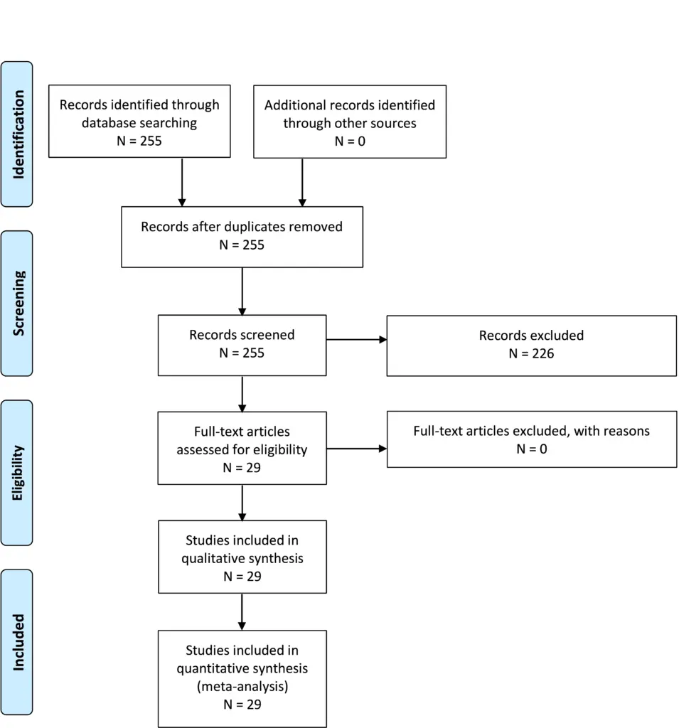
PICO 1-2.
PICO 1-3.
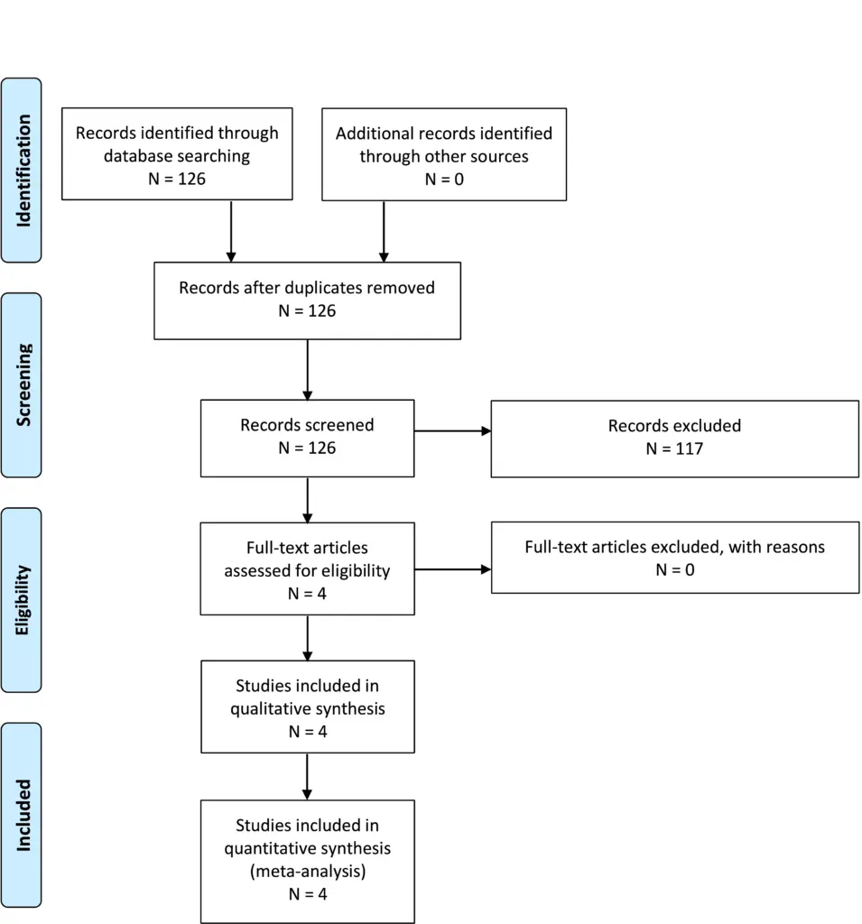
Appendix IV. Evidence tables
| PICO | Author, Year | Type of Evidence | Study Type | Class of Evidence | Reviewer’s Conclusions |
|---|---|---|---|---|---|
| 1 | Strahle et al, 20152 | Patient assessment | Retrospective case series | III | CIM may not be independently associated with scoliosis, and scoliosis may not be a symptom of CIM. The aim was to see if CIM is associated with scoliosis independent of syrinx; included patients: 509 Chiari, 1740 scoli, and 243 syrinx |
| 1 | Milhorat et al, 20093 | Therapy/patient assessment | Retrospective case control | III | Determines if section film was beneficial for patients with CIM. Evaluates association between CIM and TCS. discusses sectioning of the filum terminal. found improved symptoms of TCS and improvement in the cerebellar tonsils |
| 1 | Leung, V et al, 20164 | Diagnostic | Retrospective case control | III | Measures motion of cerebellum in Chiari/control patients; in patients with CIM and cardiac gated Cine MRI, cerebellar tonsil motion may be related to CIM; looks at the motion of the cerebellar tonsils in control patients, preop CIM patients with and without syringomyelia and postop patients. Includes Chiari decompression |
| 1 | Taylor et al, 20205 | Therapy/patient assessment | Retrospective case series | III | Evaluated subarachnoid space at foramen magnum for syrinx resolution; shows correlation between syringogenesis and diminution of shubarachnoid space in patients with CIM; lack of follow-up |
| 1 | Milhorat et al, 19996 | Patient assessment | Prospective case series | III | Chiari may be related to underdevelopment of posterior fossa; symptoms may be due to CSF flow blockage or direct compression; there may be a familial component for some patients; includes posterior fossa size and associated symptoms; includes information about the origins and etiology in patients with CIM |
| 1 | Elster et al, 19927 | Patient assessment | Prospective case series | III | Correlate symptoms with radiographic findings; no treatment was included (observational study); cerebellar tonsillation and presence of a syrinx |
| 1 | Bollo et al, 20128 | Patient assessment | Retrospective case series | III | Determines radiographic parameters needed from OC fusion; includes preoperative risk factors for patients with CIM that could potentially need occipitocervical fusion; Includes suboccipital decompression and or fusion; Shows need for fusion due to basilar invagination |
| 1 | Tubbs et al, 20119 | Patient assessment | Retrospective case series | III | Patients with CIM might present with headache/ neck pain and scoliosis. Other imaging findings or symptoms are described. This is a retrospective view of 500 surgically treated patients; results include symptom improvement and resolution of syrinx |
| 1 | Kennedy et al, 201610 | Patient assessment | Retrospective case series | III | Includes 12 patients with isolated thoracic syrinx, 50% syrinx; objective of this study was to determine the number of patients with a CIM that have an isolated thoracic syrinx; No specific treatments were identified; results include symptoms associated with isolated thoracic syrinx |
| 1 | Tubbs et al, 200011 | Patient assessment | Retrospective case series | III | Evaluates conus position, syrinx, Chiari relationship; compares observational vs no treatment; to see whether there is a relationship between conus even and patients with a CIM; level of conus and syrinx |
| 1 | Sadler et al, 202012 | Patient assessment | Retrospective case series | III | Identify underlying other DX with CIM to determine if other comorbidities are present in patients with CIM |
| 1 | Menezes et al, 199513 | Patient assessment | Prospective case series | III | Identify ways to treat complex CIM including craniocervical fixation and fusion, various ways; posterior decompression and fusion; resolution of syringohydromelia and resterablishment of CSF flow |
| 1 | Strahle et al, 202014 | Patient assessment | Retrospective/prospective case series | III | Determines associated factors between Chiari and scoliosis; determines the clinical and radiologic predictors of curve regression after PFD in patients with CIM; curve progression >5 degrees; regression of the scoliosis curve |
| 1 | McGirt, et al, 200615 | Patient assessment | Retrospective case series | III | Association between Cine flow and improvement after Chiari decompression; explores whether or not CSF flow dynamics assessed by Cine phase contrast MRI could independently predict response to posterior fossa decompression for CIM; follow-up was 1 month and 1 year after surgery—no mean follow-up was included |
| 1 | Caldarelli et al, 200716 | Therapy/patient assessment | Retrospective case series | III | Role of limited posterior fossa crani in CIM; determine if PFD works for CIM; extradural only procedure with C1 laminectomy |
| 1 | Krieger et al, 201117 | Patient assessment | Retrospective case series | III | Evaluates effect of CIM decompression on scoliosis; to see the association between CIM and scoliosis in children; needing further orthopedic procedure to correct the curvature; craniocervical decompression in a standard fashion |
| 1 | Brockmeyer et al 200318 | Patient assessment | Retrospective case series | III | Identifies relationship between CIM and scoliosis; effect of subocciptal decompression on curve progression; curve improvement or necessity of a fusion |
| 1 | Bhangoo et al, 200619 | Patient assessment | Retrospective case series | III | Determines association between CIM and scoliosis; whether or not the scoliosis curve improved after suboccipital decompression |
| 1 | Muhonen et al, 199220 | Patient assessment | Retrospective case series | III | Association between CIM and scoliosis; effect of treatment of CIM and scoliosis; PFDD, transoral, fusion; transoral and suboccipital decompression; population is heterogeneous |
| 1 | O’Neill et al 202121 | Patient assessment | Retrospective case series | III | Chiari decompression in asymptomatic patient with scoliosis may not be helpful to change outcome of curve progression or need for spine fusion; association between CIM and scoliosis; scoliosis in patients with CIM without a syrinx; curve progression or stabilization |
| 1 | Mauer et al 201122 | Patient assessment | Retrospective case series | III | CINE flow in patients pre- and postdecompression; demonstration of CSF pulsations can indicate surgical outcomes |
| 1 | Fan et al, 201723 | Patient assessment | Retrospective case series | III | Evaluates blockage location and treatment for CIM with syrinx; different surgical approaches are hypothesized based on CSF flow dynamics; includes PFD and PFD subarachnoid manipulation; authors were subjective with respect to Cine imaging; population was heterogeneous |
| 1 | Lee et al, 201424 | Patient assessment | Retrospective case series | III | Evaluation of optimal treatment of CIM; shows improvement in symptoms and syrinx |
| 1 | Villa et al, 201925 | Patient assessment | Retrospective case series | III | Evaluates efficacy of surgery; shows improvement in symptoms and syrinx |
| 1 | Menezes et al, 201826 | Patient assessment | Retrospective case series | III | Includes characteristics of CIM patients with syringobulbia; assesses if the syringobulbia resolved after PFD in patients with CIM; results show that in patients with syringobulbia and Chiari, posterior fossa decompression with intradural exploration and duraplasty may treat symptoms and imaging |
| 1 | Gad et al, 201727 | Patient assessment | Retrospective case series | III | Foramen magnum osseous abnormalities may contribute to syrinx formation |
| 1 | Lara-Reyna et al, 202028 | Patient assessment | Retrospective case series | III | There is variable reduction in syrinx after Chiari decompression in patients with Chiari and syrinx |
| 1 | Strahle et al, 201529 | Patient assessment | Retrospective case series | III | Location of CIM syrinx typically have cranial extent in cervical spine, and may be wider than other etiology for syrinx |
| 1 | Xie et al, 201530 | Patient assessment | Retrospective case series | III | In patients with CIM and syrinx, PFD may improve syrinx, and patients with improved syrinx may have upward shift of tonsils |
| 2 | Sadique et al, 202031 | Diagnostic test | Prospective/retrospective case series | III | There is improvement in peak velocity after decompression, but the authors do not comment on the relationship of changes in velocity to symptoms; it does not answer the question about benefit from decompression as mentioned in the PICO question |
| 2 | Bapuraj et al, 201632 | Diagnostic test | Prospective/retrospective case series | III | AMV at aqueduct improves after surgery, but no change in APV. This may correlate with symptom improvement |
| 2 | McGirt et al, 200533 | Diagnostic test | Retrospective case series | III | Patients with occipital headaches had obstructed flow and decompression helped those patients but there is no data on whether there is improvement of CSF flow parameters in these patients; only headache improvement is included |
| 2 | McGirt et al, 200834 | Diagnostic test | Prospective/retrospective case series | III | Those with ventral and dorsal CSF flow abnormalities showed highest likelihood of symptom improvement/lack of symptom recurrence; does not include postoperative CSF flow data |
| 2 | McGirt et al, 200615 | Diagnostic test | Retrospective case series | III | Lack of preoperative CSF obstruction predicts symptom recurrence, but age of each patient is unclear; mean age is 16 ± 13 years, no other details regarding age are provided |
| 2 | Ventureyra et al, 200335 | Diagnostic test | Retrospective case series | III | Patients with syrinx, absent flow, and CIM improved after posterior fossa decompression; those with no syrinx may improve with bone only decompression |
| 2 | Lara-Reyna et al, 202028 | Diagnostic test | Prospective/retrospective case series | III | Does not consider the value of advanced imaging at predicting benefit from decompression |
| 2 | Radmanesh et al, 201536 | Diagnostic test | Prospective/retrospective case series | III | No change in CCOS after surgery even though there was change in the tonsillar pulsatility |
| 2 | Ellenbogen et al, 200037 | Diagnostic test | Prospective/retrospective case series | III | All patients had improvement in CSF flow parameters, and pediatric patients mostly improved symptomatically (all except 1) |
| 3 | Bollo et al, 20128 | Patient assessment/therapy | Retrospective case series | III | CM 1.5, basilar invagination, CXA <125 degrees are at increased risk of instability and requirement for fusion |
| 3 | Ravindra et al 202038 | Diagnostic test | Prospective case series | III | C-C2SVA >5 mm may be predictive of the need for ventral decompression or OC fusion to better understand the anatomic load-bearing relationship between the atlantooccipital joint and the upper cervical spine and its influence on the clinical behavior of patients with CIM and craniocervical pathology |
| 3 | CreveCoeur et al 202139 | Diagnostic test/therapy | Prospective case series | III | Platybasia, Klippel–Feil, BV predictive of OCF; BV predictive of OCF/VD; CXA lower in OCF and OCF/VD groups compared with PFD only to examine factors influencing the use of OCF and OCF/VD in a multicenter cohort of pediatric CIM and SM subjects treated with PFD; both retrospective and prospective |
| 3 | Grabb et al, 199940 | Diagnostic test | Retrospective case series | III | pb-C2 >9 mm may require stabilization due to neurologic symptoms and instability but very few patients in this study that meet that criterion; 1) to determine the incidence and degree of VBSC in pediatric and young adult patients with CIM and 2) to correlate VBSC with other imaging and clinical factors to help determine what amount of VBSC is successfully treated with a posterior decompressive procedure alone |
AMV, amplitude of mean velocity; APV, amplitude of peak velocity; C-C2SVA, C2 sagittal vertebral alignment; CCOS, Chicago Chiari Outcome Scale; CM, Chiari malformation; CSF, cerebrospinal fluid;
CXA, clivoaxial angle; DX, diagnosis; MRI, magnetic resonance imaging; OC, occipitocervical; OCF, posterior fossa decompression and fusion; TCS, tethered cord syndrome; VBSC, ventral brain stem compression; VD, ventral decompression.
Appendix V. Conflicts of interest
| Name | Affiliation | Type of COI |
|---|---|---|
| Toba Niazi, MD | Live Like Bella Foundation, Nicklaus Children’s Hospital | Grants/Research Support |
| Laurie Ackerman, MD | Park Reeves Consortium | Grants/Research Support |
| David Bauer, MD | None | |
| Brandon G. Rocque, MD, MS, FAANS | None | |
| Carolyn S. Quinsey, MD | None | |
| Eric Jackson, MD | None | |
| Jogi V. Pattisapu MD FAAP FAANS | J&J, Integra | Consultant |
| Rabia Qaiser, MD | None | |
| Cormac O. Maher, MD, FAAP, FACS, FAANS | None | |
| Shobhan H. Vachhrajani MD, PhD, FRCSC | None | |
| Libby Infinger, MD | None | |
| Howard Silberstein, MD | None | |
| Sarah Jernigan, MD | None | |
| Jeffrey S. Raskin MS MD FAANS FAAP | None | |
| Dorothy Poppe | None | |
| Kaitlyn Esposito | None |
Congress of Neurological Surgeons Systematic Review and Evidence-Based Guidelines for Chiari Malformation: Symptoms
Sponsored by: Congress of Neurological Surgeons (CNS) and the Section on Pediatrics
Endorsed by: The Congress of Neurological Surgeons (CNS), American Association of Neurological Surgeons (AANS) and the Bobby Jones Chiari and Syringomyelia Foundation (Bobby Jones CSF)
Authors:
Eric M. Jackson, MD1, Sarah Jernigan, MD, MPH2, Jeffrey S. Raskin MS MD3, Laurie L Ackerman, MD4, Libby Kosnik Infinger, MD, MPH5, Cormac O. Maher, MD, FAAP, FACS, FAANS6, Toba Niazi, MD7, Jogi V. Pattisapu MD FAAP FACS FAANS8, Rabia Qaiser, MD9, Carolyn Quinsey, MD10, Brandon G. Rocque, MD, MS11, Howard Silberstein, MD12, Shobhan Vachhrajani MD, PhD, FRCSC13, David F. Bauer, MD, MPH14
Departmental and institutional affiliations:
- Department of Neurosurgery, Johns Hopkins University School of Medicine, Baltimore, MD
- Carolina Neurosurgery & Spine Associates, Charlotte, NC
- Department of Neurological Surgery, Northwestern University Feinberg School of Medicine, Chicago, IL
- Department of Neurological Surgery, Indiana University Health, Indianapolis, IN
- Department of Neurosurgery, Medical University of South Carolina (MUSC), Charleston, SC
- Department of Neurosurgery, Stanford Medicine, Palo Alto, CA
- Department of Neurological Surgery, Nicklaus Children’s Hospital, Miami, FL
- Pediatric Neurosurgery, University of Central Florida College of Medicine, Orlando FL
- Department of Neurological Surgery, Indiana University School of Medicine, Indianapolis, IN
- Department of Neurosurgery, University of North Carolina Chapel Hill, Chapel Hill, NC
- Division of Pediatric Neurosurgery, Department of Neurosurgery, University of Alabama at Birmingham, Birmingham, AL
- Department of Neurosurgery, University of Rochester School of Medicine and Dentistry, Rochester, NY
- Department of Pediatrics, Wright State University Boonshoft School of Medicine, Dayton, OH
- Department of Neurosurgery, Baylor College of Medicine, Division of Pediatric Neurosurgery, Texas Children’s Hospital, Houston, TX
Corresponding Author contact information:
Eric M. Jackson, MD
Department of Neurosurgery, Johns Hopkins University School of Medicine
Baltimore, MD
No part of this article has been published or submitted for publication elsewhere.
Keywords:
Abbreviations: CIM, Chiari type I malformation; CSF, cerebrospinal fluid; PFD, polysomnography (PSG); sleep disordered breathing (SDB)
ABSTRACT
Background: Chiari I malformation (CIM) is characterized by descent of the cerebellar tonsils through the foramen magnum, potentially causing symptoms from compression or obstruction of the flow of cerebrospinal fluid (CSF). Diagnosis and treatment of CIM is varied, and guidelines produced through systematic review may be helpful for clinicians.
Objective: We performed a systematic review of the medical literature to answer specific questions on the diagnosis and treatment of CIM.
Methods: PubMed and Embase were queried between 1946 and January 23, 2021 using the search strategies provided in Appendix I.
Results: The literature search yielded 430 abstracts, of which 79 were selected for full-text review, 44 were then rejected for not meeting the inclusion criteria or for being off-topic, and 35 were included in this systematic review.
Conclusion: Four Grade C recommendations were made based on Class III evidence and 1 question had insufficient evidence.
RECOMMENDATIONS
2-1. In patients operated for symptomatic CIM, what symptoms are most likely to improve after surgery?
Recommendation: Clinicians may perform foramen magnum decompression surgery on symptomatic patients with CIM to improve pain associated with strain-related headaches. Other symptoms demonstrate more variable response to decompression.
Strength of recommendation: Grade C
Level III evidence
2-2. In patients with asymptomatic CIM without syrinx, is prophylactic surgery indicated to prevent future need for surgery? What is the chance of developing symptoms in the future?
Recommendation: Clinicians should not perform prophylactic surgery on patients with asymptomatic CIM without syrinx. There is a small percentage of patients who develop new or worsening symptoms in the future.
Strength of recommendation: Grade C
Level III evidence
2-3. In patients with asymptomatic CIM without syrinx, should the patient have any activity restrictions to prevent future harm?
Recommendation: Clinicians should not recommend activity restrictions for patients with asymptomatic CIM without syrinx, as there is no evidence of future harm prevention.
Strength of recommendation: Grade C
Level III evidence
2-4. In patients with CIM, should sleep or swallow studies be routinely performed to evaluate for sleep apnea or dysphagia?
Recommendation: There is insufficient evidence to support routine sleep and swallow studies in patients with CIM without sleep or swallow symptoms.
Strength of recommendation: Grade insufficient
2-5. In patients with CIM, should siblings or first-degree relatives be screened for CIM?
Recommendation: Clinicians should not routinely screen asymptomatic siblings or first-degree relatives of patients with CIM.
Strength of recommendation: Grade C
Level III evidence
INTRODUCTION
Goals and Rationale
This clinical guideline has been created to improve patient care by outlining the appropriate information gathering and decision-making processes involved in the treatment of patients with Chiari I malformation (CIM). Care for patients with CIM is provided in many different settings by many different providers. This guideline has been created as an educational tool to guide qualified physicians through a series of diagnostic and treatment decisions to improve the quality and efficiency of care.
Objectives
CIM is a structural abnormality related to the anatomy of the base of the skull and the cerebellum. CIM is defined as descent of the cerebellar tonsils ≥3 to 5 mm below the foramen magnum. Based on a definition of a tonsillar position of ≥5 mm below the foramen magnum, imaging studies estimate a prevalence ranging from 0.24% to 2.6% of the population,1–5 including children and adults. Patients may have varied symptoms and responses to treatment for their CIM, with controversy about what symptoms may relate to the underlying malformation. This chapter aims to provide guidelines based on the literature regarding the symptoms most likely to relate to patients with CIM and thus respond to treatment for CIM, as well as the need for prophylactic surgery, activity restrictions, symptom evaluation, and familial screening.
METHODOLOGY
The guidelines task force initiated a systematic review of the literature and evidence-based guideline relevant to the treatment of patients with CIM. Through objective evaluation of the evidence and transparency in the process of making recommendations, this evidence-based clinical practice guideline was developed for the diagnosis and treatment of patients with CIM. These guidelines are developed for educational purposes to assist practitioners in their clinical decision-making processes. Additional information about the methods used in this systematic review is provided below.
Literature Search
Task force members identified search terms/parameter and a medical librarian implemented the literature search, consistent with the literature search protocol (see Appendix I), using the National Library of Medicine/PubMed database and Embase for the period from 1946 to January 23, 2021 using the search strategies provided in Appendix I.
Inclusion/Exclusion Criteria
Articles were retrieved and included only if they met specific inclusion/exclusion criteria. To reduce bias, these criteria were specified before conducting the literature searches.
Articles that do not meet the following criteria, for the purposes of this evidence-based clinical practice guideline, were excluded. To be included as evidence in the guideline, an article had to be a report of a study that:
- Investigated patients with CIM;
- Studies that enrolled ≥80% of CIM (we included studies with mixed patient populations if they reported results separately for each group/patient population);
- Was a full article report of a clinical study;
- Was not a medical records review, meeting abstract, historical article, editorial, letter, or commentary;
- Appeared in a peer-reviewed publication or a registry report;
- Enrolled a minimum of 10 patients;
- Was of humans;
- Was published in or after 1946;
- Quantitatively presented results;
- Was not an in vitro study;
- Was not a biomechanical study;
- Was not performed on cadavers;
- Was published in English;
- Was not a systematic review, meta-analysis, or guideline developed by others
Systematic reviews or meta-analyses conducted by others, or guidelines developed by others were not included as evidence to support this review because of the differences in article inclusion/exclusion criteria specified compared with the criteria specified by the Guidelines Task Force. Although these articles were not included as evidence to support the review, these articles were recalled for full-text review for the Guidelines Task Force to conduct manual searches of the bibliographies.
Assessment for Risk of Bias
The methodological quality of randomized controlled trials and the risk of bias were assessed using the following 6 criteria:
- Sequence generation (Was the allocation sequence adequately generated?)
- Allocation concealment (Was allocation adequately concealed such that it could not be foretold?)
- Blinding (Were participants, treatment providers and/or outcome assessors blinded to the treatment allocations?)
- Incomplete reporting of data (Were incomplete outcome data adequately addressed?)
- Selective reporting of outcomes (Were all the outcomes specified reported?)
- Other potential threats to validity (Was the randomized controlled trial free of other issues that could put it at a high risk of bias?)
1The guideline task force did not include systematic reviews, guidelines, or meta-analyses conducted by others. These documents are developed using different inclusion criteria than those specified in this guideline; therefore, they may include studies that do not meet the inclusion criteria specific to this guideline. In cases where these types of documents’ abstract suggested relevance to the guideline’s recommendations, the task force searched their bibliographies for additional studies.
In the case of nonrandomized observational evidence, potential threats to the validity of the data were assessed by examining for:
- Bias due to selective case choice for study and selective result reporting
- Bias due to lack or loss of information over time
- The biases of the interpreting investigator regarding the study
- Publication bias regarding positive studies or positive cases
- Misclassification
- Survivorship bias
- Publication bias
- Recognition that in data collected in a retrospective or prospective manner correlation does not imply causation
- Election bias
- Attrition bias
- Bias of change in methods over time
- Ascertainment bias
Rating Quality of Evidence
The quality of evidence was rated using an evidence hierarchy for each of 4 different study types; therapeutic, prognostic, diagnostic, and decision modeling. These hierarchies are shown in Appendix II: Rating Evidence Quality. Additional information regarding the hierarchy classification of evidence can be located here: https://www.cns.org/guidelines/guideline-procedures-policies/guideline-development-methodology.
Revision Plans
In accordance with the Institute of Medicine’s standards for developing clinical practice guidelines and criteria specified by the National Guideline Clearinghouse, the task force will monitor related publications after the release of this document and will revise the entire document and/or specific sections “if new evidence shows that a recommended intervention causes previously unknown substantial harm; that a new intervention is significantly superior to a previously recommended intervention from an efficacy or harms perspective; or that a recommendation can be applied to new populations.”6 In addition, the task force will confirm within 5 years from the date of publication that the content reflects current clinical practice for treatment of CIM.
RESULTS
The literature search yielded 430 abstracts. Task force members reviewed all abstracts yielded from the literature search and identified the literature for full-text review and extraction, addressing the clinical questions, in accordance with the literature search protocol (Appendix I). Task force members identified the best research evidence available to answer the targeted clinical questions. When class I, II and or III literature was available to answer specific questions, the task force did not review class IV studies.
The task force selected 79 full-text articles for full-text review. Of these, 44 were rejected for not meeting the inclusion criteria or for being off-topic. Thirty-five were selected for this systematic review (Appendix III).
DISCUSSION
Question 2-1. In patients operated for symptomatic CIM, what symptoms most likely improve after surgery?
Recommendation: Clinicians may perform foramen magnum decompression surgery on symptomatic patients with CIM to improve pain associated with strain-related headaches. Other symptoms demonstrate more variable response to decompression.
Strength of recommendation: Grade C
Class III Evidence
There is an extensive literature on symptoms related to CIM and likelihood of response to intervention. Studies were all class III evidence and had varying objectives, but symptom response data were abstracted on review. Manuscripts focused on patient outcomes, outcome scores, or clinical decision rules were excluded from analysis based on a lack of focus on specific symptoms for answering this question. Notably, manuscripts often included patients with syringomyelia, but syrinx as a separate entity was not considered to be a symptom for this question. Syringomyelia may affect symptoms attributed to Chiari with 1 study reporting7 variable improvement in symptoms in patients with and without syrinx. For this question, symptoms that may be related to either the CIM or syrinx, including numbness or weakness, are included in the general discussion, as they are difficult to separate. Syrinx is discussed as a separate entity in Chapter 1 of these guidelines.
After review of the literature, no symptoms demonstrated uniform improvement. Headache was the symptom most likely to respond to intervention; strain-induced occipital headache was the most likely to respond. Other headaches, including frontal headaches and non–strain-induced headaches were also noted to show improvement in some cases but to a lesser degree than occipital strain-induced headaches, which have been classically termed “Chiari headaches.”8 Other symptoms including drop attacks; visual symptoms or changes; vestibular dysfunction including hearing loss, tinnitus, or vertigo; extremity numbness, weakness, or dysesthetic pain; and myelopathy and ataxia likewise showed a variable response across the literature but with less consistent improvement, either due to smaller sample size or a much lower percentage of improvement reported.7–27
Age-related differences in symptomatology were identified, with children noted to be more likely to have oropharyngeal symptoms including sleep apnea and reflux.28–30 These symptoms did appear to improve with intervention in patients across different studies.
Although not always discussed as a symptom, 1 study in a limited patient set (11 patients) demonstrated improvement in some patients in neuropsychological testing in areas including executive function, verbal learning, psychomotor speed, and color naming speed.31
The literature on symptoms in CIM is extensive. As all the studies reviewed were class III data, it is difficult to draw significant conclusions. The variability of response of different symptoms in different studies is subject to many confounding factors. As such, there is not adequate evidence to provide specific recommendations about most symptoms that patients may describe in the setting of CIM nor to say whether the symptoms may in fact be attributable to the CIM. Regardless, across the literature reviewed, strain-induced occipital headaches were the symptom most consistently demonstrated to improve with treatment of CIM.
Question 2-2. In patients with asymptomatic CIM without syrinx, is prophylactic surgery indicated to prevent future need for surgery? What is the chance of developing symptoms in the future?
Recommendation: Clinicians should not perform prophylactic surgery on patients with asymptomatic CIM without syrinx. There is a small percentage of patients who develop new or worsening symptoms in the future.
Strength of recommendation: Grade C
Class III Evidence
There were 3 studies identified by the methodology that compared operative and nonoperative treatment of patients with CIM and were thus used in formulating the recommendation. Of note, although the papers may include patients with syrinx, most of the patients were treated nonoperatively in the literature and did not have what was determined to be a clinically significant syrinx. As such, the recommendations are for asymptomatic patients without syrinx and should not be generalized to patients with syrinx.
One study32 identified 226 pediatric patients seen for initial consultation for CIM over a 5-year period, with symptoms and/or with syrinx. Of these patients, 34 had surgery and 192 patients were treated nonoperatively. Of the 34 patients who had surgery, 15 had surgery >6 months after the initial consultation, of which only 5 were delayed because of new symptoms/syrinx (n = 4) or symptom progression (n = 1). In total, authors identified 2 patients with worsening symptoms, 1 patient who had symptoms that failed to improve, and 2 who had worsening syrinx. No patients had surgery >2 years after initial consultation. The authors concluded from their data that patients treated nonoperatively are unlikely to progress, suggesting a benign natural history.
An additional pediatric study33 reviewed 95 patients over a 10-year period. Seventy patients were managed conservatively and 25 had surgery (either dural splitting or duraplasty). They noted a higher percentage of improvement in surgical patients, but 41.7% (20/48) of symptomatic patients treated nonsurgically demonstrated improvements in symptoms. Of the conservatively managed group, 45 showed no change in symptoms or new symptoms if asymptomatic and 5 (7.1%) exhibited worsening symptoms over time. They indicated that their study was consistent with the literature suggesting against prophylactic decompression, as the development of new symptoms or deficits was uncommon.
One additional study17 identified was a mixed population study (pediatric and adults) that reviewed patients evaluated from 2000 to 2011 with long-term follow-up. The population was approximately 30% pediatric (<18 years of age). One-hundred nine patients had surgery with 236 recommended for nonsurgical therapy. Of the 236 nonsurgical patients, 78 were able to be contacted and consented to long-term follow-up questions. Of those, 10 were excluded including 8 that had surgery at an outside institution. Of the 68 remaining patients, they calculated that 73% (50/68) of their patients treated without surgery demonstrated stability or improvement in symptoms with 47.1% (32/68) of those patients showing improvement over an average of 4.9 years. Eighteen patients (26.5%) noted worsening of any symptom with 5 of those patients having improvement in another symptom, suggestive of mixed etiology.
These studies were not limited to asymptomatic patients but still provide class III evidence that most patients that are treated conservatively for CIM remain stable or improve, suggesting a benign natural history.
There were 4 additional studies identified below that, while they did not meet inclusion criteria due to a focus on natural history (and/or do not explicitly address this PICO question), suggest similar conclusions.
Novegno et al34 reviewed patients evaluated for Chiari at their institution from 1988 to 2007. Of a total of 94 children, there were 22 patients with mild or absent clinical symptoms that were followed for ≥3 years (mean 5.9 years). Eleven of the patients were asymptomatic and the other 11 had mild symptoms not felt to warrant intervention. Of the 22 patients, 17 (77.3%) remained asymptomatic or had improvement in the mild symptoms. Five patients had worsening symptoms, of which 2 were mild and were still observed and 3 ultimately had surgery. Of those patients, 2 had endoscopic third ventriculostomy performed for worsening hydrocephalus, suggesting secondary Chiari. The third patient had a Chiari decompression (1 of 22 patients treated directly for Chiari [4.5%]). Based on their data and review of the literature, the authors conclude that conservative treatment is appropriate for asymptomatic and slightly symptomatic patients with Chiari malformation.
Strahle et al35 reviewed the natural history of patients at their institution following a decision to treat conservatively. They included 147 patients who had a CIM diagnosed on magnetic resonance imaging that were not offered surgery and had ≥1 year of follow-up. They had a mean clinical follow-up of 4.6 years and mean imaging follow-up of 3.8 years. One hundred thirty-three of the 147 patients (90.5%) remained asymptomatic or minimally symptomatic. Fourteen patients (9.5%) progressed to surgery with indications including new syrinx or syrinx progression, worsening and refractory headaches, sleep apnea, concern for neurologic decline, and progression of scoliosis. In addition to patients with worsening syrinx, there were 3 patients with resolution of the syrinx as well. They reviewed the data on cerebrospinal fluid (CSF) flow studies on 74 patients with adequate data and noted improvement in CSF flow in 23, no change in 39, and worsening in 12. There were not significant anatomic differences in the patients who later required surgery from those that did not. Based on their data, the authors concluded that the natural history for patients selected for nonoperative management of Chiari is typically benign, with a small percentage of patients having changes.
Benglis et al36 retrospectively reviewed 124 cases of pediatric patients treated nonoperatively and seen over a 10-year period. Eighty-one patients were symptomatic, with 67 felt to have symptoms not typical of Chiari malformation. Of the 14 with symptoms felt typical for Chiari malformation, 9 had symptoms not frequent or severe enough to recommend intervention and 5 were offered surgery. Of the 14 patients with Chiari symptoms, 6 experienced symptom improvement over time, 4 had stable symptoms over time, and 4 had worsening symptoms. No new neurologic deficits were noted among the patients. They concluded that most patients with CIM followed over time do not progress clinically, suggesting a benign natural history.
Another more recent study37 reviewed prospectively collected data on patients with incidentally discovered CIM ≤18 years of age between 2009 and 2019 with at least 12 months of follow-up. They reviewed 218 consecutive patients with a mean follow-up of 40.6 months. Thirty-six patients (16.5%) underwent decompression surgery. Twenty-two of the patients underwent surgery within 6 months of diagnosis, while 14 patients had surgery >6 months after diagnosis. Indications included development of a syrinx (n = 6), syrinx and symptom progression (n = 3), and symptom progression alone (n = 5). Thus, for the patients that did not have surgery within the first 6 months, 7.1% (14/196) progressed to requiring treatment within the review period. The studies that met inclusion criteria, as well as others identified in discussion, support that patients diagnosed with CIM that is not symptomatic enough to warrant intervention generally have a benign natural history, which does not support prophylactic surgery. There is a small percentage of patients that can worsen over time; therefore, clinical and imaging follow-up should be considered on a case-by-case basis and patients can be followed for changes to ensure they are not in a subset that requires treatment in the future.
Question 2-3. In patients with asymptomatic CIM without syrinx, should the patient have any activity restrictions to prevent future harm?
Recommendation: Clinicians should not recommend activity restrictions for patients with asymptomatic CIM without syrinx, as there is no evidence of future harm prevention.
Strength of recommendation: Grade C
Four studies met criteria for inclusion regarding activity restrictions in patients with asymptomatic CIM. Two studies looked at sports participation and risk of injury related to Chiari. Two other studies provide more indirect evidence of potential worsening of symptoms in CIM patients who have a traumatic injury.
Class II Evidence
Strahle et al38 performed a prospective dual site survey study of 503 patients with CIM including 328 sports participants. There was no difference in disease severity with tonsillar ectopia on average 11 mm in both sports participants and sports abstaining cohorts; 74% of all patients had pegged tonsillar morphology and 74% of available CSF flow imaging showed diminished flow. Respondents played a wide variety of high impact sports and over 4641 seasons there were no catastrophic or permanent neurologic injuries.
Class III Evidence
Meehan et al39 performed a single-institution retrospective cohort study over 3 years in 147 patients with an average tonsillar ectopia of 11 mm, again with the majority exhibiting pegged tonsils and crowding at the foramen magnum. Similar results were found including no deaths, coma, or paralysis in 1627 athletic seasons.
In terms of more indirect evidence of worsening with trauma, Wan et al40 performed a single-center retrospective series identifying 85 patients seen with CIM over 21 years. They noted that patients can have onset of symptoms after a minor injury. Freeman et al41 reviewed the cervical spine magnetic resonance imaging scans of 1200 individuals with neck pain, 600 with a history of a whiplash type injury and 600 without a traumatic injury. They noted a statistically significant increase in cerebellar ectopia in patients with a history of whiplash type injury, suggesting a connection between the trauma and worsening cerebellar ectopia or symptoms.
Although there is potentially indirect evidence of possible worsening with trauma, the 2 studies that directly address need for restrictions, 1 prospective38 and 1 retrospective survey study,39 similarly found no poor outcomes in pediatric patients with CIM related to sports participation. Taken together, the studies recommend against activity restrictions for these patients.
It is important to note that this discussion is relevant to patients with asymptomatic CIM and does not apply to patients with symptomatic CIM or with significant skull base abnormalities such as basilar invagination. Overall, although there is some evidence of patients developing symptoms after trauma, there were no patients presented with known Chiari that were followed and worsened. As such, there is no direct evidence of worsening based on activity. Despite a potential increased theoretical risk relative to the general population, the reviewed literature demonstrated no significant injuries in patients with known asymptomatic Chiari from sports participation. The literature supports appropriate counseling of potential risks and individualized discussion with patients and family members but does not provide evidence to support routinely restricting the activity of patients with asymptomatic CIM.
Question 2-4. In patients with CIM, should sleep or swallow studies be routinely performed to evaluate for sleep apnea or dysphagia?
Recommendation: There is insufficient evidence to support routine sleep and swallow studies in patients with CIM without sleep or swallow symptoms.
Strength of recommendation: Grade insufficient
Class III Evidence
There were no studies that met inclusion criteria to address routine swallow studies with CIM. There were 2 retrospective single-institution studies identified that investigated sleep disordered breathing (SDB) in patients with CIM.
Amin et al42 performed a retrospective review on patients with CIM who underwent baseline polysomnography (PSG). They identified 68 children. They noted a 49% prevalence of SDB based on the apnea-hypopnea index, with obstructive apnea being the predominant type of SDB. Tonsillar descent did not predict the presence of SDB in their cohort but was significantly correlated with the obstructive apnea-hypopnea index but not the central apnea index. Using a cutoff of 20 mm herniation, there was a statistically significant association of the level of tonsillar descent with the presence of obstructive sleep apnea.
Khatwa et al43 reviewed 22 children with CIM and SDB on PSG. Seventeen patients had known Chiari before the PSG study, including 5 patients that were asymptomatic. Five patients had symptoms that led to the sleep study prior to the diagnosis of Chiari. They found that the extent of herniation was significantly greater in patients with SDB than those with normal PSG (16.0 vs 8.2 mm mean descent). There were 4 patients with preoperative testing that underwent decompression. All patients showed improvement, but 1 still required treatment for residual sleep apnea. In addition to the demonstration of improvement, they conclude that imaging parameters may correlate with the presence of SDB.
One additional study was excluded based on not having enough patients but did look at benefits of treatment of Chiari in patients with sleep apnea. Addo et al44 showed significant benefit in measured numbers of central sleep apnea postdecompression in 5 patients with known sleep apnea in the setting of CIM and syndromic synostosis, indicating that treating the Chiari malformation can improve SDB.
SDB and swallowing dysfunction may be present in patients with CIM. When these symptoms are present, many providers obtain sleep and swallow studies for further evaluation. The literature reviewed does support the possibility of improvement in SDB after decompression surgery with patients across studies demonstrating improvement. The reviewed literature also suggests that breathing changes may be more prevalent with increasing tonsillar descent on imaging, especially with very significant tonsillar descent (>20 mm). Thus, further clinical workup may be appropriate on an individual basis for many patients with CIM based on clinical suspicion or significant imaging findings. There is no evidence in the literature to support the need for sleep or swallow studies in the routine evaluation of all patients with CIM.
Question 2-5. In patients with CIM, should siblings or first-degree relatives be screened for CIM?
Recommendation: Clinicians should not routinely screen asymptomatic siblings or first-degree relatives of patients with CIM.
Strength of recommendation: Grade C
Class III Evidence
There are different data that suggest a familial association of CIM. Case series identify families with multiple members with CIM,45 suggesting an inherited basis. There are studies that look at the presence of CIM in patients with other genetic disorders, such as neurofibromatosis type 1.46 There is also ongoing research investigating the underlying genetic basis of CIM. One recent study47 performed whole-exome screening on 51 unrelated surgical patients with CIM. They also tested the parents of the patients. They identified multiple genetic variants, including a high number in chromatin-remodeling genes. They highlighted that CIM is likely underdiagnosed in the population as 21 patients had a parent with the same genetic variant, who on imaging were documented to have a CIM despite only 4 of the 51 patients having a known family member with CIM before enrollment in the study.
Based on the data, it seems likely that the family members of patients with CIM are more likely to have the diagnosis than the general population. There is not sufficient evidence to determine a relative risk, however. Although the evidence does support an increased likelihood of having the diagnosis, there is no evidence of a benefit to screening asymptomatic family members or first-degree relatives. Question 2-2 reviews the data on the role of prophylactic surgery on asymptomatic patients and there was no evidence to support prophylactic surgery in those patients. Therefore, without evidence that patients would benefit from intervention, there is no support for routine screening of asymptomatic siblings or first-degree relatives of patients with CIM. It is important to indicate that the guideline recommendation is based on family members being asymptomatic. If family members are symptomatic with concerns for symptoms possibly related to CIM, providers may wish to pursue further evaluation because of the apparent underlying familial predisposition.
Future Research
Review of the literature for the guidelines highlights the lack of Class I evidence to make strong recommendations. It highlights the need for multicenter prospective data collections regarding symptoms and natural history as well as surgical studies. The randomized posterior fossa decompression versus posterior fossa decompression with duraplasty study recently completed will provide some additional data for the pediatric population, but more such studies will be needed to better clarify the optimal management for patients with CIM.
Future studies and collaborative efforts may offer more insight to improve our management approach. Patient-centered studies evaluating patient-reported outcomes may be helpful to inform future clinical decision making and recommendations. It is imperative to explore these questions are help improve care of our patients with CIM and syringomyelia.
CONCLUSIONS
There was primarily Class III evidence for these questions as well as differences across studies, highlighting the need for better data to generate stronger conclusions and recommendations. As such, the guidelines are worded to provide guidance but allow for practitioners to assess and treat patients on an individual basis, based on individual symptoms and characteristics.
Conflicts of Interest
All Guideline Task Force members were required to disclose all potential COIs prior to beginning work on the guideline, using the COI disclosure form of the AANS/CNS Joint Guidelines Review Committee. The CNS Guidelines Committee and Guideline Task Force Chair reviewed the disclosures and either approved or disapproved the nomination and participation on the task force. The CNS Guidelines Committee and Guideline Task Force Chair may approve nominations of task force members with possible conflicts and restrict the writing, reviewing, and/or voting privileges of that person to topics that are unrelated to the possible COIs. See Appendix V for a complete list of disclosures.
Disclosure of Funding
These evidence-based clinical practice guidelines were funded exclusively by the Congress of Neurological Surgeons, which received no funding from outside commercial sources to support the development of this document.
Disclaimer of Liability
This clinical systematic review and evidence-based guideline was developed by a physician volunteer task force as an educational tool that reflects the current state of knowledge at the time of completion. Each chapter is designed to provide an accurate review of the subject matter covered. This guideline is disseminated with the understanding that the recommendations by the authors and consultants who have collaborated in their development are not meant to replace the individualized care and treatment advice from a patient’s physician(s). If medical advice or assistance is required, the services of a competent physician should be sought. The proposals contained in these guidelines may not be suitable for use in all circumstances. The choice to implement any particular recommendation contained in these guidelines must be made by a managing physician in light of the situation in each particular patient and on the basis of existing resources.
Acknowledgments
The guidelines task force would like to acknowledge the CNS Guidelines Committee for their contributions throughout the development of the guideline, the AANS/CNS Joint Guidelines Review Committee, as well as the contributions of Kirsten Aquino, contracted project manager for the CNS, Trish Rehring, MPH, Associate Director for Evidence-Based Practice Initiatives for the CNS, and Janet Waters, MLS, BSN, RN, for assistance with the literature searches. The guidelines task force would also like to acknowledge the contributions of Dorothy Poppe, Kaitlyn Esposito, MPH and Mary Poppe, as well as the Bobby Jones Chiari and Syringomyelia Foundation for serving as patient advocates on this guideline task force. Throughout the review process, the reviewers and authors were blinded from one another. At this time the guidelines task force would like to acknowledge the following individual peer reviewers for their contributions: Jennifer Sweet, MD, Andrew Carlson, MD, MS, Matthew Reynolds, MD, PhD, Alexandra D. Beier, D.O., FACOS, FAAP, Jonathan Pindrik, MD and Patti Raksin, MD.
REFERENCES
1. Aitken LA, Lindan CE, Sidney S, et al. Chiari type I malformation in a pediatric population. Pediatric Neurology. 2009;40(6):449-454.
2. Meadows J, Kraut M, Guarnieri M, Haroun RI, Carson BS. Asymptomatic Chiari type I malformations identified on magnetic resonance imaging. Journal of neurosurgery. 2000;92(6):920-926.
3. Morris Z, Whiteley WN, Longstreth Jr WT, et al. Incidental findings on brain magnetic resonance imaging: Systematic review and meta-analysis. BMJ (Online). 2009;339(7720):547-550.
4. Strahle J, Muraszko KM, Kapurch J, Bapuraj JR, Garton HJ, Maher CO. Chiari malformation type I and syrinx in children undergoing magnetic resonance imaging. Journal of neurosurgery Pediatrics. 2011;8(2):205-213.
5. Vernooij MW, Ikram MA, Tanghe HL, et al. Incidental findings on brain MRI in the general population. New England Journal of Medicine. 2007;357(18):1821-1828.
6. Ransohoff DF, M. Pignone, and H.C. Sox, . How to decide whether a clinical practice guideline is trustworthy. . JAMA. 2013;309(2):139-140.
7. Dones J, De Jesús O, Colen CB, Toledo MM, Delgado M. Clinical outcomes in patients with Chiari I malformation: a review of 27 cases. Surgical neurology. 2003;60(2):142-147; discussion 147-148.
8. Grangeon L, Puy L, Gilard V, et al. Predictive factors of headache resolution after Chiari type 1 malformation surgery. World neurosurgery. 2017;110:e60-e66.
9. Massimi L, Frassanito P, Chieffo D, Tamburrini G, Caldarelli M. Bony decompression for Chiari malformation type I: long-term follow-Up. Acta neurochirurgica Supplement. 2019;125:119-124.
10. Ma J, You C, Chen H, Huang S, Ieong C. Cerebellar tonsillectomy with suboccipital decompression and duraplasty by small incision for Chiari I malformation (with syringomyelia): long term follow-up of 76 surgically treated cases. Turkish neurosurgery. 2012;22(3):274-279.
11. Straus D, Foster K, Zimmerman F, Frim D. Chiari drop attacks: surgical decompression and the role of tilt table testing. Pediatric neurosurgery. 2009;45(5):384-389.
12. Kumar A, Patni AH, Charbel F. The Chiari I malformation and the neurotologist. Otology & neurotology : official publication of the American Otological Society, American Neurotology Society [and] European Academy of Otology and Neurotology. 2002;23(5):727-735.
13. Beretta E, Vetrano IG, Curone M, et al. Chiari malformation-related headache: outcome after surgical treatment. Neurological sciences : official journal of the Italian Neurological Society and of the Italian Society of Clinical Neurophysiology. 2017;38(Suppl 1):95-98.
14. Spena G, Bernucci C, Garbossa D, Valfrè W, Versari P. Clinical and radiological outcome of craniocervical osteo-dural decompression for Chiari I-associated syringomyelia. Neurosurgical review. 2010;33(3):297-303; discussion 303-294.
15. Kumar A, Ghosh SN, Sadique SI. Clinicoradiological study of adult Chiari malformation type 1 patients with emphasis on cerebrospinal fluid peak flow velocity at foramen magnum level. Neurology India. 2019;67(3):744-748.
16. Jia C, Li H, Wu J, et al. Comparison decompression by duraplasty or cerebellar tonsillectomy for Chiari malformation-I complicated with syringomyelia. Clinical neurology and neurosurgery. 2018;176:1-7.
17. Chavez A, Roguski M, Killeen A, Heilman C, Hwang S. Comparison of operative and non-operative outcomes based on surgical selection criteria for patients with Chiari I malformations. Journal of clinical neuroscience : official journal of the Neurosurgical Society of Australasia. 2014;21(12):2201-2206.
18. Parker SL, Godil SS, Zuckerman SL, et al. Comprehensive assessment of 1-year outcomes and determination of minimum clinically important difference in pain, disability, and quality of life after suboccipital decompression for Chiari malformation I in adults. Neurosurgery. 2013;73(4):569-581; discussion 581.
19. McGirt MJ, Nimjee SM, Floyd J, Bulsara KR, George TM. Correlation of cerebrospinal fluid flow dynamics and headache in Chiari I malformation. Neurosurgery. 2005;56(4):716-721; discussion 716-721.
20. Raza-Knight S, Mankad K, Prabhakar P, Thompson D. Headache outcomes in children undergoing foramen magnum decompression for Chiari I malformation. Archives of disease in childhood. 2017;102(3):238-243.
21. Hayhurst C, Richards O, Zaki H, Findlay G, Pigott TJ. Hindbrain decompression for Chiari-syringomyelia complex: an outcome analysis comparing surgical techniques. British journal of neurosurgery. 2008;22(1):86-91.
22. Tisell M, Wallskog J, Linde M. Long-term outcome after surgery for Chiari I malformation. Acta neurologica Scandinavica. 2009;120(5):295-299.
23. De Vlieger J, Dejaegher J, Van Calenbergh F. Multidimensional, patient-reported outcome after posterior fossa decompression in 79 patients with Chiari malformation type I. Surgical neurology international. 2020;10:242.
24. Liu Z, Hao Z, Hu S, Zhao Y, Li M. Predictive value of posterior cranial fossa morphology in the decompression of Chiari malformation type I: A retrospective observational study. Medicine. 2019;98(19):e15533.
25. Caldarelli M, Novegno F, Vassimi L, Romani R, Tamburrini G, Di Rocco C. The role of limited posterior fossa craniectomy in the surgical treatment of Chiari malformation type I: experience with a pediatric series. Journal of neurosurgery. 2007;106(3 Suppl):187-195.
26. McGirt MJ, Attenello FJ, Atiba A, et al. Symptom recurrence after suboccipital decompression for pediatric Chiari I malformation: analysis of 256 consecutive cases. Child’s nervous system : ChNS : official journal of the International Society for Pediatric Neurosurgery. 2008;24(11):1333-1339.
27. Albert GW, Menezes AH, Hansen DR, Greenlee JD, Weinstein SL. Chiari malformation type I in children younger than age 6 years: presentation and surgical outcome. Journal of neurosurgery Pediatrics. 2010;5(6):554-561.
28. Yates C, Campbell R, Wood M, Chaseling R, Tollesson G, Ma N. Chiari 1 malformation: age-based outcomes in a paediatric surgical cohort. Child’s nervous system : ChNS : official journal of the International Society for Pediatric Neurosurgery. 2020;36(11):2807-2814.
29. Greenlee JD, Donovan KA, Hasan DM, Menezes AH. Chiari I malformation in the very young child: the spectrum of presentations and experience in 31 children under age 6 years. Pediatrics. 2002;110(6):1212-1219.
30. Grahovac G, Pundy T, Tomita T. Chiari type I malformation of infants and toddlers. Child’s nervous system : ChNS : official journal of the International Society for Pediatric Neurosurgery. 2018;34(6):1169-1176.
31. Almotairi FS, Hellström P, Skoglund T, Nilsson Å L, Tisell M. Chiari I malformation-neuropsychological functions and quality of life. Acta neurochirurgica. 2019;162(7):1575-1582.
32. Carey M, Fuell W, Harkey T, Albert GW. Natural history of Chiari I malformation in children: a retrospective analysis. Child’s nervous system : ChNS : official journal of the International Society for Pediatric Neurosurgery. 2020.
33. Pomeraniec IJ, Ksendzovsky A, Awad AJ, Fezeu F, Jane JA, Jr. Natural and surgical history of Chiari malformation type I in the pediatric population. Journal of neurosurgery Pediatrics. 2015;17(3):343-352.
34. Novegno F, Caldarelli M, Massa A, et al. The natural history of the Chiari type I anomaly. Journal of neurosurgery Pediatrics. 2008;2(3):179-187.
35. Strahle J, Muraszko KM, Kapurch J, Bapuraj JR, Garton HJ, Maher CO. Natural history of Chiari malformation type I following decision for conservative treatment. Journal of neurosurgery Pediatrics. 2011;8(2):214-221.
36. Benglis D, Jr., Covington D, Bhatia R, et al. Outcomes in pediatric patients with Chiari malformation Type I followed up without surgery. Journal of neurosurgery Pediatrics. 2011;7(4):375-379.
37. Davidson L, Phan TN, Myseros JS, et al. Long-term outcomes for children with an incidentally discovered Chiari malformation type 1: what is the clinical significance? Child’s nervous system : ChNS : official journal of the International Society for Pediatric Neurosurgery. 2020.
38. Strahle J, Geh N, Selzer BJ, et al. Sports participation with Chiari I malformation. Journal of neurosurgery Pediatrics. 2015;17(4):403-409.
39. Meehan WP, 3rd, Jordaan M, Prabhu SP, Carew L, Mannix RC, Proctor MR. Risk of athletes with Chiari malformations suffering catastrophic injuries during sports participation is low. Clinical journal of sport medicine : official journal of the Canadian Academy of Sport Medicine. 2014;25(2):133-137.
40. Wan MJ, Nomura H, Tator CH. Conversion to symptomatic Chiari I malformation after minor head or neck trauma. Neurosurgery. 2008;63(4):748-753; discussion 753.
41. Freeman MD, Rosa S, Harshfield D, et al. A case-control study of cerebellar tonsillar ectopia (Chiari) and head/neck trauma (whiplash). Brain injury. 2010;24(7-8):988-994.
42. Amin R, Sayal P, Sayal A, et al. The association between sleep-disordered breathing and magnetic resonance imaging findings in a pediatric cohort with Chiari 1 malformation. Canadian respiratory journal. 2014;22(1):31-36.
43. Khatwa U, Ramgopal S, Mylavarapu A, et al. MRI findings and sleep apnea in children with Chiari I malformation. Pediatric neurology. 2013;48(4):299-307.
44. Addo NK, Javadpour S, Kandasamy J, Sillifant P, May P, Sinha A. Central sleep apnea and associated Chiari malformation in children with syndromic craniosynostosis: treatment and outcome data from a supraregional national craniofacial center. Journal of neurosurgery Pediatrics. 2012;11(3):296-301.
45. Milhorat TH, Chou MW, Trinidad EM, et al. Chiari I malformation redefined: clinical and radiographic findings for 364 symptomatic patients. Neurosurgery. 1999;44(5):1005-1017.
46. Tubbs RS, Rutledge SL, Kosentka A, Bartolucci AA, Oakes WJ. Chiari I malformation and neurofibromatosis type 1. Pediatric neurology. 2004;30(4):278-280.
47. Provenzano A, La Barbera A, Scagnet M, et al. Chiari 1 malformation and exome sequencing in 51 trios: the emerging role of rare missense variants in chromatin-remodeling genes. Human genetics. 2020.
Appendix I. Literature searches
Literature searches can be found in Chapter 1, Appendix I.
Appendix II. Rating evidence quality
Classification of Evidence on Therapeutic Effectiveness and Levels of Recommendation
| Class I evidence Level I (or A) recommendation | Evidence from one or more well-designed, randomized controlled clinical trial, including overviews of such trials |
| Class II evidence Level II (or B) recommendation | Evidence from one or more well-designed comparative clinical studies, such as non-randomized cohort studies, case-control studies, and other comparable studies, including less well-designed randomized controlled trials |
| Class III evidence Level III (or C) recommendation | Evidence from case series, comparative studies with historical controls, case reports, and expert opinion, as well as significantly flawed randomized controlled trials |
Classification of Evidence on Prognosis and Levels of Recommendation
| Class I evidence Level I (or A) recommendation | All 5 technical criteria above are satisfied |
| Class II evidence Level II (or B) recommendation | Four of 5 technical criteria are satisfied |
| Class III evidence Level III (or C) recommendation | Everything else |
Classification of Evidence on Diagnosis and Levels of Recommendation
| Class I evidence Level I (or A) recommendation | Evidence provided by one or more well-designed clinical studies of a diverse population using a “gold standard” reference test in a blinded evaluation appropriate for the diagnostic applications and enabling the assessment of sensitivity, specificity, positive and negative predictive values, and, where applicable, likelihood ratios |
| Class II evidence Level II (or B) recommendation | Evidence provided by one or more well-designed clinical studies of a restricted population using a “gold standard” reference test in a blinded evaluation appropriate for the diagnostic applications and enabling the assessment of sensitivity, specificity, positive and negative predictive values, and, where applicable, likelihood ratios |
| Class III evidence Level III (or C) recommendation | Evidence provided by expert opinion or studies that do not meet the criteria for the delineation of sensitivity, specificity, positive and negative predictive values, and, where applicable, likelihood ratios |
Classification of Evidence on Clinical Assessment and Levels of Recommendation
| Class I evidence Level I (or A) recommendation | Evidence provided by one or more well-designed clinical studies in which interobserver and/or intraobserver reliability is represented by a kappa statistic >0.60 |
| Class II evidence Level II (or B) recommendation | Evidence provided by one or more well-designed clinical studies in which interobserver and/or intraobserver reliability is represented by a kappa statistic >0.40 |
| Class III evidence Level III (or C) recommendation | Evidence provided by one or more well-designed clinical studies in which interobserver and/or intraobserver reliability is represented by by a kappa statistic <0.40 |
Appendix III. PRISMA flowchart
Question 2-1.
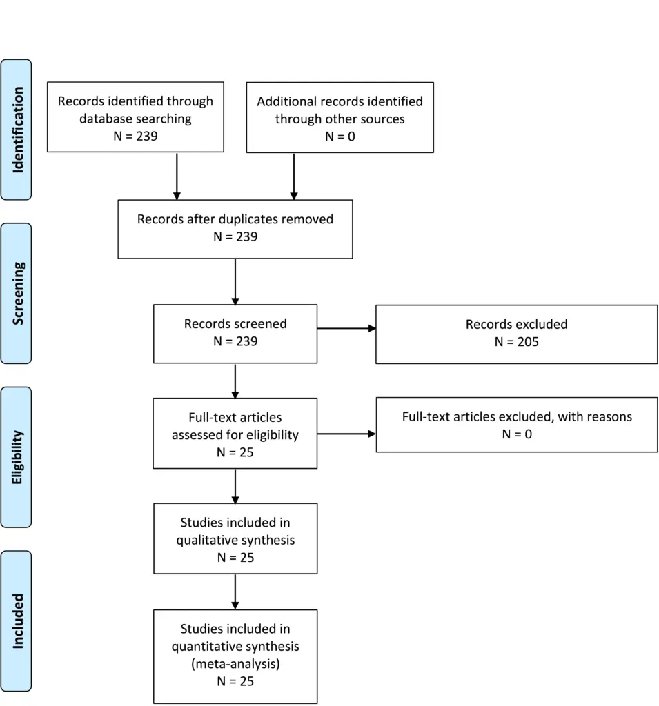
Question 2-2.
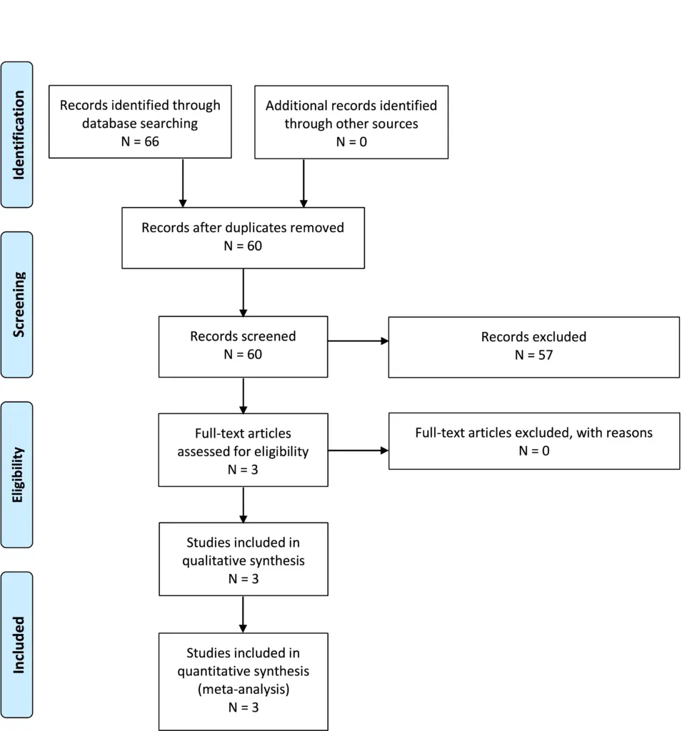
Question 2-3.
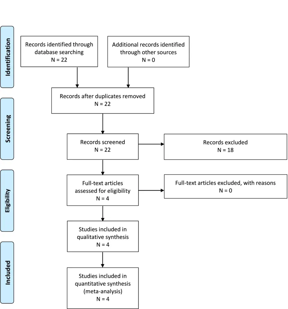
Question 2-4.
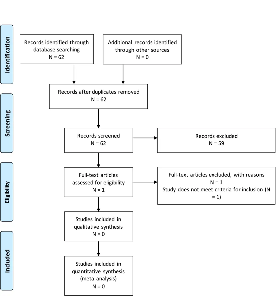
Question 2-5.
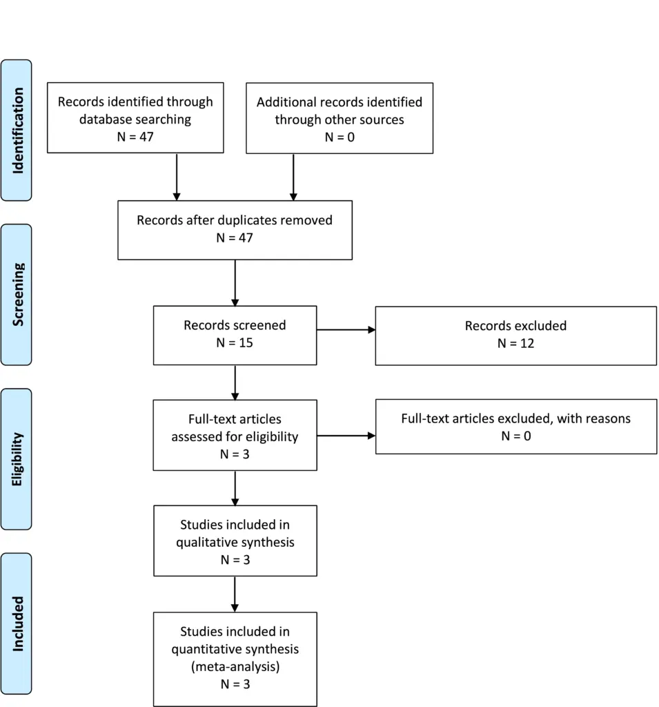
Appendix IV. Evidence Tables
| PICO | Author, Year | Literature Type | Study Design | Class of Evidence | Author Conclusions |
|---|---|---|---|---|---|
| 1 | Massimi et al, 20199 | Patient assessment | Retrospective case series | III | Retrospective review of 42 children with CIM and symptoms including headache (81%), neck pain (40%), vertigo (40%), ataxia (26%), and upper and lower extremity paraesthesia (26%). Resolution and significant improvement of preoperative symptoms was achieved in 36.5% and 21.5% after bone-only decompression, respectively |
| 1 | Ma et al, 201210 | Patient assessment | Retrospective case series | III | Retrospective review of 76 pediatric and adult patients with neurologic symptoms and combined structural diagnoses of CIM and syringomyelia, or either alone. In this mixed surgical population 80% of patients improved, with 16% stabilized and 4% worsened; 98% of patients with syringomyelia improved or stabilized while expansion occurred in 2% |
| 1 | Yates et al, 202028 | Patient assessment | Retrospective comparative | III | Retrospective review for 65 pediatric patients with CIM who underwent PFD. Important differences were found between very young patients aged ≤6 and children ages 7-18 years; very young children scored significantly lower on the CCOS, returned to the operating room more frequently for revision surgery, and presented differently with more common oropharyngeal and motoric symptomatology |
| 1 | Straus et al 200911 | Diagnostic test | Retrospective case series | III | Retrospective review of a subpopulation of patient with CIM and drop attacks with a negative cardiac workup for syncope, tilt table test, and surgical decompression. Ten patients met inclusion criteria and half had a positive tilt test. Following PFD, 7/10 patients improved, and the tilt test accuracy was determined to be 40% with a recommendation that it has poor value predicting clinical response in Chiari drop attack patients |
| 1 | Kumar et al, 200212 | Patient assessment | Retrospective case series | III | Retrospective review of 77 patients with CIM who were evaluated for dizziness, hearing loss, and tinnitus at a vestibular laboratory. Following a battery of vestibular tests, surgical decompression was performed on 33 patients. Surgery-dependent improvement in SNHL and vestibular dysfunction was not strictly reported but the conclusions are that this battery may help guide neurosurgeons for a subset of patients |
| 1 | Greenlee et al, 200229 | Patient assessment | Retrospective case series | III | Retrospective review of 31 children <6 years of age with CIM and symptoms including oropharyngeal function (35%), scoliosis (23%), headache or neck pain (23%), sensory disturbance (6%), weakness (3%), and other (10%). Preoperative symptoms resolved in 31%, improved in 42%, and were unchanged in 27% after PFD |
| 1 | Almotairi et al, 202031 | Patient assessment | Prospective case series | III | Prospectively reviewed 11 adult patients with CIM who underwent pre- and postoperative neuropsychiatric and QOL testing. CIM patients reported lower life satisfaction compared with normal control subjects before and after surgery; however, visual analogue and descriptive responses indicated that their QOL was significantly improved after surgery. Surgery also improved responses in dimensions testing in executive functioning, verbal learning, psychomotor speed, and color naming speed |
| 1 | Albert et al, 201027 | Patient assessment | Retrospective case series | III | Retrospective review of 39 CIM patients aged <6 years identifying age-dependent symptomatology upon presentation. Early recognition and treatment by different surgical approaches lead to good outcomes, including complete resolution of gastroesophageal reflux, and significant amelioration of headache |
| 1 | Beretta et al, 201713 | Patient assessment | Retrospective case series | III | Retrospective review of 135 patients assessing headache improvement after Chiari decompression, noting improvement in 93% with typical headache and 85% of patients with atypical headache |
| 1 | Spena et al, 201014 | Patient assessment | Retrospective case series | III | Retrospective review of 39 CIM patients who presented with variable symptoms. Headache symptoms improved in 80% of patients, whereas neuropathic pain or motor weakness responded less frequently to treatment |
| 1 | Dones et al, 20037 | Patient assessment | Retrospective case series | III | Retrospective review of 27 patients treated for CIM over a 9-year period with variable improvement in symptoms. Some differences in symptoms that improved pending presence of syrinx |
| 1 | Kumar et al, 201915 | Patient assessment | Retrospective case series | III | Retrospective review of 30 CIM patients postoperatively for clinical features and peak flow velocity. Overall, 8 (25.6%) of the 32 patients had complete resolution of their symptoms, 18 (56.25%) patients reported partial resolution, and 6 reported no improvement in their symptoms. Headaches responded the best with 16 (88.9%) patients noted some improvement in their headache; 6 had complete resolution, 5 had decreased frequency, 2 were able to control their headache with over-the counter analgesics, and 4 (including 1 of the patients with decreased headache frequency) had resolution of some, but not all, headache subtypes. Other symptoms that were alleviated postoperatively included upper and lower extremity sensory changes (n = 7), neck pain (n = 4), dizziness/vertigo (n = 5), visual symptoms (n = 1), and dysphagia (n = 2). Two patients did not report any improvement in symptoms within 1 year of follow-up. Patients who did not report an immediate clinical improvement continued to experience headaches (n = 2), neck pain/stiffness (n = 2), dizziness (n = 1), and photophobia (n = 1). No patient had a worsening of symptomatology following surgery |
| 1 | Jia et al, 201916 | Patient assessment | Retrospective case series | III | Retrospective review of 115 adult CIM patients underwent PFD or PFDD with tonsillar resection. Symptoms were grouped into pain, dysesthesias, motor weakness, and gait ataxia. Overall, symptoms were reported improved in 83-88%, unchanged in 9-13%, and worse in 3%. Pain improved the most and there were not significant differences between procedures |
| 1 | Chavez et al, 201417 | Patient assessment | Retrospective case series | III | Retrospective review of 177 CIM patients, 109 treated surgically and 68 conservatively. Risk factors for clinical improvement were identified and a propensity score defined. The propensity score-adjusted odds for overall improvement was 16.5 times higher for improvement with surgery than patients managed conservatively. Cough headache, migraine or other headache, paresthesia, and ataxia were most highly predictive |
| 1 | Parker et al, 201318 | Patient assessment | Prospective case series | III | One-year longitudinal cohort study of 50 CIM patients at a single institution. Headache severity improved in 37 patients (74%), remained the same in 11 (22%), and worsened in 2 (4%).Twenty patients (40%) presented with syringomyelia, 19/20 (95%) had postoperative MRI; 12/19 patients (63%; 4 cervical, 8 thoracic) demonstrated improvement in syrinx size, whereas the remaining 7 patients (37%; 3 cervical, 4 thoracic) showed no change in syringomyelia. Twelve patients (60%) had improved myelopathy. Baseline ventriculomegaly improved in 1 of 3 patients (33.3%) |
| 1 | McGirt et al, 200519 | Patient assessment | Retrospective case series | III | Retrospective review of 38 patients with headache alone and CIM. Seventeen patients underwent surgical decompression with 7 reporting frontal and 10 reporting occipital headaches. Radiographic CSF obstruction at the foramen magnum was present in 2/7 frontal headaches and 10/10 occipital headache patients. At follow-up 12 months after surgery, 4 (57%) of 7 patients with frontal headaches experienced recurrent headaches versus none (0%) of 10 patients originally presenting with occipital headaches. In the frontal headache group, the 2 patients with obstructed CSF flow had no headache recurrence at follow-up. Furthermore, decompressive treatment failed in 4 (80%) of the 5 patients with nonobstructed flow. Regardless of the degree of tonsillar ectopia, occipital headaches were strongly associated with hindbrain CSF flow abnormalities, whereas frontal and generalized headaches were not. Normal magnetic resonance imaging-cine CSF flow in the setting of a Chiari I malformation and frontal headaches alone suggests that frontal headaches are not pathologically or causatively associated with the Chiari I malformation in most patients. Frontal headaches with obstructed flow may respond to surgery |
| 1 | Raza-Knight et al, 201720 | Patient assessment | Retrospective case series | III | Retrospective review of 102 CIM patients, 57 (55.9%) presented with headache. Forty-two of 57 (73.7%) were classified as CIM headache, and 32/39 with 3-month follow-up sustained improvement. Duraplasty improved headaches in 32/38 (84.2%) patients receiving such therapy compared with 9/16 (56.3%) treated by bone-only decompression |
| 1 | Hayhurst et al, 200821 | Patient assessment | Retrospective case series | III | Retrospective review of 96 patients with average follow-up 3.6 years. Postoperative resolution or improvement in symptoms was seen in 75 patients (78%). Drop attacks and headaches were the most likely to respond to hindbrain decompression, showing improvement or resolution in 100% and 92% of cases. Dysaesthetic arm pain and weakness carried the worse prognosis with only 20% having symptom resolution. Sixteen patients had only bony decompression leaving the dura intact. In 8 patients (66%), headaches resolved following bony decompression alone but unchanged in 25% of cases. Dysaesthetic pain and weakness were unchanged in 60%. Restoration of CSF flow dynamics at the foramen magnum by surgical decompression does not consistently result in resolution of symptoms in all patients |
| 1 | Tisell et al, 200922 | Patient assessment | Retrospective case series | III | Retrospective review of 24 consecutive patients who were contacted about long-term follow-up after Chiari decompression. Seventy-five percent noted an improvement in headache, and 88% noted an improvement in associated neurologic symptoms |
| 1 | Khatwa et al, 201343 | Patient assessment | Retrospective case series | III | Retrospective review of 22 children with CIM who underwent PFD and polysomnography studies. Diagnoses included central sleep apnea (3), obstructive sleep apnea (5), and both obstructive and central sleep apnea (1). Children with sleep-disordered breathing had excessive crowding of the brainstem structures at the foramen magnum and were more likely to have a greater length of herniation compared with those children without sleep-disordered breathing (p = .046). Patients with central sleep apneas received surgical decompression, and their conditions were significantly improved on follow-up polysomnography |
| 1 | De Vlieger et al, 201923 | Patient assessment | Retrospective case series | III | Retrospective review of 79 CIM patients who were interviewed about symptom improvement following surgery. Fifty-four patients (68%) reported at least some improvement, 46 (58%) important improvement, 13 (16%) worsening, and 12 stabilization (15%). Any improvement as well as important improvement were significantly more often reported in the nonsyringomyelia group (85% vs 57%, p = .01 and 76% vs 46%, p = .01, respectively). Forty-five of 59 (76%) patients with headaches reported some improvement with 4 (7%) worsening. Sixty-two patients (78%) were satisfied or very satisfied with the results of surgery and 8 (11%) were unsatisfied or very unsatisfied. Up to 71 patients (90%) would consent to surgery again |
| 1 | Grangeon et al, 20188 | Patient assessment | Retrospective case series | III | Retrospective review of 49 CM1 patients and preoperative headaches. Clinical predictors for patients achieving >50% decrease in headache days included duration <5 min, occipital location, associated with Valsalva maneuver, severe intensity, and greater number of headaches per month; there were no predictive radiological factors. Postoperative improvement was inversely correlated with the Chiari severity index |
| 1 | Liu et al 201924 | Patient assessment | Retrospective Case series | III | Retrospective review of 39 patients following PFD. Overall, 24 (61.5%) showed improvement and 15 (38.5%) showed no improvement. Symptoms of motor weakness, lower limb symptoms, muscular atrophy, dizziness, and gait instability were not likely to improve, whereas headache and pain symptoms were likely to improve |
| 1 | Caldarelli et al, 200725 | Patient assessment | Retrospective case series | III | Retrospective review of 30 CIM pediatric patients who underwent bone-only PFD, although 11 patients also had serial incision of the outer layer of dura. The most frequent symptoms and signs were head and/or neck pain (56.7%), followed by vertigo (27.7%), upper and lower-extremity weakness (20.0%), and ataxia (20.0%). Improvement or resolution in symptoms was found in all patients without any change in tonsillar position and syrinx reduction in half the cases |
| 1 | McGirt et al, 200826 | Patient assessment | Retrospective case series | III | Retrospective review of 256 patients over 10 years to identify predictors of persistence of symptoms. 192 (75%) had headaches and 68 (27%) had brainstem or cranial nerve symptoms. Fifty-seven patients (22%) experienced mild to moderate symptom recurrence and this was less likely in patients who were treated with concurrent brainstem or cranial nerve involvement. Vertigo and frontal headache were more likely associated with symptom recurrence, and length of time for headache preceding treatment increased symptom recurrence by 15% per year |
| 2 | Chavez et al, 201417 | Patient assessment/therapy | Retrospective case series | III | Retrospective mixed population study for patients evaluated from 2000-2011 with long-term follow-up. The population was approximately 30% pediatric (<18 years of age). One hundred nine patients had surgery with 236 managed nonoperatively. Of those, 78 were able to be contacted and consented to long-term follow-up questions. Of those, 10 were excluded including 8 that had surgery at an outside institution. Of the 68 remaining patients, they calculated that 73% (50/68) of their patients treated without surgery demonstrated stability or improvement in symptoms over an average of 4.9 years |
| 2 | Pomeraniec et al, 201633 | Patient assessment | Retrospective case series | III | Retrospective review of 95 pediatric CIM patients managed conservatively (70) or with PFD with dural splitting or duraplasty (25). Seventy-five percent of operated patients had significant improvement in clinical symptoms. At the same time, 92.9% of nonoperatively-treated patients did not show progression and 41.7% of those patients showed improvement in symptoms without intervention. There were no differences between the surgical groups |
| 2 | Carey et al, 202132 | Patient assessment/therapy | Retrospective case series | III | Retrospective study of 226 pediatric patients seen for initial consultation for Chiari malformation over a 5-year period. The study includes patients with symptoms and patients with syrinx (26). Thirty-four were managed surgically and 192 patients were managed nonoperatively. Fifteen of 34 had surgery greater than 6 months after the initial consultation, 3 due to new symptoms and 1 due to new syrinx. Of those patients, 3 had a Chiari decompression and 1 had a shunt placed. As the study was not limited to asymptomatic patients without, they also had 2 patients with worsening symptoms, 1 patient had symptoms that failed to improve and 2 had worsening syrinx. No patients had surgery >2 years after initial consultation |
| 3 | Freeman et al, 201041 | Patient assessment | Retrospective case control | III | Retrospective study comparing scans of 1200 patients with neck pain including 600 with trauma and 600 without trauma. They identified a statistically significantly increased percentage of patients with cerebellar tonsillar ectopia in the group with trauma |
| 3 | Wan et al, 200840 | Patient assessment | Retrospective case series | III | Retrospective series of 85 patients over 21 years. The authors note that minor head or neck trauma can precipitate the onset of symptoms in a small number of previously asymptomatic patients with CIM |
| 3 | Meehan et al 201539 | Patient assessment | Retrospective comparative | III | Retrospective cohort study over 3 years in 147 patients with an average tonsillar ectopia of 11mm. Most patients exhibited pegged tonsils and crowding at the foramen magnum. Results demonstrated no deaths, coma, or paralysis in 1627 athletic seasons, indicating that the risk of injury is low |
| 3 | Strahle et al, 201638 | Patient assessment | Prospective comparative | II | Prospective dual site survey study of 503 patients including 328 sports participants. There was no difference in disease severity in both sports participants and non-sports participants. Respondents played a wide variety of high impact sports and over 4641 seasons there were no catastrophic or permanent neurologic injuries |
| 5 | Provenzano et al, 202047 | Patient assessment | Prospective case series | III | Prospective series of patients where they performed whole-exome sequencing of 51 unrelated patients with Chiari malformation. They also tested parents. They noted abnormalities in chromatin remodeling genes. They also found that many patients had parents with the same gene mutation who also were noted to have Chiari malformation on further testing. The authors conclude that in most cases CIM is a dominant, Mendelian inherited trait |
| 5 | Tubbs et al, 200446 | Patient assessment | Retrospective case series | III | Retrospective evaluation of 2 groups, 1 with CIM who underwent PFD for symptoms, and the second group is patients observed in clinic with NF-1. 5.4% of 130 patients in group 1 also had NF-1. 8.6% of 198 patients in group 2 with NF-1 also had posterior fossa decompression for symptoms. The authors conclude that there is an association between CIM and NF-1 |
| 5 | Milhorat et al, 199945 | Patient assessment | Prospective case series | III | A prospective cohort of 364 symptomatic patients and 50 patients and 50 age- and gender-matched control subjects underwent posterior fossa volumetric analysis using the Cavalieri methodology. Families of 21 patients participated in a study of familial aggregation. There were 275 female and 89 male patients. Age of symptom onset was 24.9 years, and 89 patients cited an immediate history of trauma. Forty-three (12%) patients had a family history of CIM or syringomyelia. The authors conclude that symptoms are associated with a volumetric reduction in CSF while brain volumes may be normal, and that tonsillar ectopia <5 mm does not obviate similar symptomatology. They conclude there is demonstration of familial aggregation that suggests a genetic component |
CCOS, Chicago Chiari Outcome Scale; CIM, Chiari type I malformation; CSF, cerebrospinal fluid; NF-1, neurofibromatosis type 1; PFD, posterior fossa decompression; PFDD, posterior fossa decompression with duraplasty; SNHL, sensorineural hearing loss; QOL, quality of life.
Appendix V. Conflicts of interest
| Name | Affiliation | Type of COI |
|---|---|---|
| Toba Niazi, MD | Live Like Bella Foundation, Nicklaus Children’s Hospital | Grants/Research Support |
| Laurie Ackerman, MD | Park Reeves Consortium | Grants/Research Support |
| David Bauer, MD | None | |
| Brandon G. Rocque, MD, MS, FAANS | None | |
| Carolyn S. Quinsey, MD | None | |
| Eric Jackson, MD | None | |
| Jogi V. Pattisapu MD FAAP FAANS | J&J, Integra | Consultant |
| Rabia Qaiser, MD | None | |
| Cormac O. Maher, MD, FAAP, FACS, FAANS | None | |
| Shobhan H. Vachhrajani MD, PhD, FRCSC | None | |
| Libby Infinger, MD | None | |
| Howard Silberstein, MD | None | |
| Sarah Jernigan, MD | None | |
| Jeffrey S. Raskin MS MD FAANS FAAP | None | |
| Dorothy Poppe | None | |
| Kaitlyn Esposito, MPH | None |
Congress of Neurological Surgeons Systematic Review and Evidence-Based Guidelines for Chiari Malformation: Surgical Interventions
Sponsored by: Congress of Neurological Surgeons (CNS) and the Section on Pediatrics
Endorsed by: The Congress of Neurological Surgeons (CNS), American Association of Neurological Surgeons (AANS) and the Bobby Jones Chiari & Syringomyelia Foundation (Bobby Jones CSF)
Authors:
Jogi V. Pattisapu MD FAAP FACS FAANS1, Laurie L Ackerman, MD2, Libby Kosnik Infinger, MD, MPH3, Cormac O. Maher, MD, FAAP, FACS, FAANS4, Carolyn Quinsey, MD5, Brandon G. Rocque, MD, MS6, Howard Silberstein, MD7, Eric M. Jackson, MD8, Sarah Jernigan, MD, MPH9, Toba Niazi, MD10, Rabia Qaiser, MD11, Jeffrey S. Raskin MS MD12, Shobhan Vachhrajani MD, PhD, FRCSC13, David F. Bauer, MD, MPH14
Departmental and institutional affiliations:
- Pediatric Neurosurgery, University of Central Florida College of Medicine, Orlando FL
- Department of Neurological Surgery, Indiana University Health, Indianapolis, IN
- Department of Neurosurgery, Medical University of South Carolina (MUSC), Charleston, SC
- Department of Neurosurgery, Stanford Medicine, Palo Alto, CA
- Department of Neurosurgery, University of North Carolina Chapel Hill, Chapel Hill, NC
- Division of Pediatric Neurosurgery, Department of Neurosurgery, University of Alabama at Birmingham, Birmingham, AL
- Department of Neurosurgery, University of Rochester School of Medicine and Dentistry, Rochester, NY
- Department of Neurosurgery, Johns Hopkins University School of Medicine, Baltimore, MD
- Carolina Neurosurgery & Spine Associates, Charlotte, NC
- Department of Neurological Surgery, Nicklaus Children’s Hospital, Miami, FL
- Department of Neurological Surgery, Indiana University School of Medicine, Indianapolis, IN
- Department of Neurological Surgery, Northwestern University Feinberg School of Medicine, Chicago, IL
- Department of Pediatrics, Wright State University Boonshoft School of Medicine, Dayton, OH
- Department of Neurosurgery, Baylor College of Medicine, Division of Pediatric Neurosurgery, Texas Children’s Hospital, Houston, TX
Corresponding Author contact information:
No part of this article has been published or submitted for publication elsewhere.
Keywords:
Abbreviations: Chiari malformation type I, CIM; Chicago Chiari Outcome Scale, CCOS; intraoperative brainstem auditory evoked potentials, BAEPs; intraoperative neuromonitoring, IONM; posterior fossa decompression, PFD; posterior fossa decompression with duraplasty, PFDD; somatosensory evoked potentials, SSEPs
ABSTRACT
Background: Chiari malformation type I (CIM) diagnoses have increased in recent years. Patients may develop delayed symptoms or syringomyelia requiring surgical intervention, while mild symptoms can be managed conservatively. Controversy regarding the best operative management prompted a review of literature to offer guidance on surgical interventions.
Objective: This evidence-based clinical practice guidelines assessed literature to determine: 1) whether posterior fossa decompression or posterior fossa decompression with duraplasty is more effective in preoperative symptom resolution; 2) if there is benefit from cerebellar tonsillar resection/reduction; 3) the role of intraoperative neuromonitoring; 4) in patients with a syrinx, how long should a syrinx be observed for improvement before additional surgery is performed; and 5) what is the optimal duration of follow-up care after preoperative symptom resolution.
Methods: A systematic review was performed using the National Library of Medicine/PubMed and Embase databases for studies on CIM in children and adults. The most appropriate surgical interventions, the use of neuromonitoring, and clinical improvement during follow-up were reviewed for studies published between 1946 and January 23, 2021.
Results: A total of 80 studies met inclusion criteria and there was no Class I evidence in the literature. Posterior fossa decompression with or without duraplasty or cerebellar tonsil reduction all appeared to show some benefit for symptom relief and syrinx reduction. There was insufficient evidence to determine if duraplasty or cerebellar tonsil reduction was needed for specific patient groups. There was no strong correlation between symptom relief and syringomyelia resolution. Many surgeons follow patients for 6 to 12 months before considering reoperation for persistent syringomyelia. No benefit or harm was seen with the use of neuromonitoring.
Conclusion: This evidence-based clinical guidelines for the treatment of CIM provide 1 Class II and 4 Class III recommendations. In patients with CIM with or without syringomyelia, treatment options include bone decompression with or without duraplasty or cerebellar tonsil reduction. Improved syrinx resolution may potentially be seen with dural patch grafting. Symptom resolution and syrinx resolution did not correlate directly. Reoperation for a persistent syrinx was potentially beneficial if the syrinx had not improved 6 to 12 months after the initial operation.
RECOMMENDATIONS
3-1. In patients with CIM (with or without syrinx), what type of surgery most often improves preoperative symptoms: posterior fossa decompression (PFD) or posterior fossa decompression with duraplasty (PFDD)?
Recommendation: In patients with symptomatic CM1 malformation (with or without syrinx), either posterior fossa decompression (PFD) or posterior fossa decompression with duraplasty (PFDD) may be utilized as a first line treatment to improve pre-operative symptoms.
Strength of recommendation: Grade C
Class III evidence
3-2. In patients with CIM, is cerebellar tonsil reduction beneficial?
Recommendation: In patients undergoing posterior fossa decompression surgery for treatment of CIM and syrinx, surgeons may perform resection or reduction of cerebellar tonsil tissue to improve syrinx and/or symptoms.
Strength of recommendation: Grade C
Class III evidence
3-3. Is there a role for intraoperative neuromonitoring in patients undergoing decompression for Chiari I malformation?
Recommendation: Intraoperative neuromonitoring in patients undergoing decompression for CIM may be used during surgery.
Strength of recommendation: Grade C
Class III evidence
3-4. In patients with CIM and syrinx, how long should you wait to evaluate for syrinx reduction prior to performing additional surgery?
Recommendation: Surgeons may perform additional neurosurgical intervention 6 to 12 months following surgical treatment of CIM with syringomyelia in patients that have not demonstrated radiographic improvement.
Strength of recommendation: Grade B
Class II evidence
3-5. In surgically treated patients with CIM who have improved, is long term follow up needed?
Recommendation: Patients undergoing surgery for CIM who experience symptom resolution may be monitored for symptom or imaging changes.
Strength of recommendation: Grade C
Class III evidence
INTRODUCTION
Goals and Rationale
Approximately 0.24% to 2.6% of the population,1–5 including children and adults, is affected by Chiari malformation type I (CIM). CIM is defined as descent of the cerebellar tonsils ≥3 to 5 mm below the foramen magnum. Not all patients are symptomatic, and there are various ways to diagnose and treat patients with CIM. CIM may cause syringomyelia, and some patients with CIM may have craniocervical instability requiring decompression and/or fusion of the craniocervical junction. Symptoms result from blockage of the flow of cerebrospinal fluid (CSF) or from compression of the brainstem or cranial nerves. In some cases, other neurologic or orthopedic conditions are also present, which can make diagnosis or management challenging for clinicians and their patients.
This guideline was developed to determine the most appropriate diagnostic and surgical management in adult and pediatric patients with CIM (with or without syrinx), based on current literature. Procedures likely to improve preoperative symptoms or clinical findings were reviewed, as well as ancillary interventions, including cerebellar tonsil reduction or intraoperative neuromonitoring (IONM). Timing for reimaging or repeat intervention is also included to inform long term assessment and follow-up.
This guideline is intended to help improve patient care by outlining appropriate information gathering and decision-making processes involved in the treatment of patients with CIM. Surgical care is provided by different clinicians, and they were created as an educational tool to navigate through a series of diagnostic and treatment decisions to this condition.
The ultimate surgical judgment regarding any specific procedure or treatment must be made in view of the patient’s presenting circumstances. Therefore, the guidelines should not be construed as all-inclusive or excluding any reasonable methods directed towards obtaining the same results.
METHODOLOGY
The guidelines task force initiated a systematic review of the evidence-based literature relevant to the treatment of patients with CIM. Through objective evaluation of the evidence and transparency in the process of making recommendations, these evidence-based clinical practice guidelines were developed for the diagnosis and treatment of patients with CIM, mainly as an educational resource to assist practitioners during clinical decision-making processes. Additional information about the methods used in this systematic review is provided below.
Literature Search
Task force members identified search terms/parameter and a medical librarian implemented the literature search, consistent with the literature search protocol (see Appendix I), using the National Library of Medicine/PubMed database and Embase for the period from 1946 to January 23, 2021 using the search strategies provided in Appendix I.
Inclusion/Exclusion Criteria
Articles were retrieved and included only if they met specific inclusion/exclusion criteria. To reduce bias, these criteria were specified before conducting the literature searches.
Articles that do not meet the following criteria were, for the purposes of this evidence-based clinical practice guideline, excluded. To be included as evidence in the guideline, an article had to be a report of a study that:
- Investigated patients with CIM;
- Studies that enrolled ≥80% of CIM (studies with mixed patient populations were included if results were separately reported for each group/patient population);
- Was a full article report of a clinical study;
- Was not a medical records review, meeting abstract, historical article, editorial, letter, or a commentary;
- Appeared in a peer-reviewed publication or a registry report;
- Enrolled a minimum of 10 patients;
- Was of humans;
- Was published between 1946 and January 23, 2021;
- Quantitatively presented results;
- Was not an in vitro study;
- Was not a biomechanical study;
- Was not performed on cadavers;
- Was published in English;
- Was not a systematic review, meta-analysis, or guideline developed by others1
Systematic reviews or meta-analyses conducted by others, or guidelines developed by others were not included as evidence to support this review due to the differences in article inclusion/exclusion criteria compared to those criteria specified by the Guidelines Task Force. Although these articles were not included as evidence to support the review, they were recalled for full-text discussion and conduct manual searches of the bibliographies.
Assessment for Risk of Bias
The methodological quality of randomized controlled trials and the risk of bias were assessed using the following 6 criteria:
1. Sequence generation (Was the allocation sequence adequately generated?)
2. Allocation concealment (Was allocation adequately concealed such that it could not be foretold?)
3. Blinding (Were participants, treatment providers and/or outcome assessors blinded to the treatment allocations?)
4. Incomplete reporting of data (Were incomplete outcome data adequately addressed?)
5. Selective reporting of outcomes (Were all the outcomes specified reported?)
6. Other potential threats to validity (Was the randomized controlled trial free of other issues that could put it at a high risk of bias?)
In the case of nonrandomized observational evidence, potential threats to the validity of the data were assessed by examining for:
1. Bias due to selective case choice for study and selective result reporting,
2. Bias due lack or loss of information over time,
3. The biases of the interpreting investigator regarding the study
4. Publication bias regarding positive studies or positive cases
5. Misclassification
6. Survivorship bias
7. Publication bias
8. Recognition that in data collected in a retrospective or prospective manner correlation does not imply causation
9. Election bias
10. Attrition bias
11. Bias of change in methods over time
12. Ascertainment bias
Rating Quality of Evidence
The quality of evidence was rated using an evidence hierarchy for each of 4 different study types; therapeutic, prognostic, diagnostic, and decision modeling. These hierarchies are shown in Appendix II: Rating Evidence Quality. Additional information regarding the hierarchy classification of evidence can be located here: https://www.cns.org/guidelines/guideline-procedures-policies/guideline-development-methodology.
Revision Plans
In accordance with the Institute of Medicine’s standards for developing clinical practice guidelines and criteria specified by the National Guideline Clearinghouse, the task force will monitor related publications following the release of this document and will revise the entire document and/or specific sections “if new evidence shows that a recommended intervention causes previously unknown substantial harm; that a new intervention is significantly superior to a previously recommended intervention from an efficacy or harms perspective; or that a recommendation can be applied to new populations.”6 In addition, the task force will confirm within five years from the date of publication that the content reflects current clinical practice and the available technologies for the evaluation and treatment for patients with CIM.
RESULTS
The literature search yielded 760 abstracts. Task force members reviewed all abstracts yielded from the literature search and identified articles for full-text review and extraction to address the clinical questions, in accordance with the literature search protocol (Appendix I). Task force members identified the best research evidence available to answer the targeted clinical questions. When class I, II, and or III literature was available to answer specific questions, the task force did not review class IV studies.
Of the 760 abstracts, 680 did not meet inclusion criteria or were off-topic and the remaining 80 were selected for systematic review (Appendix III).
DISCUSSION
In general, extradural treatment of CIM consists of suboccipital decompression, C1 laminectomy, with or without resection of occipital cervical ligament with possible scoring of the dura. Intradural or “duraplasty” consists of the standard suboccipital decompression with C1 laminectomy, in addition to opening of the dura with potential reduction of the cerebellar tonsisl with cautery or resection, with possible exploration of fourth ventricle outflow, and finally duraplasty to expand the dura. Duraplasty can consist of either autograft or allograft.
Question 3-1. In patients with CIM (with or without syrinx), what type of surgery most often improves preoperative symptoms (syrinx, headache, etc): posterior fossa decompression (PFD) or posterior fossa decompression with duraplasty (PFDD)?
Recommendations: In patients with symptomatic CIM malformation (with or without syrinx), either PFD or PFDD may be used as a first-line treatment to improve preoperative symptoms.
Strength of recommendation: Grade C
Most studies (n = 17) met the criteria for class III evidence with 1 study meeting criteria for class II evidence. No studies met criteria for class I evidence.
An article by the Park-Reeves Syringomyelia consortium7 was published after the literature search was completed for this guideline and therefore was not included in the initial systematic review or guideline recommendations. Nevertheless, the results are pertinent and are included in this discussion. This study compared outcomes in 117 patients that had PFD to 575 patients that had PFDD. All patients had tonsil position ≥5 mm below the foramen magnum and a syrinx diameter >3 mm. The mean postoperative follow-up interval was 2.7 years, with a minimum of 1 year. PFDD was strongly associated with improved outcomes with respect to headache (89.6% vs 80.8%) and a significantly greater reduction in syrinx diameter (47.7% vs 26.9%), and had a higher rate of pseudomeningocele (7.7% vs 2.6%).
Class II Evidence
One class II study was identified. Pisapia et al8 performed a retrospective cohort study of 189 patients, comparing intradural and extradural PFDs. The surgical technique was selected by the surgeon and the extradural approach was associated with a high rate of occipital headache resolution compared with the intradural method. Otherwise, there was no difference in rates of symptom resolution or syrinx size based on these 2 surgical approaches. The intradural group had statistically significant more pseudomeningocele formation (18% vs 0%) and chemical meningitis (10% vs 0%). However, there was no difference in the need for reoperation in these patients.
Class III Evidence
Most authors included the degree of symptom resolutions, syrinx resolution, need for reoperation, and complication rates.
Symptom Resolution
Multiple authors reported an improvement in symptom improvement (such as headaches, neck pain, or visual complaints) in the PFDD over the PFD approach.9–11 Gurbuz et al9 found 93% improved with PFDD versus 50% with PFD if symptoms were <36 months’ duration. Gallo et al10 also reported better outcomes in the PFDD group, although the changes were not statistically significant. Yilmaz et al11 also reported a 56.3% recovery rate and 89.6% improvement with PFDD versus a 51.9% recovery rate and 79.1% improvement rate with PFD. Interestingly, Pandey et al12 and Shimoji et al13 found no significant differences between the groups; however, Pandey et al had several missing data points. Jiang et al14 found no significant difference in the Chicago Chiari Outcome Scale (CCOS) between PFD and PFDD in their small group. Butensky et al15 performed a large retrospective study and found no difference in symptom resolution, and other similar studies reported impressive symptom improvement rates by PFDD alone.16,17 A small, retrospective study found that patients with PFDD had a greater range of cervical motion compared with patients with PFD.18
Syrinx Resolution
Most articles identified improved syrinx resolution in patients undergoing PFDD versus PFD,9–12,15,19,20 although 1 series reported a slight advantage in PFD.13 Others found no difference with relatively small comparison groups.14,21 Gurbuz et al9 noted a syrinx regression rate of 92.3% with PFDD versus 12.5% with PFD. Gallo et al10 reported 92% improvement with PFDD versus 80% with PFD. Butensky et al15 found that 93% of syrinxes improved after PFDD versus 62% of the small number receiving PFD alone.15 Yilmaz et al11 reported 91.1% improvement with PFDD versus 84.2% with PFD alone. Interestingly Pandey et al12 noted the best results with PFD with dural splitting (86%) versus PFDD (60%) and Chotai et al17 documented a mean time to 50% resolution of syrinx in PFDD of 8 months. Although Jiang et al14 had a slightly higher rate of syrinx resolution after PFDD than PFD, the patient numbers in that series were too small to draw any conclusions.
Complications
Reported complications such as pseudomeningocele or CSF leak were more frequently cited with PFDD versus PFD.9–11,13–15,17,21,22 Gurbuz et al9 reported a 28.6% complication rate in PFDD versus 5.6% in PFD. Although not statistically significant, CSF leak, infection, and pseudomeningocele were more frequently seen in PFDD. Similarly, Jiang et al14 found that a higher percentage of patients with PFDD experienced CSF complications, while Shimoji et al13 reported 1 CSF leak and 2 pseudomeningocles in 11 PFDD operations. There were 10 complications in 85 cases reported by Chotai et al,17 although details were not readily available. In this series with PFD and PFDD, 3 patients had aseptic meningitis, 4 patients developed CSF leaks, 1 with hydrocephalus, and 2 had persistent headaches. Klekamp et al23 reported a complication rate of 21.8% in 371 patients undergoing PFDD, with CSF fistula in 5.5% of cases, aseptic meningitis in 4.3%, and hydrocephalus in 3.1%. This article also noted that severe arachnoid scarring tended to portend higher complication and reoperation rates. Yilmaz et al11 also reported a statistically significant increase in complications (wound infection, CSF leak, etc) in PFDD compared with PFD.
Need for Reoperation
Reoperations occurred more frequently in the PFD group, but this was not statistically significant.9,10,24 Gurbuz et al9 reported a 22.2% reoperation rate in PFD versus 14.3% in PFDD. Gallo et al10 reported 15% in his series and 6% in the PFDD group. Conversely, Shimoji et al13 noted 1 reoperation in 11 cases of PFDD group, and Klekamp23 reported 45 reoperations in his series of 371 PFDDs and noted severe arachnoid scarring as a reason for reoperation. Yilmaz et al11 reported a 3.6% rate of reoperation for CSF fistula in the PFDD group versus a 9.5% reoperation rate for inadequate decompression in the PFD group. The literature suggested mixed results with symptom or syrinx resolution, complications, and need for reoperations. Since there was no class I evidence to support the use of either PFD or PFDD, either approach may be used as a first-line treatment for CIM.
Question 3-2. In patients with CIM, is cerebellar tonsil reduction beneficial?
Recommendation: In patients undergoing PFD surgery for treatment of CIM and syrinx, surgeons may perform resection or reduction of cerebellar tonsil tissue to improve syrinx and/or symptoms.
Strength of recommendation: Grade C
Several retrospective studies and a single prospective study assessed outcomes after CIM decompression with or without manipulation of the cerebellar tonsils. All studies included for review considered syrinx resolution as the primary outcome, therefore an independent assessment of clinical symptom resolution was not possible. Accordingly, this recommendation included syringomyelia reduction as the outcome. Some studies reported better syrinx resolution with tonsil manipulation, while other studies reported higher complication rates with tonsil manipulation. Overall, there was not an obvious benefit or harm from tonsil manipulation apparent across studies.
Class III Evidence
There were 10 studies that directly compared CIM PFDD with and without manipulation of the cerebellar tonsils (including tonsil dissection, resection, cauterization, or some combination thereof). In 6 studies, there was no difference in outcomes with either option.25–30 In 3 studies,17,31,32 resolution of syringomyelia was more likely when tonsil manipulation was performed. Conversely, 1 study suggested patients who did not undergo tonsil manipulation had a higher likelihood of resolution of syringomyelia.33
Seven studies17,25,28,29,31–33 reported no difference in complication rates, regardless of tonsillar manipulation, while 3 studies reported higher complication rates with tonsil manipulation.26,27,30 In the single prospective and nonrandomized study, no difference in outcomes or complications was reported.29
Question 3-3. Is there a role for IONM in patients undergoing decompression for CIM?
Recommendation: IONM in patients undergoing decompression for CIM may be used during surgery.
Strength of recommendation: Grade C
All studies met the criteria for class III evidence. None met the criteria for class II or I evidence.
Class III Evidence
Anderson et al34 performed a retrospective study to determine if intraoperative brainstem auditory evoked potentials (BAEPs) changes might determine the extent of decompression necessary and if changes occurred during operative positioning. They reported that conduction through the brainstem improves substantially after bony opening, but only marginally after dural opening. The authors suggested both BAEPs and somatosensory evoked potentials (SSEPs) might identify early changes during operative positioning and may help prevent adverse outcomes. In support of these findings, Barzilai et al35 found that IONM can be useful in PFD, particularly during patient positioning. Class III evidence supports the use of IONM, but it is not necessary in patients undergoing decompression of a CIM.
Anderson et al36 went on to perform a IONM prospective study to determine which patients may respond to bony decompression alone. The authors performed a continuous study of intraoperative BAEPs in patients undergoing PFDD and compared conduction at 3 points: 1) at baseline when patient is supine before positioning, 2) immediately after bony decompression and release of the atlanto-occipital membrane, and 3) after dural opening. They concluded that significant improvement in brainstem conduction immediately occurs after bone decompression and division of the atlanto-occipital membrane, rather than dural opening.
Data from Zamel et al37 further support this finding of intraoperative BAEP changes during different surgical stages of CIM repair. They reviewed variations during different aspects of surgery and correlated them with clinical or radiologic findings. In both groups (with or without syringomyelia) the predominant improvement in central conduction occurred during the period of bony decompression without significant additional improvement after the duraplasty.
Another group38 reviewed their experience with IONM during pediatric CIM surgery and did not identify a definite correlation between clinical outcomes or syrinx improvement.
Subtle nonsignificant changes in SSEP/MEPs during suboccipital decompression (without clinical correlation) were reported in adults by Roser et al.39 The authors felt that IONM is not considered a prerequisite for a safe suboccipital decompression when surgery is performed by an experienced team. In support of these findings, Barzilai et al35 found that IONM can be useful in CIM surgery, particularly during patient positioning.
Class III evidence supports the use of IONM, but it is not necessary in patients undergoing decompression of a CIM.
Question 3-4. In patients with CIM and syrinx, how long should you wait to evaluate for syrinx reduction before performing additional surgery?
Recommendation: Surgeons may perform additional neurosurgical intervention for 6 to 12 months after surgical treatment of CIM with syringomyelia in patients that have not demonstrated radiographic improvement.
Strength of recommendation: Grade B
The decision to reoperate on a patient with CIM and syringomyelia will require consideration of multiple factors (such as presenting clinical symptoms/signs or radiographic evidence of syrinx improvement/resolution).
Reported timetables for observing postoperative syrinx improvement by MRI imaging vary widely. While some studies report syrinx improvement as early as 3 to 4 months, most studies suggest follow-up for 6 to 7 months; the remaining studies report further syrinx resolution by 1 year in 80% of cases. Beyond 12 months, patients will likely experience late stability, resolution, or expansion of their syrinx (often without symptom changes).
Numerous retrospective publications that discussed syringomyelia reduction and resolution after neurosurgical intervention were included. The degree of granularity and emphasis on this radiographic outcome varied among these reports.
Hale et al40 reported on time to syrinx resolution comparing PFD and PFDD approaches. A similar study evaluating use of tonsillar coagulation was done by Stanko et al.32 El-Ghandour41 reported a group of patients treated with a variety of approaches: PFD, PFD with fourth ventricle stent, and patients treated with syrinx shunting. Soleman et al42 reported syrinx reduction times (mean 3.4 months) for 21 patients with CIM and syringomyelia treated by syrinx shunting.
The remaining 16 retrospective studies17,32,43–58 reported times for syrinx response to surgical PFD procedures. They did not compare differences in surgical techniques.
Available evidence suggests additional neurosurgical interventions after surgical treatment of patients with CIM with syringomyelia may be needed for patients that have not demonstrated radiographic improvement within 6 to 12 months postsurgery.
Question 3-5. In surgically treated patients with CIM who have clinically improved, is long-term follow-up needed?
Recommendation: Patients undergoing surgery for CIM who experience symptom resolution may be monitored for symptom or imaging changes.
Strength of recommendation: Grade C
Neurosurgeons routinely follow patients with CIM postoperatively at varied scheduled intervals within 12 months, according to their practice routine. Long-term follow-up (follow-up >1 year) varies widely.
Many reports included patients who initially improve after CIM surgery but who require another operative intervention for recurrence of symptoms or a syrinx years later.
Twenty-six publications8,17,51,52,57–78 reported long-term follow-up and clinical status after Chiari decompression surgery. All are retrospective case series, with inconsistent long-term criteria for reoperation or intervention. It is unclear if planned long-term follow-up or an unscheduled return for symptom evaluation yield different reoperation rates or clinical outcomes.
In these retrospective studies, patients were evaluated clinically or for syrinx recurrence, but do not include details for the repeat assessment (ie, routine/scheduled long-term follow-up or return of symptoms). Five studies found significantly elevated reoperation rates (12-50%), mostly occurring after 6 to 12 months.59,62,67,75,76 Nine studies noted a 1.1% to 9% reoperation rate for symptoms or imaging findings, consistent with previous reports,8,17,58,60,64,65,68,71,73 and 8 studies had long-term follow-up without clinical indication for reoperation.61,63,66,70,72,74,77,78 Two studies reported patient deterioration or syrinx progression after 1 year; however, patients did not undergo surgery.51,52
Two studies identified long-term scoliosis progression referred for surgical management; however, it was unclear if the initial neurosurgeon or scoliosis physician performed the evaluations.69,73
Future Directions
This systematic review of the literature highlighted variations in clinical practices. No specific recommendations can be made on the best surgical approach, the need for dural patch grafting, or cerebellar tonsillar resection. There is significant need for prospective studies to evaluate the use of duraplasty and cerebellar tonsil reduction to determine which patients may potentially benefit from these practices. In most instances, symptoms and syrinx improved within 6 to 12 months of a successful operation. Some clinicians follow patients for 6 to 12 months, and other clinicians follow for much longer. Further research into the age of a patient at initial decompression and the optimal duration of follow up care is needed. Recent data7–11 suggest better outcomes using a dural patch graft without increase in complication rates. Further studies are necessary to confirm these results, and more data are expected for the planned update of these guidelines in 5 years.
Future studies and collaborative efforts may offer more insights to improve our management approach. Patient-centered studies evaluating patient-reported outcomes may be helpful to inform future clinical decision making and recommendations. It is imperative to explore these questions to help improve care of our patients with CIM and syringomyelia.
CONCLUSIONS
Mostly class III and some class II evidence informed our recommendations for the treatment of CIM. Decompression with or without duraplasty and with or without cerebellar tonsil reduction seemed to provide symptom relief and syrinx resolution. Recent evidence (published after evidence review) suggests improved outcomes with duraplasty. Benefit of intraoperative monitoring was inconclusive, and it is common to wait 6 to 12 months for symptom or syrinx resolution before considering a repeat operation.
The guidelines offer a review of current evidence in the literature. We anticipate upcoming high-quality studies to develop revision of these guidelines to allow improved care for our patients with CIM.
Conflicts of Interest
All Guideline Task Force members were required to disclose all potential COIs prior to beginning work on the guideline, using the COI disclosure form of the AANS/CNS Joint Guidelines Review Committee. The CNS Guidelines Committee and Guideline Task Force Chair reviewed the disclosures and either approved or disapproved the nomination and participation on the task force. The CNS Guidelines Committee and Guideline Task Force Chair may approve nominations of task force members with possible conflicts and restrict the writing, reviewing, and/or voting privileges of that person to topics that are unrelated to the possible COIs. See Appendix V for a complete list of disclosures.
Disclosure of Funding
These evidence-based clinical practice guidelines were funded exclusively by the Congress of Neurological Surgeons, which received no funding from outside commercial sources to support the development of this document.
Disclaimer of Liability
This clinical systematic review and evidence-based guideline was developed by a physician volunteer task force as an educational tool that reflects the current state of knowledge at the time of completion. Each chapter is designed to provide an accurate review of the subject matter covered. This guideline is disseminated with the understanding that the recommendations by the authors and consultants who have collaborated in their development are not meant to replace the individualized care and treatment advice from a patient’s physician(s). If medical advice or assistance is required, the services of a competent physician should be sought. The proposals contained in these guidelines may not be suitable for use in all circumstances. The choice to implement any particular recommendation contained in these guidelines must be made by a managing physician in light of the situation in each particular patient and on the basis of existing resources.
Acknowledgments
The guidelines task force would like to acknowledge the CNS Guidelines Committee for their contributions throughout the development of the guideline, the AANS/CNS Joint Guidelines Review Committee, as well as the contributions of Kirsten Aquino, contracted project manager for the CNS, Trish Rehring, MPH, Associate Director for Evidence-Based Practice Initiatives for the CNS, and Janet Waters, MLS, BSN, RN, for assistance with the literature searches. The guidelines task force would also like to acknowledge the contributions of Dorothy Poppe, Kaitlyn Esposito, MPH and Mary Poppe, as well as the Bobby Jones Chiari and Syringomyelia Foundation for serving as patient advocates on this guideline task force1. Throughout the review process, the reviewers and authors were blinded from one another. At this time the guidelines task force would like to acknowledge the following individual peer reviewers for their contributions: Jennifer Sweet, MD, Andrew Carlson, MD, MS, Matthew Reynolds, MD, PhD, Alexandra D. Beier, D.O., FACOS, FAAP, Jonathan Pindrik, MD and Patti Raksin, MD.
1The guideline task force did not include systematic reviews, guidelines, or meta-analyses conducted by others. These documents are developed using different inclusion criteria than those specified in this guideline; therefore, they may include studies that do not meet the inclusion criteria specific to this guideline. In cases where these types of documents’ abstract suggested relevance to the guideline’s recommendations, the task force searched their bibliographies for additional studies.
REFERENCES
1. Aitken LA, Lindan CE, Sidney S, et al. Chiari type I malformation in a pediatric population. Pediatric Neurology. 2009;40(6):449-454.
2. Meadows J, Kraut M, Guarnieri M, Haroun RI, Carson BS. Asymptomatic Chiari Type I malformations identified on magnetic resonance imaging. Journal of neurosurgery. 2000;92(6):920-926.
3. Morris Z, Whiteley WN, Longstreth Jr WT, et al. Incidental findings on brain magnetic resonance imaging: systematic review and meta-analysis. BMJ (Online). 2009;339(7720):547-550.
4. Strahle J, Muraszko KM, Kapurch J, Bapuraj JR, Garton HJ, Maher CO. Chiari malformation type I and syrinx in children undergoing magnetic resonance imaging. Journal of neurosurgery Pediatrics. 2011;8(2):205-213.
5. Vernooij MW, Ikram MA, Tanghe HL, et al. Incidental findings on brain MRI in the general population. New England Journal of Medicine. 2007;357(18):1821-1828.
6. Ransohoff DF, M. Pignone, and H.C. Sox, . How to decide whether a clinical practice guideline is trustworthy. . JAMA. 2013;309(2):139-140.
7. Akbari SHA, Yahanda AT, Ackerman LL, et al. Complications and outcomes of posterior fossa decompression with duraplasty versus without duraplasty for pediatric patients with Chiari malformation type I and syringomyelia: a study from the Park-Reeves Syringomyelia Research Consortium. Journal of Neurosurgery: Pediatrics. 2022:1-13.
8. Pisapia JM, Merkow MB, Brewington D, et al. External validity of the chiari severity index and outcomes among pediatric chiari I patients treated with intra- or extra-Dural decompression. Child’s nervous system : ChNS : official journal of the International Society for Pediatric Neurosurgery. 2016;33(2):313-320.
9. Gurbuz MS, Karaaslan N, Caliskan T, Unal E, Berkman MZ. Comparison of the surgical results for foramen magnum decompression with and without duraplasty in Chiari malformation type 1. Turkish neurosurgery. 2015;25(3):419-424.
10. Gallo P, Sokol D, Kaliaperumal C, Kandasamy J. Comparison of three different cranio-cervical decompression procedures in children with Chiari malformation type I: does the surgical technique matter? Pediatric neurosurgery. 2017;52(5):289-297.
11. Yilmaz A, Kanat A, Musluman AM, et al. When is duraplasty required in the surgical treatment of Chiari malformation type I based on tonsillar descending grading scale? World neurosurgery. 2011;75(2):307-313.
12. Pandey S, Li L, Wan RH, Gao L, Xu W, Cui DM. A retrospective study on outcomes following posterior fossa decompression with dural splitting surgery in patients with Chiari type I malformation. Clinical neurology and neurosurgery. 2020;196:106035.
13. Shimoji K, Hara T, Ohara Y. Controversies related to pediatric Chiari I malformation. Child’s nervous system : ChNS : official journal of the International Society for Pediatric Neurosurgery. 2019;35(10):1695-1699.
14. Jiang E, Sha S, Yuan X, et al. Comparison of clinical and radiographic outcomes for posterior fossa decompression with and without duraplasty for treatment of pediatric Chiari I malformation: a prospective study. World neurosurgery. 2017;110:e465-e472.
15. Butensky S, Rodgers S, Baron S, Schneider S, Mittler M. Comparison of surgical outcomes in patients with Chiari type I malformation receiving posterior fossa decompression with and without duraplasty. Child’s nervous system : ChNS : official journal of the International Society for Pediatric Neurosurgery. 2020;36(7):1399-1405.
16. Bao CS, Liu L, Wang B, et al. Craniocervical decompression with duraplasty and cerebellar tonsillectomy as treatment for Chiari malformation-I complicated with syringomyelia. Genetics and molecular research : GMR. 2015;14(1):952-960.
17. Chotai S, Chan EW, Ladner TR, et al. Timing of syrinx reduction and stabilization after posterior fossa decompression for pediatric Chiari malformation type I. Journal of neurosurgery Pediatrics. 2020:1-7.
18. Yilmaz A, Urgun K, Aoun SG, et al. Adding expansile duraplasty to posterior fossa decompression may restore cervical range of motion in grade 3 Chiari malformation type 1 patients. World neurosurgery. 2016;98:98-103.
19. Ito K, Yamada M, Horiuchi T, Hongo K. Appropriate surgical procedures for Chiari type 1 malformation and associated syrinx based on radiological characteristics of the craniovertebral junction. Neurosurgical review. 2019;43(2):575-580.
20. Oral S, Yilmaz A, Kucuk A, Tumturk A, Menku A. Comparison of dural splitting and duraplasty in patients with Chiari type I malformation: relationship between tonsillo-dural distance and syrinx cavity. Turkish neurosurgery. 2019;29(2):229-236.
21. Chen J, Li Y, Wang T, et al. Comparison of posterior fossa decompression with and without duraplasty for the surgical treatment of Chiari malformation type I in adult patients: A retrospective analysis of 103 patients. Medicine. 2017;96(4):e5945.
22. Del Gaudio N, Vaz G, Duprez T, Raftopoulos C. Comparison of dural peeling versus duraplasty for surgical treatment of Chiari type I malformation: results and complications in a monocentric patients’ cohort. World neurosurgery. 2018;117:e595-e602.
23. Klekamp J. Surgical treatment of Chiari I malformation-analysis of intraoperative findings, complications, and outcome for 371 foramen magnum decompressions. Neurosurgery. 2012;71(2):365-380.
24. Grahovac G, Pundy T, Tomita T. Chiari type I malformation of infants and toddlers. Child’s nervous system : ChNS : official journal of the International Society for Pediatric Neurosurgery. 2018;34(6):1169-1176.
25. da Silva JA, Holanda MM. Basilar impression, Chiari malformation and syringomyelia: a retrospective study of 53 surgically treated patients. Arquivos de neuro-psiquiatria. 2003;61(2b):368-375.
26. Vidal CHF, Brainer-Lima AM, Valença MM, Farias RL. Chiari 1 malformation surgery: comparing non-violation of the arachnoid versus arachnoid opening and thermocoagulation of the tonsils. World neurosurgery. 2018;121:e605-e613.
27. Kunert P, Janowski M, Zakrzewska A, Marchel A. Comparison of results between two different techniques of cranio-cervical decompression in patients with Chiari I malformation. Neurologia i Neurochirurgia Polska. 2009;43(4):337-345.
28. Park YS, Kim DS, Shim KW, Kim JH, Choi JU. Factors contributing improvement of syringomyelia and surgical outcome in type I Chiari malformation. Child’s nervous system : ChNS : official journal of the International Society for Pediatric Neurosurgery. 2008;25(4):453-459.
29. Koueik J, Sandoval-Garcia C, Kestle JRW, et al. Outcomes in children undergoing posterior fossa decompression and duraplasty with and without tonsillar reduction for Chiari malformation type I and syringomyelia: A pilot prospective multicenter cohort study. Journal of Neurosurgery: Pediatrics. 2020;25(1):21-29.
30. Jia C, Li H, Wu J, et al. Comparison decompression by duraplasty or cerebellar tonsillectomy for Chiari malformation-I complicated with syringomyelia. Clinical neurology and neurosurgery. 2018;176:1-7.
31. Wang L, Zhao H, Zhu W, Yan P, Teng YD. A combinatorial approach with cerebellar tonsil suspension to treating symptomatic Chiari malformation type I in adults: a retrospective study. World neurosurgery. 2020;143:e19-e35.
32. Stanko KM, Lee YM, Rios J, et al. Improvement of syrinx resolution after tonsillar cautery in pediatric patients with Chiari type I malformation. Journal of neurosurgery Pediatrics. 2015;17(2):174-181.
33. Asgari S, Engelhorn T, Bschor M, Sandalcioglu IE, Stolke D. Surgical prognosis in hindbrain related syringomyelia. Acta neurologica Scandinavica. 2003;107(1):12-21.
34. Anderson RC, Dowling KC, Feldstein NA, Emerson RG. Chiari I malformation: potential role for intraoperative electrophysiologic monitoring. Journal of clinical neurophysiology : official publication of the American Electroencephalographic Society. 2003;20(1):65-72.
35. Barzilai O, Roth J, Korn A, Constantini S. The value of multimodality intraoperative neurophysiological monitoring in treating pediatric Chiari malformation type I. Acta neurochirurgica. 2015;158(2):335-340.
36. Anderson RC, Emerson RG, Dowling KC, Feldstein NA. Improvement in brainstem auditory evoked potentials after suboccipital decompression in patients with Chiari I malformations. Journal of neurosurgery. 2003;98(3):459-464.
37. Zamel K, Galloway G, Kosnik EJ, Raslan M, Adeli A. Intraoperative neurophysiologic monitoring in 80 patients with Chiari I malformation: role of duraplasty. Journal of clinical neurophysiology : official publication of the American Electroencephalographic Society. 2009;26(2):70-75.
38. Rasul FT, Matloob SA, Haliasos N, Jankovic I, Boyd S, Thompson DNP. Intraoperative neurophysiological monitoring in paediatric Chiari surgery-help or hindrance? Child’s nervous system : ChNS : official journal of the International Society for Pediatric Neurosurgery. 2019;35(10):1769-1776.
39. Roser F, Ebner FH, Liebsch M, Tatagiba MS, Naros G. The role of intraoperative neuromonitoring in adults with Chiari I malformation. Clinical neurology and neurosurgery. 2016;150:27-32.
40. Hale AT, Adelson PD, Albert GW, et al. Factors associated with syrinx size in pediatric patients treated for Chiari malformation type I and syringomyelia: a study from the Park-Reeves Syringomyelia Research Consortium. Journal of neurosurgery Pediatrics. 2020:1-11.
41. El-Ghandour NM. Long-term outcome of surgical management of adult Chiari I malformation. Neurosurgical review. 2012;35(4):537-546; discussion 546-537.
42. Soleman J, Roth J, Bartoli A, Rosenthal D, Korn A, Constantini S. Syringo-subarachnoid shunt for the treatment of persistent syringomyelia following decompression for Chiari type I malformation: surgical results. World neurosurgery. 2017;108:836-843.
43. Arnautovic KI, Qaladize BF, Pojskic M, Gienapp AJ, Splavski B, Boop FA. The 270° circumferential microsurgical decompression of the foramen magnum in adult Chiari malformation type I: single surgeon series of 130 patients with syringomyelia, neurologic, and headache outcomes. World neurosurgery. 2020.
44. Ghanem IB, Londono C, Delalande O, Dubousset JF. Chiari I malformation associated with syringomyelia and scoliosis. Spine. 1997;22(12):1313-1317; discussion 1318.
45. Spena G, Bernucci C, Garbossa D, Valfrè W, Versari P. Clinical and radiological outcome of craniocervical osteo-dural decompression for Chiari I-associated syringomyelia. Neurosurgical review. 2010;33(3):297-303; discussion 303-294.
46. Kemerdere R, Akgun MY, Cetintas SC, Kacira T, Tanriverdi T. Clinical and radiological outcomes of arachnoid-preseving suboccipital decompression for adult chiari I malformation with and without syringomyelia. Clinical neurology and neurosurgery. 2019;188:105598.
47. Kennedy BC, Nelp TB, Kelly KM, et al. Delayed resolution of syrinx after posterior fossa decompression without dural opening in children with Chiari malformation type I. Journal of neurosurgery Pediatrics. 2015;16(5):599-606.
48. Nagoshi N, Iwanami A, Toyama Y, Nakamura M. Factors contributing to improvement of syringomyelia after foramen magnum decompression for Chiari type I malformation. Journal of orthopaedic science : official journal of the Japanese Orthopaedic Association. 2014;19(3):418-423.
49. Isu T, Sasaki H, Takamura H, Kobayashi N. Foramen magnum decompression with removal of the outer layer of the dura as treatment for syringomyelia occurring with Chiari I malformation. Neurosurgery. 1993;33(5):845-849; discussion 849-850.
50. Raftopoulos C, Sanchez A, Matos C, Baleriaux D, Bank WO, Brotchi J. Hydrosyringomyelia-Chiari I complex. Prospective evaluation of a modified foramen magnum decompression procedure: preliminary results. Surgical Neurology. 1993;39(2):163-169.
51. Alfieri A, Pinna G. Long-term results after posterior fossa decompression in syringomyelia with adult Chiari type I malformation. Journal of neurosurgery Spine. 2012;17(5):381-387.
52. Attenello FJ, McGirt MJ, Gathinji M, et al. Outcome of Chiari-associated syringomyelia after hindbrain decompression in children: analysis of 49 consecutive cases. Neurosurgery. 2008;62(6):1307-1313; discussion 1313.
53. Takayasu M, Takagi T, Hara M, Anzai M. A simple technique for expansive suboccipital cranioplasty following foramen magnum decompression for the treatment of syringomyelia associated with Chiari I malformation. Neurosurgical review. 2004;27(3):173-177.
54. Bao C, Yang F, Liu L, et al. Surgical treatment of Chiari I malformation complicated with syringomyelia. Experimental and therapeutic medicine. 2012;5(1):333-337.
55. Wu T, Zhu Z, Jiang J, et al. Syrinx resolution after posterior fossa decompression in patients with scoliosis secondary to Chiari malformation type I. European spine journal : official publication of the European Spine Society, the European Spinal Deformity Society, and the European Section of the Cervical Spine Research Society. 2011;21(6):1143-1150.
56. Xie D, Qiu Y, Sha S, et al. Syrinx resolution is correlated with the upward shifting of cerebellar tonsil following posterior fossa decompression in pediatric patients with Chiari malformation type I. European spine journal : official publication of the European Spine Society, the European Spinal Deformity Society, and the European Section of the Cervical Spine Research Society. 2014;24(1):155-161.
57. Wetjen NM, Heiss JD, Oldfield EH. Time course of syringomyelia resolution following decompression of Chiari malformation type I. Journal of neurosurgery Pediatrics. 2008;1(2):118-123.
58. Hidalgo ET, Dastagirzada Y, Orillac C, et al. Time to resolution of symptoms after suboccipital decompression with duraplasty in children with Chiari malformation type I. World neurosurgery. 2018;117:e544-e551.
59. Paul KS, Lye RH, Strang FA, Dutton J. Arnold-Chiari malformation. Review of 71 cases. Journal of neurosurgery. 1983;58(2):183-187.
60. Zhang ZQ, Chen YQ, Chen YA, Wu X, Wang YB, Li XG. Chiari I malformation associated with syringomyelia: a retrospective study of 316 surgically treated patients. Spinal cord. 2007;46(5):358-363.
61. Ene CI, Wang AC, Collins KL, et al. Expansile duraplasty and obex exploration compared with bone-only decompression for Chiari malformation type I in children: retrospective review of outcomes and complications. Journal of neurosurgery Pediatrics. 2020:1-8.
62. Krishna V, McLawhorn M, Kosnik-Infinger L, Patel S. High long-term symptomatic recurrence rates after Chiari-1 decompression without dural opening: a single center experience. Clinical neurology and neurosurgery. 2014;118:53-58.
63. Tubbs RS, Beckman J, Naftel RP, et al. Institutional experience with 500 cases of surgically treated pediatric Chiari malformation Type I. Journal of neurosurgery Pediatrics. 2011;7(3):248-256.
64. Balestrino A, Consales A, Pavanello M, et al. Management: opinions from different centers-the Istituto Giannina Gaslini experience. Child’s nervous system : ChNS : official journal of the International Society for Pediatric Neurosurgery. 2019;35(10):1905-1909.
65. De Vlieger J, Dejaegher J, Van Calenbergh F. Multidimensional, patient-reported outcome after posterior fossa decompression in 79 patients with Chiari malformation type I. Surgical neurology international. 2020;10:242.
66. Pomeraniec IJ, Ksendzovsky A, Awad AJ, Fezeu F, Jane JA, Jr. Natural and surgical history of Chiari malformation type I in the pediatric population. Journal of neurosurgery Pediatrics. 2015;17(3):343-352.
67. Klekamp J. Neurological deterioration after foramen magnum decompression for Chiari malformation type I: old or new pathology? Journal of neurosurgery Pediatrics. 2012;10(6):538-547.
68. Kennedy BC, Kelly KM, Phan MQ, et al. Outcomes after suboccipital decompression without dural opening in children with Chiari malformation Type I. Journal of neurosurgery Pediatrics. 2015;16(2):150-158.
69. Flynn JM, Sodha S, Lou JE, et al. Predictors of progression of scoliosis after decompression of an Arnold Chiari I malformation. Spine. 2004;29(3):286-292.
70. Feghali J, Xie Y, Chen Y, Li S, Huang J. The SHORE score: a novel predictive tool for improvement after decompression surgery in adult Chiari malformation type I. World neurosurgery. 2020;142:e195-e202.
71. Attenello FJ, McGirt MJ, Garcés-Ambrossi GL, Chaichana KL, Carson B, Jallo GI. Suboccipital decompression for Chiari I malformation: outcome comparison of duraplasty with expanded polytetrafluoroethylene dural substitute versus pericranial autograft. Child’s nervous system : ChNS : official journal of the International Society for Pediatric Neurosurgery. 2008;25(2):183-190.
72. Gilmer HS, Xi M, Young SH. Surgical decompression for Chiari malformation type I: an age-based outcomes study based on the Chicago Chiari Outcome Scale. World neurosurgery. 2017;107:285-290.
73. Singhal GD, Singhal S, Agrawal G, Singhal D, Arora V. Surgical experience in pediatric patients with Chiari-I malformations aged ≤18 years. journal of neurosciences in rural practice. 2019;10(1):85-88.
74. Pepper J, Elhabal A, Tsermoulas G, Flint G. Symptom outcome after craniovertebral decompression for Chiari type 1 malformation without syringomyelia. Acta neurochirurgica. 2020;163(1):239-244.
75. Guyotat J, Bret P, Jouanneau E, Ricci AC, Lapras C. Syringomyelia associated with type I Chiari malformation. A 21-year retrospective study on 75 cases treated by foramen magnum decompression with a special emphasis on the value of tonsils resection. Acta neurochirurgica. 1998;140(8):745-754.
76. Soleman J, Bartoli A, Korn A, Constantini S, Roth J. Treatment failure of syringomyelia associated with Chiari I malformation following foramen magnum decompression: how should we proceed? Neurosurgical review. 2018;42(3):705-714.
77. Alzate JC, Kothbauer KF, Jallo GI, Epstein FJ. Treatment of Chiari I malformation in patients with and without syringomyelia: a consecutive series of 66 cases. Neurosurgical focus. 2006;11(1):E3.
78. Imperato A, Seneca V, Cioffi V, Colella G, Gangemi M. Treatment of Chiari malformation: who, when and how. Neurological sciences : official journal of the Italian Neurological Society and of the Italian Society of Clinical Neurophysiology. 2011;32 Suppl 3:S335-339.
Appendix I. Literature searches
See Chapter 1, Appendix I.
Appendix II. Rating evidence quality
Classification of Evidence on Therapeutic Effectiveness and Levels of Recommendation
| Class I evidence Level I (or A) recommendation | Evidence from one or more well-designed, randomized controlled clinical trial, including overviews of such trials. |
| Class II evidence Level II (or B) recommendation | Evidence from one or more well-designed comparative clinical studies, such as non-randomized cohort studies, case-control studies, and other comparable studies, including less well-designed randomized controlled trials. |
| Class III evidence Level III (or C) recommendation | Evidence from case series, comparative studies with historical controls, case reports, and expert opinion, as well as significantly flawed randomized controlled trials. |
Classification of Evidence on Prognosis and Levels of Recommendation
| Class I evidence Level I (or A) recommendation | All 5 technical criteria above are satisfied |
| Class II evidence Level II (or B) recommendation | Four of 5 technical criteria are satisfied |
| Class III evidence Level III (or C) recommendation | Everything else |
Classification of Evidence on Diagnosis and Levels of Recommendation
| Class I evidence Level I (or A) recommendation | Evidence provided by one or more well-designed clinical studies of a diverse population using a “gold standard” reference test in a blinded evaluation appropriate for the diagnostic applications and enabling the assessment of sensitivity, specificity, positive and negative predictive values, and, where applicable, likelihood ratios |
| Class II evidence Level II (or B) recommendation | Evidence provided by one or more well-designed clinical studies of a restricted population using a “gold standard” reference test in a blinded evaluation appropriate for the diagnostic applications and enabling the assessment of sensitivity, specificity, positive and negative predictive values, and, where applicable, likelihood ratios |
| Class III evidence Level III (or C) recommendation | Evidence provided by expert opinion or studies that do not meet the criteria for the delineation of sensitivity, specificity, positive and negative predictive values, and, where applicable, likelihood ratios |
Classification of Evidence on Clinical Assessment and Levels of Recommendation
| Class I evidence Level I (or A) recommendation | Evidence provided by one or more well-designed clinical studies in which interobserver and/or intraobserver reliability is represented by a kappa statistic >0.60 |
| Class II evidence Level II (or B) recommendation | Evidence provided by one or more well-designed clinical studies in which interobserver and/or intraobserver reliability is represented by a kappa statistic >0.40 |
| Class III evidence Level III (or C) recommendation | Evidence provided by one or more well-designed clinical studies in which interobserver and/or intraobserver reliability is represented by a kappa statistic <0.40 |
Appendix III. PRISMA flowcharts
Question 3-1
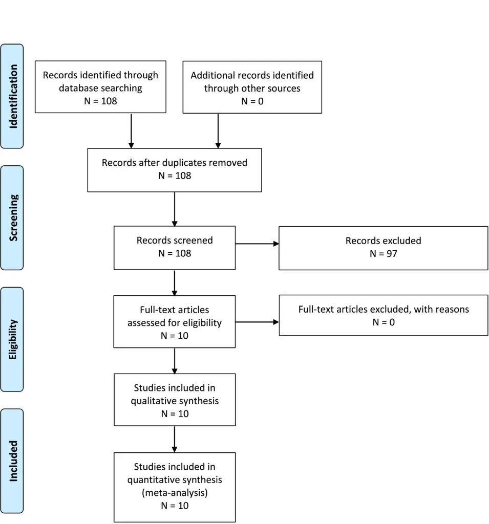
Question 3-2
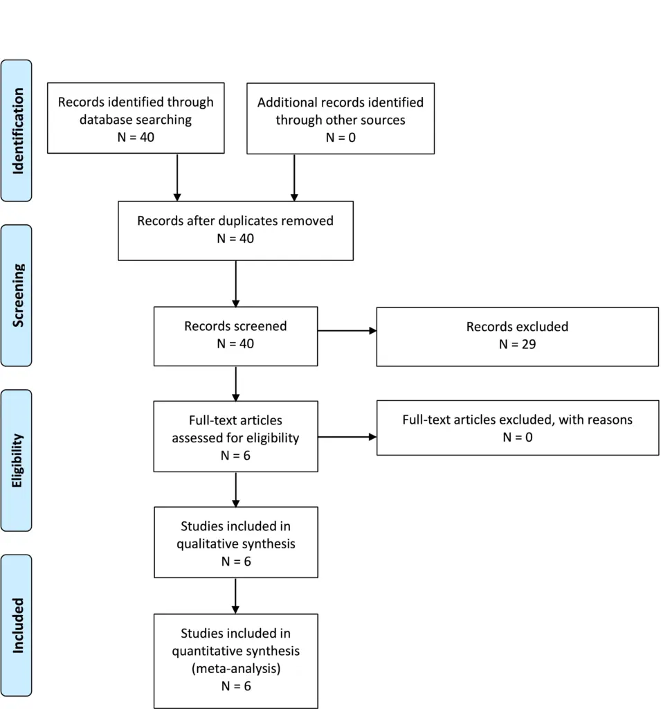
Question 3-3
Question 3-4
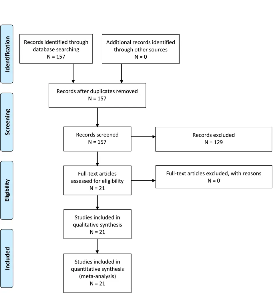
Question 3-5
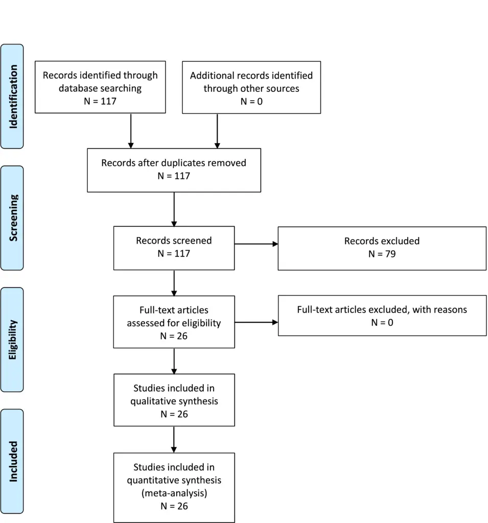
Appendix IV. Evidence tables
CI, confidence interval; CMD, circumferential microsurgical decompression; CSF, cerebrospinal fluid; CTC, cerebellar tonsil coagulation; CTS, cerebellar tonsil suspension; FMD, foramen magnum decompression; IQR, interquartile range; IOM, intraoperative monitoring; IONM, intraoperative neuromonitoring; MRI, magnetic resonance imaging; PFD, posterior fossa decompression; PFDD, posterior fossa decompression and duraplasty; PFDD-T, posterior fossa decompression and duraplasty with tonsillar resection; RCT, randomized controlled trial; SM, syringomyelia; SSS, syringo-subarachnoid shunt.
| PICO | Author | Literature Type | Description of the study | Class of Evidence | Author Conclusions |
|---|---|---|---|---|---|
| 1 | Yilmaz et al, 201718 | Therapy | Retrospective case series | III | Single-center retrospective review that showed there was no difference in outcome between PFD and PFDD, except in patients with severe tonsillar herniation who had an improvement in their cervical motility without compromising their spinal stability |
| 1 | Ito et al, 202019 | Therapy | Retrospective case series | III | This study shows increased complications with PFDD, and PFD is not as effective with less syrinx resolution and increased need for reoperation. However, very small subgroups were reported (n = 8 in the duraplasty group) |
| 1 | Grahovac et al, 201824 | Therapy | Retrospective comparative | III | In young patients (<3 years of age) with CIM, recurrence rates were higher after a PFD than after a PFDD. The authors reported that PFDD at primary or redo surgery provides for better decompression and long-term outcome. However, this was a small study (n = 16) |
| 1 | Jiang et al, 201814 | Therapy | Prospective comparative RCT | III | Found that PFD produces comparable radiologic and clinical outcomes and is associated with a lower risk of complications (higher incidence of CSF leak in the PFDD group) |
| 1 | Del Gaudio et al, 201822 | Therapy | Retrospective case series | III | Small numbers (n = 28) but concluded that PFD (with dural peeling) was less risk than PFDD but had a lower response rate (66.7% vs 100%) |
| 1 | Oral et al, 201920 | Therapy | Retrospective case control | III | Found that a syrinx cavity is more likely to regress in patients who undergo PFDD, but there are complications with a PFDD. Therefore, they concluded PFD should be initially preformed in patients with CIM and a small syrinx cavity, or those without syringomyelia |
| 1 | Chen et al, 201721 | Therapy | Retrospective case control | III | No statistically significant differences were found between PFD and PFD groups with regard to demographics, preoperative symptoms, radiographic characteristics, and clinical outcomes. However, postoperative aseptic meningitis occurred more frequently in the PFDD group than the PFD group |
| 1 | Butensky et al, 202015 | Therapy | Retrospective case control | III | Found that there is a role for PFDD in patients with severe syringomyelia, but overall PFD may be safely offered as the initial surgical intervention for symptomatic CIM patients |
| 1 | Gurbuz et al, 20159 | Therapy | Retrospective case control | III | Reported that in CIM cases with syringomyelia and tonsillar herniation >10 mm and whose symptoms lasted <36 months, PFDD is a more reliable choice than PFD despite a slightly higher rate of complications |
| 1 | Gallo et al, 201710 | Therapy | Retrospective case control | III | PFD has comparable clinical and radiologic outcomes to PFDD in children with CIM. But there was a higher risk of postoperative complications with PFDD that was not statistically significant |
| 1 | Shimoji et al, 201913 | Therapy | Retrospective case control | III | Small study (n = 26), but PFD was similar to PFDD in terms of the results of surgery and had a lower risk of complications for the treatment of noncomplicated CIM |
| 1 | Bao et al, 201516 | Therapy | Retrospective case series | III | Did not compare 2 techniques but did show that PFDD is a rational surgical approach with beneficial clinical effects |
| 1 | Pisapia et al, 20178 | Therapy | Retrospective comparative | II | Large study from 1 center. Concluded that equivalent rates of symptom resolution and reoperation after PFDD and PFD support the PFD approach as a first-line surgical option for pediatric CIM patients |
| 1 | Pandey et al, 202012 | Therapy | Retrospective case series | III | Showed more optimal syrinx resolution, shorter operative time, and shorter postoperative stay with the PFDD group. Both PFD and PFDD had similar outcome scores |
| 1 | Klekamp et al, 201223 | Therapy | Retrospective case series | III | Series of 371 patients only included PFDD, but it offered a favorable long-term prognosis. However, there was a high complication rate of 21.8%, permanent morbidity rate of 3/2%, and mortality rate of 1.3% |
| 1 | Chotai et al, 202017 | Therapy | Retrospective case series | III | Maximal reduction in syrinx size can be expected within 3 months after PFD in patients with SM and a syrinx; however, the syringes continue to regress over time. In patient with CIM and SM, the median time to >50% regression in maximal syrinx diameter was 8 months |
| 1 | Yilmaz et al, 201111 | Therapy | Retrospective comparative | III | In patients with tonsillar descent below C1, PFDD may lead to a more reliable decrease in SM and outcome scores, but with less severe tonsillar descent, PFD alone may be performed |
| 1 | da Silva et al, 200325 | Therapy | Case series | III | Included 53 patients 24 without tonsil manipulation. Symptom improvement was shown |
| 2 | Vidal et al, 201926 | Therapy | RCT | III | Included 32 patients, with 16 in each group. There were more complications in the tonsil coagulation group |
| 2 | Wang et al, 202031 | Therapy | Comparative | III | Included 42 patients, 12 of whom had PFDD, 13 had PFDD with coagulation and 17 had PFDD with coagulation and suspension. Patients who underwent PFDD + CTC + CTS improved more than comparison group |
| 2 | Kunert et al, 200927 | Therapy | Comparative | III | Included 38 patients with 6 months postoperative MRI. Results of PFD and PFDD are comparable, but the risk of post op complications after PFD were significantly lower |
| 2 | Park et al, 200928 | Therapy | Case series | III | Included 57 patients 40 of whom had PFD. Seventeen patients had PFD with tonsil management. There was no difference in outcomes |
| 2 | Stanko et al, 201632 | Therapy | Case series | III | Included 171 patients, 43 with tonsil cautery and 128 without. Syrinx resolution more likely with cautery, no increase in complications |
| 2 | Koueik et al, 202029 | Therapy | Comparative | III | Included 75 patients, and 42 had PFDD-T, while 26 had PFDD. This was a prospective, nonrandomized study and there was no difference in outcomes or complications |
| 2 | Chotai et al, 202017 | Therapy | Case series | III | Included 85 patients, 5 of whom had bone one only, 21 had tonsil cauterization and 15 had arachnoid veil transection. Resolution of syringomyelia was more likely when tonsil manipulation was performed |
| 2 | Jia et al, 201830 | Therapy | Retrospective comparative | III | In 115 adult patients, there were 37 with posterior fossa decompression with duraplasty and 78 who had duraplasty and cerebellar tonsil resection. There was no difference in symptom resolution or syrinx reduction between groups. There was higher complication rate for subjects who had tonsil resection |
| 2 | Asgari et al, 200333 | Therapy | Retrospective comparative | III | Nonrandomized. Technique difference was due to different preferences by different surgeons. No overlap |
| 3 | Anderson et al, 200334 | Therapy | Retrospective case series | III | There was no comparison |
| 3 | Anderson et al, 200336 | Therapy | Prospective case series | III | No comparison. Authors concluded that in pediatric patients the majority of improvement occurs after bone decompression and division of the atlanto-occipital membrane, rather than opening of the dura |
| 3 | Zamel et al 200937 | Therapy | Retrospective case series | III | No comparison. Data revealed that for both groups of patients, with or without associated syringomyelia, the predominant improvement in central conduction in most cases occurred during the period of bony decompression without significant additional improvement after the duraplasty procedure |
| 3 | Rasul et al, 201938 | Therapy | Retrospective case series | III | No comparison. Found no link between clinical outcomes and IONM, nor did syrinx outcome correlate with IONM |
| 3 | Roser et al, 201639 | Patient assessment | Retrospective case series | III | A 39-patient case series with no comparison group. All patients had IOM with CMD. Authors fail to show much if any effectiveness |
| 3 | Barzilai et al, 201635 | Diagnostic test | Retrospective case series | III | There was no comparison group |
| 4 | Arnautovic et al, 202043 | Patient assessment | Prospective case series | II | Reports all syrinx cases stable or reduced in size by 8 months. No patients in the syrinx subgroup required additional surgery. Of 43 patients with syrinx, 35 resolved and 8 improved. MRI was performed at 2, 6, and 12 months. The median time to resolving/improving syrinx was 4 months (IQR, 3-8 months) |
| 4 | Ghanem et al, 199744 | Patient assessment | Case series | III | In 11 of 12 patients, syrinxes improved by 1 year. The 1 case without radiographic improvement had clinical improvement. There were no reoperations for failure to improve syrinx |
| 4 | Spena et al, 201045 | Patient assessment | Retrospective case series | III | Postoperative MRI was performed at 3 and 6 months and then annually. All patients had at least 3 postoperative MRI scans. Syringomyelia improved in 29 patients (80.5%) and remained unchanged in 7 (19.4%). The mean time for syrinx reduction was 8 months (range, 6-26 months). |
| 4 | Kemerdere et al, 202046 | ? | Retrospective case series | III | Follow-up MRI performed at 3 and 12 months. There was a decrease in size in all syrinxes 3 months after surgery. Long-term follow-up showed persistent improvement in 16 of 21 patients (76.1%) |
| 4 | Kennedy et al, 201547 | ? | Retrospective case series | III | Fifty-seven patients with available images had mean radiographic follow-up of 32 months. Of these, 70% (40) demonstrated radiographic improvement on MRI |
| 4 | Hale et al, 202040 | ? | Retrospective case series | I | Kaplan-Meier analysis of time to syrinx resolution (<2 mm diameter) shows 50% resolution by slightly over 3 years. By 6 years, nearly all syringomyelia had improved |
| 4 | Nagoshi et al, 201448 | ? | Retrospective case series | III | Authors recommend a minimum of 12 months’ follow-up before considering additional intervention |
| 4 | Isu et al, 199349 | ? | Retrospective case series | III | In a series of 7 patients, 5 syrinx decreased over weeks and 2 decreased over months |
| 4 | Raftopoulos et al, 199350 | ? | Retrospective case series | III | MRI follow-up was performed 9 days, 2 months, 6 months after surgery, and then annually. By 2 months, all 8 patients had decreased syrinx volume. All were then stable or further decreased on subsequent imaging |
| 4 | Stanko et al, 201632 | ? | Retrospective case series | III | MRI images were obtained 3–6 months after surgery. If there was no improvement, imaging was repeated 3–4 months after the first postoperative scan. The mean time to syrinx improvement was 11 months. |
| 4 | El-Ghandour et al, 201241 | ? | Retrospective case series | III | Postoperative MRI performed every 3 months until syrinx resolution or until 18 months. The mean time to syrinx resolution was 7.4 months |
| 4 | Alfieri et al, 201251 | ? | Retrospective case series | II | MRI performed at 6 weeks and 6 months after surgery, and then annually. At 1 year, 29% had syrinx resolution; 57% had unchanged syrinx size. Results were similar at last follow-up |
| 4 | Attenello et al, 200852 | ? | Retrospective case series | II | Three-month postoperative MRI, usually repeated 12 to 18 months after surgery. The median time to radiographic improvement was 14 months after surgery |
| 4 | Takayasu et al, 200453 | ? | Retrospective case series | III | Postoperative MRI was performed between 1 week and 3 months after surgery. The second postoperative MRI was performed at 3, 4, and 7 months. Syrinx reduced in size in all patients by 7 months |
| 4 | Bao et al, 201354 | ? | Retrospective case series | III | One hundred forty-eight patients received MRI within 2 weeks after operation. Seventy-five patients (50.7%) had a reduced spinal syrinx |
| 4 | Wu et al, 201255 | ? | Retrospective case series | III | Follow-up MRI was conducted at 6 months and 2, 4, and 6 years postsurgery. Thirty-six of 44 patients (81.8%) had significant improvement within 6 months after surgery, and 97.7% (43/44) had significant improvement by the final follow-up |
| 4 | Xie et al, 201556 | ? | Retrospective case series | III | Of 87 patients, 31 cases who had both 6-month and a >12-month follow-up were selected to evaluate the time course of syrinx resolution. Most syrinx improvement noted by 6 months then slow improvement over the next several years |
| 4 | Wetjen et al, 200857 | ? | Retrospective case series | II | The median time to syrinx narrowing (>50% reduction in syrinx diameter) was 3.6 months (95% CI, 3-6.5 months) |
| 4 | Hidalgo et al, 201858 | ? | Retrospective case series | III | MRI was obtained at 3-, 6-, 12-, 18-, and 24-month intervals. The median time to syrinx improvement was 3 months (range, 3-72 months) |
| 4 | Chotai et al, 202017 | ? | Retrospective | II | Maximum syrinx regression was seen at 3 months but noted to improve for as long as 12 years |
| 4 | Soleman et al, 201742 | ? | Retrospective case series | III | The average time from FMD to SSS for patients in this study was 3 year ± 3 years (range, 134-4104 days) |
| 5 | Paul et al, 198359 | Therapy | Retrospective case series | III | Seventy-one patient series showed 21% symptom recurrence/deterioration after initial improvement 2-3 years out from surgery, no reoperation data |
| 5 | Zhang et al, 200860 | Therapy | Retrospective case series | III | Three hundred sixteen patient series with 7% syrinx enlargement at ≥2 years of follow-up |
| 5 | Ene et al, 202061 | Therapy | Retrospective case series | III | Two hundred seventy-six patients followed for symptom and syrinx resolution and scoliosis outcomes mean follow-up 35 months without reoperation data |
| 5 | Pisapia et al, 20178 | Therapy | Retrospective case series | III/II | One hundred eighty-nine patients followed up 1-75 months showed 8% reoperation. Unclear what findings for reoperation were found in short- vs. long-term follow-up |
| 5 | Krishna et al, 201462 | Therapy | Retrospective case series | III | Forty-seven patients showed 31.9% of patients had redo decompression (<1 year to 11 years), 22.9% reoperation for Chiari associated; 31.9% reoperation for recurrent symptoms at mean 2.6 years 1-11 postoperative from initial surgery FMD |
| 5 | Tubbs et al, 201163 | Therapy | Retrospective case series | III | Five hundred cases followed 2 months to 15 years, mean 5 years, 15 patients (3%) reoperated for syrinx or symptoms |
| 5 | Alfieri et al, 201251 | Therapy | Retrospective case series | III | One hundred nine patients median follow-up of 12.7 years, followed-up at 1 year and last follow-up for spinal sx, pain, cranial sx, and syrinx. Beyond 1 year at last follow-up, 2 syrinx expansions and 1 worsening pain without reoperation |
| 5 | Balestrino et al, 201964 | Therapy | Retrospective case series | III | One hundred seventy-two patients mean follow-up 5.1 years. Six patients had (3.5%) redo decompression without timeline details short vs. long |
| 5 | De Vlieger et al, 201965 | Therapy | Retrospective case series | III | Seventy-nine patients were interviewed, with median follow-up 5.8 years. 2 (2.5%) patients with syrinx recurrence at 2 years were reoperated |
| 5 | Pomeraniec et al, 201666 | Therapy | Retrospective case series | III | Mean follow-up 66 months, 25 surgical patients. There was no reoperation data |
| 5 | Klekamp et al, 201267 | Therapy | Retrospective case series | III | Of 371 patients, 45 (12%) had reoperations. Patients were followed long term 5 and 10 years with patient deterioration |
| 5 | Attenello et al, 200852 | Therapy | Retrospective case series | III | In 49 patients, all but 1 patient improved by 12 months. 1 patient did not go on to have additional surgery |
| 5 | Kennedy et al, 201568 | Therapy | Retrospective case series | III | Study included 156 patients with 32 months’ follow-up. Of 14 cases, 9% not improved by 22 months were reoperated on at 3-57 months |
| 5 | Flynn et al, 200469 | Therapy | Retrospective case series | III | Patients followed long-term for scoliosis progression after Chiari decompression. Patients were followed 2-15 years |
| 5 | Feghali et al, 202070 | Therapy | Retrospective case series | III | Of 149 patients, mean follow-up was 1.9 years. There was no reoperation for syrinx progression |
| 5 | Attenello et al, 200971 | Therapy | Retrospective case series | III | Of 67 patients, 4 (6%) patients required revision on average 16 months postoperatively |
| 5 | Gilmer et al, 201772 | Therapy | Retrospective case series | III | One hundred forty-four patients had a mean follow-up 27 months following symptoms. There was no reoperation data |
| 5 | Singhal et al, 201973 | Therapy | Retrospective case series | III | Of 50 patients, 8 (1.6%) patients were reoperated on at 24 months. Two patients referred for scoliosis surgery |
| 5 | Pepper et al, 202174 | Therapy | Retrospective case series | III | One hundred twenty-nine patients had at least 24 months average follow-up. There were no revision data |
| 5 | Guyotat et al, 199875 | Therapy | Retrospective case series | III | Of 75 patients, 27 (36%) reoperated patients in the follow-up timeframe |
| 5 | Wetjen et al, 200857 | Therapy | Retrospective case series | III | In 29 patients, 96% showed symptoms improved by 6 months, no deterioration >1 year |
| 5 | Hidalgo et al, 201858 | Therapy | Retrospective case series | III | In 105 patients, there were 12 (1.1%) reoperations at mean 8.9 months postoperatively (range, 1.3-77 months). Median time to syrinx improvement was 3 months (range, 3-72 months) |
| 5 | Chotai et al, 202017 | Therapy | Retrospective case series | III | In 85 patients, there was reoperation in 3 (3.5%) patients |
| 5 | Soleman et al, 201976 | Therapy | Retrospective case series | III | Of 48 patients, 24 (50%) patients had additional surgery at mean 21.4 months (range, 12 days to 34.9 months) |
| 5 | Alzate et al, 200177 | Therapy | Retrospective case series | III | Sixty-six patients had a mean follow- up of 24 months with no mention of reoperations |
| 5 | Imperato et al, 201178 | Therapy | Retrospective case series | III | Thirty-six patients were followed up to 18 months from surgery with no reoperations |
Appendix V. Conflicts of Interest
| Name | Affiliation | Type of COI |
|---|---|---|
| Toba Niazi, MD | Live Like Bella Foundation, Nicklaus Children’s Hospital | Grants/Research Support |
| Laurie Ackerman, MD | Park Reeves Consortium | Grants/Research Support |
| David Bauer, MD | None | |
| Brandon G. Rocque, MD, MS, FAANS | None | |
| Carolyn S. Quinsey, MD | None | |
| Eric Jackson, MD | None | |
| Jogi V. Pattisapu MD FAAP FAANS | J&J, Integra | Consultant |
| Rabia Qaiser, MD | None | |
| Cormac O. Maher, MD, FAAP, FACS, FAANS | None | |
| Shobhan H. Vachhrajani MD, PhD, FRCSC | None | |
| Libby Infinger, MD | None | |
| Howard Silberstein, MD | None | |
| Sarah Jernigan, MD | None | |
| Jeffrey S. Raskin MS MD FAANS FAAP | None | |
| Dorothy Poppe | None | |
| Kaitlyn Esposito, MPH | None |