






Part 1: Introduction and Methodology
Reposted with permission from ©AANS, 2014
J Neurosurg Pediatrics (Suppl) 14:3–7, 2014
AANS, 2014
(Original text of the guideline was edited to reflect the update. Please click here for the original publication.)
UPDATE
1Department of Neurological Surgery, Saint Louis University, St. Louis, Missouri, and 2Guidelines Department, Congress of Neurological Surgeons; Schaumburg, Illinois
This clinical systematic review of and evidence-based guidelines for the treatment of pediatric hydrocephalus were developed by a physician volunteer task force. They are provided as an educational tool based on an assessment of current scientific and clinical information as well as accepted approaches to treatment. They are not intended to be a fixed protocol, because some patients may require more or less treatment.
In Part 1, the authors introduce the reader to the complex topic of hydrocephalus and the lack of consensus on its appropriate treatment. The authors describe the development of the Pediatric Hydrocephalus Systematic Review and Evidence-Based Guidelines Task Force charged with reviewing the literature and recommending treatments for hydrocephalus, and they set out the basic methodology used throughout the specific topics covered in later chapters. (http://thejns.org/doi/abs/10.3171/2014.7.PEDS14321)
Keywords: hydrocephalus, systematic review, practice guidelines
Abbreviations used in this paper: AANS = American Association of Neurological Surgeons; CNS = Congress of Neurological Surgeons; JGC = Joint Guidelines Committee.
Pediatric hydrocephalus is the most common surgically correctable neurological problem in infants, children, and adolescents. It is estimated that hydrocephalus may occur with the frequency of 1 in every 500 children. There are multiple causes of this disorder: genetic causes such as X-linked aqueductal stenosis; other congenital causes including myelomeningocoele and Chiari malformation; and acquired causes such as intra-ventricular hemorrhage, trauma, tumors, and infection. The burden of disease is substantial: hydrocephalus can have an effect on development as well as an impact on overall quality of life. Children with hydrocephalus rep- resent a disproportionate share of all children admitted to hospitals.5 There are effective surgical interventions that can preserve and improve quality of life; however, these are not without side effects and failures. The need for improvements in surgical interventions is well understood. Patients, families, and treating physicians acknowledge that, while the problems of hydrocephalus appear simple, they are actually quite complex. Current treatment methods are insufficient, and there is currently little agreement on the “best” treatment, even among leading practitioners. Nevertheless, there is consensus that current management may result in “frequent complications, poor rate of shunt survival, and poor quality of life for patients leading to unsatisfactory outcomes.”8
Recent publications on readmission rates associated with pediatric hospitalization have only served to high- light the need for close scrutiny of current interventions. Ventriculoperitoneal shunt procedures account for only a small number of the total admissions analyzed, but they have the second highest readmission prevalence. As the focus on prevention of hospital readmission increases, this information further emphasizes the importance of discerning safe and effective treatments for hydrocephalus.1
This review was conducted to evaluate the best available evidence to aid clinicians and to guide clinical practice by determining the best options for the management of pediatric hydrocephalus. The details of specific topics selected will be discussed in the following papers, but, in general, in their analyses members of the Pediatric Hydrocephalus Systematic Review and Evidence-Based Guidelines Task Force looked at available treatments, the effects and complications of those treatments, and strategies to avoid and manage complications. In undertaking this review, we define the condition “hydrocephalus” for purposes of the literature search and use the term “pediatric” to specify the care of infants, children, and adolescents younger than 18 years of age. The scope of this inquiry will include patients with either congenital or acquired hydrocephalus, including communicating and noncommunicating hydrocephalus for which the causes are undetermined. This review was undertaken under the auspices of the Pediatric Section of the American Association of Neurological Surgeons (AANS) and the Congress of Neurological Surgeons (CNS).
The recommendations contained in this supplement to the Journal of Neurosurgery: Pediatrics deliberately eschew the use of expert opinion, relying strictly on information available in the literature. Studies have reported that expert opinions may not use evaluable evidence, if the papers containing that evidence do not support the “expert” point of view.2 Throughout the development of these guidelines, the Task Force used evidence-based methodologies and adhered to strict criteria that had been defined a priori as specified by the Institute of Medicine’s standards for conducting systematic reviews and clinical evidence-based guidelines, as well as the methodologies described below.
This effort was begun by a small study group that convened at the Pediatric Section Annual Meeting in Austin, Texas, in 2011. At that time the basic topics were considered, and over the course of several months these were further refined. The search strategies we used will be discussed in detail in a later section. Members of the Task Force involved in the creation of this document were recruited from a variety of institutions and subspecialty disciplines in an effort to have as broad a representation of opinions and expertise as possible. Pediatric neurosurgeons, their patients, and patients’ family members hope that someday hydrocephalus will be entirely preventable or curable. Until that time, efforts must continue to refine and improve treatment as well as the evaluation of the effectiveness of treatment.
The Task Force followed protocols established by the Joint Guidelines Committee (JGC) of the AANS and the CNS. A conscientious effort was also made to be sure that conflict of interest was avoided. Members who had published extensively in certain areas were mindfully as- signed to evaluate evidence in other topics. Every effort was made to ensure that the work product would be trans- parent and trustworthy.4
Methods
Process Overview
The Task Force and the Pediatric Section of the AANS/CNS conducted a systematic review of the literature relevant to the management of hydrocephalus in infants and children. Additional details of the systematic review are provided below. During the development process, the panel participated in a series of conference calls and meetings. Multiple iterations of the written review were conducted by individuals in the Task Force and various AANS/CNS committees (Fig. 1). In accordance with the Institute of Medicine’s recommended best practice of reviewing guidelines every 5 years, the Congress of Neurological Surgeons is committed to ensuring timeliness and accuracy for its guidelines. The authors performed a planned five-year review of the medical literature and subsequently updated the “Pediatric hydrocephalus: systematic literature review and evidence-based guidelines.”
Selection of Clinical Topics
The goals of this effort were to discern the most effective strategies for a variety of hydrocephalus-related problems, including acquired hydrocephalus of the pre- mature neonate. We also considered the use of technical adjuvants such as antibiotic-impregnated catheters, endoscopic placement of shunt catheters, electromagnetic guidance for shunt catheter placement, and ultrasound guidance for shunt catheter placement. It was hoped that these adjuvants would lead to improvements in outcome and a reduction in the frequency of revision.
Complications associated with ventriculoperitoneal shunts and endoscopic third ventriculostomies are known, and these interventions’ effects and long-term successes are useful to evaluate. Complications associated with infection are of particular significance. Therefore, the prevention and treatment of infection occupies a significant portion of the hydrocephalus literature. Finally, the correlation of ventricle size to outcome in a child is a source of great interest as an indicator of the success of the intervention.
Following the identification of hydrocephalus-related problems, the Task Force developed preliminary recommendations that were formatted similarly to the PICO (patients, interventions, comparisons, and outcomes) formula to aid in the determination of the overall scope of the review and the terminology used to formulate the literature search strategies described below.
Literature Search
The Task Force worked with a research librarian and methodologist to assist with the formulation of search terms and strategies used to search the US National Library of Medicine PubMed/MEDLINE database and the Cochrane Database of Systematic Reviews for relevant literature published between January 1966 and March 2012. Four to five Task Force members used the article inclusion/exclusion criteria described below to screen abstracts and provide a list of relevant articles for a full-text review. Each Task Force member was blinded to the lists of abstracts provided by others. Staff compiled all lists together for review and final approval by all Task Force members. The literature search was updated using the same search strategies to include studies published from March 2012 through November 2019.
The searches were supplemented with manual screenings of bibliographies from all retrieved articles. In addition, the bibliographies of potentially relevant systematic reviews were screened for potentially relevant articles. All literature identified either by searches of the electronic database or by manual searches were subject to the article inclusion/exclusion criteria listed below. Specific search strategies used by Task Force members are provided within the Methods sections of the topics evaluated later in this supplement.
Article Inclusion/Exclusion Criteria
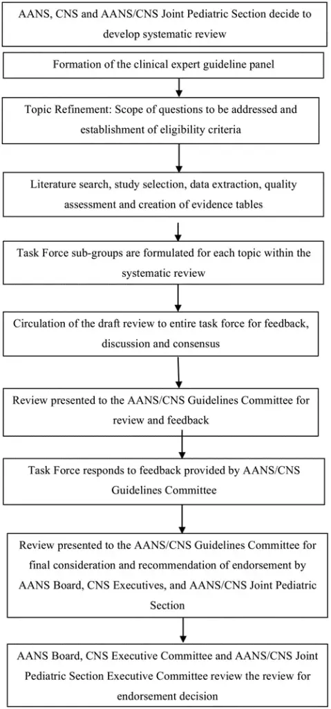
Fig. 1. Flowchart showing the overall development process.
Articles were retrieved and included as evidence to support the topics discussed in this review if they met specific inclusion/exclusion criteria. These criteria were also applied to articles provided by Task Force members who supplemented the electronic database searches with articles obtained from manual searches of bibliographies from the original articles. To reduce bias, the criteria were specified before conducting the literature searches. For the purposes of the systematic review and guidelines, articles that did not meet the following criteria were not deemed evidence and were not considered as potential evidence to support the topics and clinical recommendations.
To be included in our review, an article had to meet the following criteria:
- Studies that combined results in patients (younger than 18 years of age) who had congenital and acquired hydrocephalus with results in patients with “normal” pressure hydrocephalus were excluded if the study enrolled fewer than 80% of the target patient population
- Studies that enrolled mixed patient populations were included only if separate results were reported for the target The results of the target population were the only results considered as evidence to sup- port our recommendations.
- The study was a full article report of a clinical study.
- The study was not a meeting abstract, editorial, letter, or a commentary.
- Prospective case series had to report baseline values.
- Case series studies with nonconsecutive enrollment of patients were (This was determined by a review of the Methods section in the relevant article.)
- Studies had to have appeared in a peer-reviewed publication or a registry report.
- Studies had to enroll at least 10 patients for each distinct outcome that was measured. If a comparative study, a minimum enrollment of five patients per treatment arm for each outcome was necessary.
- The study involved humans.
- The study was published in or after 1966.
- The study presented results quantitatively.
- The study did not involve “in vitro” or “biomechanical” data or results obtained in cadavers.
- The study was published in English.
- Papers reporting the results of systematic reviews, meta-analyses, or guidelines developed by others were excluded.
Articles presenting systematic reviews or meta-analyses conducted by others, as well as guidelines developed by others, were not included as evidence to support this review due to differences in inclusion/exclusion criteria between those specified in such articles and those established by the Task Force. Although such articles were not included as evidence to support the review, they were re- called for full-text review so that the Task Force could conduct manual searches of the articles’ bibliographies.
Statistical Methods
For some topics, the available literature provided sufficient quality and quantity of data to allow more detailed statistical analysis going beyond the basic methods described in this paper. For those topics, including the ones shown in Part 64 and Part 7,3 the methods that were used will be described separately. In brief, a forest plot was created and a meta-analysis was conducted to determine the overall effect of the intervention, such as preoperative antibiotics, after the sources had been selected using evidence-based criteria, as described above.
Revision Plans
In accordance with the Institute of Medicine’s standards for developing clinical practice guidelines and criteria specified by the National Guideline Clearinghouse, following the release of this document the Task Force will monitor related publications and will revise the entire document and/or specific sections “if new evidence shows that a recommended intervention causes previously unknown substantial harm; that a new intervention is significantly superior to a previously recommended intervention from an efficacy or harms perspective; or that a recommendation can be applied to new populations.”6 In addition, the Task Force will conduct another review in 5 years following the date of publication of this updated version that the content is up to date with current clinical practice and available technologies for the treatment of pediatric hydrocephalus.
Rating the Quality of Evidence
The quality of evidence was rated using an evidence hierarchy developed by the AANS/CNS Guidelines Committee for each of the four different study types: therapeutic, diagnostic, prognostic, and clinical assessment. Additional information regarding the hierarchy classification of evidence can be located here: JGC Guideline Development Methodology (PDF)
Strength of the Recommendations Rating Scheme
The Task Force used methodologies endorsed by the AANS/CNS Guidelines Committee to assign a strength category to each recommendation in this review. Linking evidence to recommendations through the use of evidentiary tables has been endorsed by the American Medical Association, the AANS, and the CNS. This process validates and supports the relationship between the strength of evidence and the strength of recommendations. Demonstrating the highest degree of clinical certainty, Class I evidence is used to support recommendations of the strongest type, defined as Level I recommendations. Level II recommendations reflect a moderate degree of clinical certainty and are supported by Class II evidence or a strong consensus of Class III evidence. Level III recommendations denote clinical uncertainty, which is sup- ported by inconclusive or conflicting evidence or expert opinion.
Voting on the Recommendations
The Task Force used a structured voting technique to finalize and approve the final recommendations, language, and strength of the recommendations presented in this review. The voting technique is referred to as the “nominal group technique” and is described in an article by Murphy et al.5 This technique includes up to 3 rounds of voting using secret ballots to ensure that each Task Force member is blinded to the responses of other Task Force members. All the recommendations in this review were approved following the first round of voting and no further discussion was needed to finalize the following recommendations. During the course of editing and finalizing the document, changes were made to allow recommendations to conform to the rules of evidence and language as described earlier. When this occurred, the changes were reviewed and approved by the group.
Guideline Panel Consensus and Practice Guideline Approval Process
Topic subtask forces were created from the larger Task Force. Each subtask force took part in the literature selection, review of the literature, creation of the evidence tables, and creation and editing of the final review. The final draft review was then circulated to the entire Task Force for feedback, discussion, and, ultimately, approval. Following Task Force approval, the completed systematic review was presented to the JGC of the AANS and CNS for consideration and recommendation of endorsement on behalf of the CNS Executive Committee and the AANS Board of Directors. As part of the evaluation process, the JGC reviewers could provide input on the content and methodologies used to create the systematic review.
Development of this review was editorially independent from the funding agencies (the CNS Executive Committee and the AANS/CNS Joint Pediatric Section Executive Committee). The funding agencies’ review of these guideline papers, following JGC approval but prior to submission for publication, was limited to whether to endorse or reject the body of work. See Fig. 1 for an out- line of key steps in the process of developing the systematic review and evidence-based guidelines.
Acknowledgments
We acknowledge the JGC of the AANS and CNS for the members’ reviews, comments, and suggestions; Pamela Shaw, research librarian, for her assistance with the literature searches; Kevin Boyer for his assistance with data analysis; and Sue Ann Kawecki for her assistance with editing. We also acknowledge the following peer reviewers for their contributions: Timothy C. Ryken, M.D., David P. Adelson, M.D.; Brian L. Hoh, M.D.; Mark D. Krieger, M.D.; Mark E. Linskey, M.D.; Jeffrey J. Olson, M.D.; Patricia Raskin, M.D.; Krystal L. Tomei, M.D.; and Monica Wehby, M.D. We also acknowledge the following peer reviewers for their contributions to review the update to the guidelines: Jennifer Sweet, MD, Brandon Rocque, MD, Christoph Greissenauer, MD, Jeffrey Olson, MD.
Disclosure
The authors report no conflict of interest. All Task Force members declared any potential conflicts of interest prior to beginning work on this systematic review and evidence-based guidelines.
Sources of Support: The systematic review and evidence-based clinical practice guidelines were funded exclusively by the AANS/CNS, which received no funding from outside commercial sources to support the development of this document unless otherwise stated in each part of these guidelines.
Author contributions to the study and manuscript preparation include the following. Conception and design: AANS/CNS Joint Section on Pediatrics. Acquisition of data: both authors. Analysis and interpretation of data: Flannery. Drafting the article: Flannery. Critically revising the article: both authors. Reviewed submitted version of manuscript: both authors. Approved the final version of the manuscript on behalf of both authors: Flannery. Administrative/technical/material support: both authors. Study supervision: Flannery.
References
- Berry JG, Toomey SL, Zaslavsky AM, Jha AK, Nakamura MM, Klein DJ, et al: Pediatric readmission prevalence and variability across hospitals. JAMA 309:372–380, 2013 (Erratum in JAMA 309:986, 2013)
- Eddy DM, Hasselblad V, Shachter RD: Meta-Analysis by the Confidence Profile Method: The Statistical Synthesis of Evidence. Boston: Academic Press, 1992
- Klimo P Jr, Thompson CJ, Baird LC, Flannery AM: Pediatric hydrocephalus: systematic literature review and evidence- based guidelines. Part 7: Antibiotic impregnated shunt systems versus conventional shunts in children: a systematic review and meta-analysis. J Neurosurg Pediatr 14 Sup- pl:53–59, 2014
- Klimo P Jr, Van Poppel M, Thompson CJ, Baird LC, Du- haime AC, Flannery AM: Pediatric hydrocephalus: systematic literature review and evidence-based guidelines. Part 6: Preoperative antibiotics for shunt surgery in children with hydrocephalus: a systematic review and meta-analysis. J Neu- rosurg Pediatr 14 Suppl:44–52, 2014
- Murphy MK, Black NA, Lamping DL, McKee CM, Sanderson CF, Askham J, et al: Consensus development methods, and their use in clinical guideline development. Health Technol Assess 2:i–iv, 1–88, 1998
- Ransohoff DF, Pignone M, Sox HC: How to decide whether a clinical practice guideline is trustworthy. JAMA 309:139– 140, 2013
- Simon TD, Riva-Cambrin J, Srivastava R, Bratton SL, Dean JM, Kestle JR: Hospital care for children with hydrocephalus in the United States: utilization, charges, comorbidities, and deaths. J Neurosurg Pediatr 1:131–137, 2008
- Williams MA, McAllister JP, Walker ML, Kranz DA, Bergs- neider M, Del Bigio MR, et al: Priorities for hydrocephalus re- search: report from a National Institutes of Health-sponsored workshop. J Neurosurg 107 (5 Suppl):345–357, 2007
Part 2: Management of posthemorrhagic hydrocephalus in premature infants
Reposted with permission from ©AANS, 2014
J Neurosurg Pediatrics (Suppl) 14:8–23, 2014
AANS, 2014
(Original text of the guideline was edited to reflect the update. Please click here for the original publication.)
UPDATE
Catherine A. Mazzola, MD1, Asim F. Choudhri MD2,3, Kurtus I. Auguste, MD4, David D. Limbrick Jr., MD, PhD5, Marta Rogido, MD6, Laura Mitchell, MA7, Ann Marie Flannery, MD8
1Division of Pediatric Neurological Surgery, Goryeb Children’s Hospital, Morristown, New Jersey; 2Departments of Radiology and Neurosurgery, University of Tennessee Health Science Center, and 3Le Bonheur Neuroscience Institute, Le Bonheur Children’s Hospital, Memphis, Tennessee; 4Department of Neurosurgery, University of California, San Francisco, California; 5Division of Pediatric Neurosurgery, St. Louis Children’s Hospital, St. Louis, Missouri; 6Division of Neonatology, Department of Pediatrics, Goryeb Children’s Hospital, Morristown; and Rutgers New Jersey Medical School, Newark, New Jersey; 7Congress of Neurological Surgeons, Schaumburg, Illinois; and 8Department of Neurological Surgery, Saint Louis University, St. Louis, Missouri
Object. The objective of this systematic review and analysis was to answer the following question: What are the optimal treatment strategies for posthemorrhagic hydrocephalus (PHH) in premature infants?
Methods. Both the US National Library of Medicine and the Cochrane Database of Systematic Reviews were queried using MeSH headings and key words relevant to PHH. Two hundred thirteen abstracts were reviewed, after which 98 full-text publications that met inclusion criteria that had been determined a priori were selected and reviewed.
Results. Following a review process and an evidentiary analysis, 68 full-text articles were accepted for the evidentiary table and 30 publications were rejected. The evidentiary table was assembled linking recommendations to strength of evidence (Classes I–III).
Conclusions. There are 7 recommendations for the management of PHH in infants. Three recommendations reached Level I strength, which represents the highest degree of clinical certainty. There were two Level II and two Level III recommendations for the management of PHH.
Recommendation concerning Surgical temporizing measures: I. Ventricular access devices (VADs), external ventricular drains (EVDs), ventriculosubgaleal (VSG) shunts, or lumbar punctures (LPs) are treatment options in the management of PHH. Clinical judgment is required. Strength of Recommendation: Level II, moderate degree of clinical certainty.
Recommendation concerning Surgical temporizing measures: II. The evidence demonstrates that VSG shunts reduce the need for daily CSF aspiration compared with VADs. Strength of Recommendation: Level II, moderate degree of clinical certainty.
Recommendation concerning Routine use of Serial lumbar puncture: The routine use of serial lumbar puncture is not recommended to reduce the need for shunt placement or to avoid the progression of hydrocephalus in premature infants. Strength of Recommendation: Level I, high clinical certainty.
Recommendation concerning nonsurgical temporizing Agents: i. Intraventricular thrombolytic agents including tissue plasminogen activator (tPA), urokinase, or streptokinase are not recommended as methods to reduce the need for shunt placement in premature infants with PHH. Strength of Recommendation: Level I, high clinical certainty.
Recommendation concerning nonsurgical temporizing Agents. ii. Acetazolamide and furosemide are not recommended as methods to reduce the need for shunt placement in premature infants with PHH. Strength of Recommendation: Level I, high clinical certainty.
Recommendation concerning timing of Shunt placement: There is insufficient evidence to recommend a specific weight or CSF parameter to direct the timing of shunt placement in premature infants with PHH. Clinical judgment is required. Strength of Recommendation: Level III, unclear clinical certainty.
Recommendation concerning endoscopic third Ventriculostomy: There is insufficient evidence to recommend the use of endoscopic third ventriculostomy (ETV) in premature infants with posthemorrhagic hydrocephalus. Strength of Recommendation: Level III, unclear clinical certainty.
Recommendation: Neuro-endoscopic lavage is a feasible and safe option and therefore may be used for the removal of intraventricular clots and may lower the rate of shunt placement. Strength of Recommendation: Level III, unclear clinical certainty.
(http://thejns.org/doi/abs/10.3171/2014.7.PEDS14322)
Key Words: hydrocephalus, infant, case management, magnetic resonance imagining, posthemorrhagic hydrocephalus, premature infant, preterm infant, ventriculomegaly, intraventricular hemorrhage, meningitis, ventricular dilation, ventricular index, head circumference, in utero, shunt, reservoir, endoscopic third ventriculostomy, ventriculoperitoneal shunt, practice guidelines
Abbreviations used in this paper: AANS = American Association of Neurological Surgeons; CDC = Centers for Disease Control and Prevention; CNS = Congress of Neurological Surgeons; ELBW = extremely low birth weight; ETV = endoscopic third ventriculostomy; EVD = external ventricular drain; HUS = head ultrasound; IVH = intraventricular hemorrhage; LBW = low birth weight; LP = lumbar puncture; OFC = occipitofrontal circumference; PHH = posthemorrhagic hydrocephalus; PHVD = posthemorrhagic ventricular dilation; tPA = tissue plasminogen activator; VAD = ventricular access device; V/BP = ventricular/biparietal; VP = ventriculoperitoneal; VSG = ventriculosubgaleal.
Although reviews have been recently published, there exists a paucity of guidelines or evidence based recommendations for the management of posthemorrhagic hydrocephalus (PHH) in infants.1 According to 2007 data provided by the Division of Vital Statistics of the Centers for Disease Control and Prevention (CDC), infants born with very low birth weight and gestational age have a significantly higher risk of mortality.2 In fact, more than 50% of all infant deaths in 2007 occurred in infants born before 32 weeks’ gestation.2 In 2008, the reported preterm birth rate declined for the second consecutive year to 12.3%, but this decrease primarily involved those infants born in the later preterm period (34–36 weeks).3 Low birth weight (LBW) also contributes to increased infant mortality, and the CDC has reported that the percentage of LBW infants, or infants born weighing less than 2500 g, increased by 24% between 1984 and 2006.3
A recent study of 15,454 extremely low birth weight (ELBW) infants, each weighing between 401 g and 1000 g, was undertaken to assess neurodevelopmental outcome.4 More than 5000 infants died while in the hospital or before the follow-up visit. Among the 7693 children in whom follow-up studies were available, 2530 (33%) had a history of intraventricular hemorrhage (IVH). The IVH was Grade III or IV for 998 (13%) of the 7693 infants. Remarkably, in only 246 (3%) of the 7693 ELBW infants with follow-up was a shunt placed for PHH.4 There are still many questions about the optimal time to intervene for infants with PHH, and there are many different opinions about the best temporizing mechanism for symptomatic infants too small or unstable for permanent shunt placement.
The objective of this systematic review and analysis was to answer the following question: What are the optimal treatment strategies for posthemorrhagic hydrocephalus (PHH) in premature infants? We evaluated the current literature and constructed evidence-based recommendations supported by the strength of the available data for the management of PHH in premature infants. Specifically, we wanted to investigate relevant evidence for the following:
- Use of surgical temporizing methods such as ventricular reservoirs, external ventricular drains (EVDs), ventriculosubgaleal (VSG) shunts, and lumbar punctures (LPs).
- Routine use of serial LPs to reduce the need to shunt or to avoid the progression of hydrocephalus in premature infants.
- Use of intraventricular thrombolytic agents, including tissue plasminogen activator (tPA), urokinase, and streptokinase, to reduce the need for shunt placement in premature infants with PHH.
- Use of acetazolamide or furosemide to reduce the need for shunt placement in premature infants with PHH.
- Efficacy of endoscopic third ventriculosomy (ETV) in this population.
- Specific CSF parameters to direct the timing of shunt placement in premature infants with PHH.
Methods
Search Criteria
Both the US National Library of Medicine and the Cochrane Database of Systematic Reviews were queried using MeSH headings and key words relevant to PHH.
Key Words. The following key words were used in this study: (((preterm[All Fields] AND Intraventricular[All Fields] AND (“haemorrhage”[All Fields] OR “hemorrhage”[MeSH Terms] OR “hemorrhage”[All Fields])) OR ((“infant, premature”[MeSH Terms] OR (“infant”[All Fields] AND “premature”[All Fields]) OR “premature infant”[All Fields] OR (“preterm”[All Fields] AND “infant”[All Fields]) OR “preterm infant”[All Fields]) AND (“hydrocephalus”[MeSH Terms] OR “hydro cephalus”[All Fields]))) OR ((preterm[All Fields] AND (“heart ventricles”[MeSH Terms] OR (“heart”[All Fields] AND “ventricles”[All Fields]) OR “heart ventricles”[All Fields] OR “ventricular”[All Fields]) AND reservoir[All Fields])) AND shunt[All Fields].
Strategy
Two hundred thirteen abstracts were reviewed, after which 98 publications that met the inclusion criteria were selected. In addition to the overall inclusion/exclusion criteria specified in the Methods section of the Guidelines (Part 1), additional inclusion criteria included studies in which infants younger than 12 months with all forms of hydrocephalus—both congenital and acquired—were evaluated to ensure that the maximum number of studies were reviewed. The analysis focused on studies evaluating infants with PHH because of the treatment strategies and challenges unique to this patient population.
As a result of the US National Library of Medicine’s search engine functionalities, additional search terms (heart ventricles) not relevant to topics addressed in this chapter were added to the search strategy. Although these search terms remained in the search strategy, we did not recall any references retrieved using them for full-text review. We excluded those references because they were not relevant to the overall scope of this project or the patient population addressed in this chapter and, therefore, did not meet the article inclusion criteria specified in the methodology section of this guideline (Part 1).5
Following an evidentiary analysis and a review of the 98 full-text articles, 68 publications were accepted for inclusion in the evidentiary table and 30 publications were excluded.1,6-33 The evidentiary table was assembled linking recommendations to the strength of the evidence (Levels I–III).
Authors performed an updated literature search (in PubMed and Cochrane Central) for this guideline chapter through a medical librarian at the Congress of Neurological Surgeons Guidelines office using the below-mentioned existing search terms to update the original search through November 30, 2019.
Search Results
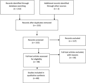
Fig. 1. Flowchart showing the process involved in identifying relevant literature. The criteria for “records excluded” and “fulltext articles excluded with reasons” are detailed in Part 1 of the Guidelines.
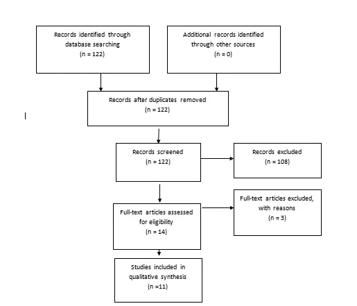
Fig. 2. Flowchart showing the process involved in identifying relevant literature for the 2020 Update. The criteria for “records excluded” and “full text articles excluded with reasons” are detailed in Part 1 of the Guidelines.
Of the 98 full-text articles selected for review, 30 full-text publications were rejected based on the criteria listed above and only 68 articles were used to construct the evidentiary table (Fig. 1). An additional 11 studies out of the 122 yielded by the 2020 update met inclusion criteria from the original guideline and were included (Figure 2). The criteria for the decision to treat were quite variable among different institutions and different study groups. For example, we evaluated 1 Class II study in which hydrocephalus was defined as the atrium of the lateral ventricle measuring > 10 mm on the horizontal plane of a head ultrasound (HUS) study or the body of the lateral ventricle at the level of the midthalamus measuring > 10 mm on a sagittal ultrasound image.34 We reviewed another Class III study in which hydrocephalus was defined as anterior cortical mantle thickness < 20 mm at an average postnatal age of 21 days along with increasing occipitofrontal circumference (OFC) as an indicator of hydrocephalus that should be treated.35 Bada et al35 reported that of 10 infants requiring shunts, 5 (50%) experienced normal development, which was defined by physical and neurological assessment and evaluation using the Denver developmental screening tool. Evan’s ratio, which is described as the lateral measurement of the ventricle across the frontal horns divided by the lateral measurement across the brain (biparietal diameter; also known as the ventricular/biparietal [V/BP] ratio) can also be used to describe the severity of PHH.36 The majority of studies that were evaluated based on an initial diagnosis of PHH on HUS, CT, and MRI studies were also used. Choudhury described mild hydrocephalus as a V/ BP ratio of 0.26–0.40, moderate hydrocephalus as a V/ BP ratio of 0.40–0.60, severe hydrocephalus as a V/BP ratio of 0.60–0.90, and extreme hydrocephalus as a V/ BP ratio of 0.91–1.0.36 These authors also reported that the thickness of the cortical mantle was not a statistically significant indicator of outcome because several infants with extreme hydrocephalus displayed normal motor development.36 One Class II and 1 Class III study indicated that when ventriculoperitoneal (VP) shunts were placed, even in cases of severe or extreme hydrocephalus, there were some infants with normal development and motor outcome (50 of 82 patients in the Choudhury study).35,36 Numerous studies have reported that good neurodevelopmental outcomes may be seen if and when infants with hydrocephalus are aggressively treated and cortical mantle thickness is restored.
Results
Surgical Temporizing Measures
Recommendation: Ventricular access devices (VADs), external ventricular drains (EVDs), ventriculosubgaleal (VSG) shunts, or lumbar punctures (LPs) are treatment options in the management of PHH. Clinical judgment is required. Strength of Recommendation: Level II, moderate degree of clinical certainty.
Recommendation: The evidence demonstrates that VSG shunts reduce the need for daily CSF aspiration compared with VADs. Strength of Recommendation: Level II, moderate degree of clinical certainty.
The evidence demonstrates that VADs reduce morbidity and mortality compared with EVDs. Three Class II and 7 Class III studies were included as evidence to support the first recommendation, and these lower-quality studies documented the safety and efficacy of VADs, or Ommaya reservoirs, for the aspiration of CSF, ventricular decompression, and lowering of intracranial pressure.37-46 The authors of 2 Class II studies reported that ventricular reservoirs may reduce the incidence of shunt infection as well as noninfectious shunt complications.38,41 In one Class II study and one class III study, repeated aspiration of CSF from a VAD did not significantly increase the risk of infection.41,44 Three Class III studies reported that ventricular reservoirs did not significantly reduce the need for permanent shunt placement.42,43,47 One Class III study reported that the use of VADs, compared with the use of continuous ventricular drainage, significantly reduced morbidity and mortality rates that were associated with the surgical treatment of PHH in LBW infants with reservoirs, instead of EVDs (Table 1).41
The placement of an EVD has also been used to treat hydrocephalus in preterm infants with PHH and is an option for these children, as shown in 1 Class II and 7 Class III studies.48-55 Three Class III studies reported that an EVD obviated the need for VP shunt placement in fewer than one-third of infants treated.48,52,54 More than 50% of preterm infants with PHH did require permanent VP shunt placement following removal of an EVD (95 out of 132 survivors required a shunt).48,52-55
It has been reported that placement of a VSG shunt may reduce the need for permanent shunt placement. The authors of Class II and Class III studies reported trends toward shunt independence, but the studies only enrolled 32 and 95 patients, respectively, and the results were not statistically significant.56,57 In their report of a Class II, retrospective historical cohort study, Lam and Heilman demonstrated that VSG shunting significantly reduced the need for daily CSF aspiration, which may decrease the risk of introducing a de novo CSF infection.56 A chi square test performed on their data indicated that a VSG shunt did significantly reduce the need for daily CSF aspiration when compared with a VAD (c2 = 19.2, df = 1, p = 0.000016, p < 0.05).56 This may reduce the risk of infection or other complications. A larger, prospective study reported a statistically significant decreased need for permanent CSF diversion in infants treated with VSG shunts.57 This study reported that 66% of infants (20 of 30) treated with VSG shunts required VP shunts and 33% (10 of 30) remained shunt free; this was compared with a group of infants treated with VADs in which 75% (49 of 65) required VP shunts and only 25% of infants (16 of 65) remained shunt free.57
In 2 studies, 1 intervention was compared to another with specific recommendations about the timing of the intervention for temporizing measures for the treatment of PHH in very LBW infants. In 1 Class III study, the authors compared early versus late intervention, as assessed by ventricular dilation in 5 collaborating neonatal centers.58 Ninety-five patients were subdivided into early intervention or late intervention groups, depending on their ventricular index at the time of initial treatment. Early treatment was safe and effective regardless of whether LP and/or reservoir placement was used. Early intervention was associated with a reduced requirement for a VP shunt (OR = 0.22) and reduced risk of moderate-to-severe disability.58 Additionally, there was a single Class III observational study of outcomes in which LPs, EVD, VSG shunts, and reservoirs were used.24 All interventional studies were found to be safe and effective.24
Routine Use of Serial Lumbar Puncture
Recommendation: The routine use of serial lumbar puncture (LP) is not recommended to reduce the need for shunt placement or to avoid the progression of hydrocephalus in premature infants. Strength of Recommendation: Level I, high degree of clinical certainty.
One Class I study was included, and it reported no statistical differences in outcomes of preterm infants with PHH treated with observation alone or infants treated with daily LP (Table 2).59 Lumbar puncture is often used early in the treatment of PHH, despite the fact that there is no statistically significant reduction in the need for a shunt or progression of PHH.59,60 In fact, LP neither predicts nor prevents the need for a permanent VP shunt.51 A second study, a Class III study, also reported no difference in adverse outcome regardless of whether infants were untreated or treated with serial LP.61 Without aggressive treatment of hydrocephalus and with persistent ventricular dilation, outcome was poor.61 Additionally, there was a single Class III study that concluded that repeated LPs may cause or contribute to subsequent shunt infection.62 Although LP may be useful for drawing off CSF as an immediate treatment for elevated intracranial pressure in infants with PHH, or for sampling CSF, we do not recommend the routine use of LP to eliminate the need for a VP shunt.61
Nonsurgical Temporizing Agents
Intraventricular Thrombolytic Agents. Recommendation: Intraventricular thrombolytic agents including tissue plasminogen activator (tPA), urokinase, or streptokinase are not recommended as methods to reduce the need for shunt placement in premature infants with PHH. Strength of Recommendation: Level I, high clinical certainty.
Based on 1 high-quality Class I study, the DRIFT procedure—Drainage, Irrigation, and Fibrinolytic Therapy (intraventricular tPA)—is not recommended for PHH.63 DRIFT did not significantly reduce shunt surgery or death, but it was associated with an increased rate of secondary IVH (Table 3).63 Forty-four percent (15 of 34) of infants in the DRIFT group died or required a shunt, compared with 50% (19 of 36) of infants who received standard treatment. Thirty-five percent (12 of 34) of preterm infants in the DRIFT study had secondary IVH, compared with 8% (3 of 34) who received standard treatment.63 These results differ from those of earlier Class II and Class III studies in which a decreased rate for the need for permanent shunt placement was reported when low-dose urokinase or fibrinolytic therapy with tPA was used for ventricular irrigation and clot reduction.33,64-66
Reviews conducted by Whitelaw and Odd63 have also revealed that intraventricular injection of streptokinase has not been shown to be beneficial.67 A single case report of intravenous streptokinase, published in 1998, suggested that there may be some benefit.68 This report was followed by an early Class III study that found benefit in a nonrandomized cohort of preterm infants with PHH who were treated with intravenous low-dose streptokinase.69 However, data from a later Class II study led to the conclusion that routine use of intraventricular streptokinase in PHH was not recommended.70 These studies were included in the 2007 Whitelaw and Odd Cochrane review,63,67 which argues against intravenous streptokinase for the treatment of PHH in preterm infants (Table 3).
Despite increased short-term morbidity and recurrent IVH, some benefits were noted in the DRIFT survivors.71 In the most recent Whitelaw study,71 the reduction in the primary long-term outcome—death or severe disability— at 2 years in the DRIFT group reached statistical significance when adjusted for sex, birth weight, and grade of IVH. Severe cognitive disability also was reduced, and this improvement in cognitive function was statistically significant. There was also a reduction in severe sensorimotor disability with DRIFT, but this clinical improvement did not reach statistical significance. The authors hypothesized that the greater effect on cognitive rather than sensorimotor function may be attributed to parenchymal infarction in the periventricular white matter, which was seen in about half of the infants enrolled in the trial.71
Acetazolamide and Furosemide.
Recommendation: Acetazolamide and furosemide are not recommended as methods to reduce the need for shunt placement in premature infants with PHH. Strength of Recommendation: Level I, high clinical certainty.
After our review of the literature, we found two Class I studies that reported that preterm infants with a diagnosis of PHH who were treated with acetazolamide and furosemide demonstrated higher risks of neurological complications, morbidity, and mortality (Table 4).72,73 The International Posthemorrhagic Ventricular Dilation (PHVD) Drug Trial Group reported that administration of acetazolamide plus furosemide leads to higher rates of shunt placement (relative risk 1.42) and morbidity (84% vs 60%) compared with standard therapy.72 Kennedy etal.73 reported that treatment of PHVD with acetazolamide and furosemide did not decrease the rate of shunt placement (64% in the acetazolamide/furosemide group vs 52% in the control group, not treated with acetazolamide/furosemide).73 However, treatment was associated with an increased rate of neurological morbidity (81% vs 66%).73
Treatment of PHVD with acetazolamide and furosemide was not recommended.73 One Class III study reported this treatment was not associated with VP shunt placement, but the severity of IVH (based on IVH grade) and the patient age at the time of IVH were significantly associated with the need for permanent CSF diversion.51 Kennedy et al. also noted that the ventricular index at time of entry into trial was the only factor significantly predictive of death or need for shunt, after multiple logistic regression analysis.73
Timing of Shunt Placement
Recommendation: There is insufficient evidence to recommend a specific infant weight or CSF parameter to direct the timing of shunt placement in premature infants with PHH. Strength of Recommendation: Level III, unclear degree of clinical certainty.
There were two Class III studies which evaluated the lower limits of infant weight at time of initial shunt insertion (Table 5).38,59 A weight of 1500 g was safely used as a criterion for VP shunt placement in the Benzel study.38 A single Class III study showed that CSF cell count, protein, and glucose levels were not statistically related to the occurrence of shunt failure or infection in the study population.74 The authors recommended that placement of the shunt be timed when the infant’s age, weight, and overall stability allow.74
Endoscopic Third Ventriculostomy
Recommendation: There is insufficient evidence to recommend the use of endoscopic third ventriculostomy (ETV) in premature infants with PHH. Strength of Recommendation: Level III, unclear degree of clinical certainty.
Although ETV was discussed in several full-text articles that we reviewed, there was insufficient evidence available for us to make a recommendation for or against its use for the treatment of PHH in premature infants (Table 6). Endoscopic third ventriculostomy for the treatment of hydrocephalus in infants and children will be discussed more thoroughly in subsequent chapters (in particular, Part 4).75
2020 Update
There was one new recommendation yielded by the update stating that that neuro-endoscopic lavage is a feasible and safe option for the removal of intraventricular clots and may lower the rate of shunt placement.76 The remaining new literature confirmed the previous recommendations.76-83 (Table 7)
There was no change in the Level I recommendation against the routine use of serial lumbar punctures (LP) to reduce the need to shunt or to avoid the progression of HC in premature infants. In the management of PHH, there is still insufficient evidence to recommend one surgical temporizing method over another. Authors suggested that ventriculo-subgaleal shunts (VSG) reduce the need of daily CSF aspiration as compared to other ventricular access devices (VAD) and that Ommaya type ventricular access reservoirs reduce in morbidity and mortality compared to external ventricular drains (EVD) (Level II). There were some benefits of VSG over VAD identified in 2014 and affirmed in 2019 (Level II).80-82 Intraventricular thrombolytic agents are still not recommended as a method to reduce the need for shunt placement in premature infants with PHH; there was no change in this Level I recommendation. Acetazolamide/ Furosemide are also not recommended as methods to reduce the need for shunt placement in premature infants with PHH (Level I).
Excluded Studies
We excluded 1 Class III study for low “preterm” patient representation (7 patients); in the review of 52 consecutive ETV procedures in 49 infants with hydrocephalus, most infants (31 patients) had aqueductal stenosis.84 Of the 7 infants with preterm PHH, 6 required a shunt even after ETV. Infants with PHH from premature birth did not benefit from ETV.84 We excluded another Class III study including patients with different etiologies for hydrocephalus.85 Although ETV was successful in 57% of patients (8 of 14), the majority of those infants had congenital aqueductal stenosis without PHH. In the remaining 6 patients, a VP shunt was needed. In 1 Class III single-institution retrospective case series, 18 preterm infants with PHH were treated initially with Ommaya reservoir placement: 1 patient died, 5 patients received a VP shunt, and 9 patients underwent ETV.86 Three patients did not require any further intervention. While overall, 59% were shunt free at the last follow-up, 5 of the 9 patients who were treated with ETV had to undergo repeated surgery for VP shunt placement. The authors recommended combining placement of an Ommaya reservoir with ETV to reduce shunt dependency for preterm infants with PHH.86 There was a large (101 patients) Class III multicenter, retrospective study evaluating the success rate of ETV in patients with hydrocephalus from subarachnoid hemorrhage, IVH, and/or CSF infection; a minority of the patients (25% [25 of 101]) had PHH of prematurity.87 Overall, ETV was successful in 52% of the infants with PHH of prematurity. Endoscopic third ventriculostomy was successful in 100% (13 of 13) of children with a history of preterm PHH, even though these patients were initially treated with a shunt. Endoscopic third ventriculostomy was unsuccessful in 12 of 12 infants treated with ETV as the first-line treatment, following preterm PHH. In patients with both hemorrhage and infection, ETV was not successful.87
Three studies were excluded from the update after full text review because (although the populations studied and results that were reported included some data about premature infants with PHH) there were mixed populations included and results for premature infants with PHH could not be separately identified, tracked or otherwise determined.88-90
Conclusions
Surgical Temporizing Measures
Recommendation: Ventricular access devices (VADs), external ventricular drains (EVDs), ventriculosubgaleal (VSG) shunts, or lumbar punctures (LPs) are treatment options in the management of posthemorrhagic hydrocephalus (PHH). Clinical judgment is required. Strength of Recommendation: Level II, moderate degree of clinical certainty.
Recommendation: The evidence demonstrates that VSG shunts reduce the need for daily CSF aspiration compared with VADs. Strength of Recommendation: Level II, moderate degree of clinical certainty.
The evidence demonstrates that VADs reduce morbidity and mortality compared with EVDs.
Routine Use of Serial Lumbar Punctures
Recommendation: The routine use of serial lumbar puncture (LP) is not recommended to reduce the need for shunt placement or to avoid the progression of hydrocephalus in premature infants. Strength of Recommendation: Level I, high clinical certainty.
Nonsurgical Temporizing Measures
Recommendation: Intraventricular thrombolytic agents including tissue plasminogen activator (tPA), urokinase, or streptokinase are not recommended as methods to reduce the need for shunt placement in premature infants with PHH. Strength of Recommendation: Level I, high clinical certainty.
Recommendation: Acetazolamide and furosemide are not recommended as methods to reduce the need for shunt placement in premature infants with PHH. Strength of Recommendation: Level I, high clinical certainty.
Timing of Shunt Placement
Recommendation: There is insufficient evidence to recommend a specific weight or CSF parameter to direct the timing of shunt placement in premature infants with PHH. Clinical judgment is required. Strength of Recommendation: Level III, unclear clinical certainty.
Endoscopic Third Ventriculostomy
Recommendation: There is insufficient evidence to recommend the use of endoscopic third ventriculostomy (ETV) in premature infants with PHH. Strength of Recommendation: Level III, unclear clinical certainty.
Recommendation: Neuro-endoscopic lavage is a feasible and safe option and therefore may be used for the removal of intraventricular clots and may lower the rate of shunt placement. Strength of Recommendation: Level III, unclear clinical certainty.
Acknowledgments
We acknowledge the American Association of Neurological Surgeons (AANS)/Congress of Neurological Surgeons (CNS) Joint Guidelines Committee for the members’ reviews, comments, and suggestions; the Hydrocephalus Association and Debby Buffa, patient advocate representative, for participation and input throughout the guidelines development; Pamela Shaw, research librarian, for her assistance with the literature searches; Kevin Boyer for his assistance with data analysis; and Sue Ann Kawecki for her assistance with editing.
We acknowledge the following individuals for their contributions throughout the review process: Timothy Ryken, M.D.; Kevin Cockroft, M.D.; Sepideh Amin-Hanjani, M.D.; Steven N. Kalkanis, M.D.; David P. Adelson, M.D.; Brian L. Hoh, M.D.; Mark D. Krieger, M.D.; Mark E. Linskey, M.D.; Jeffrey J. Olson, M.D.; Patricia Raskin, M.D.; Krystal L. Tomei, M.D.; and Monica Wehby, M.D. We also acknowledge the following peer reviewers for their contributions to review the update to the guidelines: Jennifer Sweet, MD, Brandon Rocque, MD, Christoph Greissenauer, MD, Jeffrey Olson, MD.
Disclosure
Dr. Limbrick receives research funding from the National Institute of Neurological Disorders and Stroke. The systematic review and evidence-based guidelines were funded exclusively by the CNS and AANS Pediatric Section, which received no funding from outside commercial sources to support the development of this document.
Conflict(s) of Interest: None. All Pediatric Hydrocephalus Systematic Review and Evidence-Based Guidelines Task Force members declared any potential conflicts of interest prior to beginning work on this systematic review and evidence-based guidelines.
Conflict(s) of Interest: None. All Pediatric Hydrocephalus Systematic Review and Evidence-Based Guidelines Update Task Force members declared any potential conflicts of interest prior to beginning work on this systematic review and evidence-based guidelines.
Author contributions to the study and manuscript preparation include the following. Conception and design: AANS/CNS Joint Section on Pediatrics. Acquisition of data: all authors. Analysis and interpretation of data: all authors. Drafting the article: Mazzola. Critically revising the article: all authors. Reviewed submitted version of manuscript: all authors. Approved the final version of the manuscript on behalf of all authors: Flannery. Administrative/technical/material support: all authors. Study supervision: Flannery.
Table 1. Surgical Temporizing Measures Evidence Table
| First Author & Year | Study Description | Data Class, Quality and Reasons | Results and Conclusions |
| Cornips., et al 1997 | Retrospective review of 14 patients with Grade III or Grade IV IVH diagnosed on HUS, treated with EVD. These 14 were compared to a historical cohort of 15 infants with similar Grade III/ IV IVH. | Class IIRetrospective review of 2 cohorts, those premature infants treated with EVD versus those treated medically. | Ventricular drainage is a safe option for infants with PHH. |
| Gurtner., et al 1992 | Retrospective consecutively enrolled study of 736 low-birth-weight infants (< 1500 g). Twenty-seven infants were excluded because of incomplete records and 162 infants were not examined by cranial ultrasound because of early postnatal death, or if they were discharged before cranial ultrasound was done. The remaining 547 infants were reviewed in retrospective consecutive fashion. Criteria for shunt placement were clearly defined the presence of progressive HC documented by serial cranial ultrasounds, and OFC greater than the 95th percentile despite repeated lumbar punctures. All surgery was done in an operating room. All infants were followed for morbidity, mortality and infection. Data were examined for differences between the 3 years of the study, analyses of variance and Duncan’s mean comparison tests were calculated. Chi-squared analyses were performed for yearly differences in discrete variables such as rates of complication and mortality. Student t test with Bonferroni corrections. Spearman correlation coefficients were computed when appropriate. Quantitative data were presented. | Class IIAlthough this was a consecutive study of a high number of low birth weight infants, this was not a randomized, controlled study, but a non-random historical cohort, compared by year of treatment and treatment type. The temporizing method used in the first year was that of external ventricular drainage, whereas in subsequent years, a subcutaneous reservoir was used to intermittently drain the ventricles. Outcomes evaluated include: morbidity, mortality, and need for shunt revision. | Frequency and mortality of grades III and IV hemorrhage in infants weighing between 500 and 700 grams remained relatively constant over the 3 year period. Weights of these infants at time of surgery were rather low and did correlate with an increased rate of complication in 1988. However, That year, a high complication rate was associated with usage of external ventricular drains. The 3-month revision rate was 20% in 1987 and 50% in 1988; no shunts required revision in 1989. The group concluded that there was a significant reduction in morbidity and mortality associated with the surgical treatment of PHH in low-birth weight infants when they began using reservoirs, instead of external ventricular drains. |
| Hudgins et al., 1997 | Use of urokinase via reservoir to treat PHH in n=18 patients. Four different doses of urokinase; ultimately grouped into “high” (n=9) and “low” dose (everyone else, n=9). Both groups compared to historical control group with respect to outcome, need for shunt. Prospective, case control. | Class IIProspective, non-randomized, case-control series. Division of 9 patients into “low” dose group would appear to dilute statistical power, despite statistical significance obtained. | “Low dose” urokinase reduced shunt rate (71% vs 92%) compared to historical controls. Fewer shunt revisions in both groups compared to control group |
| Lam et al., 2009 | Single institution, retrospective historical cohort study of 32 preterm infants with PHH. This study compared 2 cohorts of infants: those treated with ventricular access device (VAD)/ Ommaya placement versus those treated with ventrciulosubgaleal shunts (VSG). There were no statistical differences in age or birth weight of the infants in the two groups. The groups were studied for IVH grade, need for daily CSF withdrawl, CSF leak from the scalp, CSF infection, and need for a VPS. | Class IIA chi squaretest was performed (X w =19.2, df=1, p value=0.000016, p<0.05) which showed that VSG significantly reduced the need of daily CSF aspiration as compared to VAD. The higher rate of complicationsof VSG was not statistically significant tothe VAD group (p=0.17). 93.75% (15 of 16 patients) of the VAD group required VPS while 71.42% (10 of 14 patients) in the VSG group needed VPS. | There was a trend towards VPS independence in the VSG group, as compared to the VAD group, but it did not reach significance.VSG did decrease the need for daily taps. There was a slightly higher rate of complications in the VSG group, but it was not significant. |
| Anwar et al., 1986 | Consecutive, non-randomized study of 19 preterm infants with PHH who underwent placement of reservoirs for symptomatic HC. Symptomatic HC was defined as infants with rapidly increasing OFC, ventriculomegaly and signs of increased intracranial pressure were present, such as tense fontanelle, splayed sutures, apnea, bradycardia, seizure, feeding difficulties, or lethargy. | Class III study was a case series study of the infants who were less than 200 grams, with clear CSF, and who were treated with reservoirs. There was only limited presentation of qualitative and quantitative data. Data were presented including: morbidity, mortality and need for shunt placement in these infants. There was no comparison to a cohort of non-treated infants or infants treated with ventricular drains. | The authors concluded that reservoirs provide safe and effective treatment for infants with PHH and symptomatic HC. |
| Benzel et al., 1993 | 41 patients requiring ventricular drainage for HC/ PHH were evaluated retrospectively. All drainage procedures were performed on patients with IVH with HC (Grade III [25 patients]) and IVH and IPH (Grade IV [16 patients]) who failed medical management. | Class IIIRetrospective case series of 41 consecutive premature infants. 26 ventricular reservoirs (Rickham or McComb reservoirs) were placed in neonates weighing less than 1500 grams, allowing for a safe but intermittent ventricular access. 18 of these reservoirs were subsequently converted to VPS. 32 % required a VPS and/or reservoir infection and 59% required a shunt revision during the first year of life.No Grade IV patients achieved a normal functional level, while 10 Grade III patients did. The incidence of severe developmental delay (44% versus 28%) and death (38% versus 12%) was greater in the grade IV than the Grade III patients. | The placement of ventricular reservoirs is acceptable as an alternative to the early placement of ventriculo-peritoneal shunts. This approach may reduce the incidence of shunt infection as well as noninfectious shunt complications. |
| Berger et al., 2000 | Retrospective review of outcomes after EVDs, n=37 preemies (n=51 drains), PHH diagnosed by ultrasound. | Class III:Single institution retrospective review | Neurodevelopmental outcome dependent on extent of parenchymal injury. Infection rate: 5.4%/patients, 3.9%drain. 11/37 did not require shunting. |
| Brouwer et al., 2007 | Single center retrospective review of 76 preterm infants treated for PHVD with ventricular reservoirs. Infection rates were measured in two successive 6-year intervals. Number of reservoir punctures also examined. | Class IIISingle center retrospective review | While the number of reservoir punctures did not change, the infection rate was lower in the second, more recent interval (2/50 or 4% versus 5/26 or 19.2%). Conclusion: Risks associated with ventricular reservoirs are within acceptable limits. |
| de Vries et al., 2002 | Retrospective review of consecutive preterm infants (EGA≤34 weeks) with Grade III IVH treated for post-hemorrhagic ventricular dilatation in 5 collaborating NICUs (n=95). Subjects were subdivided into early intervention or late intervention groups, depending on their ventricular index at the time of initial treatment. | Class IIIWhile this was a multi-center study, it was a retrospective case series. Treatments were not standardized (infants treated variably with LPs, reservoir, shunt) and neurodevelopmental outcome measures were limited. | Early treatment was associated with a reduced requirement for VP shunt (OR=0.22) and reduced risk of moderate-severe disability. |
| Gaskill et al., 1998 | The use of a subcutaneous reservoir was studied in a consecutive, non-randomized series of 38 infants with preterm IVH and PHH. All infants had failed LP and medical treatment. | Class IIIThis was a retrospective study of a series of premature infants who required temporizing measures (reservoir placement) after failing medical treatment/ LP for PHH. There were 28 survivors overall (8 died before a shunt, 2 died after a shunt). Four survivors (15%) did not require a shunt. | The authors concluded that early reservoir placement is feasible, safe and effective treatment of PHH associated with preterm IVH. |
| Harbaugh et al., 1981 | Retrospective review of 11 premature infants with IVH and PHH were managed with tunneled EVD. The mean duration of drainage for this group was 20.7 days. No morbidity or mortality occurred as a result. 7/11 patients required a shunt. 2/11 have not required VPS. | Class IIISmall retrospective case review. | EVD via a subcutaneously tunneled catheter has been found to be a safe and reliable initial method of treating posthemorrhagic hydrocephalus in premature infants. |
| Heep et al., 2007 | Safety/efficacy of Rickham reservoir placement for PHH patients. | Class IIIRetrospective review. Broad inclusion criteria for reservoir placement. No comparison to patients managed with other methods. | Ommaya / Rickham reservoir is a safe, effective option for managing PHH until ready for shunt. 5% infection rate, 85% of patients needed shunt. |
| Hudginsn et al., 1998 | Use of VAD in n=149 PHH patients. Daily taps for first ‘several’ days (10-15 cm3/kg). | Class IIIRetrospective, case series from a single institution retrospective review. Shunts placed at 2kg if still symptomatic, but criteria not otherwise clear on when to stop VAD aspirations. | 8% infection, 20% revision rates. 88% shunt implantation rate. |
| Kazan et al., 2005 | Single-center, retrospective review of preterm and low birth weight infants diagnosed with intraventricular hemorrhage by ultrasound (n=42). 11 infants who required VP shunt were compared to 31 who did not. All subjects received acetazolamide and furosemide as an initial medical treatment. | Class IIISmall, retrospective case series with grouping of subjects despite variable treatments. | Risk factors for VP shunt included IVH grade, later EGA at birth, and age (days) at time of IVH, but not treatment for IVH/PHH (acetazolamide, furosemide, LP, and external ventricular drainage). |
| Kormanik et al., 2010 | Kormanik et al. reported a retrospective review of the outcome of infants receiving a ventricular reservoir for PHH. | Class IIIRetrospective observational study, review of medical records of all infants receiving ventricular reservoir in one center between 2000 and 2007. | Ventricular reservoirs were placed in 35 infants during study period for management of hydrocephalus. Six (17%) were excluded. The remaining 29 infants had VR placement for PHVD. Serial tapping was performed on 681 occasions in 29 infants before placement of VP shunt or transport back to referring hospital. There were no cases of CSF culture-proven reservoir infection related to repeated taps, except for one case of Candida albicans CSF culture. |
| Kormanik et al., 2010 | Kormanik et al. reported a retrospective review of the outcome of infants receiving a ventricular reservoir for PHH. | Class IIIRetrospective observational study, review of medical records of all infants receiving ventricular reservoir in one center between 2000 and 2007. | Ventricular reservoirs were placed in 35 infants during study period for management of hydrocephalus. Six (17%) were excluded. The remaining 29 infants had VR placement for PHVD. Serial tapping was performed on 681 occasions in 29 infants before placement of VP shunt or transport back to referring hospital. There were no cases of CSF culture-proven reservoir infection related to repeated taps, except for one case of Candida albicans CSF culture. |
| Limbrick et al., 2010 | Large, single center retrospective review of 325 preterm infants with Grade 3 or 4 IVH. The development of PHH and the need for a temporizing device (VAD or VSG) were studied. Infections, complications and need for VPS were analyzed, as was mortality rate. | Class IIIRetrospective analysis showed 75.4% of the 65 infants treated with VAD needed a shunt; 66.7% of the 30 treated with VSG required a shunt. There was no significant difference in the infection rate between VAD and VSG, revision rate, or VPS infection afterwards, | There was no significant difference in outcome between infants treated with VAD or VSG. |
| Rahman et al., 1993 | Single institution, small retrospective review of 37 patients with PHH, 31 of which required VPS. | Class IIIObservational study of outcomes; LP, EVD, VSG, and Ommaya reservoirs were used. No statistical data available. | Suggested LP, VSG, Ommaya and VPS are safe and effective. |
| Rhodes et al., 1987 | Thirty-seven premature infants with PHH were treated with an EVD. Complications, including morbidity, were presented. 32 did not require a permanent shunt. Neurodevelopmental outcomes were presented along with neuromuscular outcomes. | Class IIIThis was a retrospective, consecutive case series. | Level III Ventricular drainage is a safe and effective mechanism for treating infants with PHH and may obviate the need for a shunt. |
| Rhodes et al., 1987 | Thirty-seven premature infants with PHH were treated with an EVD. Complications, including morbidity, were presented. 32 did not require a permanent shunt. Neurodevelopmental outcomes were presented along with neuromuscular outcomes. | Class IIIThis was a retrospective, consecutive case series. | Level III Ventricular drainage is a safe and effective mechanism for treating infants with PHH and may obviate the need for a shunt. |
| Weninger et al., 1992 | Study of 27 consecutive infants with an average gestational age of 31 weeks, with PHH and increased ICP who were treated with a tunneled EVD. PHH was defined as ventricular dilation, progressively increasing OFC, bulging fontanel, widening of the sutures, apnea or bradycardia. | Class IIIThe study is a case series report. | PHH was successfully treated in all patients; the EVD was left in situ for an average of 23 +/- 9 days. 4 patients died of unrelated causes, and 23patients survived. 16 required shunts. Neurological outcome correlated with severity of the Grade of IVH. Grade 4 IVH infants had the worst neurological outcomes, despite treatment. The authors conclude that EVD is a safe and effective treatment for PHH in premature infants. |
| Willis et al., 2009 | Thirty-two premature infants with PHH treated with shunts were retrospectively reviewed to analyze the complications and outcome with respect to shunt revisions. Retrospective, consecutive case series of 32 infants who needed treatment for PHH. Multivariate analysis and time series were used to identify factors that influence the outcome in terms of shunt revisions. | Class III | Reservoir placement suspended progression of hydrocephalus in only 2 patients, while permanent shunts were needed in 90.6% of cases. CSF reservoirs were a safe and effective method of treatment in infants considered too small for VP shunt placement but did NOT obviate the need for a shunt. |
| Yu et al., 2009 | The authors performed a retrospective case study of 11 premature infants with PHH all treated with a subcutaneous reservoir for CSF aspiration. | Class III Retrospective case series. | The authors concluded that CSF reservoir treatment is safe and effective for infants with PHH. |
Table 2. Serial Lumbar Punctures Evidence Table
| Author | Study Description | Data Class, Quality and Reasons | Results and Conclusions |
| Anwar et al., 1986 | Randomized controlled study of 47 consecutive pre-term infants with PHH with Grade 3 or Grade 4 IVH. Infants enrolled in the study were randomized to observation only or daily LP. Cohorts were studied for morbidity, mortality and need for a shunt. | Class IConsecutively enrolled infants randomized to treatment (daily LP) (N= 24) or observation only (N=23). Ten infants treated with LP (10/24) required shunts and 9 of the observation-only group (9/23) required shunt placement for progressive PHH and HC. | There were no statistical differences of outcomes studied in infants treated with observation alone or infants treated with daily LP. Although LP was safe, there was no statistically significant reduction in the need for shunt or progression of PHH. |
| Behjati et al., 2011 | Case series study that investigated risk factors for ventriculoperitoneal (VP) shunting in infants with HC following IVH in 97 consecutive pre-term infants with IVH. | Class IIICase series of 97 infants with IVH associated with prematurity. The risks factors associated with the need for a shunt were investigated. Infants were followed for one year. Morbidities and mortalities were reported in a quantitative fashion. Patients treated medically with acetazolamide showed no benefit; however, infants treated with repeated CSF drainage through LP did have a higher shunt infection rate, once shunted. | Infants with Grade 3 or 4 IVH are at highest risk of PHH and HC and that the 11 of 31 patients who required a shunt developed shunt infection, which was significantly associated with repeat LP’s. |
| Anwar et al., 1986 | Randomized controlled study of 47 consecutive pre-term infants with PHH with Grade 3 or Grade 4 IVH. Infants enrolled in the study were randomized to observation only or daily LP. Cohorts were studied for morbidity, mortality and need for a shunt. | Class IConsecutively enrolled infants randomized to treatment (daily LP) (N= 24) or observation only (N=23). Ten infants treated with LP (10/24) required shunts and 9 of the observation-only group (9/23) required shunt placement for progressive PHH and HC. | There were no statistical differences of outcomes studied in infants treated with observation alone or infants treated with daily LP. Although LP was safe, there was no statistically significant reduction in the need for shunt or progression of PHH. |
| Chaplin et al., 1980 | Retrospective review of 22 consecutive, low birth weight infants with PHH. All developed HC after 2 weeks of age. The first 12 required VPS. In 10 infants born after September 1974, an attempt was first made to control the HC with repeated lumbar puncture and diuretics prior to placing a shunt. In 7/10 the hydrocephalus was successfully arrested by medical therapy alone. | Class IIIRetrospective review of 22 infants with PHH. There were two cohorts: 12 treated with VPS, and 10 treated with LP and diuretics. | Follow-up at 1 to 8 years of age were done on 18 infants. 2/ 12 treated by permanent shunts and 3/6 treated medically had an IQ score of 85 or greater. These results indicate a poor long-term outlook for the low birth weight infant who develops clinically overt hydrocephalus after intracranial bleeding. |
| Kazan et al. 2005 | Single-center, retrospective review of preterm and low birth weight infants diagnosed with intraventricular hemorrhage by ultrasound (n=42). 11 infants who required VP shunt were compared to 31 who did not. All subjects received acetazolamide and furosemide as an initial medical treatment. | Class IIISmall, retrospective case series with grouping of subjects despite variable treatments. | Risk factors for VP shunt included IVH grade, later EGA at birth, and age (days) at time of IVH, but not treatment for IVH/PHH (acetazolamide, furosemide, LP, external ventricular drainage). |
| Muller et al., 1998 | Effect of aggressive LP schedule on PHH. LPs started 0-4 days, on average 11LPs performed per patient, 15ml/kg or end of CSF flow per LP. Used protein, RBC count, glucose, ventricular size to determine endpoint. | Class IIINon-randomized, prospective study, single institution. 16% complete resolution, 65% ventriculomegaly but not shunted, 19% shunted.. | Serial LP should be started early for treatment of HC. |
Table 3. Intraventricular Thrombolytic Agents Evidence Table
| Author | Study Description | Data Class, Quality and Reasons | Results and Conclusions |
| Whitelaw et al., 2007 | Randomized multicenter clinical trial examining standard treatment to DRIFT. 70 infants enrolled (34: DRIFT, 36: standard treatment). Outcomes at 6 months of age or hospital discharge: death or VP shunt surgery, secondary IVH, and infection. | Class IMulticenter randomized controlled trial. | 15/34 (44%) subjects in the DRIFT group expired or required a shunt, compared with 19/36 (50%) receiving standard treatment. 12/34 (35%) in DRIFT had secondary IVH, compared with 8% receiving standard treatment. Conclusion: DRIFT did not reduce shunt surgery or death but was associated with an increased rate of secondary IVH. |
| Hudgins et al., 1997 | Use of urokinase via reservoir to treat PHH in n=18 patients. 4 different doses of urokinase; ultimately grouped into “high ” (n=9) and “low” dose (everyone else, n=9). Both groups compared to historical control group with respect to outcome, need for shunt. Prospective, case control. | Class IIProspective, non-randomized, case-control series. Division of 9 patients into “low” dose group would appear to dilute statistical power, despite statistical significance obtained. | “Low dose” urokinase reduced shunt rate (71% vs 92%) compared to historical controls. Fewer shunt revisions in both groups compared to control group. |
| Whitelaw et al., 2007 | Review and meta-analysis of 2 prospective case-control studies (Luciano 1997, Yapicioglu 2003). Both source studies included 12 subjects total: 6 cases, 6 controls. meta-analysis. | Class IIBoth sources studies were Class II (both were small randomized, prospective case-control studies). | No difference in mortality or VP shunt rate was observed with intraventricular streptokinase. Intraventricular fibrinolytic therapy cannot be recommended for infants following IVH. |
| Yapicioglu et al., 2003 | Single blind, prospective, study. Twelve preterm infants who developed posthaemorrhagic hydrocephalus were randomly assigned to the control group (no treatment) or to receive intraventricular streptokinase (x 3 days). Note: the streptokinase group also had an LP (10-15 cc) prior to treatment and then daily LPs (5-10cc). They also received intraventricular vancomycin. Primary outcome: VP shunt placement. | Class IISmall randomized, prospective study | Five of 6 infants in the streptokinase group and 3 of 6 in the control group required VP shunts. No complications were noted. Routine use of intraventricular streptokinase in PHH was not recommended. |
| Richard et al., 2001 | Single institution experience with Ommaya reservoir in n=64 patients. N=17 patients received fibrinolytic therapy through Ommaya. | Class IIIRetrospective case series. Statistics performed on fibrinolytic therapy subgroup that consists of 2 different agents with multiple doses. Fibrinoytic subgroup then mixed back into overall outcome analysis. | Fibrinolytic therapy led to statistically significant lower rate of shunt (31% versus 87%). 22% infection rate. |
| Whitelaw et al., 2003 | Prospective Phase I trial of new treatment methodology (DRIFT) for prevention of PHH of prematurity. Data from 24 subjects compared with historical controls. Outcome measures: death, need for shunt, secondary IVH, infection, neurodevelopmental outcome. | Class IIIProspective Phase I trial in 24 subjects and compared with historical controls. | One subject expired. 17 of 23 (74%) did not require a shunt. Two subjects experienced secondary IVH, and 2 experienced infections. 19 subjects >12 months had ND testing: 8 (42%) were normal, 7 (37%) had a single disability; 4 (21%) had multiple disabilities. Conclusion: Compared with historical controls, DRIFT reduced the need for shunts and showed a trend toward lower rates of mortality and disability. |
| Whitelaw et al., 1992 | Prospective study of 9 preterm infants with progressive post-hemorrhagic ventricular dilation who underwent a 48-72 hour continuous intraventricular infusion of streptokinase. Outcomes: death, need for shunt, secondary IVH, and infection. | Class IIISmall, prospective, non-randomized cohort study (Phase I trial). | All subjects survived; only 1 of 9 required a shunt prior to discharge (later reports indicated that a total of 4 of 9 ultimately required shunts). No infections, one re-hemorrhage. |
| Whitelaw et al., 1996 | Phase I study l to evaluate safety of tPA in 22 preterm infants with post-hemorrhagic ventricular dilation. Dose-finding data reported. Outcome measures: death and need for shunt prior to discharge and secondary IVH. | Class IIISmall, prospective, non-randomized cohort study (Phase I trial). | Dose-finding and pharmacokinetic data reported ([tPA], half-life tPA). 21 of 22 (95%) of subjects survived, 9/21 (45%) required shunts. One subject experienced secondary IVH. Conclusion: tPA resulted in survival without shunting in most subjects. |
Table 4. Acetazolamide/Furosemide Evidence Table
| Author | Study Description | Data Class, Quality and Reasons | Results and Conclusions |
| International PHVD Drug Trial Group, 1998 | Use of ACTZ and furosemide in PHH patients. Comparison with standard therapy for shunt placement and neurologic outcome. | Class IRandomized, controlled, multicenter, well-designed | ACTZ+furosemide leads to higher rates of shunt placement (RR 1.42) and higher morbidity (84% versus 60%) compared with standard therapy |
| Kennedy et al., 2001 | Multicenter, randomized, controlled trial designed to test the hypothesis that treatment of PHVD with acetazolamide and furosemide (vs standard therapy) would reduce: 1) risk of shunt placement or death before 1 year, 2) death or disability at 1 year. 177 subjects recruited from 55 centers worldwide. | Class IMulticenter RCT.Positive: Excellent subject retention. Therapeutic CSF removal in 56% of subjects (equivalent in both groups). Negative: Acetazolamide and furosemide were stopped in many subjects due to adverse effects. Also, furosemide was given in the std therapy group in some cases. | Treatment of PHVD with acetazolamide and furosemide did not decrease the rate of shunt placement (64% in acetazol/furosemide group versus 52%; RR=1.23, CI=0.95-1.59) and was associated with increased neurological morbidity (81% vs 66%). Treatment of PHVD with acetazolamide and furosemide cannot be recommended. |
| Kazan et al., 2005 | Single-center, retrospective review of preterm and low birth weight infants diagnosed with intraventricular hemorrhage by ultrasound (n=42). Eleven infants who required VP shunt were compared to 31 who did not. All subjects received acetazolamide and furosemide as an initial medical treatment. | Class IIISmall, retrospective case series with grouping of subjects despite variable treatments. | Risk factors for VP shunt included IVH grade, later EGA at birth, and age (days) at time of IVH, but not treatment for IVH/PHH (acetazolamide, furosemide, LP, external ventricular drainage). |
Table 5. Timing of Shunt Placement: Specific Weight or CSF Parameter Evidence Table
| Author | Study Description | Data Class, Quality and Reasons | Results and Conclusions |
| Anwar et al., 1986 | Consecutive, non-randomized study of 19 preterm infants with PHH who underwent placement of reservoirs for symptomatic HC. Symptomatic HC was defined as infants with rapidly increasing OFC,ventriculomegaly and signs of increased intracranial pressure were present, such as tense fontanelle, splayed sutures, apnea, bradycardia, seizure, feeding difficulties, or lethargy. | Class IIICase series study of the infants who were less than 200 grams, with clear CSF, and who were treated with reservoirs. There was only limited presentation of qualitative and quantitative data. Data were presented including: morbidity, mortality and need for shunt placement in these infants. There was no comparison to a cohort of non-treated infants or infants treated with ventricular drains. | The authors concluded that reservoirs provide safe and effective treatment for infants with PHH and symptomatic HC. |
| Benzel et al., 1992 | Forty-one patients requiring ventricular drainage for HC/ PHH were evaluated retrospectively. All drainage procedures were performed on patients with IVH with HC (Grade III [25 patients]) and IVH and IPH (Grade IV [16 patients]) who failed medical management. | Class III: Retrospective case series of 41 consecutive premature infants. 26 ventricular reservoirs (Rickham or McComb reservoirs) were placed in neonates weighing less than 1500 grams, allowing for a safe but intermittent ventricular access. Eighteen of these reservoirs were subsequently converted to VPS. 32 % required a VPS and/or reservoir infection and 59% required a shunt revision during the first year of life. No grade IV patients achieved a normal functional level, while 10 grade III patients did. The incidence of severe developmental delay (44% versus 28%) and death (38% versus 12%) was greater in the grade IV than the grade III patients. | The placement of ventricular reservoirs is acceptable as an alternative to the early placement of ventriculo-peritoneal shunts. This approach may reduce the incidence of shunt infection as well as noninfectious shunt complications. |
| Elgamal et al., 2011 | Review of 52 consecutive ETV procedures for 49 infants with HC NOT necessarily associated with preterm IVH. Most infants (N=31) had aqueductal stenosis. The remainder of infants with HC had other causes of HC including Chiari II, Dandy-Walker cysts, quadrigeminal lipoma, CPA arachnoid cyst. Only 6 had PHH caused by preterm IVH. | Class IIICase series of infants treated with an ETV. Infants were followed for 68 months on average. Six of the seven infants with PHH from premature birth required a shunt. | The authors concluded that the success rate of 69.4% indicates that ETV is safe and effective in infants with HC not associated with PHH and prematurity. Infants with PHH from premature birth did not benefit from ETV. |
| Fulkerson et al., 2011 | Premature infants with PHH have a high risk of shunt obstruction and infection. Risk factors for complications include grade of IVH and age at shunt insertion. There is anecdotal evidence that the amount of red blood cells or protein levels in the CSF may also increase shunt complications. This study examined whether any relationship exists between the CSF constituents and shunt malfunction or infection. | Class IIIThis was a retrospective, cohort study evaluating the risk factors for shunt failure in preterm infants with IVH and PHH. Inclusion criteria and pre-intervention data points (baselines) were well documented. Outcomes reported were: early shunt failure or infection within 3 months of shunt. Statistical analysis was done using SAS version 9.2 for Windows (SAS Institute). Ordinary binary log regression was performed in which each CSF parameter was modeled as a possible predictor of the presence or absence of shunt malfunction or infection. Statistical significance was set at a probability level < 0.05. | The authors concluded that neither CSF cell count nor protein or glucose levels were statistically related to the occurrence of shunt failure or infection in the study population. The authors recommend that the placement of the shunt be timed when age, weight, and overall stability of the infant allow. |
Table 6. Endoscopic Third Ventriculostomy for PHH in Premature Infants Evidence Table
| Author | Study Description | Data Class, Quality and Reasons | Results and Conclusions |
| Elgamal et al., 2011 | Review of 52 consecutive ETV procedures for 49 infants with HC NOT necessarily associated with preterm IVH. Most infants (N=31) had aqueductal stenosis. The remainder of infants with HC had other causes of HC including Chiari II, Dandy-Walker cysts, quadrigeminal lipoma, CPA arachnoid cyst. Only 6 had PHH caused by preterm IVH. | Class IIICase series of infants treated with an ETV. Infants were followed for 68 months on average. Six of the seven infants with PHH from premature birth required a shunt. | The authors concluded that the success rate of 69.4% indicates that ETV is safe and effective in infants with HC not associated with PHH and prematurity. Infants with PHH from premature birth did not benefit from ETV. |
| Lipina et al., 2008 | Retrospective, consecutive case series of 14 infants less than 6 month of age presenting with obstructive hydrocephalus. Eight of the 14 patients had post-hemorrhagic hydrocephalus. ETV was considered successful when VP shunt was not necessary. | Class IIIThis study included a small number of patients with very different etiologies for hydrocephalus. | ETV was successful in 57%, the majority of them with primary aqueductal stenosis. In the remaining six patients, a VP shunt was needed. |
| Peretta et al., 2007 | Single institution retrospective review of 18 consecutive preterm infants with PHH. Subjects were treated with placement of an Ommaya reservoir for temporizing ventricular decompression. When necessary, subjects later underwent VP shunt placement (5) or ETV (9). | Class IIISmall, single-institution retrospective case series with variable treatment patterns. Three of the surviving 17 infants (17.6%) treated with Ommayas did not require additional surgery. 14 of 17 required VP shunt (5) or ETV (9). While additional surgeries were required in the majority of cases, 59% were shunt-free at last follow-up. | Recommended combining Ommaya placement with ETV reduces shunt-dependency in this condition. |
| Siomin et al., 2002 | Multicenter, retrospective case series of 101 patients who had ETV for hemorrhage or infection. Both pediatric and adult subjects included. Of the 101 subjects, 25 were treated for PHH of prematurity, and specific data was reported for this cohort. Successful ETV defined as no further hydrocephalus operations required. | Class III multi-center study with a minority of subjects (25%) with PHH of prematurity. | ETV was successful in 52% of subjects with PHH of prematurity. Note that ETV was successful in 13/13 of PHH subjects previously treated with a shunt, whereas it was unsuccessful in 12/12 treated with ETV as the first-line treatment. ETV was not successful in subjects with both hemorrhage and infection. |
Table 7. New evidence included in 2020 Update
| Author | Study Description | Data Class, Quality and Reasons | Results and Conclusions |
| Bassan et al, 2012 | 32 premature infants with PHH retrospectively put into two groups: early (n ¼ 10) or late (n ¼ 22) EVD. The Battelle Developmental Inventory II and neuro-motor exam was then done at the median age of 73months. | Class III | In premature infants with PHH and Grade 1-3 IVH, early EVD placement was associated with lower rates of cognitive, communication and social disabilities. |
| Bock et al, 2018 | Retrospective analysis; of premature infants w PHH. VAD or VPS. 72 patients. Time to first shunt revision and the mean number of shunt revisions through a span of 5 years after initial treatment were studied. Gestational age (GA), extent of IVH, and timing and placement of VAD or VPS were analyzed. | Class III | Low GA and high grade IVH in preterm neonates with PHH with VPS show no significant impact on time to first shunt revision (i.e., revision-free shunt survival), but older GA infants had lower revision rates after 5 years of follow up. Temporizing measures may delay permanent VPS insertion and decrease revision rates through 5 years after VPS placement. |
| Chamiraju et al, 2014 | Seventeen (63%) of 27 infants w PHH required a VPS after ETV/CPC. Several factors studied were associated with a higher rate of failure: Grade IV hemorrhage,weight lessthan 3 kg, age younger than 3 months, need for reservoir placement, and presence of a normal cerebral aqueduct on MRI. Two factors were statistically significant: the patient’s corrected gestational age of less than 0 weeks at surgery and a narrow pre-pontine cistern on MRI. The majority (83%) of ETV/CPC failures occurred in the first 3 months after the procedure. | Class III | ETV/ CPC is feasible but failed in 63% of infants with PHH. |
| Christian et al, 2016 | Retrospective study of 91 preterm infants with PHH. 50 received VAD that was serially tapped. 41 received shunt placement. Of the infants with VAD, 5/50 (10%) did not undergo subsequent shunt placement. Number of shunt revisions and the rates of loculated HC and infection did not differ between the 2 groups. | Class III. | Infants who required a VAD had earlier surgery and 10% were able to avoid VPS. There were no differences in the number of shunt revisions, loculated hydrocephalus, and shunt infection among those infants who had VAD first, as compared to VPS as an initial treatment. |
| De Vries et al, 2019 | Multi-center RCT of 126 preterm infants ≤34 weeks gestation with VM after grade III–IV IVH. Infants randomized to low threshold (LT) (ventricular index (VI) >p97 and anterior horn width (AHW) >6 mm) or higher threshold (HT) (VI>p97+4 mm and AHW>10 mm). | Class III | NO significant difference in VPS placement or death in infants with PHH who were treated at a lower threshold. Infants treated at the lower threshold had more invasive procedures. Assessment of neurodevelopmental outcomes will provide information in defining the risks and benefits of both options. |
| Riva-Cambrin et al, 2012 | Use of temporizing devices and conversion to VPS was examined in 110 consecutive neonates surgically treated for IVH related to prematurity from the 4 clinical centers of the Hydrocephalus Clinical Research Network (HCRN). Clinical, imaging, and other care factors were analyzed. Seventy-three (66%) of the patients underwent temporization surgeries, including 50 VAD and 23 SGS. Center (p < 0.001), increasing ventricular size (p = 0.04), and bradycardia (p = 0.07) were associated with the use of a temporizing device, whereas apnea, occipitofrontal circumference (OFC), and fontanel were not. Implanted temporizing devices were converted to VPS in 65 (89%) of the 73 neonates. Only a full fontanel (p < 0.001) and VM (p =0.002) were associated with conversion to VPS. | Class III | Center variability exists in temporization of IVH in prematurity within the HCRN; however, variability between centers is not seen with VPS. VM—rather than clinical findings such as increasing OFCs—represents the threshold for either temporization or VPS. |
| Schulz et al, 2014 | Retrospective cohort study of twho groups of premature infants w PHH. 19 neonates treated w neuro-endoscopic lavage and removal of clot. 10 PHH neonates were treated conventionally, initially using temporary CSF diversion via lumbar punctures, VAD, or EVD. Complications and VPS rates were evaluated. Patient groups did not differ regarding gestational age and birth weight. In the endoscopic lavage group, no procedure-related complications were observed. After endoscopic lavage, 11 (58%) of 19 patients had VPS, as compared with 100% of infants treated conventionally (p < 0.05). Endoscopic lavage infants had fewer numbers of procedures (median 2 vs 3.5per patient, respectively; p =0.08), significantly fewer infections (2 vs 5 patients, respectively; p < 0.05), or multi-loculated HC (0 vs 4), respectively; p < 0.01). | Class III | Feasibility and safety of neuro-endoscopic lavage for the treatment of PHH in neonates are presented. The nominally improved results (decreased shunt, infection and loculation rates) deserve further study. |
| Tian et all, 2012 | Retrospective, single center study of 310 premature infants w IVH. Of these, 28 required VAD. There were no infections associated with VAD and a very low rate of complications (repositioning 7.4%) or replacement (3.75%). | Class III | VAD is very safe, with few complications. |
| Wang et al, 2014 | Retrospective analysis of 90 infants with IVH and PHH treated with VAD (n = 44) or VSGS (n = 46). The mean GA and weight were lower for VSGS patients (30.1 ± 1.9 weeks, 1.12 ± 0.31 kg) than for reservoir patients (31.8 ± 2.9 weeks, 1.33 ± 0.37 kg; p = 0.002 and p = 0.004, respectively). VAD was predictive of more taps prior to VPS compared withVSGS placement (10 ± 8.7 taps vs 1.6 ± 1.7 taps, p < 0.001). VPS placement was more delayed in VSGS patients as compared to AVD infants(80.8 ± 67.5 days vs 48.8 ± 26.4 days, p = 0.012), so that VSGS patients weighed more at time of VPS (3.31 ± 2.0 kg vs 2.42 ±0.63 kg, p = 0.016). VAD infants had more infections than VSGS (n = 9 [20.5%] vs n = 5 [10.9%], p = 0.21). | Class III | VAD and VSGS infection rates were similar but VSGS patients were significantly older and had achieved greater weights at the time of VPS. VSGS requires less taps.The potential differences in long-term developmental and neurological outcomes between VSGS and reservoir placement warrant further study. |
| Wellons et al, 2017 | Multicenter HCRN prospective cohort study of 145 premature infants w Grade 3 or 4 IVH and PHH. Treatment decisions were standardized regarding timing of initial surgical treatment, upfront VPS versus temporization procedure (VR or VSGS), and when to convert a VR or VSGS to a VPS. The primary outcome was the proportion of infants who underwent conversion to a VPS. The secondary outcomes of interest included infection and complication rates. | Class III | Using standardized decision rubrics, 28 infants never reached the threshold for treatment, 11 initially received VPS, 4 were treated with ETV and 102 underwent a temporization (36 with VSGSs and 66 with VRs). The 2 temporization cohorts were similar in terms of sex, race, IVH grade, head (orbitofrontal) circumference, and ventricular size at temporization. There were statistically significant differences noted between groups in gestational age, birth weight, and bilaterality of clot burden that were controlled for in post hoc analysis. By Kaplan-Meier analysis, the 180-day rates of conversion to permanent shunts were 63.5% for VSGS and 74.0% for VR (p = 0.36, log-rank test). The infection rate for VSGS was 14% (5/36) and for VR was 17% (11/66; p = 0.71). The overall compliance with standardized decision process was 90% for all surgeons. VAD/ VR or VSGS have similar VPS conversion rates. |
| Zuchelli et al, 2016 | 10 ELBW infants w PHH (5 cases < 700 g, range for all cases 550–1000 g) were treated with a PTT EVD that was implanted at bedside. AVG procedure time was 7 minutes, and there was no blood loss. EVD stayed in place for ave of 24 days (range 8–45 days). In all cases early control of HC was achieved. There was one CSF leak and one contamination. One patient died of sepsis. Once a patient reached 1 kg a VPS was implanted if needed. | Class III | The introduction of PTT EVD placement for the management of PHH is feasible and safe. |
References
- Robinson S. Neonatal posthemorrhagic hydrocephalus from prematurity: pathophysiology and current treatment concepts. Journal of neurosurgery Pediatrics. 2012;9(3):242-258.
- Mathews TJ, MacDorman MF. Infant mortality statistics from the 2007 period linked birth/infant death data set. National vital statistics reports : from the Centers for Disease Control and Prevention, National Center for Health Statistics, National Vital Statistics System. 2011;59(6):1-30.
- Martin JA, Hamilton BE, Sutton PD, Ventura SJ, Mathews TJ, Osterman MJ. Births: final data for 2008. National vital statistics reports : from the Centers for Disease Control and Prevention, National Center for Health Statistics, National Vital Statistics System. 2010;59(1):1, 3-71.
- Adams-Chapman I, Hansen NI, Stoll BJ, Higgins R. Neurodevelopmental outcome of extremely low birth weight infants with posthemorrhagic hydrocephalus requiring shunt insertion. Pediatrics. 2008;121(5):e1167-1177.
- Flannery AM, Mitchell L. Pediatric hydrocephalus: systematic literature review and evidence-based guidelines. Part 1: Introduction and methodology. 2014;14(Supplement_1):3.
- Albright ALH, S. J.; Taylor, F. H. Function of parietal and frontal shunts in childhood hydrocephalus. Journal of neurosurgery. 1988;69(88):883-886.
- Benzel EC, Reeves JD, Kesterson L, Hadden TA. Slit ventricle syndrome in children: clinical presentation and treatment. Acta neurochirurgica. 1992;117(1-2):7-14.
- Bruinsma N, Stobberingh EE, Herpers MJ, Vles JS, Weber BJ, Gavilanes DA. Subcutaneous ventricular catheter reservoir and ventriculoperitoneal drain-related infections in preterm infants and young children. Clinical microbiology and infection : the official publication of the European Society of Clinical Microbiology and Infectious Diseases. 2000;6(4):202-206.
- Burstein J, Papile LA, Burstein R. Intraventricular hemorrhage and hydrocephalus in premature newborns: a prospective study with CT. AJR American journal of roentgenology. 1979;132(4):631-635.
- Chaparro MJ, Pritz MB, Yonemura KS. Broviac ventriculostomy for long-term external ventricular drainage. Pediatric neurosurgery. 1991;17(4):208-212.
- Felderhoff-Mueser U, Buhrer C, Groneck P, Obladen M, Bartmann P, Heep A. Soluble Fas (CD95/Apo-1), soluble Fas ligand, and activated caspase 3 in the cerebrospinal fluid of infants with posthemorrhagic and nonhemorrhagic hydrocephalus. Pediatr Res. 2003;54(5):659-664.
- Fernell E, Hagberg G, Hagberg B. Infantile hydrocephalus in preterm, low-birth-weight infants–a nationwide Swedish cohort study 1979-1988. Acta Paediatrica. 1993;82(1):45-48.
- Haines SJ, Lapointe M. Fibrinolytic agents in the management of posthemorrhagic hydrocephalus in preterm infants: the evidence. Child’s nervous system : ChNS : official journal of the International Society for Pediatric Neurosurgery. 1999;15(5):226-234.
- Horinek D, Cihar M, Tichy M. Current methods in the treatment of posthemorrhagic hydrocephalus in infants. Bratislavske lekarske listy. 2003;104(11):347-351.
- Inagaki T, Kawaguchi T, Yamahara T, et al. Management of intraventricular hemorrhage in preterm infants with low birth weight. Acta neurochirurgica Supplement. 2012;113:173-175.
- James HE. Spectrum of the syndrome of the isolated fourth ventricle in posthemorrhagic hydrocephalus of the premature infant. Pediatric neurosurgery. 1990;16(6):305-308.
- Korinth MC, Weinzierl MR, Gilsbach JM. Experience with a new concept to lower non-infectious complications in infants with programmable shunts. Eur J Pediatr Surg. 2003;13(2):81-86.
- Martinez-Lage JF, Almagro MJ, Del Rincon IS, et al. Management of neonatal hydrocephalus: feasibility of use and safety of two programmable (Sophy and Polaris) valves. Child’s nervous system : ChNS : official journal of the International Society for Pediatric Neurosurgery. 2008;24(5):549-556.
- McCullough DC. A critical evaluation of continuous intracranial pressure monitoring in pediatric hydrocephalus. Child’s brain. 1980;6(5):225-241.
- Ment LR, Duncan CC, Ehrenkranz RA, et al. Randomized low-dose indomethacin trial for prevention of intraventricular hemorrhage in very low birth weight neonates. The Journal of pediatrics. 1988;112(6):948-955.
- Miranda P. Intraventricular hemorrhage and posthemorrhagic hydrocephalus in the preterm infant. Minerva pediatrica. 2010;62(1):79-89.
- Morimoto K, Hayakawa T, Yoshimine T, Wakayama A, Kuroda R. Two-step procedure for early neonatal surgery of fetal hydrocephalus. Neurologia medico-chirurgica. 1993;33(3):158-165.
- Paraicz E. Successful treatment of perinatal intraventricular haemorrhage. Acta paediatrica Academiae Scientiarum Hungaricae. 1979;20(2-3):211-214.
- Perlman JM, Lynch B, Volpe JJ. Late hydrocephalus after arrest and resolution of neonatal post-hemorrhagic hydrocephalus. Developmental medicine and child neurology. 1990;32(8):725-729.
- Pople IK, Griffith HB. Control of hydrocephalus by endoscopic choroid plexus coagulation–long-term results and complications. Eur J Pediatr Surg. 1993;3 Suppl 1:17-18.
- Sasidharan P, Marquez E, Dizon E, Sridhar CV. Developmental outcome of infants with severe intracranial-intraventricular hemorrhage and hydrocephalus with and without ventriculoperitoneal shunt. Child’s nervous system : ChNS : official journal of the International Society for Pediatric Neurosurgery. 1986;2(3):149-152.
- Scavarda D, Bednarek N, Litre F, et al. Acquired aqueductal stenosis in preterm infants: an indication for neuroendoscopic third ventriculostomy. Child’s nervous system : ChNS : official journal of the International Society for Pediatric Neurosurgery. 2003;19(10-11):756-759.
- Tubbs RS, Banks JT, Soleau S, et al. Complications of ventriculosubgaleal shunts in infants and children. Child’s nervous system : ChNS : official journal of the International Society for Pediatric Neurosurgery. 2005;21(1):48-51.
- Tubbs RS, Smyth MD, Wellons JC, 3rd, Blount JP, Grabb PA, Oakes WJ. Alternative uses for the subgaleal shunt in pediatric neurosurgery. Pediatric neurosurgery. 2003;39(1):22-24.
- Warf BC, Campbell JW, Riddle E. Initial experience with combined endoscopic third ventriculostomy and choroid plexus cauterization for post-hemorrhagic hydrocephalus of prematurity: the importance of prepontine cistern status and the predictive value of FIESTA MRI imaging. Child’s nervous system : ChNS : official journal of the International Society for Pediatric Neurosurgery. 2011;27(7):1063-1071.
- Whitelaw A. Repeated lumbar or ventricular punctures in newborns with intraventricular hemorrhage. The Cochrane database of systematic reviews. 2001(1):CD000216.
- Whitelaw A, Kennedy CR, Brion LP. Diuretic therapy for newborn infants with posthemorrhagic ventricular dilatation. The Cochrane database of systematic reviews. 2001(2):CD002270.
- Whitelaw A, Pople I, Cherian S, Evans D, Thoresen M. Phase 1 trial of prevention of hydrocephalus after intraventricular hemorrhage in newborn infants by drainage, irrigation, and fibrinolytic therapy. Pediatrics. 2003;111(4 Pt 1):759-765.
- Boynton BR, Boynton CA, Merritt TA, Vaucher YE, James HE, Bejar RF. Ventriculoperitoneal shunts in low birth weight infants with intracranial hemorrhage: neurodevelopmental outcome. Neurosurgery. 1986;18(2):141-145.
- Bada HS, Salmon JH, Pearson DH. Early surgical intervention in posthemorrhagic hydrocephalus. Child’s brain. 1979;5(2):109-115.
- Choudhury AR. Infantile hydrocephalus: management using CT assessment. Child’s nervous system : ChNS : official journal of the International Society for Pediatric Neurosurgery. 1995;11(4):220-226.
- Anwar M, Doyle AJ, Kadam S, Hiatt IM, Hegyi T. Management of posthemorrhagic hydrocephalus in the preterm infant. Journal of pediatric surgery. 1986;21(4):334-337.
- Benzel EC, Reeves JP, Nguyen PK, Hadden TA. The treatment of hydrocephalus in preterm infants with intraventricular haemorrhage. Acta neurochirurgica. 1993;122(3-4):200-203.
- Brouwer AJ, Groenendaal F, van den Hoogen A, et al. Incidence of infections of ventricular reservoirs in the treatment of post-haemorrhagic ventricular dilatation: a retrospective study (1992-2003). Archives of disease in childhood Fetal and neonatal edition. 2007;92(1):F41-43.
- Gaskill SJ, Marlin AE, Rivera S. The subcutaneous ventricular reservoir: an effective treatment for posthemorrhagic hydrocephalus. Child’s nervous system : ChNS : official journal of the International Society for Pediatric Neurosurgery. 1988;4(5):291-295.
- Gurtner P, Bass T, Gudeman SK, Penix JO, Philput CB, Schinco FP. Surgical management of posthemorrhagic hydrocephalus in 22 low-birth-weight infants. Child’s nervous system : ChNS : official journal of the International Society for Pediatric Neurosurgery. 1992;8(4):198-202.
- Heep A, Engelskirchen R, Holschneider A, Groneck P. Primary intervention for posthemorrhagic hydrocephalus in very low birthweight infants by ventriculostomy. Child’s nervous system : ChNS : official journal of the International Society for Pediatric Neurosurgery. 2001;17(1-2):47-51.
- Hudgins RJ, Boydston WR, Gilreath CL. Treatment of posthemorrhagic hydrocephalus in the preterm infant with a ventricular access device. Pediatric neurosurgery. 1998;29(6):309-313.
- Kormanik K, Praca J, Garton HJ, Sarkar S. Repeated tapping of ventricular reservoir in preterm infants with post-hemorrhagic ventricular dilatation does not increase the risk of reservoir infection. Journal of Perinatology. 2010;30(3):218-221.
- Willis B, Javalkar V, Vannemreddy P, et al. Ventricular reservoirs and ventriculoperitoneal shunts for premature infants with posthemorrhagic hydrocephalus: an institutional experience. Journal of neurosurgery Pediatrics. 2009;3(2):94-100.
- Yu B, Li S, Lin Z, Zhang N. Treatment of posthemorrhagic hydrocephalus in premature infants with subcutaneous reservoir drainage. Pediatric neurosurgery. 2009;45(2):119-125.
- !!! INVALID CITATION !!! {}.
- Berger A, Weninger M, Reinprecht A, Haschke N, Kohlhauser C, Pollak A. Long-term experience with subcutaneously tunneled external ventricular drainage in preterm infants. Child’s nervous system : ChNS : official journal of the International Society for Pediatric Neurosurgery. 2000;16(2):103-109; discussion 110.
- Cornips E, Van Calenbergh F, Plets C, Devlieger H, Casaer P. Use of external drainage for posthemorrhagic hydrocephalus in very low birth weight premature infants. Child’s nervous system : ChNS : official journal of the International Society for Pediatric Neurosurgery. 1997;13(7):369-374.
- Harbaugh RE, Saunders RL, Edwards WH. External ventricular drainage for control of posthemorrhagic hydrocephalus in premature infants. Journal of neurosurgery. 1981;55(5):766-770.
- Kazan S, Gura A, Ucar T, Korkmaz E, Ongun H, Akyuz M. Hydrocephalus after intraventricular hemorrhage in preterm and low-birth weight infants: analysis of associated risk factors for ventriculoperitoneal shunting. Surg Neurol. 2005;64 Suppl 2:S77-81; discussion S81.
- Kreusser KL, Tarby TJ, Taylor D, et al. Rapidly progressive posthemorrhagic hydrocephalus. Treatment with external ventricular drainage. Am J Dis Child. 1984;138(7):633-637.
- Rahman N, Murshid WR, Jamjoom ZA, Jamjoom A. Neurosurgical management of intraventricular haemorrhage in preterm infants. JPMA The Journal of the Pakistan Medical Association. 1993;43(10):195-200.
- Rhodes TT, Edwards WH, Saunders RL, et al. External ventricular drainage for initial treatment of neonatal posthemorrhagic hydrocephalus: surgical and neurodevelopmental outcome. Pediatric neuroscience. 1987;13(5):255-262.
- Weninger M, Salzer HR, Pollak A, et al. External ventricular drainage for treatment of rapidly progressive posthemorrhagic hydrocephalus. Neurosurgery. 1992;31(1):52-57; discussion 57-58.
- Lam HP, Heilman CB. Ventricular access device versus ventriculosubgaleal shunt in post hemorrhagic hydrocephalus associated with prematurity. The journal of maternal-fetal & neonatal medicine : the official journal of the European Association of Perinatal Medicine, the Federation of Asia and Oceania Perinatal Societies, the International Society of Perinatal Obstet. 2009;22(11):1097-1101.
- Limbrick DD, Jr., Mathur A, Johnston JM, et al. Neurosurgical treatment of progressive posthemorrhagic ventricular dilation in preterm infants: a 10-year single-institution study. Journal of neurosurgery Pediatrics. 2010;6(3):224-230.
- Dykes FD, Dunbar B, Lazarra A, Ahmann PA. Posthemorrhagic hydrocephalus in high-risk preterm infants: natural history, management, and long-term outcome. The Journal of pediatrics. 1989;114(4 Pt 1):611-618.
- Anwar M, Kadam S, Hiatt IM, Hegyi T. Serial lumbar punctures in prevention of post-hemorrhagic hydrocephalus in preterm infants. The Journal of pediatrics. 1985;107(3):446-450.
- Muller W, Urlesberger B, Maurer U, et al. Serial lumbar tapping to prevent posthaemorrhagic hydrocephalus after intracranial haemorrhage in preterm infants. Wiener klinische Wochenschrift. 1998;110(18):631-634.
- Chaplin ER, Goldstein GW, Myerberg DZ, Hunt JV, Tooley WH. Posthemorrhagic hydrocephalus in the preterm infant. Pediatrics. 1980;65(5):901-909.
- Behjati S, Emami-Naeini P, Nejat F, El Khashab M. Incidence of hydrocephalus and the need to ventriculoperitoneal shunting in premature infants with intraventricular hemorrhage: risk factors and outcome. Child’s nervous system : ChNS : official journal of the International Society for Pediatric Neurosurgery. 2011;27(6):985-989.
- Whitelaw A, Evans D, Carter M, et al. Randomized clinical trial of prevention of hydrocephalus after intraventricular hemorrhage in preterm infants: brain-washing versus tapping fluid. Pediatrics. 2007;119(5):e1071-1078.
- Hudgins RJ, Boydston WR, Hudgins PA, Morris R, Adler SM, Gilreath CL. Intrathecal urokinase as a treatment for intraventricular hemorrhage in the preterm infant. Pediatric neurosurgery. 1997;26(6):281-287.
- Richard E, Cinalli G, Assis D, Pierre-Kahn A, Lacaze-Masmonteil T. Treatment of post-haemorrhage ventricular dilatation with an Ommaya’s reservoir: management and outcome of 64 preterm infants. Child’s nervous system : ChNS : official journal of the International Society for Pediatric Neurosurgery. 2001;17(6):334-340.
- Whitelaw A, Saliba E, Fellman V, Mowinckel MC, Acolet D, Marlow N. Phase I study of intraventricular recombinant tissue plasminogen activator for treatment of posthaemorrhagic hydrocephalus. Archives of disease in childhood Fetal and neonatal edition. 1996;75(1):F20-26.
- Whitelaw A, Odd DE. Intraventricular streptokinase after intraventricular hemorrhage in newborn infants. The Cochrane database of systematic reviews. 2007(4):CD000498.
- Luciano R, Tortorolo L, Chiaretti A, Piastra M, Velardi F, Polidori G. Intraventricular streptokinase infusion in acute post-haemorrhagic hydrocephalus. Intensive care medicine. 1998;24(5):526-529.
- Whitelaw A, Rivers RP, Creighton L, Gaffney P. Low dose intraventricular fibrinolytic treatment to prevent posthaemorrhagic hydrocephalus. Arch Dis Child. 1992;67(1 Spec No):12-14.
- Yapicioglu H, Narli N, Satar M, Soyupak S, Altunbasak S. Intraventricular streptokinase for the treatment of posthaemorrhagic hydrocephalus of preterm. Journal of clinical neuroscience : official journal of the Neurosurgical Society of Australasia. 2003;10(3):297-299.
- Whitelaw A, Jary S, Kmita G, et al. Randomized trial of drainage, irrigation and fibrinolytic therapy for premature infants with posthemorrhagic ventricular dilatation: developmental outcome at 2 years. Pediatrics. 2010;125(4):e852-858.
- International randomised controlled trial of acetazolamide and furosemide in posthaemorrhagic ventricular dilatation in infancy. International PHVD Drug Trial Group. Lancet (London, England). 1998;352(9126):433-440.
- Kennedy CR, Ayers S, Campbell MJ, Elbourne D, Hope P, Johnson A. Randomized, controlled trial of acetazolamide and furosemide in posthemorrhagic ventricular dilation in infancy: follow-up at 1 year. Pediatrics. 2001;108(3):597-607.
- Fulkerson DH, Sivaganesan A, Hill JD, et al. Progression of cerebrospinal fluid cell count and differential over a treatment course of shunt infection. Journal of neurosurgery Pediatrics. 2011;8(6):613-619.
- Limbrick DD, Jr., Baird LC, Klimo P, Jr., Riva-Cambrin J, Flannery AM. Pediatric hydrocephalus: systematic literature review and evidence-based guidelines. Part 4: Cerebrospinal fluid shunt or endoscopic third ventriculostomy for the treatment of hydrocephalus in children. Journal of neurosurgery Pediatrics. 2014;14 Suppl 1:30-34.
- Schulz M, Buhrer C, Pohl-Schickinger A, Haberl H, Thomale UW. Neuroendoscopic lavage for the treatment of intraventricular hemorrhage and hydrocephalus in neonates. Journal of neurosurgery Pediatrics. 2014;13(6):626-635.
- Christian EA, Melamed EF, Peck E, Krieger MD, McComb JG. Surgical management of hydrocephalus secondary to intraventricular hemorrhage in the preterm infant. Journal of neurosurgery Pediatrics. 2016;17(3):278-284.
- de Vries LS, Groenendaal F, Liem KD, et al. Treatment thresholds for intervention in posthaemorrhagic ventricular dilation: a randomised controlled trial. Archives of disease in childhood Fetal and neonatal edition. 2019;104(1):F70-f75.
- Riva-Cambrin J, Shannon CN, Holubkov R, et al. Center effect and other factors influencing temporization and shunting of cerebrospinal fluid in preterm infants with intraventricular hemorrhage. Journal of neurosurgery Pediatrics. 2012;9(5):473-481.
- Tian AG, Hintz SR, Cohen RS, Edwards MS. Ventricular access devices are safe and effective in the treatment of posthemorrhagic ventricular dilatation prior to shunt placement. Pediatric neurosurgery. 2012;48(1):13-20.
- Wang JY, Amin AG, Jallo GI, Ahn ES. Ventricular reservoir versus ventriculosubgaleal shunt for posthemorrhagic hydrocephalus in preterm infants: infection risks and ventriculoperitoneal shunt rate. Journal of neurosurgery Pediatrics. 2014;14(5):447-454.
- Wellons JC, 3rd, Shannon CN, Holubkov R, et al. Shunting outcomes in posthemorrhagic hydrocephalus: results of a Hydrocephalus Clinical Research Network prospective cohort study. Journal of neurosurgery Pediatrics. 2017;20(1):19-29.
- Zucchelli M, Lefosse M, Corvaglia L, et al. Introduction of percutaneous-tunneled transfontanellar external ventricular drainage in the management of hydrocephalus in extremely low-birth-weight infants. Journal of neurosurgery Pediatrics. 2016;18(1):1-6.
- Elgamal EA, El-Dawlatly AA, Murshid WR, El-Watidy SM, Jamjoom ZA. Endoscopic third ventriculostomy for hydrocephalus in children younger than 1 year of age. Child’s nervous system : ChNS : official journal of the International Society for Pediatric Neurosurgery. 2011;27(1):111-116.
- Lipina R, Reguli S, Dolezilova V, Kuncikova M, Podesvova H. Endoscopic third ventriculostomy for obstructive hydrocephalus in children younger than 6 months of age: is it a first-choice method? Child’s nervous system : ChNS : official journal of the International Society for Pediatric Neurosurgery. 2008;24(9):1021-1027.
- Peretta P, Ragazzi P, Carlino CF, Gaglini P, Cinalli G. The role of Ommaya reservoir and endoscopic third ventriculostomy in the management of post-hemorrhagic hydrocephalus of prematurity. Child’s nervous system : ChNS : official journal of the International Society for Pediatric Neurosurgery. 2007;23(7):765-771.
- Siomin V, Cinalli G, Grotenhuis A, et al. Endoscopic third ventriculostomy in patients with cerebrospinal fluid infection and/or hemorrhage. Journal of neurosurgery. 2002;97(3):519-524.
- Bassan H, Eshel R, Golan I, et al. Timing of external ventricular drainage and neurodevelopmental outcome in preterm infants with posthemorrhagic hydrocephalus. European journal of paediatric neurology : EJPN : official journal of the European Paediatric Neurology Society. 2012;16(6):662-670.
- Bock HC, Feldmann J, Ludwig HC. Early surgical management and long-term surgical outcome for intraventricular hemorrhage-related posthemorrhagic hydrocephalus in shunt-treated premature infants. Journal of neurosurgery Pediatrics. 2018;22(1):61-67.
- Chamiraju P, Bhatia S, Sandberg DI, Ragheb J. Endoscopic third ventriculostomy and choroid plexus cauterization in posthemorrhagic hydrocephalus of prematurity. Journal of neurosurgery Pediatrics. 2014;13(4):433-439.
Part 3: Endoscopic computer-assisted electromagnetic navigation and ultrasonography as technical adjuvants for shunt placement
Reposted with permission from ©AANS, 2014
J Neurosurg Pediatrics (Suppl) 14:24–29, 2014
AANS, 2014
(Original text of the guideline was edited to reflect the update. Please click here for the original publication.)
UPDATE
Ann Marie Flannery, MD,1 Ann-Christine Duhaime, MD,2 Mandeep S. Tamber, MD, PhD,3 Joanna Kemp, MD1
1Department of Neurological Surgery, Saint Louis University, St. Louis, Missouri; 2Department of Pediatric Neurosurgery, Massachusetts General Hospital, Boston, Massachusetts; and 3Department of Pediatric Neurological Surgery, Children’s Hospital of Pittsburgh, University of Pittsburgh, Pittsburgh, Pennsylvania
Object. This systematic review was undertaken to answer the following question: Do technical adjuvants such as ventricular endoscopic placement, computer-assisted electromagnetic guidance, or ultrasound guidance improve ventricular shunt function and survival?
Methods. The US National Library of Medicine PubMed/MEDLINE database and the Cochrane Database of Systematic Reviews were queried using MeSH headings and key words specifically chosen to identify published articles detailing the use of cerebrospinal fluid shunts for the treatment of pediatric hydrocephalus. Articles meeting specific criteria that had been delineated a priori were then examined, and data were abstracted and compiled in evidentiary tables. These data were then analyzed by the Pediatric Hydrocephalus Systematic Review and EvidenceBased Guidelines Task Force to consider evidence-based treatment recommendations.
Results. The search yielded 163 abstracts, which were screened for potential relevance to the application of technical adjuvants in shunt placement. Fourteen articles were selected for full-text review. One additional article was selected during a review of literature citations. Eight of these articles were included in the final recommendations concerning the use of endoscopy, ultrasonography, and electromagnetic image guidance during shunt placement, whereas the remaining articles were excluded due to poor evidence or lack of relevance.
The evidence included 1 Class I, 1 Class II, and 6 Class III papers. An evidentiary table of relevant articles was created.
Conclusions. Recommendation: There is insufficient evidence to recommend the use of endoscopic guidance for routine ventricular catheter placement. Strength of Recommendation: Level I, high degree of clinical certainty.
Recommendation: The routine use of ultrasound-assisted catheter placement is an option. Strength of Recommendation: Level III, unclear clinical certainty.
Recommendation: The routine use of computer-assisted electromagnetic (EM) navigation is an option. Strength of Recommendation: Level III, unclear clinical certainty.
(http://thejns.org/doi/abs/10.3171/2014.7.PEDS14323)
Key Words: cerebrospinal fluid shunt, hydrocephalus, ultrasonography, ventricular catheter, ventricular shunt, computer-assisted navigation, image-guided navigation, electromagnetic guidance,
Abbreviations used in this paper: AANS = American Association of Neurological Surgeons; CNS = Congress of Neurological Surgeons; EM = electromagnetic ; ETV = endoscopic third ventriculostomy.
As navigation, imaging, and endoscopy have increasingly been used in the field of neurosurgery, they have been applied to the placement of CSF shunts. Surgeons have used these technical adjuvants in attempts to accomplish good catheter placement with the hopes of improving the longevity of the shunt as well as reducing potential complications. This specific systematic review was undertaken to answer the following question: Do technical adjuvants such as ventricular endoscopic placement, computer-assisted elect romagnetic (EM) guidance, or ultrasound guidance improve ventricular shunt function and survival? As seen in the following Methods section, we conducted a search for articles on the use of CSF shunts in pediatric patients with hydrocephalus. The original search yielded 163 abstracts, which were screened for their potential relevance to the application of technical adjuvants in shunt placement. Fifteen articles were deemed relevant. Eight of these articles were included in the final recommendations for the use of endoscopy, ultrasonography, or EM image guidance in the placement of shunts, with the remainder excluded due to poor evidence or lack of relevance.
Authors performed an update of the original search yielding 118 citations, of which 8 were selected for full text review. Four studies were included, confirming the original recommendations. The remainder were excluded.
Methods
The US National Library of Medicine PubMed/ MED LINE database and the Cochrane Database of Systematic Reviews were queried for the period January 1966 through March 2012 using MeSH headings and key words specifically chosen to identify published articles detailing the use of CSF shunts for the treatment of pediatric hydrocephalus. Please see below for the specific search terms and strategies used (Fig. 1).
Authors performed an updated literature search (in PubMed and Cochrane Central) for this guideline chapter through a medical librarian at the Congress of Neurological Surgeons Guidelines office using the below-mentioned existing search terms to update the original search through November 30, 2019 (Figure 2).
Search Terms
PubMed/MEDLINE
- (“Cerebrospinal Fluid Shunts”[MeSH]) AND “Hydrocephalus”[MeSH:noexp]
- Limit 1 to Child (0–18 years)
- 2 and ((ventricular AND (catheter OR shunt)) AND (“computer assisted” OR “image guided” OR electromagnetic OR ultrasound OR Endoscopy[MeSH] OR endoscop*))
- Limit to English and Humans
Cochrane Database
- MeSH descriptor Child
- MeSH descriptor Infant
- 1 or 2 and (MeSH descriptor Cerebrospinal Fluid
Shunts)
- 3 and (MeSH descriptor Hydrocephalus)
- 4 and (ventricular NEAR/2 (catheter OR shunt))
- (computer OR ultrasound OR endoscop*)
- 5 and 6
Search Strategies
Articles meeting specific criteria that had been delineated a priori were then examined, and the data yielded were abstracted and compiled in evidentiary tables (Tables 1–4). These data were then analyzed by the Pediatric Hydrocephalus Systematic Review and Evidence-Based Guidelines Task Force to consider evidence-based treatment recommendations.
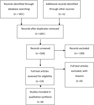
Fig. 1. Flowchart showing the process involved in identifying relevant literature.
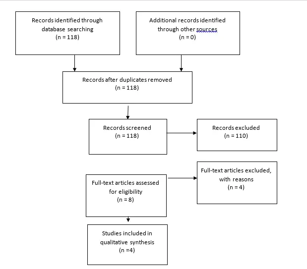
Fig. 2. Flowchart showing the process involved in identifying relevant literature for the 2020 Update. The criteria for “records excluded” and “full text articles excluded with reasons” are detailed in Part 1 of the Guidelines.
Results
Endoscopy
Recommendation: There is insufficient evidence to recommend using endoscopic guidance for routine ventricular catheter placement. Strength of Recommendation: Level I, high degree of clinical certainty.
Four studies were identified to assess the use of endoscopy in the placement of ventricular catheters (Table 1). Two of the earlier reports in the literature described a case series by Vries1 and another by Kellnar et al.2 Vries1 described the technique for insertion of endoscopically placed catheters and reported that 79% of patients for whom endoscopy was used did not require further shunt revision. However, the author did not report the follow-up period, and without a control arm there is no clear demonstration of an advantage of using the endoscope over the standard technique, which relies on anatomical landmarks. Kellnar et al.2 described a case series in which neuroendoscopy was used for placement of ventricular catheters in 14 patients. These authors demonstrated the feasibility of their technique and stated that there were no revisions due to catheter malposition during an 18-month followup period.2 Again, without a control group, no conclusions can be drawn regarding an advantage of endoscopic shunt insertion over standard techniques.
A later study by Villavicencio et al3 retrospectively compared the survival of shunts placed with neuroendoscopic guidance to that of shunts placed without guidance. The authors found no advantage to overall shunt survival between the two study groups (hazard ratio 1.08, 95% CI 0.84–1.41). They did, however, note that the risk of proximal failure was lower in the endoscopy group (odds ratio 0.56, 95% CI 0.32–0.93) and observed an increased rate of distal malfunction when compared with shunts placed without endoscopic assistance.
TABLE 1: Use of endoscopy in the placement of ventricular catheters: summary of evidence*
| First Author & Year | Study Description | Data Class, Quality and Reasons | Results and Conclusions |
| Kestle et al., 2003 | Endoscopic placement of new shunts at 16 centers, randomized to endoscope directed vs non–endoscope directed. 393 pts in study, ventricular catheter scope. | Class IRandomized, blinded, multicenter. | Incidence of shunt failure at 1 yr: Endoscope Insertion Group: 42%; Non–Endoscopic Insertion Group: 34%.Time to 1st shunt failure did not differ between groups (log-rank = 2.92, p = 0.09). |
| Villavicencio et al., 2003 | 447 children underwent total of 965 shunt placements or revisions.605 catheters (63%) placed w/ aid of endoscope; 360 catheters (37.3%) placed w/o endoscope. | Class IIIRetrospective review, historical cohort. | Neuroendoscopy did not independently affect risk of subsequent shunt failure (HR 1.08, 95% CI 0.84–1.41).Endoscopic placement:Independently decreased odds of proximal obstruction (OR 0.56, 95% CI 0.32–0.93);Increased odds of distal malfunction (OR 1.52, 95% CI 1.02–2.72);Was not associated w/ infection (OR 1.42, 95% CI0.78–2.61).Authors state, “Endoscope assisted ventricular catheter placement decreased the odds of proximal obstruction but failed to improve overall shunt survival in this 6-year experience.” |
| Vries, 1980 | 2.7-mm 0° scope used to place ventricular catheter.102 shunts in 85 pts, 67 were new shunts. | Class IIICase series, retrospective, chart review.No statistical analysis. | Max follow-up 18 mos.24 shunt malfunctions in 18 pts: 9 were ventricular catheter malfunctions. 6 infections.Author reported that 79% of the shunts were “troublefree.” |
TABLE 2: Use of ultrasound guidance in the placement of ventricular catheters: summary of evidence
| First Author & Year | Study Description | Data Class, Quality and Reasons | Results and Conclusions |
| Whitehead et al., 2007 | Authors used transcranial sonography via a 2-cm bur hole for CSF ventricular catheter insertion.10 pts w/o open fontanelles & normal-size to slitlike ventricles, July–December 2006. | Class IIICase series & technical note. | 10 of 10 catheters in ventricles.No follow-up.No data on shunt survival. |
TABLE 3: Use of electromagnetic image guidance in the placement of ventricular catheters: summary of evidence
| First Author & Year | Study Description | Data Class, Quality and Reasons | Results and Conclusions |
| Hayhurst et al.,2010 | 3 centers, new shunts, adult & pediatric pts followed up for 1 yr or to shunt failure.Graded by observer blinded to placement method.Pediatric pts: 20 w/o navigation, 15 w/ EM image navigation. | Class IICohort, good-quality prospective study.Follow-up for EM image navigation group was shorter (6 mos) than for no navigation group.Small study, <50% pediatric pts. | Pediatric standard group (no navigation): 20 pts < 18 yrs of age. 6 shunts failed (30%); in 4 cases early failures due to proximal obstruction.EM image navigation group: 15 children, 3 shunts failed (20%). Failures due to infection in 1 pt & valve blockage in 2 pts.No significant difference in overall shunt failure in the pediatric group when EM image guidance was used for shunt placement (p = 0.39, Fisher exact test). |
| Levitt et al., 2012 | 89 pts, 102 shunt surgeries: 58 initial &44 revisions. Image guidance used in 56 surgeries; standard technique used in 46 surgeries. | Class IIIRetrospective review. | 22% shunt failure rate w/ standard technique; 25% shunt failure rate w/ EM image guidance (not significant, p = 0.21, log-rank test).Ventricle size significantly smaller in pts in EM image guidance group (p < 0.02) & in the surgery revision group (p < 0.01), Student t-test. Small ventricle size did not affect shunt failure rate, even when authors controlled for technique of insertion.Image guidance significantly improved accuracy of catheter placement (p < 0.01). Shunt placement accuracy had no significant effect on shunt failure. Significant improvement in catheter position, even though pts in EM image guidance group had smaller ventricles. |
Ultrasound Guidance
Recommendation: The routine use of ultrasound-assisted catheter placement is an option. Strength of Recommendation: Level III, unclear clinical certainty.
The supporting evidence consisted of 1 Class III study with limited follow-up and no control or comparison (Table 2).
Few studies have sought to evaluate the utility of ultrasound in the placement of ventricular catheters. One Class III cohort study with a limited follow-up and no control or comparison met the inclusion criteria and was included as evidence to support this topic. Whitehead et al5 described the technique of placing the ventricular catheter with ultrasound guidance in pediatric patients with closed fontanelles. The authors described the creation of a 2-cm bur hole, followed by catheter placement and ultrasonography to confirm catheter location. There was no analysis of outcomes related to shunt longevity, but immediate postoperative imaging did confirm the expected placement of the catheter away from the choroid plexus.5 Ultrasound-assisted catheter placement may be used to confirm placement of the proximal catheter within the cerebral ventricle.
Electromagnetic Image Guidance
Recommendation: The routine use of computer assisted electromagnetic (EM) navigation is an option. Strength of Recommendation: Level III, unclear clinical certainty.
One Class II study, which did not reach significance in the pediatric subset, and 1 Class III study, which reported a decrease in proximal failure compared with historical reports, were included as evidence to support this topic.
Electromagnetic image guidance has been used (and studied in the pediatric hydrocephalus literature) as a technical adjuvant for assisting placement of ventricular catheters. In a case series by Clark et al6 published in 2008, prospectively collected data confirmed the feasibility of using EM image guidance, specifically in 23 patients with anatomy that was difficult to navigate, including those with small and slit ventricles or complex loculated hydrocephalus. These authors demonstrated a 9% proximal revision rate in the 7-month follow-up period, which they compared with a historical control group in which there was a 35% failure rate. The authors proposed a randomized controlled trial to strengthen this evidence, as their study did not have its own control, had a short follow-up period, and included a small number of patients.
An article by Hayhurst and coauthors7 described a nonrandomized prospective cohort study in which standard shunt placement was compared with EM image– guided shunt placement in both adult and pediatric patients. The end points of the study were failure rates as well as grading of the catheter position as follows: Grade 1, catheter tip floating in CSF equidistant from the ventricle walls, away from the choroid plexus, and in a straight trajectory from the bur hole; Grade 2, catheter tip touching the ventricle wall or the choroid plexus; and Grade 3, part of the catheter tip within the parenchyma or failure to cannulate the ventricle completely. The pediatric cases were analyzed separately with regard to shunt failure, and the data showed a 30% failure rate in the standard cohort and a 20% failure rate in the EM image guidance cohort. This difference was statistically insignificant. In the entire cohort, which included both adult and pediatric patients, “the rate of proximal obstruction falls … from 17 to 6% [when] using EM-navigated placement (p = 0.129, Fisher exact test).”7 Despite that, the overall failure rate was still equivalent, as other components of the system failed, even when the proximal catheter performance improved. There were no Grade 3 catheter positions in the EM image guidance group; this did have a significant impact on shunt survival compared with Grades 1 and 2. These data were not analyzed independently for the pediatric patients included in the study.7
A recent paper by Levitt et al8 also looked at the use of EM navigation versus standard shunt placement. The authors retrospectively analyzed 102 surgeries with frontal catheter placement in pediatric patients to identify the rates of subsequent shunt failure and catheter positioning with the same grading as previously described. The authors found no significant difference in the incidence of proximal obstruction in the EM guidance group (25%) compared with the standard technique group (22%). There was significantly improved catheter positioning in the EM guidance group, although this had no impact on shunt survival. Patients in the EM guidance group were more likely to have small or difficult-to-navigate ventricular anatomy.
Excluded Articles
Seven articles were identified, reviewed, and ultimately excluded because of weak evidence, incomplete evidence, or irrelevance regarding the application of technical adjuvants.9-15 Lam and colleagues12 discussed a Seldinger technique for placing ventricular catheters over endoscopes, and included only a single case. A paper by Chernov et al16 was excluded for a number of factors, including the fact that there were only 4 patients in one group (endoscopic third ventriculostomy [ETV]) and the inclusion range of 5–21 years included 5 patients who were 20 years of age or older.9 In a retrospective review, Shim and colleagues15 compared infants treated with ETV and shunts with infants treated with shunts alone. The authors found improved longevity of the shunt when combined with ETV. Gil et al11 retrospectively evaluated ventricular catheter placement with a frameless optical navigation system, but their study included only 9 patients and was excluded for having a sample size smaller than 10. McMillen et al13 used EM image guidance for a variety of conditions in children that included only 3 cases of hydrocephalus. Piatt and Garton’s paper14 was excluded because it was an analysis of shunt failure and related infection symptoms and frequencies using data derived from two large multicenter studies without direct reference to the techniques being considered in this review. Finally, Farahmand and colleagues10 presented information on a large group of patients older than 16 years of age without separately reporting individuals between the ages of 16 and 19 years, who could have been included.
2020 Update
This review yielded 118 abstracts, of which 8 were selected for full text review. Seven studies were excluded after review8,17-22. There were 4 studies17,18, relevant to ultrasound, but only Whitehead et al23 met inclusion criteria. This study is based on class II data indicating that ultrasound assisted shunt catheter placement did not result in a statistically significant improvement in shunt survival. Studies relevant to endoscopy19 and image guidance20,21 did not meet criteria for inclusion. In conclusion, Whitehead et al23 confirmed the recommendation from the original guideline that ultrasound is an option.
Conclusions
Endoscopy
Recommendation: There is insufficient evidence to recommend using endoscopic guidance for routine ventricular catheter placement. Strength of Recommendation: Level I, high degree of clinical certainty.
Ultrasound Guidance
Recommendation: The routine use of ultrasound-assisted catheter placement is an option. Strength of Recommendation: Level III, unclear clinical certainty.
Electromagnetic Image Guidance
Recommendation: The routine use of computer assisted electromagnetic (EM) navigation is an option. Strength of Recommendation: Level III, unclear clinical certainty.
The availability and quality of evidence is variable throughout the literature for the application of technical adjuvants for catheter placement in the treatment of hydrocephalus by CSF shunting. The strongest body of evidence applies to the use of endoscopy in insertion of shunts. Based on this evidence, endoscopy cannot be recommended for routine use, as no benefit of its application could be identified in the available literature. Less evidence exists for ultrasound and EM image guidance, and therefore a conclusive recommendation cannot be made. Catheter position may be optimized with the application of these technologies, although they may not have an impact on overall outcome with regard to shunt longevity. Insufficient data exist for the use of technical adjuncts in patients with more challenging anatomy, or in the situation in which the ventricle is unable to be cannulated using standard anatomical techniques.
Acknowledgments
We acknowledge the American Association of Neurological Surgeons (AANS)/Congress of Neurological Surgeons (CNS) Joint Guidelines Committee for the members’ reviews, comments, and suggestions; Laura Mitchell, Guidelines Project Manager for the CNS, for her contributions; Pamela Shaw, research librarian, for her assistance with the literature searches; Kevin Boyer for his assistance with data analysis; and Sue Ann Kawecki for her assistance with editing. We also acknowledge the following peer reviewers for their contributions to review the update to the guidelines: Jennifer Sweet, MD, Brandon Rocque, MD, Christoph Greissenauer, MD, Jeffrey Olson, MD.
Disclosure
The systematic review and evidence-based guidelines were funded exclusively by the CNS and AANS Pediatric Section, which received no funding from outside commercial sources to support the development of this document.
Conflict(s) of Interest: None. All Task Force members declared any potential conflicts of interest prior to beginning work on this evidence review.
Conflict(s) of Interest: None. All Pediatric Hydrocephalus Systematic Review and Evidence-Based Guidelines Update Task Force members declared any potential conflicts of interest prior to beginning work on this systematic review and evidence-based guidelines.
Author contributions to the study and manuscript preparation include the following. Conception and design: AANS/CNS Joint Section on Pediatrics. Acquisition of data: all authors. Analysis and interpretation of data: all authors. Drafting the article: Flannery. Critically revising the article: all authors. Reviewed submitted version of manuscript: all authors. Approved the final version of the manuscript on behalf of all authors: Flannery. Administrative/technical/material support: all authors. Study supervision: Flannery.
References
- Vries JK. Endoscopy as an adjunct to shunting for hydrocephalus. Surg Neurol. 1980;13(1):69-72.
- Kellnar S, Boehm R, Ring E. Ventriculoscopy-aided implantation of ventricular shunts in patients with hydrocephalus. Journal of pediatric surgery. 1995;30(10):1450-1451.
- Villavicencio AT, Leveque JC, McGirt MJ, Hopkins JS, Fuchs HE, George TM. Comparison of revision rates following endoscopically versus nonendoscopically placed ventricular shunt catheters. Surg Neurol. 2003;59(5):375-379; discussion 379-380.
- Kestle JR. Administrative database research. Journal of neurosurgery. 2015;122(2):441-442.
- Whitehead WE, Jea A, Vachhrajani S, Kulkarni AV, Drake JM. Accurate placement of cerebrospinal fluid shunt ventricular catheters with real-time ultrasound guidance in older children without patent fontanelles. Journal of neurosurgery. 2007;107(5 Suppl):406-410.
- Clark S, Sangra M, Hayhurst C, et al. The use of noninvasive electromagnetic neuronavigation for slit ventricle syndrome and complex hydrocephalus in a pediatric population. Journal of neurosurgery Pediatrics. 2008;2(6):430-434.
- Hayhurst C, Beems T, Jenkinson MD, et al. Effect of electromagnetic-navigated shunt placement on failure rates: a prospective multicenter study. Journal of neurosurgery. 2010;113(6):1273-1278.
- Levitt MR, O’Neill BR, Ishak GE, et al. Image-guided cerebrospinal fluid shunting in children: catheter accuracy and shunt survival. Journal of neurosurgery Pediatrics. 2012;10(2):112-117.
- Chernov MF, Kamikawa S, Yamane F, Ishihara S, Hori T. Neurofiberscope-guided management of slit-ventricle syndrome due to shunt placement. Journal of neurosurgery. 2005;102(3 Suppl):260-267.
- Farahmand D, Hilmarsson H, Hogfeldt M, Tisell M. Perioperative risk factors for short term shunt revisions in adult hydrocephalus patients. Journal of neurology, neurosurgery, and psychiatry. 2009;80(11):1248-1253.
- Gil Z, Siomin V, Beni-Adani L, Sira B, Constantini S. Ventricular catheter placement in children with hydrocephalus and small ventricles: the use of a frameless neuronavigation system. Child’s nervous system : ChNS : official journal of the International Society for Pediatric Neurosurgery. 2002;18(1-2):26-29.
- Lam S, Harris DA, Lin Y, Rocque BG, Ham S, Pan IW. Outcomes of endoscopic third ventriculostomy in adults. Journal of clinical neuroscience : official journal of the Neurosurgical Society of Australasia. 2016;31:166-171.
- McMillen JL, Vonau M, Wood MJ. Pinless frameless electromagnetic image-guided neuroendoscopy in children. Child’s nervous system : ChNS : official journal of the International Society for Pediatric Neurosurgery. 2010;26(7):871-878.
- Piatt JH, Garton HJ. Clinical diagnosis of ventriculoperitoneal shunt failure among children with hydrocephalus. Pediatric emergency care. 2008(4):201-210. http://www.mrw.interscience.wiley.com/cochrane/clcentral/articles/377/CN-00631377/frame.html.
- Shim KW, Kim DS, Choi JU. Simultaneous endoscopic third ventriculostomy and ventriculoperitoneal shunt for infantile hydrocephalus. Child’s nervous system : ChNS : official journal of the International Society for Pediatric Neurosurgery. 2008;24(4):443-451.
- !!! INVALID CITATION !!! {}.
- Heussinger N, Eyupoglu IY, Ganslandt O, Finzel S, Trollmann R, Jungert J. Ultrasound-guided neuronavigation improves safety of ventricular catheter insertion in preterm infants. Brain & development. 2013;35(10):905-911.
- Crowley RW, Dumont AS, Asthagiri AR, et al. Intraoperative ultrasound guidance for the placement of permanent ventricular cerebrospinal fluid shunt catheters: a single-center historical cohort study. World neurosurgery. 2014;81(2):397-403.
- Beez T, Sarikaya-Seiwert S, Steiger HJ, Hanggi D. Real-time ultrasound guidance for ventricular catheter placement in pediatric cerebrospinal fluid shunts. Child’s nervous system : ChNS : official journal of the International Society for Pediatric Neurosurgery. 2015;31(2):235-241.
- Roth J, Constantini S. Selective use of intra-catheter endoscopic-assisted ventricular catheter placement: indications and outcome. Child’s nervous system : ChNS : official journal of the International Society for Pediatric Neurosurgery. 2012;28(8):1163-1169.
- Hermann EJ, Capelle HH, Tschan CA, Krauss JK. Electromagnetic-guided neuronavigation for safe placement of intraventricular catheters in pediatric neurosurgery. Journal of neurosurgery Pediatrics. 2012;10(4):327-333.
- Thomale UW, Knitter T, Schaumann A, et al. Smartphone-assisted guide for the placement of ventricular catheters. Child’s nervous system : ChNS : official journal of the International Society for Pediatric Neurosurgery. 2013;29(1):131-139.
- Whitehead WE, Riva-Cambrin J, Wellons JC, 3rd, et al. No significant improvement in the rate of accurate ventricular catheter location using ultrasound-guided CSF shunt insertion: a prospective, controlled study by the Hydrocephalus Clinical Research Network. Journal of neurosurgery Pediatrics. 2013;12(6):565-574.
Part 4: Cerebrospinal fluid shunt or endoscopic third ventriculostomy for the treatment of hydrocephalus in children
Reposted with permission from ©AANS, 2014
J Neurosurg Pediatrics (Suppl) 14:30–34, 2014
AANS, 2014
(Original text of the guideline was edited to reflect the update. Please click here for the original publication.)
UPDATE
David Dr. Limbrick Jr., MD, PhD,1 Lissa C. Baird, MD,2Paul Klimo Jr., MD, MPH,3-5 Jay Riva-Cambrin, MD, MSc,6 Ann Marie Flannery, MD7
1Division of Pediatric Neurosurgery, Department of Neurological Surgery, Washington University School of Medicine, St. Louis, Missouri; 2Department of Neurological Surgery, Oregon Health & Science University, Portland, Oregon; 3Semmes-Murphey Neurologic & Spine Institute, 4Department of Neurosurgery, University of Tennessee Health Science Center, and 5Le Bonheur Children’s Hospital, Memphis, Tennessee; 6Division of Pediatric Neurosurgery, University of Utah School of Medicine, Salt Lake City, Utah; 7Department of Neurological Surgery, Saint Louis University, St. Louis, Missouri
Object. The objective of this systematic review was to examine the existing literature comparing CSF shunts and endoscopic third ventriculostomy (ETV) for the treatment of pediatric hydrocephalus and to make evidence-based recommendations regarding the selection of surgical technique for this condition.
Methods. Both the US National Library of Medicine and the Cochrane Database of Systematic Reviews were queried using MeSH headings and key words specifically chosen to identify published articles detailing the use of CSF shunts and ETV for the treatment of pediatric hydrocephalus. Articles meeting specific criteria that had been determined a priori were examined, and data were abstracted and compiled in evidentiary tables. These data were then analyzed by the Pediatric Hydrocephalus Systematic Review and Evidence-Based Guidelines Task Force to consider treatment recommendations based on the evidence.
Results. Of the 122 articles identified using optimized search parameters, 52 were recalled for full-text review. One additional article, originally not retrieved in the search, was also reviewed. Fourteen articles met all study criteria and contained comparative data on CSF shunts and ETV. In total, 6 articles (1 Class II and 5 Class III) were accepted for inclusion in the evidentiary table; 8 articles were excluded for various reasons. The tabulated evidence supported the evaluation of CSF shunts versus ETV.
Conclusions. Cerebrospinal fluid shunts and ETV demonstrated equivalent outcomes in the clinical etiologies studied.
Recommendation: Both CSF shunts and ETV are options in the treatment of pediatric hydrocephalus. Strength of Recommendation: Level II, moderate clinical certainty.
(http://thejns.org/doi/abs/10.3171/2014.7.PEDS14324)
Key Words: hydrocephalus, cerebrospinal fluid, ventriculoperitoneal, shunt, endoscopic third ventriculostomy, practice guidelines
Abbreviations used in this paper: AANS = American Association of Neurological Surgeons; CNS = Congress of Neurological Sur- geons; CPC = choroid plexus cauterization; ETV = endoscopic third ventriculostomy; VP = ventriculoperitoneal.
Selection of the appropriate surgical method—CSF shunt placement or endoscopic third ventriculostomy (ETV)—for the treatment of pediatric hydrocephalus remains a topic of considerable debate. Advocates for ETV cite low failure rates and the potential to avoid shunt placement and its inherent risks as major advantages of the procedure, while supporters of insertion of a ventriculoperitoneal (VP) shunt question the efficacy of ETV in treating hydrocephalus as well as ETV’s unknown effects on neurodevelopment and quality of life.1-3 While there is a great deal of literature on CSF shunts and ETV, there is a relative dearth of articles describing evaluation of both CSF shunt placement and ETV, and there are no randomized trials in which the 2 procedures have been compared.
Endoscopic third ventriculostomy may be beneficial in cases in which there is a clear obstruction to CSF flow and ETV provides an alternate pathway. It is less certain that ETV holds an advantage over shunts in many other causes of hydrocephalus. There is a great deal of interest in the use of ETV with choroid plexus coagulation (CPC) in the treatment of infant hydrocephalus. The Pediatric Hydrocephalus Systematic Review and Evidence-Based Guidelines Task Force anticipates that this topic, along with the effectiveness of ETV compared with shunts in treating hydrocephalus of specific etiologies, will be addressed in subsequent publications. In the current analysis, we sought to systematically review the existing literature detailing the efficacy of CSF shunts versus ETV and to generate evidence-based recommendations for the selection of surgical procedure based on the strength of the available data. Evidence for ETV and a discussion of that procedure in infants younger than 1 year of age is addressed elsewhere in the Guidelines.4
The primary objective of this work was to broadly assess treatment outcomes and review evidence that one treatment may have greater efficacy than the other.
Methods
Search Criteria
The US National Library of Medicine PubMed/ MEDLINE database and the Cochrane Database of Sys- tematic Reviews were queried using MeSH headings, key words, and terms relevant to hydrocephalus, CSF shunts, and ETV. The structure of the literature queries used to search these databases for published articles relevant to hydrocephalus, CSF shunts, and ETV is explained below and in “Part 1: Introduction and Methods.”5
Search Terms
PubMed/MEDLINE
- ((“Hydrocephalus”[Majr]) AND “Ventriculosto- my”[Majr]) AND “Endoscopy”[Mesh]
- Limit 1 to Child (0–18 years)
- Limit to English and Humans Number = 120
Cochrane Database
- MeSH descriptor Child
- MeSH descriptor Infant
- MeSH descriptor Hydrocephalus
- MeSH descriptor Ventriculostomy
- MeSH descriptor Third Ventricle
- (1 or 2) and 3 and 4
- (1 or 2) and 3 and 5
Number = 2 Systematic Reviews
Abstracts of papers that were identified using these optimized search parameters (n = 122) were screened for inclusion criteria. Electronic searches were supplemented by manual searches of article bibliographies. Of the 122 records that were screened, 52 articles were recalled for a full-text review. One additional article identified but not retrieved in the search was also reviewed. Fourteen articles contained comparative data on CSF shunts and ETV, a criterion defined a priori for the current study. Please refer to Part 1 of the Guidelines,5 which offers additional inclusion/exclusion criteria and search terms that were used.
Authors performed an updated literature search (in PubMed and Cochrane Central) for this guideline chapter through a medical librarian at the Congress of Neurological Surgeons Guidelines office using the below-mentioned existing search terms to update the original search through November 30, 2019, yielding an additional 107 abstracts. Of those, 22 were screened for full text review.
Search Strategy
An evidentiary table was constructed to facilitate data review and analysis by the Task Force. Each of the 14 articles was read and reviewed in detail by the full Task Force. Afterward 7 articles were excluded due to potential redundancy in the subject population (n = 3), potential redundancy and variable or inappropriate outcome measures (n = 2), or a clinical scenario in which hydrocephalus might reasonably be expected to resolve without ETV or shunt surgery (n = 2); and 1 article was excluded because it covered simultaneous treatment with a CSF shunt and ETV (Fig. 1).6-13 The evidentiary table was constructed to link recommendations to strengths of evidence (Levels I–III), which were assigned by a group consensus during face-to-face meetings of the Task Force.
Part 4 explores the utility of ventriculoperitoneal shunt (VPS) placement versus endoscopic third ventriculostomy (ETV) for children with hydrocephalus. Twelve new studies out of 107 abstracts resulting from this review met inclusion criteria as described in the original guideline. Of those, nine studies affirmed the previous recommendation stating that VPS and ETV are both options for the treatment of HC in children (Level II).
Search Results
Of the 122 articles identified using the optimized search parameters, 52 were recalled for full-text review. Members of the Task Force who were assigned to the current topic read and discussed all 52 articles recalled for the full-text review as well as the additional study identified and subsequently disqualified. As the primary objective of this study was to assess treatment outcomes following placement of a CSF shunt or ETV, the scope of the evidentiary review was limited to studies that reported quantitative outcomes on both procedures (n = 14). In total, 6 articles (1 Class II and 5 Class III) were accepted for inclusion in the evidentiary table (Table 1)14-19 and 8 were excluded for various reasons as detailed above.6-13 When more than 1 paper covered the same clinical material or included the same subjects, only the paper with the largest patient population and most current data was included in the evidentiary table. The tabulated evidence provided adequate data to allow our evaluation of CSF shunts versus ETV.
Two articles6,8 were excluded because they contained redundant material or constituted duplicate publication. When more than 1 paper covered the same clinical material, only the paper with the largest patient population and most current data was included in Table 1 as evidence to support the topic. Two other articles by the same group of researchers were also excluded: one because of possible subject redundancy and insufficient data to address the primary objective,9 and the other because it contained differing outcome measures.10 Two articles were excluded because they evaluated the role of ETV11 or ETV and CSF shunts7 prior to posterior fossa surgery for tumor excision, a clinical scenario in which hydrocephalus may reasonably be expected to resolve in some cases. Finally, 1 article was excluded because its topic was simultaneous ETV and CSF shunt implantation, which prohibited an assessment of the outcome of either procedure alone.12
The 2020 update yielded 107 abstracts, of which, 22 were selected for full text review and 6 new studies met inclusion criteria as described above.
Results
Recommendation: Both CSF shunts and ETV are options in the treatment of pediatric hydrocephalus. Strength of Recommendation: Level II, moderate clinical certainty.
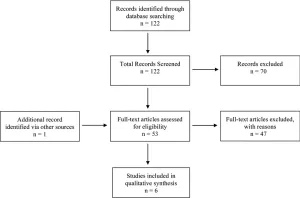
Fig. 1. Flowchart showing the process involved in identifying relevant literature. See text for exclusion criteria at each stage.
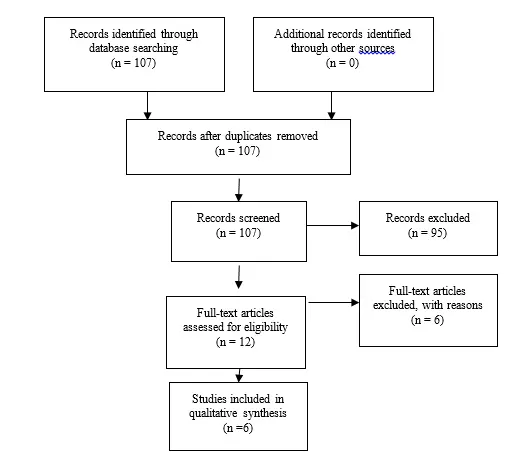
Fig. 2. Flowchart showing the process involved in identifying relevant literature for the 2020 Update. The criteria for “records excluded” and “full text articles excluded with reasons” are detailed in Part 1 of the Guidelines.
Cerebrospinal fluid shunts and ETV demonstrated equivalent outcomes in the clinical scenarios that were studied.
All relevant articles, including those planned for inclusion and exclusion, were reviewed before finalizing the evidentiary table. Each article was presented and discussed in detail, and careful deliberation was made to determine each article’s data class. In sum, there were 5 Class III articles and 1 Class II article (Table 1).
Tuli et al. (1999) published the sole article included in the evidentiary table rated as Class II.19 The authors reported results from a single-center, nonrandomized, prospective cohort study investigating outcomes in 242 consecutive patients treated with ETV (n = 32) or VP shunts (n= 210). While differences in patient ages and the etiology of hydrocephalus were noted among groups (patients who underwent ETV were older at surgery and more likely to have aqueductal stenosis), no significant difference was observed in the procedure failure rate, with a 44% failure rate for ETV and a 45% failure rate for VP shunt surgery.
The remaining 5 articles included in the evidentiary table were rated as Class III studies. Appelgren and colleagues14 (2010) reported a retrospective analysis of prospectively acquired data in 98 patients treated with VP shunts (76 patients) or ETV (22 patients). The primary outcome was failure of the surgery, which was recorded when subsequent surgery was required. Failure rates for VP shunt surgery and ETV were 58% and 55%, respectively, with a mean follow-up of 4.7 years. Study group allocation was uncontrolled, and there was variability in patient ages at surgery, the etiology of hydrocephalus, and other factors.
De Ribaupierre et al.15 (2007) reported the results of a retrospective review of 55 procedures (24 ETVs and 31 VP shunt placements) performed in 48 patients in the context of a literature review. With a median follow-up of 39 months, the authors noted a trend toward a lower failure rate in the ETV group (26% vs 42% in the VP shunt group), although this was not significant. The authors acknowledged nonsignificant differences in patient age and sex as well as in the etiology of hydrocephalus among their groups. Shimizu et al.18 (2012) presented a retrospective, 2-center review of cases in which ETV (9 patients) or VP shunt surgery (36 patients) was performed after removal of infected shunts. No significant difference was observed between the 2 groups in reinfection rates or procedural longevity. Of note, however, 7 of the 9 ETVs ultimately failed in this case series of patients being treated after shunt infection.
Garton et al.16 (2002) reported a retrospective, single- institution, matched cohort study in which ETV was com- pared with VP shunt surgery. With 28 patients in each group, no significant differences were noted between the 2 procedures in the treatment success rate or in cost-effectiveness parameters such as length of stay, operating time, or cost per patient. The authors acknowledge several limitations in this study, including modest sample sizes, a long treatment interval with practice deviation over time, and the possibility of missing late ETV failures (there was 1 hydrocephalus-related death in this group).
The largest study included in the evidentiary table was conducted by Kulkarni et al.8 (2010). This large multicenter comparative study had 2 arms: a retrospective arm for ETV (n = 489), and an arm in which prospectively acquired data for VP shunts was obtained from 2 previous clinical trials and reanalyzed for this study (n = 720). As noted previously, several related studies by the same research group were excluded, because there was likely overlap in the patients included in these studies. Although this paper was rated by the Task Force as Class III, Kulkarni et al. described a sophisticated analysis of ETV versus VP shunt surgery on a large scale. The initial unadjusted models showed lower rates of failure for ETV compared with shunt surgery, but when adjusted for patient age and hydrocephalus etiology, the comparison became more complicated: early failure was higher for ETV than for shunt placement, but at points after 3 months, the ETV failure rate was lower than that for shunt surgery. Based on these findings, the authors concluded that there may be a long-term treatment survival advantage for ETV.
Ultimately, there are a number of limitations to this systematic review. In narrowing the scope of this project to focus specifically on evaluating the method of hydrocephalus treatment—VP shunt or ETV—several key factors known to impact the success of ETV, namely patient age, etiology of hydrocephalus, and history of prior hydrocephalus surgery, were not assessed.8 Thus, the heterogeneity in subject data that we have analyzed herein inherently limits the ability of this recommendation to inform surgical decision making in any specific case. Further, emerging information regarding the role of choroid plexus cauterization (CPC) in conjunction with ETV and alternative indications for ETV with or without CPC (for example, posthemorrhagic hydrocephalus of prematurity) should be evaluated in future iterations of guidelines for the treatment of hydrocephalus as more information becomes available.13 Finally, and most significantly, there is an urgent need for large-scale randomized controlled trials to generate the Level I evidence that is required to definitively address the question of the optimal surgical technique (VP shunt, ETV, or ETV-CPC) in cases of any given hydrocephalus etiology.
2020 Update
Six studies20-25 affirmed the previous recommendation stating that VPS and ETV are both options for the treatment of HC in children (Level II).
Conclusions
Recommendation: Both CSF shunts and endoscopic third ventriculostomy (ETV) are options in the treatment of pediatric hydrocephalus. Strength of Recommendation: Level II, moderate clinical certainty.
Cerebrospinal fluid shunts and ETV demonstrated equivalent outcomes in the clinical scenarios that were studied.
Acknowledgments
We acknowledge the American Association of Neurological Surgeons (AANS)/Congress of Neurological Surgeons (CNS) Joint Guidelines Committee for the members’ reviews, comments, and suggestions; Laura Mitchell, Guidelines Project Manager for the CNS, for her contributions; Pamela Shaw, research librarian, for her assistance with the literature searches; Kevin Boyer for his assistance with data analysis; and Sue Ann Kawecki for her assistance with editing. We also acknowledge the following peer reviewers for their contributions to review the update to the guidelines: Jennifer Sweet, MD, Brandon Rocque, MD, Christoph Greissenauer, MD, Jeffrey Olson, MD.
Disclosure
Dr. Limbrick receives research funding through the National Institute of Neurological Disorders and Stroke. The systematic review and evidence-based guidelines were funded exclusively by the CNS and AANS Pediatric Section, which received no funding from outside commercial sources to support the development of this document.
Conflict(s) of Interest: None. All Task Force members declared any potential conflicts of interest prior to beginning work on this evidence review.
Conflict(s) of Interest: None. All Pediatric Hydrocephalus Systematic Review and Evidence-Based Guidelines Update Task Force members declared any potential conflicts of interest prior to beginning work on this systematic review and evidence-based guidelines.
Author contributions to the study and manuscript preparation include the following. Conception and design: AANS/CNS Joint Section on Pediatrics. Acquisition of data: all authors. Analysis and interpretation of data: all authors. Drafting the article: Limbrick. Critically revising the article: all authors. Reviewed submitted version of manuscript: all authors. Approved the final version of the manuscript on behalf of all authors: Flannery. Administrative/technical/ material support: all authors. Study supervision: Flannery.
Evidence Tables
Table 1 Evidentiary Table Evaluating CSF shunts and ETV
| First Author & Year | Study Description | Data Class, Quality and Reasons | Results and Conclusions |
| Tuli S et al., 1999 | Single center, non-randomized, prospective cohort study. Analyzed all subjects over 10 year period with hydrocephalus from AS or tumor. | Class IIProspective cohort study of consecutive subjects | 32 ETV, 210 VPS patients: 14 (44%) and 95 (45%) failed, respectively. ETV patients were older (median age = 8.1 vs. 3.6 years) and had a higher incidence of aqueductal stenosis (53 vs. 25%). No Difference observed among groups (p = 0.66, hazard ratio = 1.19, 95% confidence interval = 0.55-2.56). |
| Appelgren et al., 2010 | Retrospective analysis of a prospective case series of98 subjects treated with VP shunt or ETV. Primary outcome: procedure failure for VP shunt or ETV. Time to failure also noted. | Class IIIRetrospective analysis of an uncontrolled, prospective case series | Failure: ETV 55%, VPS 58%. Hazard ratios calculated for failure of shunts, prematurity, and length of procedure. Variability present in age, etiology of hydrocephalus, and other factors. |
| de Ribaupierre S et al., 2007 | Retrospective review of 55 procedures in 48 consecutive subjects (24 ETVs, 31 VP shunts). | Class IIIRetrospective review | Difference in failure rate not significant (26% with ETV versus 42% with VP shunt)No difference between groups at 6 months,1 year, 2 years, or 5 years after surgery. |
| Garton et al., 2002 | Matched cohort analysis of ETV vs. VP shunt with comparison of cost-effectiveness and procedure failure rate. n=28 for both groups. | Class IIIRetrospective single-institution matched cohort study. The study interval was prolonged (10 years) and contained practice variation. | ETV success rate of 54% was not significantly different than VP shunt. However, at 34 months post-op, the procedure survival curve favored ETV. No difference between procedures observed in cost or efficacy. |
| Kulkarni et al., 2010 | International, multicenter study comparing retrospective data for ETV and prospectively-acquired data for shunts (from the Shunt Design Trial and the Endoscopic Shunt insertion Trial). Primary outcome: treatment failure, defined as any subsequent surgical procedure for CSF diversion or death related to hydrocephalus management. | Class IIIMulticenter comparative study with two arms: 1) retrospective for ETV; 2) prospectively-acquired data from 2 previous trials re-analyzed in the current study. | Unadjusted models showed lower rates of failure for ETV compared to shunting. After adjusting for age and hydrocephalus etiology, early failure was higher in ETV than shunting; however, at points after 3 months post-surgery, ETV failure was lower than shunting. Conclusion: the risk of ETV failure is initially higher than shunting, but after 3 months, the risk of ETV failure is less than shunt failure. |
| Shimizu et al., 2012 | Retrospective, two-center study of ETV (n=9) or VP shunt (n=36) after removal of infected VP shunt. Compared re-infection rates after each procedure and procedure longevity. | Class IIIRetrospective review, modest sample size. | Reinfection rates were not significantly different between VP shunt (27.8%) and ETV (11.1%). Procedure longevity not different between VP shunt (658 days) and ETV (929 days). |
Table 2. New evidence included in 2020 Update
| First Author & Year | Study Description | Data Class, Quality and Reasons | Results and Conclusions |
| Fani et al, 2013 | Study of children under 2 years with hydrocephalus undergoing ETV | III | Shows no significance resulting from the use of ETV. |
| Jernigan et al, 2014 | Multi-center retrospective cohort study of infants comparing ETV to VPS. | III | This study showed failure rates were higher in ETV than VPS. |
| Kalkarni et al, 2014 | Retrospective, multicenter cohort study of children under age 2 with ETV-CPC versus VPS. | III | ETV- CPC in infants is reasonably safe in certain scenarios. The degree of CPC may be associated with surgeon experience. |
| Lam et al, 2014 | Retrospective multicenter study of children with ETV | III | ETV success was higher in children age 1 year or older and without any prior shunts. |
| Stone et al, 2014 | Prospective study of infants under 2 treated with ETV CPC | III | In this population (North American), ETV-CPC is a safe and effective treatment for hydrocephalus in infants. |
| Vogel et al, 2013 | Retrospective single center study of ETV in children | III | ETV is effective and safe in certain patents with hydrocephalus. When ETV is successful it may limit complications from implanted ventricular shunts. |
References
- Buckley RT, Yuan W, Mangano FT, et al. Longitudinal comparison of diffusion tensor imaging parameters and neuropsychological measures following endoscopic third ventriculostomy for hydrocephalus. Journal of neurosurgery Pediatrics. 2012;9(6):630-635.
- Di Rocco C, Massimi L, Tamburrini G. Shunts vs endoscopic third ventriculostomy in infants: are there different types and/or rates of complications? A review. Child’s nervous system : ChNS : official journal of the International Society for Pediatric Neurosurgery. 2006;22(12):1573-1589.
- Kulkarni AV, Warf BC, Drake JM, Mallucci CL, Sgouros S, Constantini S. Surgery for hydrocephalus in sub-Saharan Africa versus developed nations: a risk-adjusted comparison of outcome. Child’s nervous system : ChNS : official journal of the International Society for Pediatric Neurosurgery. 2010;26(12):1711-1717.
- Mazzola CA, Choudhri AF, Auguste KI, et al. Pediatric hydrocephalus: systematic literature review and evidence-based guidelines. Part 2: Management of posthemorrhagic hydrocephalus in premature infants. Journal of neurosurgery Pediatrics. 2014;14 Suppl 1:8-23.
- Flannery AM, Mitchell L. Pediatric hydrocephalus: systematic literature review and evidence-based guidelines. Part 1: Introduction and methodology. 2014;14(Supplement_1):3.
- Drake JM, Kulkarni AV, Kestle J. Endoscopic third ventriculostomy versus ventriculoperitoneal shunt in pediatric patients: a decision analysis. Child’s nervous system : ChNS : official journal of the International Society for Pediatric Neurosurgery. 2009;25(4):467-472.
- El-Ghandour NM. Endoscopic third ventriculostomy versus ventriculoperitoneal shunt in the treatment of obstructive hydrocephalus due to posterior fossa tumors in children. Child’s nervous system : ChNS : official journal of the International Society for Pediatric Neurosurgery. 2011;27(1):117-126.
- Kulkarni AV, Drake JM, Kestle JR, Mallucci CL, Sgouros S, Constantini S. Predicting who will benefit from endoscopic third ventriculostomy compared with shunt insertion in childhood hydrocephalus using the ETV Success Score. Journal of neurosurgery Pediatrics. 2010;6(4):310-315.
- Kulkarni AV, Shams I, Cochrane DD, McNeely PD. Does treatment with endoscopic third ventriculostomy result in less concern among parents of children with hydrocephalus? Child’s nervous system : ChNS : official journal of the International Society for Pediatric Neurosurgery. 2010;26(11):1529-1534.
- Kulkarni AV, Shams I, Cochrane DD, McNeely PD. Quality of life after endoscopic third ventriculostomy and cerebrospinal fluid shunting: an adjusted multivariable analysis in a large cohort. Journal of neurosurgery Pediatrics. 2010;6(1):11-16.
- Sainte-Rose C, Cinalli G, Roux FE, et al. Management of hydrocephalus in pediatric patients with posterior fossa tumors: the role of endoscopic third ventriculostomy. Journal of neurosurgery. 2001;95(5):791-797.
- Shim KW, Kim DS, Choi JU. Simultaneous endoscopic third ventriculostomy and ventriculoperitoneal shunt for infantile hydrocephalus. Child’s nervous system : ChNS : official journal of the International Society for Pediatric Neurosurgery. 2008;24(4):443-451.
- Warf BC, Campbell JW, Riddle E. Initial experience with combined endoscopic third ventriculostomy and choroid plexus cauterization for post-hemorrhagic hydrocephalus of prematurity: the importance of prepontine cistern status and the predictive value of FIESTA MRI imaging. Child’s nervous system : ChNS : official journal of the International Society for Pediatric Neurosurgery. 2011;27(7):1063-1071.
- Appelgren T, Zetterstrand S, Elfversson J, Nilsson D. Long-term outcome after treatment of hydrocephalus in children. Pediatric neurosurgery. 2010;46(3):221-226.
- de Ribaupierre S, Rilliet B, Vernet O, Regli L, Villemure JG. Third ventriculostomy vs ventriculoperitoneal shunt in pediatric obstructive hydrocephalus: results from a Swiss series and literature review. Child’s nervous system : ChNS : official journal of the International Society for Pediatric Neurosurgery. 2007;23(5):527-533.
- Garton HJ, Kestle JR, Cochrane DD, Steinbok P. A cost-effectiveness analysis of endoscopic third ventriculostomy. Neurosurgery. 2002;51(1):69-77; discussion 77-68.
- Kulkarni AV, Drake JM, Kestle JR, Mallucci CL, Sgouros S, Constantini S. Endoscopic third ventriculostomy vs cerebrospinal fluid shunt in the treatment of hydrocephalus in children: a propensity score-adjusted analysis. Neurosurgery. 2010;67(3):588-593.
- Shimizu T, Luciano MG, Fukuhara T. Role of endoscopic third ventriculostomy at infected cerebrospinal fluid shunt removal. Journal of neurosurgery Pediatrics. 2012;9(3):320-326.
- Tuli S, O’Hayon B, Drake J, Clarke M, Kestle J. Change in ventricular size and effect of ventricular catheter placement in pediatric patients with shunted hydrocephalus. Neurosurgery. 1999;45(6):1329-1333; discussion 1333-1325.
- Fani L, de Jong TH, Dammers R, van Veelen ML. Endoscopic third ventriculocisternostomy in hydrocephalic children under 2 years of age: appropriate or not? A single-center retrospective cohort study. Child’s nervous system : ChNS : official journal of the International Society for Pediatric Neurosurgery. 2013;29(3):419-423.
- Jernigan SC, Berry JG, Graham DA, Goumnerova L. The comparative effectiveness of ventricular shunt placement versus endoscopic third ventriculostomy for initial treatment of hydrocephalus in infants. Journal of neurosurgery Pediatrics. 2014;13(3):295-300.
- Kulkarni AV, Sgouros S, Constantini S. International Infant Hydrocephalus Study: initial results of a prospective, multicenter comparison of endoscopic third ventriculostomy (ETV) and shunt for infant hydrocephalus. Child’s nervous system : ChNS : official journal of the International Society for Pediatric Neurosurgery. 2016;32(6):1039-1048.
- Lam S, Harris D, Rocque BG, Ham SA. Pediatric endoscopic third ventriculostomy: a population-based study. Journal of neurosurgery Pediatrics. 2014;14(5):455-464.
- Stone SS, Warf BC. Combined endoscopic third ventriculostomy and choroid plexus cauterization as primary treatment for infant hydrocephalus: a prospective North American series. Journal of neurosurgery Pediatrics. 2014;14(5):439-446.
- Vogel TW, Bahuleyan B, Robinson S, Cohen AR. The role of endoscopic third ventriculostomy in the treatment of hydrocephalus. Journal of neurosurgery Pediatrics. 2013;12(1):54-61.
Part 5: Effect of valve type on cerebrospinal fluid shunt efficacy
Reposted with permission from ©AANS, 2014
J Neurosurg Pediatrics (Suppl) 14:35–43, 2014
AANS, 2014
(Original text of the guideline was edited to reflect the update. Please click here for the original publication.)
UPDATE
Lissa C. Baird, MD,1 Catherine A. Mazzola, MD,2 Kurtis I Auguste, MD,3 Paul Klimo Jr.MD, MPH,4–6 Ann Marie Flannery, MD7
1Department of Neurological Surgery, Oregon Health & Science University, Portland, Oregon; 2Division of Pediatric Neurological Surgery, Goryeb Children’s Hospital, Morristown, New Jersey; 3Department of Neurosurgery, University of California, San Francisco, California; 4Semmes-Murphey Neurologic & Spine Institute; 5Department of Neurosurgery, University of Tennessee Health Science Center; and 6Le Bonheur Children’s Hospital, Memphis, Tennessee; and 7Department of Neurological Surgery, Saint Louis University, St. Louis, Missouri
Object. The objective of this systematic review was to examine the existing literature to compare differing shunt components used to treat hydrocephalus in children, find whether there is a superior shunt design for the treatment of pediatric hydrocephalus, and make evidence-based recommendations for the selection of shunt implants when placing shunts.
Methods. Both the US National Library of Medicine PubMed/MEDLINE database and the Cochrane Database of Systematic Reviews were queried using MeSH headings and key words chosen to identify publications comparing the use of shunt implant components. Abstracts of these publications were reviewed, after which studies meeting the inclusion criteria were selected. An evidentiary table was compiled summarizing the selected articles and quality of evidence. These data were then analyzed by the Pediatric Hydrocephalus Systematic Review and Evidence-Based Guidelines Task Force to consider evidence-based treatment recommendations.
Results. Two hundred sixty-nine articles were identified using the search parameters, and 43 articles were re- called for full-text review. Of these, 22 papers met the study criteria for a comparison of shunt components and were included in the evidentiary table. The included studies consisted of 1 Class I study, 11 Class II studies, and 10 Class III studies. The remaining 21 articles were excluded.
Conclusions. An analysis of the evidence did not demonstrate a clear advantage for any specific shunt component, mechanism, or valve design over another.
Recommendation: There is insufficient evidence to demonstrate an advantage for one shunt hardware design over another in the treatment of pediatric hydrocephalus. Current designs described in the evidentiary tables are all treatment options. Strength of Recommendation: Level I, high degree of clinical certainty.
Recommendation: There is insufficient evidence to recommend the use of a programmable valve versus a nonprogrammable valve. Programmable and nonprogrammable valves are both options for the treatment of pediatric hydrocephalus. Strength of Recommendation: Level II, moderate degree of clinical certainty. (http://thejns.org/doi/abs/10.3171/2014.7.PEDS14325)
Key Words: hydrocephalus, cerebrospinal fluid shunt, practice guidelines, programmable valve, antisiphon device
Abbreviations used in this paper: AANS = American Association of Neurological Surgeons; CNS = Congress of Neurological Surgeons.
Hydrocephalus is the most common condition treated by pediatric neurosurgeons. Successful management with cerebrospinal fluid shunt systems began after Nulsen and Spitz placed the first implantable shunt in 1949, using a stainless steel ball-valve system.1 Over the next 2 decades, shunt systems evolved to include distal slit valves, proximal slit valves, and diaphragm valves. The subsequent development of artificial valves and silicone tubing advanced shunt design dramatically. Simple differential pressure valves were initially engineered followed by a second generation of valves that included autoregulating, adjustable, antisiphon, and gravitational components.
The objective of this systematic review is to examine literature in which differing shunt components used to treat hydrocephalus in children are compared to find whether there is a superior shunt design for the treatment of pediatric hydrocephalus and to make evidence-based recommendations regarding the selection of shunt implants when placing shunts. Currently, many shunt system components are available to the pediatric neurosurgeon, and they function with a variety of pressure, flow, and siphon control characteristics. Shunt system design has evolved along with attempts to minimize failure rates. The initial use of simple differential pressure valves led to concerns about the disadvantages of siphoning and associated shunt obstruction, subdural hematoma, slit ventricle syndrome, overdrainage, and craniosynostosis. In an attempt to minimize these complications, antisiphon devices have been developed and integrated into shunt systems as intrinsic to the valve mechanism or as separate devices. The antisiphon device is designed to provide progressive resistance to flow to counteract the siphoning that occurs when negative pressure is exerted with vertical positioning. The later development of programmable valves allowed for purposeful alterations in valve function to be made without a surgical procedure.
The purpose of this evidence-based review is to critically evaluate available data on the efficacy of comparable shunt components to determine if one shunt component is superior to another. Additionally, we created evidence-based recommendations on the selection of shunt components based on the strength of the available data. Most of the available evidence focuses on the comparison of shunt valve designs. Study outcome variables accepted for the purposes of this review included shunt survival, shunt complications, development of slit ventricle syndrome, and development of signs or symptoms of overdrainage.
Methods
Search Criteria
The US National Library of Medicine (PubMed/ MEDLINE) and the Cochrane Database of Systematic Reviews were queried for the period January 1966 through March 2012 using MeSH headings and key words relevant to shunt system components as detailed below.
Search Terms
PubMed/MEDLINE
- (“Cerebrospinal Fluid Shunts”[MeSH]) “Hydro- cephalus”[MeSH:noexp]
- 1 AND (programmable OR nonprogrammable OR non-programmable OR siphon OR
antisiphon* OR anti- siphon* OR (“differential pressure” OR “fixed pressure”) OR valve*)
- Limit 2 to Child (0–18 years)
- Limit to English and Humans
Cochrane Database
- MeSH descriptor Child
- MeSH descriptor Infant
- 1 or 2 and (MeSH descriptor Cerebrospinal Fluid Shunts)
- 3 and (MeSH descriptor Hydrocephalus)
- (programmable OR nonprogrammable)
- 4 and 5
Search Results
The search yielded 269 abstracts, which were then reviewed for relevance to the demonstration of superiority of 1 shunt component over another. Forty-three articles were recalled for full-text review. Predetermined inclusion and exclusion criteria were used to review each of these articles in detail. Twenty-two articles were included in the final evidentiary table. Reasons for exclusion of full-text articles included the absence of a valid comparison group (n = 14),2-15 the absence of a valid outcome variable (n = 4),16-19 invalid study design (n = 2),20,21 and redundant patient population (n = 1) (Fig. 1).22
For each article included in the evidentiary table, the study type, summary findings, and major conclusions were recorded, and a preliminary data class was assigned. The Pediatric Hydrocephalus Systematic Review and Evidence-Based Guidelines Task Force met to discuss the ranking of the evidence and the classification of data. Recommendations were then made based on the strength of the data in the evidentiary table (Table 1). In these discussions, if disagreement was encountered among Task Force members, a blinded vote was held and a consensus or majority opinion was reached.
Authors performed an updated literature search (in PubMed and Cochrane Central) for this guideline chapter through a medical librarian at the Congress of Neurological Surgeons Guidelines office using the above-mentioned existing search terms to update the original search through November 30, 2019. (Fig. 2)
Results
The review process identified 1 Class I study, 11 Class II studies, and 10 Class III studies. Only one included article was rated as a Class I study, Kestle et al. (2000),23 in which the investigators performed a randomized controlled trial comparing 3 kinds of valves: all types of standard differential pressure valves, a Delta valve (Medtronic) with an antisiphon mechanism, and an Orbis-Sigma valve (Cordis) with a variable-resis- tance and flow-limiting mechanism. Three hundred forty- four patients were randomly assigned to a valve type and followed up until the time of first shunt failure. Assessed outcome variables included shunt obstruction, overdrain-age, ventricular loculations, and infection. The investigators did not find a significant difference in shunt survival between the 3 valve types in either the short-term (Drake 1998)22 or extended23 follow-up.
Eleven Class II studies24-34 in which differing valve types were compared also failed to demonstrate a superior valve when shunt survival was assessed. Jain et al26 (2000) conducted a prospective cohort study in which they compared shunts using a standard differential pressure valve with a Delta (Medtronic) flow- regulating valve. The authors found no significant difference in overall shunt survival (p = 0.72), with a 5-year survival rate of 58.6% for the differential pressure valves and 58.7% for the Delta valves. The authors did note a relative difference between the 2 groups in the incidence of overdrainage and infection. The differential pressure valve was associated with 4 cases of post-shunt subdural effusion or slit ventricle syndrome, while the Delta valve was associated with only 1 case of subdural effusion. The Delta valve group had 3 infections, whereas the differential pressure valve group had no infections. Warf et al34 (2005) conducted a prospective randomized trial in which they compared the Codman-Hakim microprecision valve with the more affordable Chhabra valve. Ninety children were evaluated after randomization for shunt malfunction, shunt migration, and wound complication. No significant differences in outcome variables were demon Part 5: Effect of valve type on CSF shunt efficacy strated between the 2 groups. Smely and Van Velthoven (1997) conducted a retrospective cohort review in which they compared 66 infants who underwent placement of a ventriculoperitoneal Cordis Orbis-Sigma valve system with 53 patients who underwent placement of a ventricu- loatrial Codman Holter Valve shunt system.41 Forty-eight percent of patients with the Orbis-Sigma valve required one or more revisions while 98.1% of patients with the Holter Valve required 1 or more revisions (p < 0.001). The difference in distal placement of the shunt system is a confounding factor when comparing valve types in this study.
The updated literature review yielded 71 new studies. After the abstracts review, 20 studies were selected for full text review, after which 13 studies were excluded.
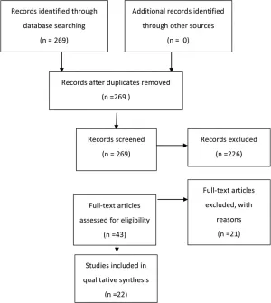
Fig. 1. Flowchart showing the process involved in identifying relevant literature.
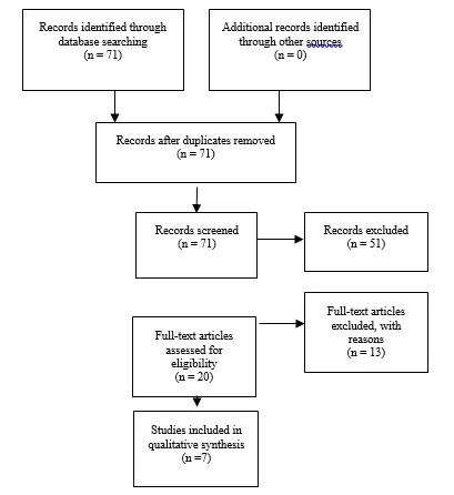
Fig. 2. Flowchart showing the process involved in identifying relevant literature for the 2020 Update. The criteria for “records excluded” and “full text articles excluded with reasons” are detailed in Part 1 of the Guidelines.
Antisiphon Mechanism
Three Class II studies evaluating the antisiphon mechanism were included in our review. Liniger et al28 (2003) studied 27 infants in a prospective cohort study in which a PS Medical medium pressure, flow-controlled valve was compared with a PS Medical 1.0 Delta valve with an antisiphon mechanism. The authors found a lower incidence of slit ventricle syndrome in the Delta valve group (6.25%) than in the flow-controlled valve group (9); however this finding did not reach statistical significance (p > 0.99). The incidence of shunt revision was also lower in the Delta group (0.12 revisions/patient/year) than in the flow-controlled valve group (0.19 revisions/ patient/year), a finding that also failed to reach statistical significance (p = 0.75). Khan et al27 (2010) studied the role of the antisiphon mechanism in a randomized con- trolled trial. Forty patients undergoing shunt placement were randomized to receive a differential pressure valve with an antisiphon device (Vygon shunt) or a differential valve without an antisiphon device (Chhabra or Ceredrain shunts). Shunt blockage, shunt infection, overdrainage, loculated ventricles, and occipitofrontal circumference were assessed in the 2 groups. No end point variables demonstrated a statistically significant difference. Over- drainage complications occurred in 10% of the patients in the group without an antisiphon device as opposed to 0% in the group with an antisiphon device (p = 0.48). A slightly higher infection and obstruction rate was noted in the antisiphon group. In a retrospective cohort study of 475 patients, Davis et al.4 (2000) assessed shunt survival and the development of subdural collection in patients treated with a Delta valve shunt with antisiphon function and in patients treated with one of 2 differential pressure valves without antisiphon control. In a comparison of the 3 groups, no significant difference was found.
The Class III studies that assessed the antisiphon mechanism include a retrospective review by Gruber et al35(1984), in which the authors evaluated 41 patients before and after primary or secondary placement of an antisiphon device. In the secondary placement group fewer complications and proximal catheter obstructions were noted after placement of such a device. However, no statistical analysis was provided by the authors to demonstrate the significance of their findings. In a retrospective cohort review of 101 patients who underwent shunt placement, Virella et al36 (2002) reported no significant differences between patients who underwent placement of a distal slit valve and patients who underwent placement of a Delta valve with an antisiphon component. The authors assessed the number of revisions, infections, and evidence of overdrainage, and reported that 31% of patients in the distal slit valve group required a single shunt revision and 8% required a second revision, whereas 30% of patients in the Delta valve group required a single revision and 20% required a second. Kaiser et al37(1997) reported a prospective but incompletely described comparison study between a conventional medium pressure valve and the Delta valve. The authors found no difference in the number of shunt revisions.
Slit Ventricles
Kan et al38(2007) conducted a retrospective review of 244 patients with at least 1 year of follow-up after primary shunt placement with a differential pressure valve, a Delta valve, or an Orbis-Sigma valve. Variables associated with the development of slitlike ventricles included patient age (younger age at insertion was associated with a higher incidence of slitlike ventricles; p = 0.09), etiology (trauma, infection, and aqueductal stenosis were associated with a higher incidence of slitlike ventricles), and valve type (10.8% of patients with differential pressure valves, 10.5% with Delta valves, and 3.6% with Orbis-Sigma valves developed slitlike ventricles; p = 0.007). This article suggests that a slower reduction in ventricle size and slower flow may lead to larger ventricles after shunt placement. Slit ventricle syndrome was not directly assessed; rather, the radiographic appearance of slitlike ventricles was used as a surrogate outcome.
Programmable Valves
Five Class II studies25,29-32 evaluated programmable valves. Pollack et al32 (1999) conducted a multicenter randomized controlled trial in which they compared the programmable Codman Hakim valve to the surgeon’s choice of any conventional valve. The authors demonstrated comparable efficacy and safety with no statistically significant difference in shunt survival between the experimental and control groups.
Hatlen et al. (2012) published an analysis of programmable and nonprogrammable valve survival.25 The programmable Strata and Codman Hakim valves were com- pared with multiple nonprogrammable valves and found to have significantly lower survival rates (19.8% vs. 45.8%, p = 0.0001). Another retrospective comparison between programmable valves (Strata or Codman-Medos) and non-programmable valves (Medtronic PS Medical) was under- taken by Mangano et al29 (2005). In that study 11% of the programmable valves malfunctioned compared with 0% of the nonprogrammable valves. The authors demonstrated a trend toward longer valve survival and shunt survival in the nonprogrammable group; however, neither reached statistical significance. McGirt et al. (2007) retrospectively reviewed 279 patients who had undergone shunt placement procedures involving either a programmable (Strata or Codman Hakim) or nonprogrammable (PS Medical Delta) valve.30 The authors found that programmable valve placement was associated with a reduced risk of both over- all shunt revision (35% vs 54% in the nonprogrammable group; p = 0.016) and proximal shunt obstruction (12% vs 28% in the nonprogrammable group; p = 0.006). Notarianni et al31 (2009) found no significant difference in a retrospective review of 253 patients who underwent shunt placement with either a programmable (Strata or Codman Hakim) or nonprogrammable (pressure-controlled or not specified) valve. The failure rate among the programmable valve group was 76.1%, and that among the differential pressure valve group was 80.0% (p = 0.11).
Other Comparison Groups
Several Class III studies comparing variable shunt valves were included in the review. Miranda et al39 (2011) describe a retrospective review of 103 patients who received shunts for preterm-related posthemorrhagic hydrocephalus. The authors reported a significantly higher rate of obstruction in patients weighing more than 2000 g who were treated with a fixed medium pressure valve (6 of 8 patients) than in those who were treated with a fixed low pressure valve (12 of 39 patients) (p = 0.040). Contrary findings were reported by Robinson et al. (2002) in a retrospective analysis of shunt malfunction variables in 158 patients.40 Valve opening pressure was the only significant controllable factor found to be associated with shunt malfunction. The 5-year shunt failure rate was 72% in the no valve or low pressure valve group and 47% in the medium or high pressure valve group (p = 0.0005).
Sainte-Rose et al41 (1991) reviewed the charts of 1719 patients with shunted hydrocephalus to assess mechanical complications. These authors found that the flanged ventricular catheter was associated with a higher risk of proximal occlusion (p < 0.04), open-ended distal catheters were associated with fewer distal obstructions (log-rank p < 0.0003), and shunts with proximal medium pressure valves were less likely to malfunction than shunts with distal slit valves (p < 0.000002). Tuli et al42 (2000) did not find valve type to be associated with shunt malfunction in a post hoc analysis of a prospective cohort of 839 patients who underwent primary shunt insertions. No association between shunt malfunction and any component of the shunt hardware was reported in that study.
Ramadwar et al43 (1997) retrospectively compared the efficacy of the Delta valve with the Heyer-Shulte Multi-Purpose valve in 28 patients. Sixty-nine percent of patients with the Delta valve required revision, compared with 53% of patients with the Multi-Purpose valve. The sample size in that study was small, and the data did not reach statistical significance. In an older paper by Serlo et al44 (1986), a retrospective review of 148 children was conducted to compare the Pudenz-Heyer valve with the Cordis Hakim valve. No significant difference was found in overall shunt efficacy, although significance was demonstrated in a higher rate of valve patency on the part of the Pudenz-Heyer valve (p < 0.001).
2020 update
Two studies supported the prior recommendations, including a large prospective cohort study by Riva-Cambrin et al45 which determined valve type had no impact on shunt survival and a retrospective comparison study between the paediGav and Codman Hakim valves by Beez et al46 which showed that valve type did not influence risk of shunt failure. Three studies25,47,48 were published demonstrating significant impact of valve type on shunt survival, however all three studies were class III data and therefore do not affect the original recommendations. Two class III studies49,50 were published demonstrating significance for unique outcome variables.
Excluded Studies
The Task Force excluded 21 articles recalled for full-text review from the final evidentiary table. The majority of excluded papers did not include a comparison group or control group.2-15 Other reasons for exclusion included invalid study design (questionnaire survey),20,21 redundant patient population22 (only the paper with the longest reported follow-up was included), and absence of a valid outcome variable (change in ventricle size, development of spinal canal stenosis, historical description, and frequency of hospital visits).16-19
Conclusions
Recommendation: There is insufficient evidence to demonstrate an advantage of one shunt hardware design over another for the treatment of pediatric hydrocephalus. Current designs described in the evidentiary tables are all treatment options. Strength of Recommendation: Level I, high degree of clinical certainty.
Recommendation: There is insufficient evidence to recommend the use of a programmable valve versus a nonprogrammable valve. Programmable and nonprogrammable valves are both options for the treatment of pediatric hydrocephalus. Strength of Recommendation: Level II, moderate degree of clinical certainty.
The available literature in which one shunt component is compared with another does not demonstrate a clear superiority of one over another. Higher rates of overdrainage were seen with standard differential pressure valves; however, the outcome variables studied in the comparisons of these groups with other shunt mechanisms failed to demonstrate statistical significance. While valves with antisiphon mechanisms may be superior in preventing overdrainage complications, no statistically significant data exist in the current medical literature to support this trend.
The studies assessing programmable versus nonprogrammable valves demonstrated either no statistically significant differences or contrary outcomes, pointing to the need for long-term prospective controlled analysis of this issue. Class III data demonstrating poorer function of distal slit valves in comparison with a proximal valve are described and are consistent with the contemporary decrease in utilization of the former type of shunt system. Many contemporary valve designs exist despite major deficiencies in long-term clinical evaluation. Well-de-signed comparison studies with clearly defined outcome variables and appropriate stratification of patient variables are needed to further investigate the appropriate clinical utilization of these valves. Accessing the necessary patient volume required to reach significance and balancing industry interests with trial integrity may be significant barriers to pursuing needed studies; however, as increasingly expensive and complex valves become available for clinical use, these studies will become imperative.
Based on the 2020 update, there is no new evidence to support a change in the original guideline recommendations.
Acknowledgments
We acknowledge the American Association of Neurological Surgeons (AANS)/Congress of Neurological Surgeons (CNS) Joint Guidelines Committee for the members’ reviews, comments, and suggestions; Laura Mitchell, Guidelines Project Manager for the CNS, for her contributions; Pamela Shaw, research librarian, for her assistance with the literature searches; Kevin Boyer for his assistance with data analysis; and Sue Ann Kawecki for her assistance with editing.
We also acknowledge the following peer reviewers for their contributions to review the update to the guidelines: Jennifer Sweet, MD, Brandon Rocque, MD, Christoph Greissenauer, MD, Jeffrey Olson, MD.
Disclosure
The systematic review and evidence-based guidelines were funded exclusively by the CNS and AANS Pediatric Section, which received no funding from outside commercial sources to support the development of this document.
Conflict(s) of Interest: None. All Task Force members declared any potential conflicts of interest prior to beginning work on this evidence review.
Conflict(s) of Interest: None. All Pediatric Hydrocephalus Systematic Review and Evidence-Based Guidelines Update Task Force members declared any potential conflicts of interest prior to beginning work on this systematic review and evidence-based guidelines.
Author contributions to the study and manuscript preparation include the following. Conception and design: AANS/CNS Joint Section on Pediatrics. Acquisition of data: all authors. Analysis and interpretation of data: all authors. Drafting the article: Baird. Critically revising the article: all authors. Reviewed submitted version of manuscript: all authors. Approved the final version of the manuscript on behalf of all authors: Flannery. Administrative/technical/material support: all authors. Study supervision: Flannery.
Table 1. Valve Type Evidence Table
| Author | Title | Study Description | Data Class, Quality and Reasons | Results and Conclusions |
| Kestle et al., 2000 | Long-Term Follow-Up Data from the Shunt Design Trial | Multicenter randomized trial comparing differential pressure valve, Delta valve, and a Sigma valve. 344 patients with time to shunt failure as endpoint. | Class I: Randomized controlled trial | No clear advantage of one valve over another. |
| Khan et al., 2010 | Role of Shunts with Antisiphon Device in Treatment of Pediatric Hydrocephalus | Role of antisiphon device. 40 patients randomly assigned to shunt with ASD or differential valve. | Class II: Prospective, randomized. Small study with short follow-up (<6 months). | No overdrainage in antisiphon group, 2 patients with overdrainage in non-antisiphon group; higher occlusion and infection in ASD group. No endpoint variables reached statistical significance. |
| Jain H, et al., 1999 | The treatment of infantile hydrocephalus: “differential-pressure” or “flow-control” valves. A pilot study | Prospective data from 50 consecutive first-time shunt insertions. Comparison of shunt survival between differential pressure and flow-regulating (Delta) valves. | Class II: prospective cohort study | No significant difference in shunt survival between two groups: 5-year survival = 58.6% in DPV group and 58.7% in Delta valve group. Higher incidence of overdrainage in DPV group and higher rate of infection in Delta valve group though neither statistically significant. |
| Kestle et al., 2000 | Long-Term Follow-Up Data from the Shunt Design Trial | Multicenter randomized trial comparing differential pressure valve, Delta valve, and a Sigma valve. 344 patients with time to shunt failure as endpoint. | Class I: Randomized controlled trial | No clear advantage of one valve over another. |
| Liniger et al., 2003 | Flow Control versus Antisiphon Valves: Late Results Concerning Slit-Ventricles and Slit-Ventricle Syndrome | 27 infants with flow-control vs antisiphon valves followed for development of slit ventricles and slit ventricle syndrome | Class II: prospective cohort study | No significant difference in development of slit ventricles or slit ventricle syndrome between 2 groups. Slit ventricle syndrome developed in 6.25% of antisiphon group and 9% of flow-controlled valve group. p>0.99 |
| Pollack et al., 1999 | A Randomized, Controlled Study of a Programmable Shunt Valve ersus a Conventional Valve for Patients with Hydrocephalus | 377 patients randomized to receive the Codman Hakim programmable shunt versus a conventional valve of surgeon’s choice and followed for valve explant and shunt failure. | Class II: multicenter randomized controlled trial. Control group not uniform in valve type. | Comparable efficacy and safety with no statistically significant difference in shunt survival. |
| Davis at al., 2000 | The Delta Valve: How Does its Clinical Performance Compare with Two other Pressure Differential Valves without Antisiphon Control | Retrospective cohort study of 475 patients undergoing VP shunt placement with Delta valve with antisiphon function or 2 differential pressure valves without antisiphon (Holter-Hausner and Heyer-Schulte). Endpoints included shunt survival and symptomatic subdural fluid collections | Class II: single institution retrospective cohort study | No significant difference in shunt survival at two-year follow-up: 65% Delta group 66% Holter-Hasner group 64% Heyer-Schulte valve group. No significant difference in rate of subdural fluid collection between groups. |
| Hatlen et a., 2012 | Nonprogrammable and programmable cerebrospinal fluid shunt valves: a 5-year study | Retrospective review of 523 patients undergoing 616 shunt surgeries with 2-year minimum follow-up. Patients with programmable valve placement (Strata and Codman Hakim valves) compared with non-programmable valves (Heyer Schulte, Spetz.er, Delta, and Medtronic). Valve survival was primary endpoint. | Class II: retrospective cohort study. Data obtained from prospectively collected shunt database | 5-year survival for non-programmable valves (45.8%) was significantly higher than that for programmable valves (19.8%). p=0.0001 |
| McGirt et al., 2007 | Adjustable vs set-pressure valves decrease the risk of proximal shunt obstruction in the treatment of pediatric hydrocephalus | 279 patients undergoing shunting procedures with either a programmable (Strata or Codman-Hakim) or a non-programmable valve (PS Medical Delta) were retrospectively reviewed and analyzed for time to shunt malfunction and type of malfunction. | Class II: retrospective cohort review | Programmable valves associated with reduced risk of both overall shunt revision (35% vs 54%, p=0.016) and proximal obstruction (12% vs 28%, p=0.006). |
| Notarianni et al., 2009 | Congenital hydrocephalus and ventriculoperitoneal shunts: influence of etiology and programmable shunts on revisions | 253 patients undergoing shunting procedures with either a programmable (Medtronic Strata or Codman-Hakim) or a non-programmable (pressure-controlled or not specified) valve were retrospectively reviewed and analyzed for time to shunt malfunction. | Class I: Randomized controlled trial | Failure rates (p=0.11) were not significantly different between shunts with programmable valve (76.1%), shunts with non-programmable valve (80.0%), and shunts with non-specified valve (65.0%). |
| Warf B 2005 | Comparison of 1-year outcomes for the Chhabra and Codman-Hakim Micro Precision shunt systems in Uganda: a prospective study in 195 children | 90 patients randomized to receive the Chhabra or Codman-Hakim shunt as primary treatment for hydrocephalus and 105 patients treated with Chhabra shunt after unsuccessful ETV. | Class II: prospective, randomized study. This study was downgraded from a Class I to a Class II due to nonblinded outcome assessors | No difference between two groups in incidence of shunt malfunction, shunt migration, wound complication, or death. |
| Gruberet al., 1984 | Experiences with the anti-siphon device (ASD) in shunt therapy of pediatric hydrocephalus | Retrospective review of 41 patients who had primary or secondary placement of an antisiphon device to their shunt system. Comparison of clinical course before and after placement of ASD. | Class III: retrospective case series. | Fewer shunt malfunctions after ASD placement. Complication rate per patient was four times less frequent and annual ventricular catheter obstruction rate per patient improved 12 times. No statistical analysis to determine significance. |
| Kan eta l., 2007 | Predicting slitlike ventricles in children on the basis of baseline characteristics at the time of shunt insertion | 244 children who underwent shunt placement with either a differential pressure valve, Delta valve or Orbis-Sigma valve and had one year follow-up data were reviewed for development of slit like ventricles. | Class III: retrospective review | 23 patients developed slitlike ventricles: 10.8% of differential pressure valves, 10.5% of Delta valves, and 3.6% of Orbis-Sigma valves p=0.007. Children with DP or Delta valve were 1.66 times more likely to develop slitlike ventricles than with an Orbis-Sigma valve. |
| Mangano et al., 2005 | Early programmable valve malfunctions in pediatric hydrocephalus | 189 children who underwent shunt placement with either a programmable valve (Strata or Codman-Medos with ASD) or a non-programmable valve (PS Medical) were retrospectively analyzed for time to shunt malfunction and CSF protein levels. | Class III: retrospective cohort review, short follow-up (mean=9 months) | Programmable valves had higher rate of malfunction (11.1% compared to 0%) but did not reach statistical significance. |
| Miranda et al., 2011 | Initial Proximal Obstruction of Ventriculoperitoneal Shunt in Patients with Preterm-Related Posthaemorrhagic Hydrocephalus | Retrospective review of shunt survival in 103 patients treated for preterm-related post-hemorrhagic hydrocephalus. | Class III: retrospective review | 42 episodes of obstruction. Fixed medium pressure valves were associated with a higher rate of obstruction compared to low pressure valves, only statistically significant in those patients with a weight over 2000 g, p=0.040. |
| Ramadwar et al., 1997 | Infantile Hydrocephalus: A Comparison of the Delta Valve and Multipurpose Valve | 28 patients underwent retrospective comparison of the efficacy of the Delta valve versus the multipurpose (Heyer-Shulte) valve. | Class III: retrospective review. Small sample. | 69% of patients with Delta valve and 53% of patients with Multipurpose valve required revision. Did not reach statistical significance. |
| Robinson et al., 2002 | Outcome Analysis of Initial Neonatal Shunts: Does the Valve Make a Difference? | 158 patients retrospectively analyzed for significant factors associated with shunt malfunction. | Class III: retrospective case series | Revision rate per year was 4 times higher for patients with no valve or low-pressure valve (72% 5-year failure rate) than for patients with medium or high pressure valve (47% 5-year failure rate). p=0.0005 |
| Sainte-Rose et al., 1991 | Mechanical Complications in Shunts | Retrospective review of the mechanical complications leading to shunt malfunction in 1,719 patients with shunted hydrocephalus. Patients were treated with distal slit valves or medium pressure proximal valves. | Class III: retrospective case series | A higher risk of proximal occlusion is associated with flanged ventricular catheters (p<0.04); shunts with proximal medium pressure valves are less likely to malfunction than shunts with distal slit valves (p<0.000002); open-ended distal catheters associated with fewer distal obstructions (p<0.0003). |
| Smely C, Velthoven V 1997 | Comparative Study of Two Customary Cerebrospinal Fluid Shunting Systems in Early Childhood Hydrocephalus | Retrospective review of 66 infants treated with CordisOrbis-Sigma Valve compared to 53 children treated with Codman Holter Valve ventriculo-atrial system. | Class III: retrospective cohort review | Codman Holter Valve group demonstrated a greater than double risk of shunt complications in comparison to the ventriculoperitoneal Orbis-Sigma valve system. 48.5% of patients with OSV required one or more revisions, 98.1% of patients with HV required one or more revisions (p<0.001). |
| Tuli et al., 2000 | Risk factors for repeated cerebrospinal shunt failures in pediatric patients with hydrocephalus | Data prospectively collected on 839 patients undergoing primary shunt insertion. 1183 episodes of shunt failure occurred. Valve types included flow regulated and differential pressure. | Class III: prospective cohort study, post-hoc analysis | No evidence of an association between shunt malfunction and type of shunt hardware. |
| Virella et al., 2002 | Distal slit valve and clinically relevant CSF overdrainage in children with hydrocephlus | Retrospective review of 101 patients undergoing shunt placement with a distal slit valve or a Delta level 1 valve with antisiphon component. | Class III: retrospective case series | No significant differences were found between the DSV and Delta with AS groups in number of revisions, infections, or over drainage.. |
| Kaiser et al., 1997 | Conventional Versus Delta Valve in the Treatment of Hydrocephalus in Early Infancy | Prospective study comparing conventional medium valve with Delta level 1 valve in 25 infants younger than 6 months. | Class III: prospective randomized. Poor description of study design and data. | No difference in number of revisions. Fewer proximal revisions in Delta Valve group. No determination of significance from described data. |
| Serlo et al., 1986 | Ball and Spring or Slit and Core Valve for Hydrocephalus Shunting? | Retrospective review of consecutive series of 148 children treated with shunting procedures with either the Pudenz-Heyer valve or the Hakim-Cordis valve. | Class III: retrospective review | No significant differences in efficacy. Tendency towards increased rate of catheter rupture in Pudenz-Heyer valve and increased rate of slit ventricles in Hakim-Cordis valve. The higher patency rate of the Pudenz-Heyer valve was statistically significant (p<0.001). |
*ASD = antisiphon device; DPV = differential pressure valve; ETV = endoscopic third ventriculostomy; pt = patient; VP = ventriculoperitoneal.
Table 2. New evidence included in 2020 Update
| Author | Study Description | Data Class, Quality and Reasons | Results and Conclusions |
| Riva-Cambrin et al, 2016 | A prospective observational study to determine risk factors for shunt failure. | II | This study determined that valve type had no impact on shunt survival. |
| Beez et al, 2014 | A retrospective study of patients <16 comparing paediGav and Codman Hakim valves. | II | This study showed that valve type did not influence the risk of shunt failure. |
| Beuriat et al, 2017 | Retrospective review of children treated for hydrocephalus. | III | This study demonstrated the significant impact of valve type on shunt survival. |
| Hatlen et al, | A retrospective study of patients with CSF shunt insertion. | III | This study demonstrated the significant impact of valve type on shunt survival. |
| Alavi et al, | A retrospective study of patients who received a valve exchange towards an adjustable differential pressure valve with gravitational unit. | III | This study demonstrated the significant impact of valve type on shunt survival. |
| Kahilogullari et al, 2018 | A retrospective study of patients who had shunt surgery at the time of myelomeningocele repair. | III | This study demonstrated significance for unique outcome variables. |
| Oushy et al, 2017 | A retrospective study of infants and neonates who had multifocal intraparenchymal hemorrhages following shunt placement. Ventricular size ratios, laboratory values, clinical presentation, shunt and valve type, and operative timing and approach were analyzed. | III | This study demonstrated significance for unique outcome variables. |
References
- Nulsen FE, Spitz EB. Treatment of hydrocephalus by direct shunt from ventricle to jugular vain. Surgical forum. 1951:399-403.
- Ahn ES, Bookland M, Carson BS, Weingart JD, Jallo GI. The Strata programmable valve for shunt-dependent hydrocephalus: the pediatric experience at a single institution. Child’s nervous system : ChNS : official journal of the International Society for Pediatric Neurosurgery. 2007;23(3):297-303.
- Arnell K, Eriksson E, Olsen L. The programmable adult Codman Hakim valve is useful even in very small children with hydrocephalus. A 7-year retrospective study with special focus on cost/benefit analysis. Eur J Pediatr Surg. 2006;16(1):1-7.
- Breimer GE, Sival DA, Hoving EW. Low-pressure valves in hydrocephalic children: a retrospective analysis. Child’s nervous system : ChNS : official journal of the International Society for Pediatric Neurosurgery. 2012;28(3):469-473.
- Guthkelch AN. The treatment of infantile hydrocephalus by the Holter valve. Br J Surg. 1967;54(8):665-673.
- Haberl EJ, Messing-Juenger M, Schuhmann M, et al. Experiences with a gravity-assisted valve in hydrocephalic children. Clinical article. Journal of neurosurgery Pediatrics. 2009;4(3):289-294.
- Hahn YS. Use of the distal double-slit valve system in children with hydrocephalus. Child’s nervous system : ChNS : official journal of the International Society for Pediatric Neurosurgery. 1994;10(2):99-103.
- Hanlo PW, Cinalli G, Vandertop WP, et al. Treatment of hydrocephalus determined by the European Orbis Sigma Valve II survey: a multicenter prospective 5-year shunt survival study in children and adults in whom a flow-regulating shunt was used. Journal of neurosurgery. 2003;99(1):52-57.
- Hertle DN, Tilgner J, Fruh K, et al. Reversible occlusion (on-off) valves in shunted tumor patients. Neurosurgical review. 2010;34(2):235-242.
- Hoekstra A. Artificial shunting of cerebrospinal fluid. The International journal of artificial organs. 1994;17(2):107-111.
- Kestle JR, Walker ML. A multicenter prospective cohort study of the Strata valve for the management of hydrocephalus in pediatric patients. Journal of neurosurgery. 2005;102(2 Suppl):141-145.
- Martinez-Lage JF, Almagro MJ, Del Rincon IS, et al. Management of neonatal hydrocephalus: feasibility of use and safety of two programmable (Sophy and Polaris) valves. Child’s nervous system : ChNS : official journal of the International Society for Pediatric Neurosurgery. 2008;24(5):549-556.
- Mauer UM, Kunz U. More malfunctioning Medos Hakim programmable valves: cause for concern? Journal of neurosurgery. 2011;115(5):1047-1052.
- Meling TR, Egge A, Due-Tonnessen B. The gravity-assisted Paedi-Gav valve in the treatment of pediatric hydrocephalus. Pediatric neurosurgery. 2005;41(1):8-14.
- Piatt JH, Jr., Carlson CV. A search for determinants of cerebrospinal fluid shunt survival: retrospective analysis of a 14-year institutional experience. Pediatric neurosurgery. 1993;19(5):233-241; discussion 242.
- Jain H, Natarajan K, Sgouros S. Influence of the shunt type in the difference in reduction of volume between the two lateral ventricles in shunted hydrocephalic children. Child’s nervous system : ChNS : official journal of the International Society for Pediatric Neurosurgery. 2005;21(7):552-558.
- Keen J. Casey Holter and the Spitz-Holter valve. Eur J Pediatr Surg. 1992;2 Suppl 1:5-6.
- Kondageski C, Thompson D, Reynolds M, Hayward RD. Experience with the Strata valve in the management of shunt overdrainage. Journal of neurosurgery. 2007;106(2 Suppl):95-102.
- Nomura S, Fujii M, Kajiwara K, et al. Factors influencing spinal canal stenosis in patients with long-term controlled hydrocephalus treated with cerebrospinal fluid shunt. Child’s nervous system : ChNS : official journal of the International Society for Pediatric Neurosurgery. 2010;26(7):931-935.
- Miyake H, Ohta T, Kajimoto Y, Ogawa D. A clinical survey of hydrocephalus and current treatment for hydrocephalus in Japan: analysis by nationwide questionnaire. Child’s nervous system : ChNS : official journal of the International Society for Pediatric Neurosurgery. 1999;15(8):363-368.
- Moritake K, Nagai H, Miyazaki T, et al. Analysis of a nationwide survey on treatment and outcomes of congenital hydrocephalus in Japan. Neurologia medico-chirurgica. 2007;47(10):453-460; discussion 460-451.
- Drake JM, Kestle JR, Milner R, et al. Randomized trial of cerebrospinal fluid shunt valve design in pediatric hydrocephalus. Neurosurgery. 1998;43(2):294-303; discussion 303-295.
- Kestle J, Drake J, Milner R, et al. Long-term follow-up data from the Shunt Design Trial. Pediatric neurosurgery. 2000;33(5):230-236.
- Davis SE, Levy ML, McComb JG, Sposto R. The delta valve: how does its clinical performance compare with two other pressure differential valves without antisiphon control? Pediatric neurosurgery. 2000;33(2):58-63.
- Hatlen TJ, Shurtleff DB, Loeser JD, Ojemann JG, Avellino AM, Ellenbogen RG. Nonprogrammable and programmable cerebrospinal fluid shunt valves: a 5-year study. Journal of neurosurgery Pediatrics. 2012;9(5):462-467.
- Jain H, Sgouros S, Walsh AR, Hockley AD. The treatment of infantile hydrocephalus: “differential-pressure” or “flow-control” valves. A pilot study. Child’s nervous system : ChNS : official journal of the International Society for Pediatric Neurosurgery. 2000;16(4):242-246.
- Khan RA, Narasimhan KL, Tewari MK, Saxena AK. Role of shunts with antisiphon device in treatment of pediatric hydrocephalus. Clinical neurology and neurosurgery. 2010;112(8):687-690.
- Liniger P, Marchand S, Kaiser GL. Flow control versus antisiphon valves: late results concerning slit ventricles and slit-ventricle syndrome. Eur J Pediatr Surg. 2003;13 Suppl 1:S3-6.
- Mangano FT, Menendez JA, Habrock T, et al. Early programmable valve malfunctions in pediatric hydrocephalus. Journal of neurosurgery. 2005;103(6 Suppl):501-507.
- McGirt MJ, Buck DW, 2nd, Sciubba D, et al. Adjustable vs set-pressure valves decrease the risk of proximal shunt obstruction in the treatment of pediatric hydrocephalus. Child’s nervous system : ChNS : official journal of the International Society for Pediatric Neurosurgery. 2007;23(3):289-295.
- Notarianni C, Vannemreddy P, Caldito G, et al. Congenital hydrocephalus and ventriculoperitoneal shunts: influence of etiology and programmable shunts on revisions. Journal of neurosurgery Pediatrics. 2009;4(6):547-552.
- Pollack IF, Albright AL, Adelson PD. A randomized, controlled study of a programmable shunt valve versus a conventional valve for patients with hydrocephalus. Hakim-Medos Investigator Group. Neurosurgery. 1999;45(6):1399-1408; discussion 1408-1311.
- Smely C, Van Velthoven V. Comparative study of two customary cerebrospinal fluid shunting systems in early childhood hydrocephalus. Acta neurochirurgica. 1997;139(9):875-881; discussion 882.
- Warf BC. Comparison of 1-year outcomes for the Chhabra and Codman-Hakim Micro Precision shunt systems in Uganda: a prospective study in 195 children. Journal of neurosurgery. 2005;102(4 Suppl):358-362.
- Gruber R, Jenny P, Herzog B. Experiences with the anti-siphon device (ASD) in shunt therapy of pediatric hydrocephalus. Journal of neurosurgery. 1984;61(1):156-162.
- Virella AA, Galarza M, Masterman-Smith M, Lemus R, Lazareff JA. Distal slit valve and clinically relevant CSF overdrainage in children with hydrocephalus. Child’s nervous system : ChNS : official journal of the International Society for Pediatric Neurosurgery. 2002;18(1-2):15-18.
- Kaiser GL, Horner E, Marchand S, Jost A. Conventional versus Delta valve in the treatment of hydrocephalus in early infancy. Eur J Pediatr Surg. 1997;7 Suppl 1:45-46.
- Kan P, Walker ML, Drake JM, Kestle JR. Predicting slitlike ventricles in children on the basis of baseline characteristics at the time of shunt insertion. Journal of neurosurgery. 2007;106(5 Suppl):347-349.
- Miranda P, Simal JA, Menor F, Plaza E, Conde R, Botella C. Initial proximal obstruction of ventriculoperitoneal shunt in patients with preterm-related posthaemorrhagic hydrocephalus. Pediatric neurosurgery. 2011;47(2):88-92.
- Robinson S, Kaufman BA, Park TS. Outcome analysis of initial neonatal shunts: does the valve make a difference? Pediatric neurosurgery. 2002;37(6):287-294.
- Sainte-Rose C, Piatt JH, Renier D, et al. Mechanical complications in shunts. Pediatric neurosurgery. 1991;17(1):2-9.
- Tuli S, Drake J, Lawless J, Wigg M, Lamberti-Pasculli M. Risk factors for repeated cerebrospinal shunt failures in pediatric patients with hydrocephalus. Journal of neurosurgery. 2000;92(1):31-38.
- Ramadwar RH, Carachi R, Young DG. Infantile hydrocephalus: a comparison of the Delta valve and multipurpose valve. Eur J Pediatr Surg. 1997;7 Suppl 1:44-45.
- Serlo W, von Wendt L, Heikkinen ES, Heikkinen ER. Ball and spring or slit and core valve for hydrocephalus shunting? Annals of clinical research. 1986;18 Suppl 47:103-106.
- Riva-Cambrin J, Kestle JR, Holubkov R, et al. Risk factors for shunt malfunction in pediatric hydrocephalus: a multicenter prospective cohort study. Journal of neurosurgery Pediatrics. 2016;17(4):382-390.
- Beez T, Sarikaya-Seiwert S, Bellstadt L, Muhmer M, Steiger HJ. Role of ventriculoperitoneal shunt valve design in the treatment of pediatric hydrocephalus–a single center study of valve performance in the clinical setting. Child’s nervous system : ChNS : official journal of the International Society for Pediatric Neurosurgery. 2014;30(2):293-297.
- Beuriat PA, Puget S, Cinalli G, et al. Hydrocephalus treatment in children: long-term outcome in 975 consecutive patients. Journal of neurosurgery Pediatrics. 2017;20(1):10-18.
- Alavi S, Schulz M, Schaumann A, Schwarz K, Thomale UW. Valve exchange towards an adjustable differential pressure valve with gravitational unit, clinical outcome of a single-center study. Child’s nervous system : ChNS : official journal of the International Society for Pediatric Neurosurgery. 2017;33(5):759-765.
- Kahilogullari G, Etus V, Guler TM, Karabagli H, Unlu A. Does Shunt Selection Affect the Rate of Early Shunt Complications in Neonatal Myelomeningocele-Associated Hydrocephalus? A Multi-Center Study. Turkish neurosurgery. 2018;28(2):303-306.
- Oushy S, Parker JJ, Campbell K, et al. Frontal and occipital horn ratio is associated with multifocal intraparenchymal hemorrhages in neonatal shunted hydrocephalus. Journal of neurosurgery Pediatrics. 2017;20(5):432-438.
Part 6: Preoperative antibiotics for shunt surgery in children with hydrocephalus: a systematic review and meta-analysis
Reposted with permission from ©AANS, 2014
J Neurosurg Pediatrics (Suppl) 14:44–52, 2014
AANS, 2014
(Original text of the guideline was edited to reflect the update. Please click here for the original publication.)
UPDATE
Paul Klimo Jr. MD, MPH ,1–3 Mark Van Poppel, MD,2,3 Clinton J. Thompson, PhD,5 Lissa C. Baird, MD,4 Ann-Christine Duhaime, MD,6 Ann Marie Flannery, MD7
1Semmes-Murphey Neurologic & Spine Institute; 2Department of Neurosurgery, University of Tennessee Health Science Center; and 3Le Bonheur Children’s Hospital, Memphis, Tennessee; 4Department of Neurological Surgery, Oregon Health & Science University, Portland, Oregon; 5School of Public Health and Health Services, The George Washington University, Washington, DC; 6 Department of Pediatric Neurosurgery, Massachusetts General Hospital, Boston, Massachusetts; and 7Department of Neurological Surgery, Saint Louis University, St. Louis, Missouri
Object. The objective of this systematic review and meta-analysis was twofold: to answer the question “What is the evidence for the effectiveness of prophylactic intravenous antibiotics for infection prevention in shunt surgery?” and to make treatment recommendations based on the available evidence.
Methods. The US National Library of Medicine PubMed/MEDLINE database and the Cochrane Database of Systematic Reviews were queried using MeSH headings and key words relevant to prophylactic antibiotic use in children undergoing a shunt operation. Abstracts were reviewed to identify which studies met the inclusion criteria. An evidentiary table was assembled summarizing the studies and the quality of evidence (Classes I–III). A meta-analysis was conducted using a random-effects model to calculate a cumulative estimate of treatment effect using risk ratio (RR). Heterogeneity was assessed using chi-square and I2 statistics. A sensitivity analysis was also conducted. Based on the quality of the literature and the result of the meta-analysis, a recommendation was rendered (Level I, II, or III). Results. Nine studies (4 Class I, 3 Class II, and 2 Class III) met our inclusion criteria. Of 7 randomized controlled trials (RCTs), 3 were downgraded from Class I to Class II because of significant quality issues, and all RCTs were potentially underpowered. In only 2 Class III retrospective cohort studies were preoperative antibiotic agents found to be protective against shunt infection. When data from the individual studies were pooled together, the infection rate in the prophylactic antibiotics group was 5.9% compared with 10.7% in the control group. Using a random-effects model, the cumulative RR was 0.55 (95% CI 0.38–0.81), indicating a protective benefit of prophylactic preoperative intravenous antibiotics. A sensitivity analysis of RCTs only (n = 7) also demonstrated a statistical benefit, but an analysis of higher-quality RCTs only (n = 4) did not.
Conclusions. Within the limits of this systematic review and meta-analysis, administration of preoperative anti- biotic agents for shunt surgery in children was found to lower the infection risk (quality of evidence: Class II; strength of recommendation, Level II).
Recommendation: The use of preoperative antibiotic agents can be recommended to prevent shunt infection in patients with hydrocephalus. It was only by combining the results of the various underpowered studies (meta- analysis) that the use of preoperative antibiotics for shunt surgery in children was shown to lower the risk of shunt infection. Strength of Recommendation: Level II, moderate degree of clinical certainty.
Key Words: preoperative, perioperative, antibiotics, intravenous, shunt, cerebrospinal fluid, infection, meta-analysis, pediatrics, evidence-based guidelines, practice guidelines, hydrocephalus
Abbreviations used in this paper: AANS = American Association of Neurological Surgeons; CNS = Congress of Neurological Surgeons; RCT = randomized controlled trial; RR = risk ratio; VP= ventriculoperitoneal.
Cerebrospinal fluid shunting procedures are among the most common procedures performed by pediatric neurosurgeons, but infection remains a challenging complication to prevent. Care of patients with infected shunts often requires prolonged hospitalization, the most common procedures performed by pediatric neurosurgeons, but infection remains a challenging complication to prevent. Care of patients with infected shunts often requires prolonged hospitalization, long-term intravenous antibiotic administration, and multiple surgical procedures; therefore, it comes as no surprise that shunt infections are among the most expensive implant-related complications to treat, with an annual estimated total cost of $100–250 million.1-3 The clinical consequences of a shunt infection include seizures, psychomotor retardation, reduced intelligence quotient, and increased mortality.4-9
The rate of shunt infection reported varies considerably in the literature, but in recent studies the incidence typically ranges from 5% to 15%, with rates typically higher in high-risk groups such as preterm neonates and patients recently treated for shunt infection.10-14 Evidence of a shunt infection often manifests within 2 months after surgery. Infections are typically caused by gram-positive opportunistic pathogens that colonize the skin of the patient such as coagulase-negative Staphylococcus, S. epidermidis, and S. aureus.10
The administration of preoperative antibiotic agents, typically those providing gram-positive coverage such as a first-generation cephalosporin, nafcillin, clindamycin, or vancomycin, has been demonstrated or is assumed to lower the infection rate for all major cranial and spinal neurosurgical procedures.15-17 Numerous studies, including 3 meta-analyses,18-20 have investigated the use of preoperative antibiotics as prophylaxis against shunt infection, both for adult and pediatric patients. Many of the original trials date back to the 1980s, and it is one of the few areas in neurosurgery in which there is an abundance of randomized controlled trials (RCTs).
Currently, the use of prophylactic antibiotics for shunt surgery is almost ubiquitous but not uniform. Biyani et al.21 conducted a survey study in which they asked pediatric neurosurgeons to detail their use of prophylactic antibiotics. All 45 respondents indicated that they used antibiotics; however, there was notable variability in the choice of drug (first-generation cephalosporins [n = 23], second-generation cephalosporins [n = 10], naficillin/oxacillin [n = 4], vancomycin [n = 3], clindamycin [n = 1], amoxicillin [n = 1], or mixed protocols [n = 3]); the timing of drug delivery (in the department before transfer to the operating room [n = 5], upon arrival at the operating room [n = 11], at induction of anesthesia [n = 13], or at initial skin incision [n = 16]); and the duration of drug delivery (single dose [n = 13], 24-hour administration [n= 26], 48-hour administration [n = 2], or administration longer than 48 hours [n = 4]). Surgeons also tended to modify their protocols for certain “high-risk” patients.
The purpose of this evidence-based review is to critically examine data on the use of preoperative prophylactic antibiotics in children undergoing shunt surgery.
Methods
Search Terms
We searched the US National Library of Medicine PubMed/MEDLINE database and the Cochrane Database of Systematic Reviews for the period from January 1966 to March 2012 using the MeSH subject headings and the following PubMed search terms: 1. (“Cerebrospinal Fluid Shunts”[MeSH] OR “shunt systems” OR (“cerebrospinal fluid” AND (shunt* OR catheter*))); and then 1 AND (infection OR infections OR “shunt infection”) AND (“Anti-Bacterial Agents”[MeSH] OR (antibiotic OR antibiotics)) AND (prophylaxis OR prevention OR protective).
Search Strategy
We reviewed the retrieved articles’ titles and abstracts to identify studies addressing the rate of shunt infection in patients treated with preoperative antibiotic agents compared with those treated with no prophylaxis. Uncontrolled studies were excluded, as were studies that evaluated intrathecal antibiotics. In all papers, the authors must have stated that the only variable that changed was the administration of perioperative antibiotics; all other aspects of the surgery and technique remained unchanged. In addition, the authors must have provided details regarding their prophylaxis protocol—drug(s), dosage, and timing of administration before and, if applicable, after surgery.
Authors performed an updated literature search (in PubMed and Cochrane Central) for this guideline chapter through a medical librarian at the Congress of Neurological Surgeons Guidelines office using the above-mentioned existing search terms to update the original search through November 30, 2019.
Meta-Analysis
For each study, we identified the number of infections in the group of patients who were treated by antibiotics as well as in the group of patients who did not receive prophylactic antibiotics (control group). We then computed the risk of an infection for the treatment group relative to the control group, yielding a risk ratio (RR). An RR less than 1 is indicative of protection against infection for prophylactic antibiotics. The overall RR was computed using the method of DerSimonian and Laird.22
We conducted a random-effects meta-analysis of the selected studies. A random-effects model—as opposed to a fixed-effects model—assumes the measure of association (that is, the RR) varies around an overall average treatment effect. A random-effects model yields a more conservative estimate of the summary effect. We assessed heterogeneity by way of the chi-square test of heterogeneity and the I2 statistic, in which the former returns a chi-square distributed test statistic and corresponding p value and the latter returns a value bound between 0% and 100%, with higher values denoting increasing heterogeneity. We regarded a chi-square test of heterogeneity p value less than alpha = 0.10 and an I2 value in the range of 30% to 60% as suggestive of moderate heterogeneity.23,24
We performed a sensitivity analysis by repeating the meta-analysis using only RCTs and then further by examining only the higher-quality RCTs. An examination of publication bias was not conducted, because the number of studies included in this analysis was not large enough to provide adequate power (i.e., fewer than 10 studies).
Search Results
Our search identified 177 articles; another 7 articles were found through a search of the articles’ bibliographies (Fig. 1). One hundred sixty-two articles were excluded based on a review of the abstract. Twenty-two full-length papers were reviewed, of which 13 were rejected for the following reasons: studies either enrolled only adults or separate results for children were not provided;25-29 there was no comparison group;30 prophylactic antibiotic use was part of a shunt surgery protocol, and thus more than one variable was conceivably being altered;31,32 clinical shunt infection was not used as the outcome;33 no details regarding the prophylaxis used were provided;7 a variety of antibiotics was used (that is, there was no standardization of the prophylaxis protocol);34 or data specific to shunt surgery were not available.35,36 Therefore, 9 articles satisfied inclusion criteria for this systematic review and meta-analysis (Table 1).9,37-44
Two studies from the 92 yielded by the 2020 update were pulled for full text review but neither met inclusion criteria from the original guideline and were not included (Figure 1b).
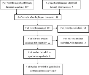
Fig. 1. Flowchart showing the selection of studies for inclusion in the review and meta-analysis.
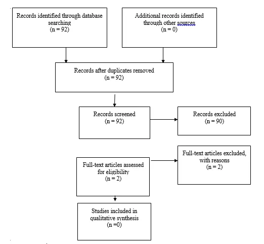
Fig. 1b Flowchart showing the process involved in identifying relevant literature for the 2020 Update. The criteria for “records excluded” and “full text articles excluded with reasons” are detailed in Part 1 of the Guidelines.
Results
The review process identified 4 Class I, 3 Class II, and 2 Class III studies. Seven of the 9 papers described RCTs, but there were notable methodological shortcomings and incomplete studies as detailed below. The primary outcome of interest—shunt infection—was defined by the authors of the individual studies but in general included signs and symptoms of a shunt malfunction or infection with an organism cultured from the CSF or shunt apparatus, purulence from the shunt wound(s), or abdominal fluid/pseudocyst in a patient after a recent shunt surgery. Some investigators also deemed that a patient had an infection if there were highly suggestive findings such as fever, redness along the shunt, or CSF pleocytosis in the absence of positive cultures. Among the studies, there was variability in which antibiotics were tested, their administration relative to the skin incision, and whether the patient received postoperative antibiotics. The only constant feature was that the children in the study groups all received preoperative antibiotics of some kind and at some time interval prior to skin incision. Overall, only 2 studies found that preoperative antibiotics were protective against shunt infection.
Seven RCTs met our inclusion criteria, but 3 of these were downgraded to Class II quality because of serious flaws, typically lack of information about issues such as blinding, randomization sequence generation, and allocation concealment.38,39,42 The trial by Blum et al38 showed no difference in the infection rate with a relatively small number of patients. The treatment group consisted of 50 patients who received preoperative cefazedone (50 mg/kg at the time of induction). The trial had significant flaws, specifically: follow-up was limited to only 8 weeks; the randomization sequence was poor (based on the patient’s date of birth); there was no attempt at blinding or allocation concealment; and although the authors mentioned “placebo” in the introduction, no details of it were provided in the methods. The trial by Djindjian et al39 was also poorly reported, and thus we downgraded it to a Class II study. Oxacillin was administered at a dose of 200 mg/kg/day beginning with induction and completing 6 doses in 24 hours. Only 19 of the 60 patients were subcategorized into an age group of younger than 6 years of age, which qualified for our analysis. The infection rate in the treatment group was 11% (1 of 9), compared with 10% (1 of 10) in the control group.
Two trials were terminated prematurely.9,42 Odio et al42 stopped their trial early because of adverse drug re- actions. The authors also failed to provide important details on their methodology; thus we downgraded the trial to Class II. The trial was a multiinstitution double-blind placebo-controlled study. Vancomycin hydrochloride (15 mg/kg) was given as a 60-minute intravenous infusion approximately 1 hour before surgery and again 6 hours later. In infants younger than 7 days of age, the interval between doses was 12 hours. Follow-up was 7–12 months after surgery. Only 37 patients were enrolled, 20 in the vancomycin group and 17 in the placebo group. The authors excluded 2 patients from the analysis because they received only 1 dose of vancomycin hydrochloride (there were adverse reactions to the first dose). Overall, the infection rates were 17% (3 of 18) and 24% (4 of 17 patients) for the vancomycin and placebo groups, respectively. The trial was stopped early because adverse reactions to vancomycin were observed in 7 (35%) of 20 patients.
Walters et al9 conducted a randomized, double-blind, placebo-controlled trial of perioperative oral rifampin–trimethoprim antibiotic treatment (20 mg/kg rifampin and 5 mg/kg trimethoprim given 2 hours preoperatively, 8 hours postoperatively, and again every 8 hours for 48 hours) in 243 patients who underwent 300 CSF shunting procedures. Patients were stratified into one of 4 groups: de novo shunt insertions in patients without spina bifida, de novo shunt insertions in patients with spina bifida, shunt revisions in patients previously enrolled in the trial, and shunt revisions in patients who had not been previously entered into the trial. After stratification, patients were then randomized into treatment or control groups by using a block randomization schema of 4 patients at a time for a given stratum. Patients were monitored for a minimum of 2 years; and the clinicians assessing the patients and the microbiologists examining the specimens were both blinded to which treatment had been received. Patients who received antibiotics had an infection rate of 12%, whereas those who received placebo had an infection rate of 19%. With respect to all surgical procedures (n= 300), the rates of infection were 9% (14 of 155) and 15% (22 of 145) for the antibiotic-treated and control groups, respectively. The trial was terminated early because the rates of infection in both groups were substantially higher than the rates of infection prior to the start of the study. No statistically significant difference in rates of infection was found between the two groups evaluated.
The other 3 Class I studies also found no difference in rates of infection. Wang et al44 performed a randomized, double-blind, placebo-controlled study with intravenous sulfamethoxazole and trimethoprim (25 mg/kg sulfamethoxazole and 5 mg/kg trimethoprim given within 1 hour of surgery and then 8 and 16 hours postoperatively) in 120 patients who underwent ventriculoperitoneal (VP) shunt surgery. Patients were not stratified into subsets, and the mean follow-up was 11 months. The incidence of infection was virtually the same in the antibiotic-treated group (7.3% [4 of 55]) and the control group (7.7% [5 of 65]). Although the authors concluded that the results of their study did not support the use of prophylactic antibiotics, the low rate and small sample size may have obscured a true difference between the groups (Type II error).
Rieder et al43 reported their results from another double-blind, placebo-controlled RCT in which cephalothin (25 mg/kg cephalothin administered prior to incision followed by 3 postoperative doses every 6 hours) was used in 63 patients undergoing elective VP shunt insertion. Patients were not stratified to subsets, and follow-up was limited to 3 months. The infection rate was 6% (2 of 32) in the treatment group and 10% (3 of 31) in the placebo group, a difference that was not significant. This study was also limited by the small sample size and the possibility of a Type II error. The final RCT that met our inclusion criteria was conducted by Haines and Taylor.40 This was a double-blind, placebo-controlled study of methicillin (12.5 mg/kg every 6 hours administered 6 hours prior to surgery, at induction of anesthesia, and for 72 hours postoperatively) in 74 patients who underwent elective VP shunt placement. Thirty-five patients were in the methicillin group and 39 were in the placebo group. The methicillin group had more previous shunt infections and intercurrent infections than the placebo group, but the placebo group had more shunt revisions. The mean follow-up was 6 months. The infection rate was 5.7% (2 of 35) for the methicillin group and 12.8% (5 of 39) for the placebo group, a nonsignificant difference.
Our search also identified 2 retrospective cohort observational studies (Class III). Ajir et al37 retrospectively analyzed the cases of 171 patients who underwent VP shunt procedures over a 6-year period. One hundred five patients did not receive antibiotics; 66 received methicillin (50 mg/kg) after induction of anesthesia and before skin incision. Interestingly, follow-up was limited to a maximum of 6 weeks after surgery. The study stratified patients into those in whom a new shunt was placed and those who received shunt revision. The infection rate was 4.5% (3 of in the treatment group, with all infections arising in the new shunt placement subset. The infection rate was 7.6% (8 of 105) in the control group, with an equal number of infections (n = 4) in each of the subsets. Although methicillin seemed to prevent infections only in cases of shunt revision surgery, the overall difference in infection rates was statistically significant, and the authors concluded that a single bolus of methicillin was warranted for shunt surgery.
In another retrospective study, McCullough and colleagues41 evaluated 257 infants and children who underwent 435 shunt operations at some point during a 9.5-year period. The authors compared 212 procedures in the first 4 years, during which patients did not receive preoperative antibiotics, and 223 procedures in the next 5.5 years, in which patients received preoperative methicillin (25 mg/kg with 1 dose before transport to the operating room, 1 dose after exposing the shunt or cannulating the ventricle, and 6 postoperative doses every 6 hours). Patients were not stratified, and the length of follow-up was not reported. The infection rates were 2.7% (6 of 223) in the treatment group and 8% (17 of 212) in the control group, a statistically significant protective benefit.
Meta-Analysis Results
There were 674 shunt operations without prophylactic antibiotics and 72 infections, yielding a pooled infection rate of 10.7%. In the treatment groups, there were 38 infections associated with 643 operations, for an overall infection rate of 5.9%. Thus, the absolute and relative risk reductions were 4.8% and 44.9%, respectively. The overall RR was 0.55 (95% CI 0.38–0.81), indicating a reduction in the risk of shunt infection with administration of prophylactic preoperative antibiotics (Fig. 2). No significant heterogeneity was detected (I2 = 0.0%).
Sensitivity Analysis
We conducted a sensitivity analysis in which we examined only the 7 RCTs; the overall RR was 0.62 (95% CI 0.40–0.96) with no heterogeneity observed (I2 = 0.0%)
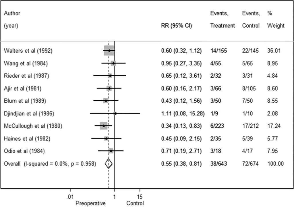
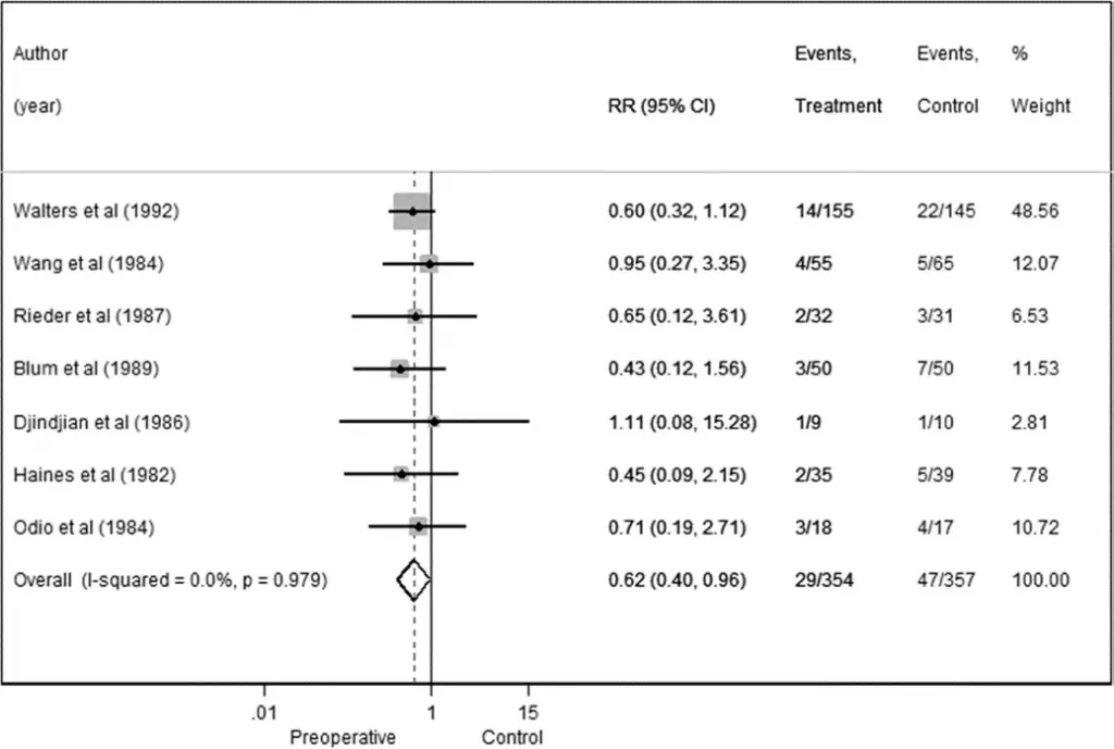
Fig. 2. Preoperative antibiotics forest plot for all studies in the meta-analysis.
Fig. 3. Preoperative antibiotics forest plot for RCTs in the meta-analysis.
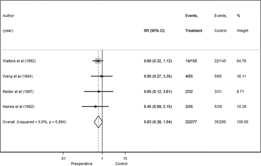
Fig. 4. Preoperative antibiotics forest plot for higher-quality RCTs.
Conclusions
Recommendation: The use of preoperative antibiotic agents can be recommended to prevent shunt infection in patients with hydrocephalus. It was only by combining the results of the various underpowered studies (meta- analysis) that the use of preoperative antibiotics for shunt surgery in children was shown to lower the risk of shunt infection. Strength of Recommendation: Level II, moderate degree of clinical certainty.
Given the available literature that met our inclusion criteria, the use of preoperative antibiotics for shunt surgery in children has not been shown to reduce the risk of developing a shunt infection. Nine trials, including 7 randomized controlled trials (RCTs), were critically evaluated. There were significant limitations and methodological flaws within the trials, most notably the potential to commit a Type II error (that is, failing to reject a false null hypothesis or, in other words, failing to detect a true therapeutic effect) because of inadequate power among the trials. Therefore, we conducted a meta-analysis to overcome the lack of power within many of the trials. Only with the meta-analysis did we demonstrate a protective benefit of preoperative antibiotics. This held true when we evaluated only the RCTs (n = 7), but lacked statistical significance when only the higher-quality RCTs (n = 4) were analyzed. The results of this systematic review and meta-analysis are, however, unlikely to spur further investigative trials within the United States, as the unwillingness of pediatric neurosurgeons to withhold prophylactic antibiotics from patients receiving shunts would be a monumental and insurmountable barrier (that is, a lack of clinical equipoise).
Acknowledgments
We acknowledge the American Association of Neurological Surgeons (AANS)/Congress of Neurological Surgeons (CNS) Joint Guidelines Committee for the members’ reviews, comments, and suggestions; Laura Mitchell, Guidelines Project Manager for the CNS, for her contributions; Pamela Shaw, research librarian, for her assistance with the literature searches; Kevin Boyer for his assistance with data analysis; and Sue Ann Kawecki and Kristin Kraus, M.Sc., for their assistance with editing. We also acknowledge the following peer reviewers for their contributions to review the update to the guidelines: Jennifer Sweet, MD, Brandon Rocque, MD, Christoph Greissenauer, MD, Jeffrey Olson, MD.
Disclosure
The systematic review and evidence-based guidelines were funded exclusively by the CNS and AANS Pediatric Section, which received no funding from outside commercial sources to support the development of this document.
Conflict(s) of Interest: None. All Task Force members declared any potential conflicts of interest prior to beginning work on this evidence review.
Conflict(s) of Interest: None. All Pediatric Hydrocephalus Systematic Review and Evidence-Based Guidelines Update Task Force members declared any potential conflicts of interest prior to beginning work on this systematic review and evidence-based guidelines.
Author contributions to the study and manuscript preparation include the following. Conception and design: AANS/CNS Joint Section on Pediatrics. Acquisition of data: all authors. Analysis and interpretation of data: all authors. Drafting the article: Klimo. Critically revising the article: all authors. Reviewed submitted version of manuscript: all authors. Approved the final version of the manuscript on behalf of all authors: Flannery. Statistical analysis: all authors. Administrative/technical/material support: all authors. Study super- vision: Flannery.
Table 1: Preoperative antibiotics: summary of evidence
| Author | Study Description | Data Class, Quality and Reasons | Results and Conclusions |
| Walters et al.,1992 | 20 mg/kg rifampin & 5 mg/kg trimethoprim oral given 2 hours preoperatively, then 8 hours postoperatively and q8 hrs for 48 hrs. | I, RCT, double-blinded, placebo given | Treatment: 22/145 (15%); control: 14/155 (9%). No statistically significant difference. Trial terminated early because higher than anticipated infection rate in both groups. |
| Wang et al., 1984 | 25 mg/kg sulfamethoxazole& 5 mg/kg trimethoprim given within 1 hour of surgery, then 8 & 16 hours postoperatively. | I, RCT, double-blinded, placebo given | Treatment: 4/55 (7.3%); control: 5/65 (7.6%). No statistically significant difference. |
| Rieder et al., 1987 | 25 mg/kg cephalothin given in OR prior to incision, then 3 times postoperatively, once every 6 hours. | I, RCT, double-blinded, placebo given | Treatment: 2/32 (6%); control: 3/31 (10%). No statistically significant difference. |
| Haines et al., 1982 | 12.5 mg/kg methicillin q6 hrs beginning 6 hours before surgery, at induction and for 72 hours postop. | I, RCT, no indication of blinding, placebo given | Treatment: 2/35 (5.7%); control: 5/39 (12.8%). No statistically significant difference. |
| Haines et al., 1982 | 12.5 mg/kg methicillin q6 hrs beginning 6 hours before surgery, at induction and for 72 hours postop. | I, RCT, no indication of blinding, placebo given | Treatment: 2/35 (5.7%); control: 5/39 (12.8%). No statistically significant difference. |
| Haines et al., 1982 | 12.5 mg/kg methicillin q6 hrs beginning 6 hours before surgery, at induction and for 72 hours postop. | I, RCT, no indication of blinding, placebo given | Treatment: 2/35 (5.7%); control: 5/39 (12.8%). No statistically significant difference. |
| Ajir et al., 1981 | 50 mg/kg methicillin given in OR prior to incision. | II, retrospective cohort | Treatment: 3/66 (4.5%); control: 8/105 (7.6%). Preoperative antibiotics were protective against infection. |
| Blum et al., 1989 | 50 mg/kg cefazedone given at time of induction. | II, RCT, single-blinded, use of placebo mentioned in introduction, but no details provided in methods, significant flaws in sequence generation and allocation concealment | Treatment: 3/50 (6%); control: 7/50 (14%). No statistically significant difference. |
| Djindjian et al., 1986 | 200 mg/kg/day oxacillin, total of 6 doses (24 hrs), first one given upon insertion of IV in OR. | II, RCT, but no methodology details provided by authors such as sequence generation, allocation concealment, blinding etc. | Treatment: 1/9 (11%); control: 1/10 (10%). No statistically significant difference. |
| McCullough et al., 1980 | 25 mg/kg methicillin given immediately prior to transport to OR, one dose after exposing the shunt or cannulating the ventricle, and 6 postoperative doses, given every 6 hours. | II, retrospective cohort | Treatment: 6/223 (2.6%); control: 17/212 (8%). Preoperative antibiotics were protective against infection. |
| Odio et al. 1984 | 15 mg/kg vancomycin hydrochloride one hour before surgery and again six hours later. | II, RCT, double-blinded, placebo given, but no methodology details provided by authors such as sequence generation, allocation concealment, blinding etc. | Treatment: 3/18 (17%); control: 4/17 (23%). No statistically significant difference. Trial terminated early because of adverse reactions to vancomycin. |
References
- Attenello FJ, Garces-Ambrossi GL, Zaidi HA, Sciubba DM, Jallo GI. Hospital costs associated with shunt infections in patients receiving antibiotic-impregnated shunt catheters versus standard shunt catheters. Neurosurgery. 2010;66(2):284-289; discussion 289.
- Darouiche RO. Treatment of infections associated with surgical implants. The New England journal of medicine. 2004;350(14):1422-1429.
- Simon TD, Riva-Cambrin J, Srivastava R, Bratton SL, Dean JM, Kestle JR. Hospital care for children with hydrocephalus in the United States: utilization, charges, comorbidities, and deaths. Journal of neurosurgery Pediatrics. 2008;1(2):131-137.
- Chadduck W, Adametz J. Incidence of seizures in patients with myelomeningocele: a multifactorial analysis. Surg Neurol. 1988;30(4):281-285.
- Jamjoom AB, Mohammed AA, al-Boukai A, Jamjoom ZA, Rahman N, Jamjoom HT. Multiloculated hydrocephalus related to cerebrospinal fluid shunt infection. Acta neurochirurgica. 1996;138(6):714-719.
- Mapstone TB, Rekate HL, Nulsen FE, Dixon MS, Jr., Glaser N, Jaffe M. Relationship of CSF shunting and IQ in children with myelomeningocele: a retrospective analysis. Child’s brain. 1984;11(2):112-118.
- Schoenbaum SC, Gardner P, Shillito J. Infections of cerebrospinal fluid shunts: epidemiology, clinical manifestations, and therapy. J Infect Dis. 1975;131(5):543-552.
- Vanaclocha V, Saiz-Sapena N, Leiva J. Shunt malfunction in relation to shunt infection. Acta neurochirurgica. 1996;138(7):829-834.
- Walters BC, Hoffman HJ, Hendrick EB, Humphreys RP. Cerebrospinal fluid shunt infection. Influences on initial management and subsequent outcome. Journal of neurosurgery. 1984;60(5):1014-1021.
- Prusseit J, Simon M, von der Brelie C, et al. Epidemiology, prevention and management of ventriculoperitoneal shunt infections in children. Pediatric neurosurgery. 2009;45(5):325-336.
- Simon TD, Hall M, Dean JM, Kestle JR, Riva-Cambrin J. Reinfection following initial cerebrospinal fluid shunt infection. Journal of neurosurgery Pediatrics. 2010;6(3):277-285.
- Simon TD, Hall M, Riva-Cambrin J, et al. Infection rates following initial cerebrospinal fluid shunt placement across pediatric hospitals in the United States. Clinical article. Journal of neurosurgery Pediatrics. 2009;4(2):156-165.
- Simon TD, Whitlock KB, Riva-Cambrin J, et al. Revision surgeries are associated with significant increased risk of subsequent cerebrospinal fluid shunt infection. The Pediatric infectious disease journal. 2012;31(6):551-556.
- Wong JM, Ziewacz JE, Ho AL, et al. Patterns in neurosurgical adverse events: cerebrospinal fluid shunt surgery. Neurosurgical focus. 2012;33(5):E13.
- Barker FG, 2nd. Efficacy of prophylactic antibiotic therapy in spinal surgery: a meta-analysis. Neurosurgery. 2002;51(2):391-400.
- Barker FG, 2nd. Efficacy of prophylactic antibiotics against meningitis after craniotomy: a meta-analysis. Neurosurgery. 2007;60(5):887-894.
- Barker FG, 2nd. Efficacy of prophylactic antibiotics for craniotomy: a meta-analysis. Neurosurgery. 1994;35(3):484-490.
- Haines SJ, Walters BC. Antibiotic prophylaxis for cerebrospinal fluid shunts: a metanalysis. Neurosurgery. 1994;34(1):87-92.
- Langley JM, LeBlanc JC, Drake J, Milner R. Efficacy of antimicrobial prophylaxis in placement of cerebrospinal fluid shunts: meta-analysis. Clin Infect Dis. 1993;17(1):98-103.
- Ratilal B, Costa J, Sampaio C. Antibiotic prophylaxis for surgical introduction of intracranial ventricular shunts: a systematic review. Journal of neurosurgery Pediatrics. 2008;1(1):48-56.
- Biyani N, Grisaru-Soen G, Steinbok P, Sgouros S, Constantini S. Prophylactic antibiotics in pediatric shunt surgery. Child’s nervous system : ChNS : official journal of the International Society for Pediatric Neurosurgery. 2006;22(11):1465-1471.
- DerSimonian R, Laird N. Meta-analysis in clinical trials. Control Clin Trials. 1986;7(3):177-188.
- Crowther M, Lim W, Crowther MA. Systematic review and meta-analysis methodology. Blood. 2010;116(17):3140-3146.
- Deeks JJ, Higgins JPT, Altman DG. Chapter 9: Analysing data and undertaking meta-analyses. Cochrane Handbook for Systematic Reviews of Interventions 2011; 5.1.0:www.cochrane-handbook.org.
- Bayston R, Bannister C, Boston V, et al. A prospective randomised controlled trial of antimicrobial prophylaxis in hydrocephalus shunt surgery. Z Kinderchir. 1990;45 Suppl 1:5-7.
- Blomstedt GC. Results of trimethoprim-sulfamethoxazole prophylaxis in ventriculostomy and shunting procedures. A double-blind randomized trial. Journal of neurosurgery. 1985;62(5):694-697.
- George R, Leibrock L, Epstein M. Long-term analysis of cerebrospinal fluid shunt infections. A 25-year experience. Journal of neurosurgery. 1979;51(6):804-811.
- Schmidt K, Gjerris F, Osgaard O, et al. Antibiotic prophylaxis in cerebrospinal fluid shunting: a prospective randomized trial in 152 hydrocephalic patients. Neurosurgery. 1985;17(1):1-5.
- Weiss SR, Raskind R. Further experience with the ventriculoperitoneal shunt. Prophylactic antibiotics. Int Surg. 1970;53(4):300-303.
- Arnaboldi L. Antimicrobial prophylaxis with ceftriaxone in neurosurgical procedures. A prospective study of 100 patients undergoing shunt operations. Chemotherapy. 1996;42(5):384-390.
- Kestle JR, Hoffman HJ, Soloniuk D, Humphreys RP, Drake JM, Hendrick EB. A concerted effort to prevent shunt infection. Child’s nervous system : ChNS : official journal of the International Society for Pediatric Neurosurgery. 1993;9(3):163-165.
- Viano JC, Tregnaghi M, Casagnas M, Suarez JC. Valvular infections in patients with hydrocephalus: preventive aspects. Child’s nervous system : ChNS : official journal of the International Society for Pediatric Neurosurgery. 1990;6(7):397-399.
- Bayston R. Antibiotic prophylaxis in shunt surgery. Dev Med Child Neurol Suppl. 1975(35):99-103.
- Yu HC, Patterson RH, Jr. Prophylactic antimicrobial agents after ventriculoatriostomy for hydrocephalus. Journal of pediatric surgery. 1973;8(6):881-885.
- Bullock R, van Dellen JR, Ketelbey W, Reinach SG. A double-blind placebo-controlled trial of perioperative prophylactic antibiotics for elective neurosurgery. Journal of neurosurgery. 1988;69(5):687-691.
- Young RF, Lawner PM. Perioperative antibiotic prophylaxis for prevention of postoperative neurosurgical infections. A randomized clinical trial. Journal of neurosurgery. 1987;66(5):701-705.
- Ajir F, Levin AB, Duff TA. Effect of prophylactic methicillin on cerebrospinal fluid shunt infections in children. Neurosurgery. 1981;9(1):6-8.
- Blum J, Schwarz M, Voth D. Antibiotic single-dose prophylaxis of shunt infections. Neurosurgical review. 1989;12(3):239-244.
- Djindjian M, Fevrier MJ, Otterbein G, Soussy JC. Oxacillin prophylaxis in cerebrospinal fluid shunt procedures: results of a randomized open study in 60 hydrocephalic patients. Surg Neurol. 1986;25(2):178-180.
- Haines SJ, Taylor F. Prophylactic methicillin for shunt operations: effects on incidence of shunt malfunction and infection. Child’s brain. 1982;9(1):10-22.
- McCullough DC, Kane JG, Harleman G, Wells M. Antibiotic prophylaxis in ventricular shunt surgery. II. Antibiotic concentrations in cerebrospinal fluid. Child’s brain. 1980;7(4):190-194.
- Odio C, Mohs E, Sklar FH, Nelson JD, McCracken GH, Jr. Adverse reactions to vancomycin used as prophylaxis for CSF shunt procedures. Am J Dis Child. 1984;138(1):17-19.
- Rieder MJ, Frewen TC, Del Maestro RF, Coyle A, Lovell S. The effect of cephalothin prophylaxis on postoperative ventriculoperitoneal shunt infections. CMAJ : Canadian Medical Association journal = journal de l’Association medicale canadienne. 1987;136(9):935-938.
- Wang EE, Prober CG, Hendrick BE, Hoffman HJ, Humphreys RP. Prophylactic sulfamethoxazole and trimethoprim in ventriculoperitoneal shunt surgery. A double-blind, randomized, placebo-controlled trial. Jama. 1984;251(9):1174-1177.
Part 7: Antibiotic-impregnated shunt systems versus conventional shunts in children: a systematic review and meta-analysis
Reposted with permission from ©AANS, 2014
J Neurosurg Pediatrics (Suppl) 14:53–59, 2014
AANS, 2014
(Used with permission from Journal of Neurosurgery: Pediatrics. Please click here for the original publication.)
UPDATE
Paul Klimo Jr. MD, MPH ,1–3 Clinton J. Thompson, PhD,4 Lissa C. Baird, MD,4 Ann Marie Flannery, MD5
1Semmes-Murphey Neurologic & Spine Institute; 2Department of Neurosurgery, University of Tennessee Health Science Center; 3Le Bonheur Children’s Hospital, Memphis, Tennessee; 4School of Public Health and Health Services, The George Washington University, Washington, DC; and 5Department of Neurological Surgery, Saint Louis University, St. Louis, Missouri
Object. The objective of this systematic review and meta-analysis was to answer the following question: Are antibiotic-impregnated shunts (AISs) superior to standard shunts (SSs) at reducing the risk of shunt infection in pediatric patients with hydrocephalus?
Methods. Both the US National Library of Medicine PubMed/MEDLINE database and the Cochrane Database of Systematic Reviews were queried using MeSH headings and key words relevant to AIS use in children. Abstracts were reviewed, after which studies meeting the inclusion criteria were selected. An evidentiary table was assembled summarizing the studies and the quality of their evidence (Classes I–III). A meta-analysis was conducted using a random-effects model to calculate a cumulative estimate of treatment effect using risk ratio (RR). Heterogeneity was assessed using the chi-square and I2 statistics. Based on the quality of the literature and the result of the meta-analysis, a recommendation was rendered (Level I, II, or III).
Results. Six studies, all Class III, met our inclusion criteria. All but one study focused on a retrospective cohort and all but one were conducted at a single institution. Four of the studies failed to demonstrate a lowered infection rate with the use of an AIS. However, when the data from individual studies were pooled together, the infection rate in the AIS group was 5.5% compared with 8.6% in the SS group. Using a random-effects model, the cumulative RR was 0.51 (95% CI 0.29–0.89, p < 0.001), indicating that a shunt infection was 1.96 times more likely in patients who received an SS.
Conclusions. We recommend AIS tubing because of the associated lower risk of shunt infection compared to the use of conventional silicone hardware (quality of evidence: Class III; strength of recommendation: Level III).
Recommendation: Antibiotic-impregnated shunt (AIS) tubing may be associated with a lower risk of shunt infection compared with conventional silicone hardware and thus is an option for children who require placement of a shunt. Strength of Recommendation: Level III, unclear degree of clinical certainty. (http://thejns.org/doi/abs/10.3171/2014.7.PEDS14327)
Key Words: antibiotic-impregnated shunt, shunt, cerebrospinal fluid, infection, pediatric patient, meta-analysis, hydrocephalus, practice guidelines
Abbreviations used in this paper: AIS = antibiotic-impregnated shunt; AANS = American Association of Neurological Surgeons; CNS = Congress of Neurological Surgeons; RR = risk ratio; SS = standard shunt.
Prevention of shunt infection is a priority for neurosurgeons, especially when treating pediatric patients. Infection can cause shunt malfunction with all the potential consequences of a nonfunctioning shunt. Shunt infection can lead to scarring and loculation of the ventricles, increasing the complexity of the patient’s hydrocephalus, and it may result in a lower intelligence quotient, increased risk of seizures, and psychomotor retardation.1-5 Treatment of shunt infections is costly, estimated to be upwards of $50,000 per infection in the United States, making it one of the most costly implant-related infections.6
The identification of modifiable risk factors or interventions to lower the risk of a shunt infection has been the topic of active research for many years. Identified factors include the duration of surgery;7,8 the skill and experience of the treating neurosurgeon;9-11 the number of personnel in the operating room;12-14 and the use of hair shaving,8,15 prophylactic systemic antibiotics,16-18 intrathecal antibiotics,19 wound irrigation,20 antibiotic-impregnated sutures,21,22 and double gloving23 (or inadvertent exposure of the shunt to breached surgical gloves).24 Antibiotic-impregnated Silastic catheters were first introduced by Roger Bayston in 1977; they were considered more specifically with shunts in 1989,25 but did not become available for clinical use in the United States until about 10 years ago. The antibiotic-impregnated shunt (AIS) systems currently on the market contain 0.054% rifampin and 0.15% clindamycin, which target the most common pathogens: Staphylococcus epidermidis and Staphylococcus aureus. Although rifampin and clindamycin do not reduce bacterial adherence, this combination of antibiotics kills bacteria and has been shown to prevent colonization for up to 56 days in in vitro studies and up to 127 days in vivo.26-28
Many studies have evaluated the efficacy of AISs compared with standard shunts (SSs) in the prevention of shunt infections, including two recent systematic reviews and meta-analyses.29-47 The purpose of this evidence-based review is to examine data on the use of AISs and SSs and compare these treatments in the prevention of shunt infections in the pediatric population.
Methods
Search Terms
We searched the US National Library of Medicine PubMed/MEDLINE database and the Cochrane Database of Systematic Reviews for the period January 1966 through March 2012 using the following MeSH subject headings: (“cerebrospinal fluid shunts” OR (“cerebro- spinal fluid” AND (shunt* OR catheter*)) OR “shunt system”) AND (“antibiotic-impregnated” OR (antibiotic AND impregnated)) AND infection.
Search Strategy
We reviewed the titles and abstracts of the papers we retrieved with attention to those titles addressing the rate of shunt infection in patients treated with AISs compared with those treated with SSs. Uncontrolled studies were excluded, as were studies that evaluated antimicrobial shunts unavailable in the US market. In all papers, we required that the authors state that the only variable that changed was the type of shunt implanted; all other aspects of the surgery and technique needed to remain unchanged.
Authors performed an updated literature search (in PubMed and Cochrane Central) for this guideline chapter through a medical librarian at the Congress of Neurological Surgeons Guidelines office using the below-mentioned existing search terms to update the original search through November 30, 2019.
Meta-Analysis
For each study, we identified the number of infections resulting from implantation of SSs and AISs and then computed the risk of an infection associated with AISs relative to that associated with SSs, yielding a risk ratio (RR). An RR less than 1 is indicative of protection against infection for the AIS. The overall RR was computed using the method of DerSimonian and Laird.48
We conducted a random-effects meta-analysis of the selected studies. A random-effects model—as opposed to a fixed-effects model—does not assume that the measure of association (that is, RR) is uniform across strata (that is, among studies) and, consequently, yields a more conservative estimate of the effect. We assessed heterogeneity by way of the chi-square test of heterogeneity and the I2 statistic, in which the former returns a chi-square distributed test statistic and corresponding p value and the latter returns a value bound between 0% and 100%, with higher values denoting increasing heterogeneity. We regarded a chi-square test of heterogeneity p value less than alpha = 0.10 and an I2 value in the range of 30% to 60% as suggestive of moderate heterogeneity.49,50 An examination of publication bias was not conducted since the number of studies included in this analysis was not large enough to provide adequate power (i.e., fewer than 10 studies).
Search Results
Our search returned 41 articles; another 3 articles were found from an examination of the articles’ bibliographies (Fig. 1). Nineteen full-length papers were reviewed, 13 of which were rejected for the following reasons: studies enrolled either adults only or enrolled mixed populations, but separate results for children were not provided,29,33,34,41-44,46 or studies contained patient data that had also been reported in separate publications.21,51-54 In fact, 1 group of researchers published no less than 9 papers on AISs that included patients from overlapping time periods.21,32,40,41,45,51,53-55 Therefore, 6 articles satisfied inclusion for this systematic review and meta-analysis (Table 1).30,31,36-38,45
An additional 5 studies out of the 122 yielded by the 2020 update met inclusion criteria from the original guideline and were included (Figure 2).
Results
The review process identified no papers providing Class I or II data specifically addressing the issue of shunt infection and the use of AISs compared with SSs in children. The 6 articles that satisfied our entry criteria were all Class III cohort studies, all but one of which were conducted within a single institution. The primary outcome of interest—shunt infection—was defined by authors of individual studies, but in general, it was a patient who underwent a recent shunt surgery and subsequently developed signs and symptoms of a shunt malfunction or an infection with an organism cultured from CSF, the shunt apparatus, purulence from the shunt wound(s), or abdominal fluid/pseudocyst. Some investigators also considered a patient to have an infection if there were highly suggestive findings such as fever, redness along the shunt, or CSF pleocytosis in the absence of a positive culture. Overall, 2 studies produced findings that AISs are protective against shunt infection, whereas the remaining studies did not.
Sciubba et al45 reported one of the earliest large series comparing AISs with SSs in a pediatric population. During an 18-month period, 208 SSs were placed; this was followed by another 18-month period during which AISs were used 145 times. The AIS patient group was younger, more frequently premature, and thus had a greater incidence of intracranial hemorrhage as the cause of hydrocephalus. The primary outcome was the development of a shunt infection, defined as clinical suspicion (fever, increased white blood cell count, and/or wound breakdown involving the shunt) with positive cultures from CSF and/or hardware. Patients who received AIS catheters had significantly fewer shunt infections: 2 patients (1.4%) with antibiotic-impregnated catheters within the 6-month follow-up period compared with 25 patients (12%) with non–antibiotic-impregnated catheters. After we adjusted for intercohort differences in primary placement compared with shunt revision, prematurity, and posthemorrhagic hydrocephalus, we found AIS catheters to be independently associated with a 2.4-fold decreased likelihood of shunt infection.
Aryan et al30 detailed their 1-year experience using the Bactiseal system (Codman, Johnson & Johnson). Al- though the rate of shunt infection was lower in the Bactiseal group (1 of 32 [3.1%]) compared with the standard group (7 of 46 [15.2%]), the difference was not statistically significant (p = 0.09). Kan and Kestler37 reported on a similar retrospective cohort in which 80 consecutive patients received the Bactiseal shunt and were compared with an earlier group of 80 patients who had received an SS. There was no statistically significant difference in the shunt infection rate (5.0% vs 8.8%), even when the authors controlled for patient age at surgery, type of revision, cause of hydrocephalus, and previous revisions or infections within the past 6 months.
In their retrospective cohort study, Hayhurst and co-workers36 looked at 4 groups of patients in whom AISs had been implanted de novo (Group 1), during noninfected revisional surgery (Group 2), and after an external ventricular drain had been replaced by the shunt (sterile CSF [Group 3] and infected CSF [Group 4]). There were 214 shunt procedures performed using the Bactiseal system in 150 children. The historical control group comprised 77 operations in 65 children. Again, there was no statistically significant difference in the infection rate (21 of 214 [9.8%] in the antibiotic group and 8 of 77 [10.4%] in the standard group). Although the authors emphasized the difference in the infection rate among neonates—27% in the standard group versus 11% in the antibiotic group— this difference too was not significant (p = 0.208). Eymann et al31 presented clinical and cost data for both adult and pediatric patients. Using Fisher’s exact test, the pediatric infection rates of 13.6% in the standard group and 3.8% in the Bactiseal group were not statistically different. However, when the authors combined both adult and pediatric outcomes, they did find a protective benefit with the Bactiseal system and a net savings of $51,651 in the 197 Bactiseal procedures.
The study with the largest number of patients was conducted by Kandasamy et al.38 This multicenter study (3 pediatric neurosurgery centers in the United Kingdom) was ambispective: patients treated with AISs were prospectively followed, whereas patients treated with SSs at earlier time periods were retrospectively reviewed (historical control). Operations were divided into those that were de novo and those that were clean revisions. There was some intercenter variability in the choice of preoperative antibiotics and surgical technique, but there was no intracenter variability. For example, centers at Leeds and Liverpool used a single dose of cefuroxime, whereas London used flucloxacillin and amikacin. The overall pooled treatment effect estimate statistically favored AISs for de novo and clean revisions combined (the incidence of infection in the AIS Group was 30 of 581 [5.2%] and that in the SS Group was 155 of 1963 [7.9%]) as well as for the subgroup of de novo shunts only and the subgroup of children younger than 1 year of age; the pooled treatment effect estimate for clean revisions only did not reach statistical significance.
Meta-Analysis Results
In total, there were 2396 procedures in which a standard catheter system had been placed and 205 infections occurred, yielding a pooled infection rate of 8.6%. In the AIS population, 59 infections occurred after 1078 shunt operations for an overall infection rate of 5.5%. Thus, the absolute and relative risk reductions were 3.1% and 36%, respectively. The overall RR was 0.51 (95% CI 0.29–0.89, p < 0.001), making a shunt infection 1.96 times more likely when an SS system is used (Fig. 2). Although the chi- square test did not indicate heterogeneity (p = 0.129), the I2 test did show moderate heterogeneity (41.5%).
To explore the uncertainty of statistical significance in the RR meta-analysis, a stepwise sensitivity analysis was performed (Table 2). When subtracting studies from the meta-analysis (and thus reducing the power of the analysis), the effect size remains relatively stable, but confidence intervals widen to the point of statistical nonsignificance. Based on significant findings in large studies comparing AISs with SSs and significant findings in a meta-analysis with a high number of studies, it is likely that the meta- analysis shown in Fig. 2 accurately represents a statistically significant effect in favor of using AISs.
Number Needed to Treat
There is a certain difficulty with interpreting an RR of 0.51, in that the number of people who benefited from AIS treatment is masked by the interpretation of an RR (i.e., a “50% reduced risk of infection”). In fact the infection rate in the AIS patient group in this meta-analysis was 5.47% compared with 8.55% in the SS patient group. These infection rates come close to approximating a “50% reduced risk of infection.”
To better understand the analysis of AISs versus SSs, absolute values calculated as the number of cases needed to treat and the number of infections avoided per 1000 cases treated with AISs were calculated (Table 2). According to the data reported in the literature, for every 24 cases treated with an AIS, 1 infection is prevented. Alternatively, 42 infections are avoided for every 1000 cases treated with AISs. As a convenience, several population infection rates (that is, infection rates unique to particular locations/practices) are presented in Table 3. Not surprisingly, the higher an infection rate in a population, the “better” the AIS becomes at preventing infection.
2020 Update
Mbabazi et al56, which was a single center RCT, provided Class II evidence that in a unique group of patients (low resource country), single center, a unique antibiotic impregnated catheter design (distal slit valve) failed to show an advantage in infection prevention. Because the population in this study is so unique, the authors concluded that this study, while meeting inclusion criteria, does not affect the recommendation. Three other publications57-59 were single center, retrospective reviews, Class III. Mallucci et al60 conducted a multicenter Class I RCT of both adult and pediatric patients, and demonstrated that antibiotic impregnated shunts do reduce the rate of infection, particularly in children. This new evidence warrants an upgrade of the level of the recommendation for this chapter.
Conclusions
Recommendation: Antibiotic-impregnated shunt (AIS) tubing reduces the risk of shunt infection compared with conventional silicone hardware and should be used for children who require placement of a shunt. Strength of Recommendation: Level I, high degree of clinical certainty.
The clinical and financial consequences of a shunt infection are substantial as is the emotional stress borne by patients and their families. Neurosurgeons have evaluated many interventions in the hopes of finding ones that can decrease the risk of developing a shunt infection. Based on the available Class III evidence, we have demonstrated that antibiotic-impregnated shunts (AISs containing rifampin and clindamycin) can lower the shunt infection risk substantially. Although only 2 of the 6 studies that met our inclusion criteria showed a protective benefit with AISs, when the data from all 6 studies were pooled together (meta-analysis), a benefit was shown, with an infection rate almost twice as high in patients receiving a standard shunt (SS). Given the large number of patients that would be needed to definitively demonstrate superior efficacy of AISs over SSs in children, it is unlikely that a clinical trial will be conducted or is even needed.
Acknowledgments
We acknowledge the American Association of Neurological Surgeons (AANS)/Congress of Neurological Surgeons (CNS) Joint Guidelines Committee for the members’ reviews, comments, and suggestions; Laura Mitchell, Guidelines Project Manager for the CNS, for her contributions; Pamela Shaw, research librarian, for her assistance with the literature searches; Kevin Boyer for his assistance with data analysis; and Sue Ann Kawecki and Kristin Kraus, M.Sc., for their assistance with editing. We also acknowledge the following peer reviewers for their contributions to review the update to the guidelines: Jennifer Sweet, MD, Brandon Rocque, MD, Christoph Greissenauer, MD, Jeffrey Olson, MD.
Disclosure
The systematic review and evidence-based guidelines were funded exclusively by the CNS and AANS Pediatric Section, which received no funding from outside commercial sources to support the development of this document.
Conflict(s) of Interest: None. All Task Force members declared any potential conflicts of interest prior to beginning work on this evidence review.
Conflict(s) of Interest: None. All Pediatric Hydrocephalus Systematic Review and Evidence-Based Guidelines Update Task Force members declared any potential conflicts of interest prior to beginning work on this systematic review and evidence-based guidelines.
Author contributions to the study and manuscript preparation include the following. Conception and design: AANS/CNS Joint Section on Pediatrics. Acquisition of data: all authors. Analysis and interpretation of data: all authors. Drafting the article: Klimo. Critically revising the article: all authors. Reviewed submitted version of manuscript: all authors. Approved the final version of the manuscript on behalf of all authors: Flannery. Statistical analysis: all authors. Administrative/technical/material support: all authors. Study supervision: Flannery.
References
- Chadduck W, Adametz J. Incidence of seizures in patients with myelomeningocele: a multifactorial analysis. Surg Neurol. 1988;30(4):281-285.
- Jamjoom AB, Mohammed AA, al-Boukai A, Jamjoom ZA, Rahman N, Jamjoom HT. Multiloculated hydrocephalus related to cerebrospinal fluid shunt infection. Acta neurochirurgica. 1996;138(6):714-719.
- Mapstone TB, Rekate HL, Nulsen FE, Dixon MS, Jr., Glaser N, Jaffe M. Relationship of CSF shunting and IQ in children with myelomeningocele: a retrospective analysis. Child’s brain. 1984;11(2):112-118.
- Vanaclocha V, Saiz-Sapena N, Leiva J. Shunt malfunction in relation to shunt infection. Acta neurochirurgica. 1996;138(7):829-834.
- Walters BC, Hoffman HJ, Hendrick EB, Humphreys RP. Cerebrospinal fluid shunt infection. Influences on initial management and subsequent outcome. Journal of neurosurgery. 1984;60(5):1014-1021.
- Darouiche RO. Treatment of infections associated with surgical implants. The New England journal of medicine. 2004;350(14):1422-1429.
- Kontny U, Hofling B, Gutjahr P, Voth D, Schwarz M, Schmitt HJ. CSF shunt infections in children. Infection. 1993;21(2):89-92.
- Ratanalert S, Musikawat P, Oearsakul T, Saeheng S, Chowchuvech V. Non-shaved ventriculoperitoneal shunt in Thailand. In: J Clin Neurosci. Vol 12. Scotland2005:147-149.
- Borgbjerg BM, Gjerris F, Albeck MJ, Borgesen SE. Risk of infection after cerebrospinal fluid shunt: an analysis of 884 first-time shunts. Acta neurochirurgica. 1995;136(1-2):1-7.
- Cochrane DD, Kestle JR. The influence of surgical operative experience on the duration of first ventriculoperitoneal shunt function and infection. In: Pediatr Neurosurg. Vol 38. Switzerland: 2003 S. Karger AG, Basel; 2003:295-301.
- Kestle JR, Cochrane DD, Drake JM. Shunt insertion in the summer: is it safe? Journal of neurosurgery. 2006;105(3 Suppl):165-168.
- Choux M, Genitori L, Lang D, Lena G. Shunt implantation: reducing the incidence of shunt infection. Journal of neurosurgery. 1992;77(6):875-880.
- Kestle JR, Hoffman HJ, Soloniuk D, Humphreys RP, Drake JM, Hendrick EB. A concerted effort to prevent shunt infection. Child’s nervous system : ChNS : official journal of the International Society for Pediatric Neurosurgery. 1993;9(3):163-165.
- Pirotte BJ, Lubansu A, Bruneau M, Loqa C, Van Cutsem N, Brotchi J. Sterile surgical technique for shunt placement reduces the shunt infection rate in children: preliminary analysis of a prospective protocol in 115 consecutive procedures. Child’s nervous system : ChNS : official journal of the International Society for Pediatric Neurosurgery. 2007;23(11):1251-1261.
- Horgan MA, Piatt JH, Jr. Shaving of the scalp may increase the rate of infection in CSF shunt surgery. Pediatric neurosurgery. 1997;26(4):180-184.
- Haines SJ, Walters BC. Antibiotic prophylaxis for cerebrospinal fluid shunts: a metanalysis. Neurosurgery. 1994;34(1):87-92.
- Ratilal B, Costa J, Sampaio C. Antibiotic prophylaxis for surgical introduction of intracranial ventricular shunts: a systematic review. Journal of neurosurgery Pediatrics. 2008;1(1):48-56.
- Zentner J, Gilsbach J, Felder T. Antibiotic prophylaxis in cerebrospinal fluid shunting: a prospective randomized trial in 129 patients. Neurosurgical review. 1995;18(3):169-172.
- Ragel BT, Browd SR, Schmidt RH. Surgical shunt infection: significant reduction when using intraventricular and systemic antibiotic agents. Journal of neurosurgery. 2006;105(2):242-247.
- Hayashi T, Shirane R, Kato T, Tominaga T. Efficacy of intraoperative wound irrigation for preventing shunt infection. Journal of neurosurgery Pediatrics. 2008;2(1):25-28.
- Sciubba DM, Lin LM, Woodworth GF, McGirt MJ, Carson B, Jallo GI. Factors contributing to the medical costs of cerebrospinal fluid shunt infection treatment in pediatric patients with standard shunt components compared with those in patients with antibiotic impregnated components. In: Neurosurg Focus. Vol 22. United States2007:E9.
- Stone J, Gruber TJ, Rozzelle CJ. Healthcare savings associated with reduced infection rates using antimicrobial suture wound closure for cerebrospinal fluid shunt procedures. In: Pediatr Neurosurg. Vol 46. Switzerland: 2010 S. Karger AG, Basel.; 2010:19-24.
- Rehman AU, Rehman TU, Bashir HH, Gupta V. A simple method to reduce infection of ventriculoperitoneal shunts. Journal of neurosurgery Pediatrics. 2010;5(6):569-572.
- Kulkarni AV, Drake JM, Lamberti-Pasculli M. Cerebrospinal fluid shunt infection: a prospective study of risk factors. Journal of neurosurgery. 2001;94(2):195-201.
- Bayston R, Grove N, Siegel J, Lawellin D, Barsham S. Prevention of hydrocephalus shunt catheter colonisation in vitro by impregnation with antimicrobials. Journal of neurology, neurosurgery, and psychiatry. 1989;52(5):605-609.
- Bayston R, Ashraf W, Bhundia C. Mode of action of an antimicrobial biomaterial for use in hydrocephalus shunts. In: J Antimicrob Chemother. Vol 53. England2004:778-782.
- Bayston R, Lambert E. Duration of protective activity of cerebrospinal fluid shunt catheters impregnated with antimicrobial agents to prevent bacterial catheter-related infection. Journal of neurosurgery. 1997;87(2):247-251.
- Pattavilakom A, Kotasnas D, Korman TM, Xenos C, Danks A. Duration of in vivo antimicrobial activity of antibiotic-impregnated cerebrospinal fluid catheters. In: Neurosurgery. Vol 58. United States2006:930-935; discussion 930-935.
- Albanese A, De Bonis P, Sabatino G, et al. Antibiotic-impregnated ventriculo-peritoneal shunts in patients at high risk of infection. Acta neurochirurgica. 2009;151(10):1259-1263.
- Aryan HE, Meltzer HS, Park MS, Bennett RL, Jandial R, Levy ML. Initial experience with antibiotic-impregnated silicone catheters for shunting of cerebrospinal fluid in children. Child’s nervous system : ChNS : official journal of the International Society for Pediatric Neurosurgery. 2005;21(1):56-61.
- Eymann R, Chehab S, Strowitzki M, Steudel WI, Kiefer M. Clinical and economic consequences of antibiotic-impregnated cerebrospinal fluid shunt catheters. Journal of neurosurgery Pediatrics. 2008;1(6):444-450.
- Farber SH, Parker SL, Adogwa O, McGirt MJ, Rigamonti D. Effect of antibiotic impregnated shunts on infection rate in adult hydrocephalus: A single institution’s experience. Neurosurgery. 2011.
- Govender ST, Nathoo N, van Dellen JR. Evaluation of an antibiotic-impregnated shunt system for the treatment of hydrocephalus. Journal of neurosurgery. 2003;99(5):831-839.
- Gutierrez-Gonzalez R, Boto GR. Do antibiotic-impregnated catheters prevent infection in CSF diversion procedures? Review of the literature. The Journal of infection. 2010;61(1):9-20.
- Gutierrez-Gonzalez R, Boto GR, Fernandez-Perez C, del Prado N. Protective effect of rifampicin and clindamycin impregnated devices against Staphylococcus spp. infection after cerebrospinal fluid diversion procedures. In: BMC Neurol. Vol 10. England2010:93.
- Hayhurst C, Cooke R, Williams D, Kandasamy J, O’Brien DF, Mallucci CL. The impact of antibiotic-impregnated catheters on shunt infection in children and neonates. Child’s nervous system : ChNS : official journal of the International Society for Pediatric Neurosurgery. 2008;24(5):557-562.
- Kan P, Kestle J. Lack of efficacy of antibiotic-impregnated shunt systems in preventing shunt infections in children. Child’s nervous system : ChNS : official journal of the International Society for Pediatric Neurosurgery. 2007;23(7):773-777.
- Kandasamy J, Dwan K, Hartley JC, et al. Antibiotic-impregnated ventriculoperitoneal shunts-a multi-centre British paediatric neurosurgery group (BPNG) study using historical controls. Child’s nervous system : ChNS : official journal of the International Society for Pediatric Neurosurgery. 2010.
- Klimo P, Jr., Thompson CJ, Ragel BT, Boop FA. Antibiotic-impregnated shunt systems versus standard shunt systems: a meta- and cost-savings analysis. Journal of neurosurgery Pediatrics. 2011;8(6):600-612.
- Parker SL, Anderson WN, Lilienfeld S, Megerian JT, McGirt MJ. Cerebrospinal shunt infection in patients receiving antibiotic-impregnated versus standard shunts. Journal of neurosurgery Pediatrics. 2011;8(3):259-265.
- Parker SL, Attenello FJ, Sciubba DM, et al. Comparison of shunt infection incidence in high-risk subgroups receiving antibiotic-impregnated versus standard shunts. Child’s nervous system : ChNS : official journal of the International Society for Pediatric Neurosurgery. 2009;25(1):77-83; discussion 85.
- Pattavilakom A, Xenos C, Bradfield O, Danks RA. Reduction in shunt infection using antibiotic impregnated CSF shunt catheters: an Australian prospective study. In: J Clin Neurosci. Vol 14. Scotland2007:526-531.
- Richards HK, Seeley HM, Pickard JD. Efficacy of antibiotic-impregnated shunt catheters in reducing shunt infection: data from the United Kingdom Shunt Registry. Journal of neurosurgery Pediatrics. 2009;4(4):389-393.
- Ritz R, Roser F, Morgalla M, Dietz K, Tatagiba M, Will BE. Do antibiotic-impregnated shunts in hydrocephalus therapy reduce the risk of infection? An observational study in 258 patients. In: BMC Infect Dis. Vol 7. England2007:38.
- Sciubba DM, Stuart RM, McGirt MJ, et al. Effect of antibiotic-impregnated shunt catheters in decreasing the incidence of shunt infection in the treatment of hydrocephalus. Journal of neurosurgery. 2005;103(2 Suppl):131-136.
- Steinbok P, Milner R, Agrawal D, et al. A multicenter multinational registry for assessing ventriculoperitoneal shunt infections for hydrocephalus. Neurosurgery. 2010;67(5):1303-1310.
- Thomas R, Lee S, Patole S, Rao S. Antibiotic-impregnated catheters for the prevention of CSF shunt infections: a systematic review and meta-analysis. British journal of neurosurgery. 2012;26(2):175-184.
- DerSimonian R, Laird N. Meta-analysis in clinical trials. Control Clin Trials. 1986;7(3):177-188.
- Crowther M, Lim W, Crowther MA. Systematic review and meta-analysis methodology. Blood. 2010;116(17):3140-3146.
- Deeks JJ, Higgins JPT, Altman DG. Chapter 9: Analysing data and undertaking meta-analyses. Cochrane Handbook for Systematic Reviews of Interventions 2011; 5.1.0:www.cochrane-handbook.org.
- Attenello FJ, Garces-Ambrossi GL, Zaidi HA, Sciubba DM, Jallo GI. Hospital costs associated with shunt infections in patients receiving antibiotic-impregnated shunt catheters versus standard shunt catheters. Neurosurgery. 2010;66(2):284-289; discussion 289.
- Eymann R, Steudel WI, Kiefer M. Infection rate with application of an antibiotic-impregnated catheter for shunt implantation in children – a retrospective analysis. Klinische Padiatrie. 2009;221(2):69-73.
- Sciubba DM, McGirt MJ, Woodworth GF, Carson B, Jallo GI. Prolonged exposure to antibiotic-impregnated shunt catheters does not increase incidence of late shunt infections. Child’s nervous system : ChNS : official journal of the International Society for Pediatric Neurosurgery. 2007;23(8):867-871.
- Sciubba DM, Noggle JC, Carson BS, Jallo GI. Antibiotic-impregnated shunt catheters for the treatment of infantile hydrocephalus. In: Pediatr Neurosurg. Vol 44. Switzerland2008:91-96.
- Farber SH, Parker SL, Adogwa O, Rigamonti D, McGirt MJ. Cost analysis of antibiotic-impregnated catheters in the treatment of hydrocephalus in adult patients. World neurosurgery. 2010;74(4-5):528-531.
- Mbabazi-Kabachelor E, Shah M, Vaughan KA, et al. Infection risk for Bactiseal Universal Shunts versus Chhabra shunts in Ugandan infants: a randomized controlled trial. Journal of neurosurgery Pediatrics. 2019;23(3):397-406.
- Raffa G, Marseglia L, Gitto E, Germano A. Antibiotic-impregnated catheters reduce ventriculoperitoneal shunt infection rate in high-risk newborns and infants. Child’s nervous system : ChNS : official journal of the International Society for Pediatric Neurosurgery. 2015;31(7):1129-1138.
- Lane JD, Mugamba J, Ssenyonga P, Warf BC. Effectiveness of the Bactiseal Universal Shunt for reducing shunt infection in a sub-Saharan African context: a retrospective cohort study in 160 Ugandan children. Journal of neurosurgery Pediatrics. 2014;13(2):140-144.
- Jaeger W, Lee S, Vineet D, Keil A, Agarwal N, Rao S. Ventriculoperitoneal shunts in neonates: a retrospective study of outcomes with antibiotic-impregnated catheters and a modified peri-operative antibiotic protocol. British journal of neurosurgery. 2017;31(6):672-676.
- Mallucci CL, Jenkinson MD, Conroy EJ, et al. Antibiotic or silver versus standard ventriculoperitoneal shunts (BASICS): a multicentre, single-blinded, randomised trial and economic evaluation. Lancet (London, England). 2019;394(10208):1530-1539.
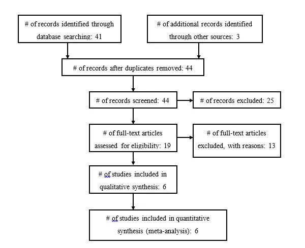
Figure 1a. Included and Excluded Articles Flowchart
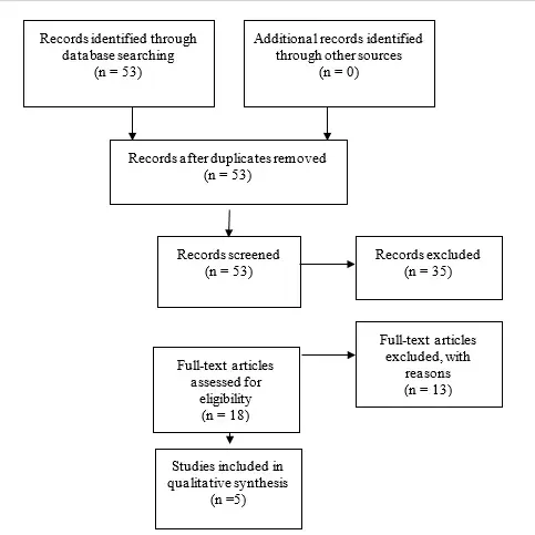
Fig. 1b. Flowchart showing the process involved in identifying relevant literature for the 2020 Update. The criteria for “records excluded” and “full text articles excluded with reasons” are detailed in Part 1 of the Guidelines.
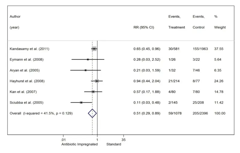
Fig. 2. Forest plot comparing AISs and SSs.
Evidence Tables
Table 1 Antibiotic Impregnated Shunt Systems vs. Conventional Shunts
| Author | Study Description | Data Class, Quality and Reasons | Results and Conclusions |
| Kandasamy et al., 2011 | ambispective, multiinstitution | Class III – Ambispective cohort with historical controls used | AIS: 30/581 (5.2%); SS: 155/1963 (7.9%). AIS reduced shunt infection rate. |
| Eymann et al., 2008 | retrospective, single institution | Class II – retrospective cohort | AIS: 1/26 (3.8%); SS: 3/22 (13.6%). No statistically significant difference.* |
| Aryan et al. 2005 | retrospective, single institution | Class II – retrospective cohort | AIS: 1/32 (3.1%); SS: 7/46 (15.2%). No statistically significant difference. |
| Hayhurst et al., 2008 | retrospective, single institution | Class III – historical controls used | AIS: 21/214 (9.8%); SS: 8/77 (10.4%). No statistically significant difference. |
| Kan et al., 2007 | retrospective, single institution | Class II – retrospective cohort | AIS: 4/80 (5%); SS: 7/80 (8.8%). No statistically significant difference. |
| Sciubba et al. 2005 | retrospective, single institution | Class II – retrospective cohort | AIS: 2/145 (1.4%); SS: 25/208 (12%). AIS reduced shunt infection rate. |
† – percentages are per shunt procedure, not per patient.
*- Fisher’s exact test used.
Table 2 Sensitivity Analysis Results
| N largest studies | Risk Ratio (95% CI) | Heterogeneity (I2) | Result |
| All studies (Figure 1) | 0.51 (0.29, 0.89) | 41.5% | Statistically Significant |
| 5 largest studies | 0.52 (0.28, 0.95) | 50.5% | Statistically Significant |
| 4 largest studies | 0.55 (0.29, 1.05) | 56.5% | Not Significant |
| 3 largest studies | 0.52 (0.23, 1.20) | 71.0% | Not Significant |
| 2 largest studies | 0.31 (0.06, 1.75) | 82.1% | Not Significant |
Table 3 Number Needed to Treat
| Assumed population Infection Rate | Number Needed to Treat | Number of Infections avoided per 1000 treated with AIS (95% CI) | |
| 5% | 41 | 24 (5, 35) | |
| 8.6%* | 24 | 42 (9, 60) | |
| 10% | 21 | 49 (11, 70) | |
| 12.5% | 17 | 61 (14, 88) | |
| 15% | 14 | 73 (16, 106) |
Table 4. New evidence included in 2020 Update
| Author | Study Description | Data Class, Quality and Reasons | Results and Conclusions |
| Mbabazi et al, 2019 | Single center, prospective RCT that attempts to determine the efficacy of bacteriseal universal shunts in Uganda patients. | II | In a unique group of patients (low resource country), a unique antibiotic impregnated catheter design (distal slit valve) failed to show an advantage in infection prevention. |
| Raffa et al, 2015 | Single center, retrospective review, comparing children who received AIS shunts and those who did not. | III | AISs reduced shunt infection in high-risk pediatric patients > 1 year old. |
| Lane et al, 2014 | Single center, retrospective review, comparing the efficacy of a Bactiseal shunt system to a non-antibiotic-impregnated system. | III | No significant different between groups. |
| Jaeger et al, 2017 | Single center, retrospective review evaluating the use of AIS shunts to decrease neonatal VP shunt infections. | III | AIS catheters and perioperative antibiotics may be helpful in neonatal hydrocephalus |
| Mallucci et al, 2019 | Multicenter, single-blinded RCT comparing antibiotic or silver to standard ventriculoperitoneal shunts (BASICS). | I | Antibiotic impregnated shunts reduce the rate of infection, particularly in children. |
Part 8: Management of cerebrospinal fluid shunt infection
Reposted with permission from ©AANS, 2014
J Neurosurg Pediatrics (Suppl) 14:60–71, 2014
AANS, 2014
(Original text of the guideline was edited to reflect the update. Please click here for the original publication.)
UPDATE
Mandeep S. Tamber, MD,1 Paul Klimo, Jr., MD, MPH,2,3 Catherine A. Mazzola, MD,4 Ann Marie Flannery, MD5
1Department of Pediatric Neurological Surgery, Children’s Hospital of Pittsburgh, University of Pittsburgh, Pittsburgh, Pennsylvania; 2Department of Neurosurgery, University of Tennessee Health Science Center, Memphis, and 3Le Bonheur Children’s Hospital, Memphis, Tennessee; 4Division of Pediatric Neurological Surgery, Goryeb Children’s Hospital, Morristown, New Jersey; and 5Department of Neurological Surgery, Saint Louis University, St. Louis, Missouri
Object. The objective of this systematic review was to answer the following question: What is the optimal treatment strategy for CSF shunt infection in pediatric patients with hydrocephalus?
Methods. The US National Library of Medicine and the Cochrane Database of Systematic Reviews were queried using MeSH headings and key words relevant to the objective of this systematic review. Abstracts were reviewed, after which studies meeting the inclusion criteria were selected and graded according to their quality of evidence (Classes I–III). Evidentiary tables were constructed that summarized pertinent study results, and based on the quality of the literature, recommendations were made (Levels I–III).
Results. A review and critical appraisal of 27 studies that met the inclusion criteria allowed for a recommendation for supplementation of antibiotic treatment using partial (externalization) or complete shunt hardware removal, with a moderate degree of clinical certainty. However, a recommendation regarding whether complete shunt removal is favored over partial shunt removal (that is, externalization) could not be made owing to severe methodological deficiencies in the existing literature. There is insufficient evidence to recommend the use of intrathecal antibiotic therapy as an adjunct to systemic antibiotic therapy in the management of routine CSF shunt infections. This also holds true for other clinical scenarios such as when an infected CSF shunt cannot be completely removed, when a shunt must be removed and immediately replaced in the face of ongoing CSF infection, or when the setting is ventricular shunt infection caused by specific organisms (for example, gram-negative bacteria).
Conclusions. Supplementation of antibiotic treatment with partial (externalization) or complete shunt hardware removal are options in the management of CSF shunt infection. There is insufficient evidence to recommend either shunt externalization or complete shunt removal as the preferred surgical strategy for the management of CSF shunt infection. Therefore, clinical judgment is required. In addition, there is insufficient evidence to recommend the combination of intrathecal and systemic antibiotics for patients with CSF shunt infection when the infected shunt hardware cannot be fully removed, when the shunt must be removed and immediately replaced, or when the CSF shunt infection is caused by specific organisms. The potential neurotoxicity of intrathecal antibiotic therapy may limit its routine use.
Recommendation: Supplementation of antibiotic treatment with partial (externalization) or with complete shunt hardware removal is an option in the management of CSF shunt infection. Strength of Recommendation: Level II, moderate degree of clinical certainty.
Recommendation: There is insufficient evidence to recommend either shunt externalization or complete shunt removal as a preferred surgical strategy for the management of CSF shunt infection. Therefore, clinical judgment is required. Strength of Recommendation: Level III, unclear degree of clinical certainty.
Recommendation: There is insufficient evidence to recommend the combination of intrathecal and systemic antibiotics for patients with CSF shunt infection in whom the infected shunt hardware cannot be fully removed or must be removed and immediately replaced, or when the CSF shunt infection is caused by specific organisms. The potential neurotoxicity of intrathecal antibiotic therapy may limit its routine use. Strength of Recommendation: Level III, unclear degree of clinical certainty.
(http://thejns.org/doi/abs/10.3171/2014.7.PEDS14328)
Key Words: cerebrospinal fluid shunt, infection, therapy, pediatrics, evidence-based guidelines, practice guidelines, hydrocephalus
Abbreviations used in this paper: EVD = external ventricular drain; VA = ventriculoatrial; VP = ventriculoperitoneal.
Cerebrospinal fluid shunt infection is one of the most common and serious complications of CSF shunt therapy. Infection admissions number approximately 2300 per year in the United States and, in aggregate, account for more than 50,000 hospital days.1 Total hospital charges related to the management of CSF shunt infection were nearly $250 million in 2003 adjusted dollars.1
Within 24 months after insertion, infections complicate approximately 11% of initial CSF shunt placements.2 Despite the high incidence of this complication, the optimal management of CSF shunt infection has yet to be defined. The existing evidence regarding the management of CSF shunt infection is of poor methodological quality. As such, current management is dictated not by evidence, but rather by physician preference and other possibly relevant patient-level factors (for example, patient surgical risk, ventricle size, and complexity of the shunt system). It is not surprising that there is significant variation in CSF shunt infection treatment protocols between centers.3
The objective of this systematic review was to answer the following question: What is the optimal treatment strategy for CSF shunt infection in pediatric patients with hydrocephalus? The successful treatment of CSF shunt infection aims to cure the infection (that is, minimize the probability of reinfection or relapse) while maintaining functional CSF diversion and minimizing morbidity, mortality, and the cost of therapy. The alternative paradigms for the management of ventricular shunt infection are illustrated well if one considers important historical milestones in the treatment of hydrocephalus. The evidentiary tables are structured somewhat accordingly (Fig. 1). The development of the Holter-Pudenz valve in 1957 and the ability to insert the distal end of a ventricular shunt into the right atrium was a major development in the treatment of hydrocephalus. Although ventriculoatrial (VA) shunts facilitated continuous and regulated CSF diversion, the fact that the distal catheter entered the heart posed logistical problems when these shunts inevitably became infected. A major issue with VA shunts was loss of limited venous access if these shunts were removed and not immediately replaced. In light of this limitation, the predominance of literature examining the treatment of CSF shunt infections in the era of VA shunts documented the outcomes of treatment with systemic antibiotics alone (Table 1) and whether the elevated CSF antibiotic concentrations achieved by intrathecal therapy conferred any additional benefit in managing the ventriculitis that often accompanied CSF shunt infection—both while leaving the infected shunt in situ or after removing the shunt and immediately replacing it in infected cerebrospinal fluid (Table 2).
A decade later, Ames developed a technique for placement of the distal catheter in the peritoneal space, and as such, made shunt removal and later replacement a feasible surgical strategy in the management of CSF shunt infection. Over time, the combined medical and surgical treatment of ventricular shunt infection became more accepted, in part because of the gradual phase-out of VA shunts and their associated limitations with respect to repeated surgical access to the heart, but perhaps more significantly because of the realization that an infected ventricular shunt, as an infected foreign body, was difficult if not impossible to sterilize using antibiotics alone. This management philosophy accepts not only that shunt removal (and eventual replacement once CSF sterility is achieved) requires multiple surgeries, but also the risk of introducing secondary infection during a variable period of external drainage. Therefore, although more contemporary literature examining the treatment of CSF shunt infection consists of studies that incorporate some form of shunt removal, variations in whether the infected shunt was partially removed (that is, externalized) (Table 3) or completely removed (see Table 4), and whether supple- mental intrathecal antibiotics were administered contribute to significant between-study heterogeneity.
A lack of rigorous comparative effectiveness studies leads to uncertainty regarding the preferred therapeutic strategy for a particular clinical circumstance. Decision analytical modeling attempts to apply statistical simulation techniques to preexisting data to rank competing therapeutic options in terms of their relative effective- ness. A decision analysis examining the treatment of CSF shunt infection using data from published studies (most included in evidentiary Tables 1–4) came to the conclusion that the best treatment modality for CSF shunt infection was antibiotic administration (systemic, with or without intrathecal administration) and complete removal of the infected shunt, with intercurrent external ventricular drainage or ventricular taps, followed by placement of a new shunt when CSF sterility is achieved. Sensitivity analyses revealed that this treatment option had the highest cure rate, the lowest failure rate, and the lowest mortality rate when compared with treatment consisting of antibiotic therapy with shunt removal and immediate replacement, or antibiotic treatment alone, over a wide range of assumptions.4
Multiple review articles on the topic also conclude that shunt infection should be ideally managed with antibiotics, complete shunt removal, and placement of a temporary external ventricular drain (EVD), followed by reimplantation after CSF sterilization.5-9 Although intrathecal administration of antibiotics appears to make theoretical sense because of enhanced CSF antibiotic concentrations, its practical application is controversial, owing in large part to the potential adverse effects of intrathecal therapy, including neurotoxicity. The indications for intrathecal therapy are not well established and presently range from use in any shunt infection, use in only those infections in which the CSF cannot be sterilized by systemic antibiotics alone (for example, persistent positive cultures), or use in those ventricular shunt infections caused by specific organisms (for example, gram- negative infections). A practice survey of board-certified members of the American Society of Pediatric Neurosurgeons revealed that most surgeons treat ventricular shunt infection with antibiotics, removal of the infected CSF shunt, and placement of an EVD, followed by delayed shunt replacement—a management paradigm that can be supported by the available evidence, as detailed below.
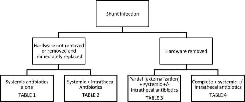
Fig. 1. Organization of evidentiary tables based on alternative paradigms for the management of CSF shunt infection.
Methods
Search Criteria
We searched the US National Library of Medicine (PubMed/MEDLINE) database and the Cochrane Data- base of Systematic Reviews for the period January 1966 through March 2012 using the following MeSH subject headings: (CSF shunts) AND (bacterial infection OR prosthesis-related infection OR catheter-related infection) AND (treatment OR outcome) AND (antibacterial agents OR injections OR antibiotics OR device removal OR ventriculostomy OR combined modality therapy). Searches were limited to studies in patients younger than 18 years of age, the management of initial (not repeat) CSF shunt infection, and to the English language.
Authors performed an updated literature search (in PubMed and Cochrane Central) for this guideline chapter through a medical librarian at the Congress of Neurological Surgeons Guidelines office using the below-mentioned existing search terms to update the original search through November 30, 2019.
Search Results
A total of 342 abstracts were screened and 69 full- text articles were retrieved for review. The details of this process are described in Part 1, the introduction and methodology section of these guidelines.5 An examination of the reference lists of these 69 full-text articles yielded an additional 24 articles that warranted full-text review (Fig. 2). Subsequent review of the full texts of these 93 articles led to the exclusion of 66 articles based on predefined criteria, leaving 27 articles as the basis for the evidentiary tables for this particular recommendation. Reasons for exclusion of full-text articles included the following: literature review (n = 19); no treatment outcomes given (n = 14); pediatric patients not reported separately (n = 6); wrong target population (n = 1); small sample size (n = 19); not a full report of a clinical study (n = 2); not relevant to the study question (n = 3); and other (n = 2).
An additional 2 studies out of the 62 new studies yielded by the 2020 underwent full text review, but were excluded. No new studies met inclusion criteria from the original guideline. (Fig 3)
Results
In general, the methodological quality of the evidence related to this recommendation was poor. The studies that met our inclusion criteria were typically descriptive series of small numbers of patients and were vulnerable to the biases and limitations of a retrospective study design. Because the studies relied on the accuracy and completeness of the medical record, the control of potentially confounding variables was nonexistent. Although most studies did compare outcomes between patient groups treated under alternative management protocols, the rationale behind why a particular treatment was assigned to a particular patient group was not clearly described, leading to significant issues with selection bias. For those studies describing the outcomes of a single management protocol, between-study comparisons of results was hampered by widely disparate management protocols and the use of nonuniform outcome measures (and definitions thereof). These limitations precluded, for the most part, any meaningful quantitative synthesis of the data; what follows is a largely qualitative review of the evidence relevant to this recommendation.
Despite the overall predominance of Class III data, the 13 studies presented in evidentiary Tables 110-16 and 217-22 are quite suggestive of the notion that in the management of CSF shunt infection, supplementation of antibiotic treatment with partial (externalization) or complete shunt hardware removal should be considered. Two Class II studies provide particularly compelling evidence in favor of a combined medical and surgical management of CSF shunt infection, and deserve to be elaborated on further.
In 1980, James et al19 published the results of a moderate-quality randomized controlled trial in which 10 patients with evidence of CSF shunt infection were randomized to each of 3 different treatment arms: 1) complete shunt removal, systemic antibiotics, and either external ventricular drainage or ventricular taps for decompression and intrathecal antibiotic administration, with delayed shunt replacement; 2) complete shunt removal and immediate shunt replacement with intrashunt and systemic antibiotics; or 3) intrashunt and systemic antibiotics without shunt removal. The outcome was negative ventricular CSF cultures 48 hours after cessation of antibiotic therapy and again within 4 months of completion of therapy. All 10 patients who underwent complete shunt removal, systemic antibiotics, and either external ventricular drainage or ventricular taps for decompression and intrathecal antibiotic administration were successfully treated. Nine of 10 patients treated with complete shunt removal and immediate shunt replacement with intrashunt and systemic antibiotics achieved therapeutic trast, only 3 of 10 patients who received systemic and intrathecal antibiotics without shunt removal were successfully treated. The treatment results in this latter group rather clearly demonstrate that shunt removal, rather than antibiotic therapy (including intrathecal therapy), was responsible for the improved outcomes seen in the comparison groups. Secondary outcomes also were consistent with a benefit toward surgical removal of the shunt, as length of hospital stay was lowest in those patients who underwent complete shunt removal with delayed shunt re placement after a course of systemic and intrathecal antibiotics. The only deaths occurred in those patients who received medical management alone. Because of the convincing inferiority of medical management alone, further randomization to this group was halted, but the study was continued as a prospective nonrandomized comparison of treatment outcomes between those patients receiving intrathecal and systemic antibiotics in conjunction with complete shunt removal and delayed versus immediate shunt replacement.18 The principal conclusions remain unchanged.
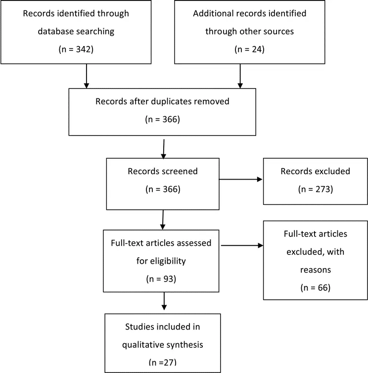
Fig. 2a. Flowchart showing the process involved in identifying relevant literature
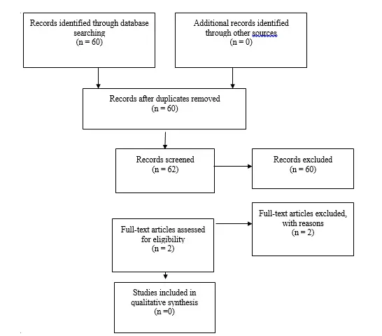
Fig. 2b. Flowchart showing the process involved in identifying relevant literature for the 2020 Update. The criteria for “records excluded” and “full text articles excluded with reasons” are detailed in Part 1 of the Guidelines.
The nearly equivalent treatment outcomes of shunt removal followed by immediate shunt replacement (that is, shunt replacement in infected CSF) versus delayed shunt replacement (that is, shunt replacement after the CSF has been sterilized) in the aforementioned studies by James and colleagues18,19 was suggestive of the potential utility of intrathecal antibiotics in those clinical circumstances in which the shunt must be removed and immediately replaced. As such, these studies provide some evidence applicable to the intrathecal antibiotic recommendation as well. As outlined earlier, it appears that most of the treatment effect comes from shunt removal, making the relative contribution of intrathecal antibiotics to improved outcomes in this scenario rather uncertain. Hence, elevating the recommendation for intrathecal antibiotics to a Level II recommendation, based on these relatively high quality data alone, appears unwarranted.
Additional evidence pertaining to the intrathecal antibiotic recommendation comes largely from Class III studies that examined the results of treatment of ventricular shunt infection in those clinical circumstances in which the infected shunt components are not removed (Table 2)17-22 or only partially removed (that is, externalized) (Table 3)23-26. There was a Class III study that documented a fairly large proportion of patients who achieved therapeutic success—comparable to the success seen in patients who underwent shunt removal—when the patients were treated with intrathecal antibiotics but their shunts were left in situ.16 In addition, Bayston and Rickwood17 documented eradication of staphylococcal VA or VP shunt infection in 5 of 43 patients who underwent antibiotic treatment alone; 4 of the 5 patients who were successfully treated received intrathecal antibiotics. In cases in which ventricular shunt infection was treated with systemic and intrathecal antibiotics along with shunt externalization, either because of the complexity of the shunt infection scenario (for example, multiloculated hydrocephalus) or surgeon preference, a prospective non-randomized study by James and Bradley24 and a Class III study by Arnell et al23 were both able to demonstrate positive treatment outcomes in all patients in their respective case series. Finally, another retrospective case series by James and Bradley27 showed convincingly high cure rates with a significantly shorter length of stay in those patients with an uncomplicated shunt infection (that is, a single shunt system) treated with complete shunt removal together with systemic and intrathecal antibiotics (Table 4). Unfortunately, the absence of a concurrent control group treated with shunt removal and systemic antibiotics alone in this and other studies listed in Table 4 limits the impact of these data to the overall body of evidence.
When examining the studies presented in evidence in Table 323-26 and Table 427-36 it is difficult to say with any degree of clinical certainty whether complete shunt removal leads to better shunt infection treatment outcomes than partial shunt removal. This is due, in part, to the paucity of outcome data comparing the 2 treatment options within the same study population, but also to the confounding effect of intrathecal antibiotic therapy, as described above.
After a full-text review of the contents of papers that were initially identified through our search strategy or our scrutiny of reference lists, predefined criteria led to the exclusion of multiple studies from the evidentiary tables. The recommendations provided above are not materially changed by the exclusion of these studies.
2020 Update
After examining these additional abstracts, two full text articles were assessed for their potential inclusion into the evidence tables. Yakut et al37 was excluded because there was non-consecutive enrollment of patients, and a study by von der Brelie et al38 did not meet inclusion criteria as pediatric patients represented less than 80% of the overall cohort that was reported.
Conclusions
Recommendation: Supplementation of antibiotic treatment with partial (externalization) or with complete shunt hardware removal is an option in the management of CSF shunt infection. Strength of Recommendation: Level II, moderate degree of clinical certainty.
Recommendation: There is insufficient evidence to recommend either shunt externalization or complete shunt removal as a preferred surgical strategy for the management of CSF shunt infection. Therefore, clinical judgment is required. Strength of Recommendation: Level III, unclear degree of clinical certainty.
Recommendation: There is insufficient evidence to recommend the combination of intrathecal and systemic antibiotics for patients with CSF shunt infection in whom the infected shunt hardware cannot be fully removed or must be removed and immediately replaced, or when the CSF shunt infection is caused by specific organisms. The potential neurotoxicity of intrathecal antibiotic therapy may limit its routine use. Strength of Recommendation: Level III, unclear degree of clinical certainty.
It appears that the optimal management of CSF shunt infection requires a multimodality approach. Re- view and critical appraisal of the available evidence regarding the management of ventricular shunt infection allow for a recommendation for the supplementation of antibiotic treatment with partial (externalization) or complete shunt hardware removal with a moderate degree of clinical certainty. However, a recommendation regarding whether complete shunt removal is favored over partial shunt removal (that is, externalization) cannot be made, owing to severe methodological deficiencies in the existing literature. Furthermore, there is insufficient evidence to recommend the use of intrathecal antibiotic therapy as an adjunct to systemic antibiotic therapy in the management of routine CSF shunt infections, or in other clinical scenarios, such as when an infected CSF shunt cannot be completely removed, must be removed and immediately replaced in the face of ongoing CSF infection, or in the setting of ventricular shunt infection caused by specific organisms (for example, gram-negative bacteria).
Deficiencies in the existing literature regarding the management of CSF shunt infection provide a strong rationale for further prospective research into the subject. Key questions that remain unanswered include, but are certainly not limited to the following:
- Defining the optimal duration of antibiotic therapy in the management of CSF shunt infection, with the aim of simultaneously maximizing the probability of successful treatment without reinfection or relapse, and minimizing the length of hospital stay and over-all cost to the health care system
- Refining the indications for intrathecal antibiotic therapy and ascertaining the risk/benefit profile of such therapy (potential adverse effects vs potential reduction in relapse/reinfection rates and shorter hospital stays).
- Definition and validation of standardized treatment outcome measures, based on microbiological or other biomarker-based criteria. This would not only facilitate a comparison of results across studies, but also potentially yield objective criteria that facilitate decision making in other contentious areas of CSF shunt infection management, such as the optimal timing of shunt reimplantation.
Perhaps the best strategy to treat ventricular shunt infection is to continue our focus on the prevention of this significant complication of CSF shunt therapy.
Based on the assessments made in the 2020 update, the authors concluded that no new literature exists to support any change or revision to the current guideline.
Acknowledgments
We acknowledge the American Association of Neurological Surgeons (AANS)/Congress of Neurological Surgeons (CNS) Joint Guidelines Committee for the members’ reviews, comments, and suggestions; Laura Mitchell, Guidelines Project Manager for the CNS, for her contributions; Pamela Shaw, research librarian, for her assistance with the literature searches; Kevin Boyer for his assistance with data analysis; and Sue Ann Kawecki for her assistance with editing. We also acknowledge the following peer reviewers for their contributions to review the update to the guidelines: Jennifer Sweet, MD, Brandon Rocque, MD, Christoph Greissenauer, MD, Jeffrey Olson, MD.
Disclosure
The systematic review and evidence-based guidelines were funded exclusively by the CNS and AANS Pediatric Section, which received no funding from outside commercial sources to support the development of this document.
Conflict(s) of Interest: None. All members of the Pediatric Hydrocephalus Systematic Review and Evidence-Based Guidelines Task Force declared any potential conflicts of interest prior to beginning work on this evidence review.
Conflict(s) of Interest: None. All Pediatric Hydrocephalus Systematic Review and Evidence-Based Guidelines Update Task Force members declared any potential conflicts of interest prior to beginning work on this systematic review and evidence-based guidelines.
Author contributions to the study and manuscript preparation include the following. Conception and design: AANS/CNS Joint Section on Pediatrics. Acquisition of data: all authors. Analysis and interpretation of data: all authors. Drafting the article: Tamber. Critically revising the article: all authors. Reviewed submitted version of manuscript: all authors. Approved the final version of the manuscript on behalf of all authors: Flannery. Administrative/technical/material support: all authors. Study supervision: Flannery.
Evidence Tables
| First Author & Year | Study Description | Data Class, Quality and Reasons | Results and Conclusions |
| Forrest et al., 1987 | 12 shunt infections with positive blood cultures (but sterile CSF) treated with IV antibiotics and complete shunt removal with immediate shunt replacement.Outcome = no evidence of recolonization at last follow-up (3-16 years) | Class IIIRetrospective case series11 patients with positive blood and CSF cultures were treated with IV antibiotics, shunt removal and EVD with delayed shunt replacement, but their treatment outcomes are not presented | 12/12 patients with positive blood cultures but sterile CSF are without evidence of recolonization at last follow-upDifficult to interpret these findings in isolation |
| Odio et al., 1984 | 59 shunt infections managed with systemic antibiotics alone (A. n=13); systemic antibiotics + immediate shunt removal (B. n=37); or systemic antibiotics + delayed shunt removal (C. n=9).Outcome = cure (absence of shunt re-infection or relapse) | Class IIIRetrospective case seriesPoor control of confoundersReasons for immediate vs. delayed shunt removal not given (selection bias)Timing of outcome assessment not given | Cure in 8/13 patients treated with antibiotics alone, 34/37 patients treated with antibiotics + immediate shunt removal, and 8/9 patients with antibiotics + delayed shunt removalResults suggest a poorer outcome without shunt removal |
| Walters et al., 1984 | 267 infections treated in 222 patients. 92 treated medically (85 systemic, 7 systemic + IT); 117 treated medical + surgical (21 shunt removed and immediately replaced under antibiotic coverage, 51 shunt removal + antibiotics + delayed shunt replacement, 20 shunt removal + EVD/shunt externalization + IT antibiotics, 25 shunt removal + antibiotics without shunt replacement); 58 no specific treatment of shunt infection (e.g. unrecognized shunt infection).Outcome = death Outcome = death | Class IIIRetrospective case seriesDefinition of cure (another tabulated outcome) not definedMultiple different permutations and combinations of treatment without clear criteria matching a particular patient with a particular treatment (selection bias)Nontraditional outcome | 37% mortality with medical management alone18% mortality with medical + surgical therapy; lowest mortality in those who had shunt removed and replaced under antibiotic coverage in a single operationAddition of surgical therapy appears to lower morbidity and mortality of shunt infection |
| Schoenbaum et al., 1975 | 98 shunt infections over 442 shunt procedures in 289 patients. Initial treatment based on shunt type – VP shunts were all completely removed and treated with IV antibiotics; some patients with VA and V-ureteral shunts were initially managed with IV antibiotics alone.Outcome = death | Class IIIRetrospective case seriesPoor control of confoundersSelection biasAlso cite “control of infection” as outcome, but not clearly definedNontraditional outcome | 1/30 patients treated with complete shunt removal and IV antibiotics died; 28/30 had control of infection6/43 patients with VA and V-ureteral shunts treated with IV antibiotics alone died; 13/43 had control of infectionShunt removal is required for improved outcome |
| Shurtleff et al., 1974 | 67 patients with shunt infection treated with IV antibiotics alone (A. n=22); IV antibiotics + shunt revision (B. n=14); IV antibiotics + complete shunt removal and replacement (C. n=12); IV + IT antibiotics + complete shunt removal and replacement (D. n=7); IV + IT/intra-shunt antibiotics alone (E. n=10); or IV + IT antibiotics with shunt revision (F. n=2).Outcome = cure (no symptoms and at least 6 negative blood cultures and two negative ventricular/shunt CSF cultures during a 6 month period off antibiotics) | Class IIIRetrospective case seriesNo control of confoundersRationale for selecting different therapy for different infections not clear (“therapy evolved during the study”) (selection bias) | Cure in 2/22 (A), 3/14 (B), 12/12 (C), 7/7 (D), 3/10 (E), 0/2 (F).Highest cure rate in those who had complete shunt removal and replacement, irrespective of whether or not they received supplemental IT antibiotics |
| Morrice et al., 1974 | Patients with colonization of VA shunt valves treated with antibiotics alone (n=14); removal and immediate replacement of shunt (n=23); or removal of shunt with a period of external drainage, followed by delayed shunt insertion (n=18).Outcome = “alive and well” at 6 months | Class IIIRetrospective case seriesPoor control of confoundersSelection biasRoute of administration of antibiotics not specifiedUnclear if patients who underwent surgical treatment (immediate or delayed shunt replacement) received supplemental antibioticsNo specific microbiological component of outcome | 2/14 patients treated with antibiotics alone are alive and well at 6 months11/23 patients treated with removal and immediate replacement of shunt are alive and well at 6 months8/18 patients treated with removal of shunt with a period of external drainage, followed by delayed shunt insertion are alive and well at 6 monthsResults suggest that shunt removal is required to optimize outcome |
| Nicholas et al., 1970 | 60 infections of VA shunts treated with IV antibiotics (also IT antibiotics if the CSF was infected) and delayed shunt replacement (n=33 infections) or with IV antibiotics and immediate shunt replacement (n=27 infections).Outcome = “subsequent good health of the patient and freedom from bacteremia” | Class IIIRetrospective case seriesNo control of confoundersSelection biasNondescript outcome | Successful treatment in 24/33 infections treated with IV antibiotics (also IT antibiotics if the CSF was infected) and delayed shunt replacement vs. 21/27 infections treated with with IV antibiotics and immediate shunt replacement.Recurrence/relapse higher in those with immediate shunt replacement |
Table 2. Systemic and Intrathecal Antibiotic Treatment with Shunt Left In Situ or Removed and Immediately Replaced
| Author | Study Description | Data Class, Quality and Reasons | Results and Conclusions |
| James et al., 1981 | 50 patients (30 reported in James 1980 RCT) with shunt infection treated with shunt removal, systemic antibiotics and either EVD or ventricular taps for decompression and antibiotic administration (A: n=22); removal and immediate replacement of shunt with intrashunt and systemic antibiotics (B: n=17); or intrashunt and systemic antibiotics without shunt removal (C: n=11).Outcome = negative ventricular CSF cultures 48h after cessation of antibiotic therapy and again within 4 months of completion of therapy. | Class IIProspective non-randomized cohortContinuation of James 1980 RCT – high incidence of failures in medical management arm (i.e. no shunt removal) made further randomization unjustified | 21/22 patients in group A were successfully treated15/17 patients in group B were successfully treated4/11 patients in group C were successfully treatedSuggests better treatment outcomes with shunt removalSuggests that IT antibiotics may be of use if shunt must be removed and immediately replaced rather than replaced in a delayed fashion (when infection has been cleared) |
| James et al., 1980 | 30 patients with shunt infection treated with shunt removal, systemic antibiotics and either EVD or ventricular taps for decompression and antibiotic administration (A: n=10); removal and immediate replacement of shunt with intrashunt and systemic antibiotics (B: n=10); or intrashunt and systemic antibiotics without shunt removal (C: n=10).Outcome = negative ventricular CSF cultures 48h after cessation of antibiotic therapy and again within 4 months of completion of therapy. | Class IIRCT with design flawsSuboptimal randomization and allocationBaseline characteristics of treatment groups not documentedUnclear if outcome assessment blindedUnderpowered (but study terminated early for harm) | 10/10 patients in group A were successfully treated9/10 patients in group B were successfully treated3/10 patients in group C were successfully treatedlength of hospital stay lowest in group Aonly deaths occurred in group CSuggests better treatment outcomes with shunt removalSuggests that IT antibiotics may be of use if shunt must be removed and immediately replaced rather than replaced in a delayed fashion (when infection has been cleared) |
| Bayston et al., 1981 | 43 children with Staphylococcal VA or VP shunt infection treated with antibiotics alone (systemic or systemic + IT)Outcome = eradication of infection (response during treatment with no clinical relapse, followed by repeated normal serological and bacteriological studies) | Class IIIRetrospective case seriesTiming of outcome assessment not clear | Eradication of infection in 5/43 patients.4/5 of those patients with eradication of infection received IT antibioticsNo eradication of S. aureus shunt infectionSuggest the utility of supplemental IT antibiotics if the shunt cannot be removed |
| Wald et al., 1980 | 20 patients with shunt infection treated with daily IT antibiotics (with systemic antibiotics) without removal of the shunt or EVD placement.Outcome = cure (2 or 3 sterile CSF cultures 72h following completion of antibiotics) | Class IIIRetrospective case seriesPharmacodynamic study | “cure” in 5/7 patients receiving at least 7 days of IT methicillin“cure” in 4/5 patients treated with IT gentamicin“cure” in 6/7 patients receiving a single 2-week course of IT cephalothinRates of “cure” appear higher than medically treated patients receiving systemic antibiotics alone |
| Sells et al., 1977 | 20 gram negative shunt infections receiving 25 total treatment trials. Treatments were no treatment (n=2); systemic antibiotics alone (n=4); systemic and intraventricular antibiotics alone (n=4); systemic antibiotics plus in situ shunt replacement (i.e. into infected tract) or incomplete shunt replacement (n=2); systemic and intraventricular antibiotics with in situ shunt replacement (i.e. into infected tract) or incomplete shunt replacement (n=4); systemic and intraventricular antibiotics with complete shunt removal or replacement in a new site (n=9).Outcome = cure (asymptomatic patient with at least 6 negative blood cultures and 2 negative ventricular or shunt CSF cultures obtained during a 6 month period off antibiotics) | Class IIIRetrospective case seriesPoor control of confoundersSelection biasVery small numbers receiving each individual treatment | Cure in 0/2 patients receiving no treatmentCure in 1/4 patients receiving systemic antibiotics aloneCure in 0/4 patients receiving systemic and intraventricular antibiotics aloneCure in 1/2 patients receiving systemic antibiotics plus in situ shunt replacement or incomplete shunt replacementCure in 0/4 patients receiving systemic and intraventricular antibiotics with in situ shunt replacement or incomplete shunt replacementCure in 9/9 patients receiving systemic and intraventricular antibiotics with complete shunt removal or replacement in a new siteClear advantage of complete vs. incomplete shunt removal No clear additional advantage of IT antibiotics in those treated medically or medically with in situ shunt replacement or incomplete shunt replacement |
| McLaurin et al., 1975 | 25 shunt infections (23 VA and 2 VP) treated with IV + IT antibiotics and delayed shunt replacement (n=4), IV + IT antibiotics with immediate shunt replacement (n=10), or IV + IT antibiotics alone (n=11).Outcome = absence of residual infection at last follow-up (6mo-5 yr) | Class IIIRetrospective case seriesCharacteristics of those patients successfully treated with IV + IT antibiotics alone (i.e. without shunt removal and replacement) not documented (selection bias)Extension of McLaurin 1973 series | Absence of residual infection at last follow-up in all 24 surviving patients (infection believed to have been cured in the one patient who died)Suggests that IT antibiotics may be of use if shunt is not removed or must be removed and immediately replacedShunt removal may not be necessary for successful treatment of shunt infection if IT antibiotics are administered. |
| James et al., 2008 | Prospective nonrandomized study of 2 protocols for treating complicated shunt infections (multiloculated, multi-organism, infection at other site in body).n=21 treated with IV (2 weeks) and IT antibiotics injected through EVD (n=10) or reservoir of externalized shunt (n=11) (2x/week for 2 weeks). Three weeks of antibiotics in total.Outcome = cure (cultures 48h after cessation of antibiotics, at time of new shunt placement, and 3-6 months later remained negative).n=18 treated with IV (2 weeks) and IT antibiotics injected through EVD or reservoir of externalized shunt (1x/week for 2 weeks). Three weeks of antibiotics in total.Outcome = cure (cultures 24h after cessation of antibiotics, at time of new shunt placement, and 3-6 months later remained negative). | Class IIINonrandomized, prospective case seriesOutcome is different for each treatment group | All patients treated according to either protocol were cured.LOS protocol A = 25.1d vs protocol B = 19.7d.No recurrent shunt infections during the follow-up period.Patients with complicated shunt infections can be successfully treated successfully with 2 weeks of once daily IT therapy concurrent with 3 weeks of IV therapy (and EVD or shunt externalization). |
| Arnell et al., 2007 | Retrospective review of 34 consecutively treated intravenricular shunt infections treated with externalization of the ventricular catheter proximal to the valve, daily IT injections (generally guided by CSF antibiotic concentrations, median 8 days) and IV antibiotics (median 10 days). Usually no antibiotics after shunt replacement.Outcome = cure (sterilization of CSF and resolution of clinical symptoms). | Class IIIRetrospective case seriesno control of confounders | CSF sterilized in 1/3, 7/8, 20/20 and 6/6 cases after 1, 2, 3, and >3 days of therapy (externalization of ventricular catheter and start of IT antibiotics). Clinical symptoms resolved in parallel with the sterilization of CSF.Despite the ventricular catheter being left in place and the short duration of therapy, the treatment protocol results in quick CSF sterilization, a low relapse rate, and survival of all patients in this series. |
| Wang et al., 1999 | 23 patients treated according to a documented management protocol (externalization of distal catheter unless failure to sterilize CSF, empiric followed by tailored antibiotics for 10 days following sterilization of CSF, reimplant shunt if cultures remain negative for 3 days off antibiotics). Comparison group 10 historical controls treated with an undisclosed regimen.Outcome = recurrence (re-infection with same organism within 6 months). | Class IIIComparative study with historical controlsNo control of confounders3 patients had a ventricular reservoir onlyDetails of treatment of historical control patients not clear (“duration of antibiotic therapy for each individual case was decided arbitrarily”) | Reinfection 0/15 patients treated under protocol (8 patients did not require shunt re-insertion) vs. 2/10 treated before protocolShorter hospital stay in those treated under the protocol.Of those treated under the protocol, patients with a “complex” shunt system required longer hospitalization.This treatment protocol may be effective in the management of shunt infection |
| Ronan et al., 1995 | 41 episodes of infection in 39 children treated with antibiotics (28 IV and oral, 11 IV + IT + oral, 4 IT + IV, 1 IT + oral) and surgical treatment (complete or partial shunt removal and immediate or delayed replacement with or without external ventricular drainage).Outcome = absence of relapse (re-infection with same organism) at 3 months, and was verified by the absence of relapse for the follow-up period (min. 1 year) | Class IIIRetrospective case seriesSelection biasOverall management approach too varied to allow for reasonable conclusions to be made | Absence of relapse in 31, relapse in 6, death in 4 (not directly related to shunt infection).Outcome not dependent on length of antibiotic treatment or use of IT antibioticsSurgical approach to treatment too varied to permit conclusions re: efficacy. Complete shunt replacement associated with lower risk of relapse vs. partial replacement, and delayed replacement had better outcomes vs. immediate replacement. |
| Kestle et al., 2006 | 70 patients from 10 centers followed prospectively for 1 year following successful treatment of shunt infection. Initial management was shunt externalization and antibiotics in 17; shunt removal, EVD insertion and antibiotics in 50; and antibiotic treatment alone in 3.Outcome = culture-proven reinfection (same or different organism). | Class IIProspective multicenter observational studyReinfection rates in those externalized versus completely removed not provided separatelyTiming of outcome assessment not clear | Reinfection occurred in 18 patients (26%) – 12 were due to the same initial organism and 6 were different organisms.Reinfection risk was not associated with length of antibiotic treatment.This study reconfirms the high reinfection rate in patients receiving treatment for shunt infection. |
| Shimuzu et al., 2012 | Retrospective chart review of 36 patients who underwent shunt removal, EVD placement (4 patients had externalization prior to EVD placement), IV antibiotics and eventual shunt replacement compared to 9 patients who underwent shunt removal, IV antibiotics and ETV for treatment of shunt infection.Outcome = recurrence of CSF infection within 6 months after shunt reinsertion or ETV. | Class IIIRetrospective case seriesno control of confoundersSelection bias | Of those treated with shunt removal then reinsertion, 10/36 experienced CSF reinfectionThis study reconfirms the high reinfection rate in patients receiving treatment for shunt infection. |
| James et al. (2), 2008 | Retrospective nonrandomized comparison of 2 protocols for treating uncomplicated shunt infections (single shunt system, single organism, noncompartmentalized hydrocephalus). A. n=25 shunt removal/EVD, IV antibiotics until clinical course and CSF values suggested cure of infection, IT antibiotics 2x/week through EVD or at times of ventricular puncture.Outcome = cure (cultures 48h after cessation of antibiotics, at time of new shunt placement, and 3-6 months later remained negative).n=15 shunt removal/EVD, IV antibiotics until clinical course and CSF values suggested cure of infection, IT antibiotics 1x/week through EVD or at times of ventricular puncture.Outcome = cure (cultures 24h after cessation of antibiotics, at time of new shunt placement, and 3-6 months later remained negative) | Class IIIRetrospective comparative studyOutcome is different for each treatment group | All patients treated according to either protocol were cured.Duration of IV antibiotics protocol A = 9.7d vs protocol B = 9.9d.Patients with a single shunt infected with a single organism and with noncompartmentalized hydrocephalus may be successfully treated without a prolonged antibiotic course and lengthy hospital stay, provided the shunt is completely removed. |
| Schuhmann et al., 2005 | 35 consecutive culture proven shunt infections were treated with antibiotics, surgery for shunt removal/EVD placement or shunt externalization and eventual reinternalization of the shunt.Outcome = shunt reinfection. | Class IIIProspective case seriesDetails of management of shunt infection not clear (e.g. number who underwent complete vs. incomplete shunt removal; number who had intrathecal supplementation to systemic antibiotic therapy, if any)Outcomes not provided separately for those externalized versus completely removed | 6/33 patients experienced a shunt reinfection.This study reconfirms the high reinfection rate in patients receiving treatment for shunt infection. |
| Turgut et al., 2005 | 37 infections in 35 patients. 31 patients treated with shunt removal, EVD and systemic + IT antibiotics. 4 patients treated with medical management alone.Outcome = death. | Class IIIRetrospective case seriesNontraditional outcomeCriteria for treating 4 patients medically not clear (selection bias) | 2/31 patients treated with shunt removal, EVD and systemic + IT antibiotics died1/4 patients treated medically diedgood outcomes with IT therapy, but no patients underwent shunt removal with systemic antibiotics alone |
| Mancao at al., 1998 | 29 consecutive shunt infections treated. 27 patients had shunt removal +/- external drainage. All had IV antibiotics, and 6 had supplemental IT antibiotics.Outcome = relapse of infection. | Class IIIRetrospective case seriesDefinition of outcome (relapse) not provided, nor was follow-up period definedCriteria for IT antibiotics not given | 27 patients had successful treatment (no relapse). 2 deaths not clearly related to shunt infectionThis study demonstrates a lower rate of reinfection than other studies, but the data is of poor quality |
| Stamos et al., 1993 | 23 consecutive gram negative shunt infections, managed with complete shunt removal and EVD, and IV antibiotics (n=19) or IT antibiotics (n=2, for persistent positive cultures).Outcome = “cure” (asymptomatic and at least 3 negative cultures off antibiotics), after which shunt was reinserted | Class IIIRetrospective case seriesNo control of confounders | All patients achieved cure with shunt removal, EVD placement, and antibiotics (19 IV, 2 IV + IT)On late follow-up of 19 patients (>6 months), 4 had subsequent coag. neg. Staph. InfectionDespite initial success, reinfection rates appear similarly high when compared to other studies |
| Kontny et al., 1993 | 28 infections in 25 patients, managed with IV antibiotics and immediate removal of the shunt system (n=24) or IV antibiotics alone (n=4).Outcome = re-infection or relapse within 1 month of completion of therapy. | Class IIIRetrospective case seriesDetails of management of infection not givenShort follow-up | All patients were without re-infection or relapseShort follow-up precludes definitive conclusions |
| James et al., 1984 | 18 infections (13 following initial shunt procedure and 5 following revisions) in low birth weight infants (<2000g) treated promptly with shunt removal and IV + IT antibiotics (see James 1980).Outcome = cure | Class IIIRetrospective case seriesOutcome not clearly defined (? Negative cultures 48h after cessation of antibiotics and within 4 months of completion of therapy, as per James 1980) | All except one patient demonstrated cure when treated according to protocolgood outcomes with IT therapy, but no patients underwent shunt removal with systemic antibiotics alone |
| Scarff et al., 1978 | 57 children with shunt-related ventricular infection treated with IV + IT antibiotics and external ventricular drainage (either shunt removal and EVD, or shunt externalization).Outcome = clearance of infection (3 consecutive cultures with negative growth at 48h) | Class IIIRetrospective case seriesNumber of patients receiving each surgical therapy (shunt removal and EVD, or shunt externalization) not documented, nor were their outcomes differentially reported | 54/57 patients demonstrated clearance of infectiongood outcomes with IT therapy, but no patients underwent shunt removal with systemic antibiotics alone |
References
- Simon TD, Riva-Cambrin J, Srivastava R, Bratton SL, Dean JM, Kestle JR. Hospital care for children with hydrocephalus in the United States: utilization, charges, comorbidities, and deaths. Journal of neurosurgery Pediatrics. 2008;1(2):131-137.
- Simon TD, Hall M, Riva-Cambrin J, et al. Infection rates following initial cerebrospinal fluid shunt placement across pediatric hospitals in the United States. Clinical article. Journal of neurosurgery Pediatrics. 2009;4(2):156-165.
- Whitehead WE, Kestle JR. The treatment of cerebrospinal fluid shunt infections. Results from a practice survey of the American Society of Pediatric Neurosurgeons. Pediatric neurosurgery. 2001;35(4):205-210.
- Schreffler RT, Schreffler AJ, Wittler RR. Treatment of cerebrospinal fluid shunt infections: a decision analysis. The Pediatric infectious disease journal. 2002;21(7):632-636.
- Anderson EJ, Yogev R. A rational approach to the management of ventricular shunt infections. The Pediatric infectious disease journal. 2005;24(6):557-558.
- Fan-Havard P, Nahata MC. Treatment and prevention of infections of cerebrospinal fluid shunts. Clinical Pharmacy. 1987;6(11):866-880.
- Gutierrez-Gonzalez R, Boto GR, Perez-Zamarron A. Cerebrospinal fluid diversion devices and infection. A comprehensive review. European journal of clinical microbiology & infectious diseases : official publication of the European Society of Clinical Microbiology. 2012;31(6):889-897.
- Treatment of infections associated with shunting for hydrocephalus. Working Party on the Use of Antibiotics in Neurosurgery of the British Society for Antimicrobial Chemotherapy. Br J Hosp Med. 1995;53(8):368-373.
- Yogev R. Cerebrospinal fluid shunt infections: a personal view. Pediatric Infectious Disease. 1985;4(2):113-118.
- Forrest DM, Tabara ZB, Towu E, Said AJ. Management of the colonised shunt. Z Kinderchir. 1987;42 Suppl 1:21-22.
- Odio C, Mohs E, Sklar FH, Nelson JD, McCracken GH, Jr. Adverse reactions to vancomycin used as prophylaxis for CSF shunt procedures. Am J Dis Child. 1984;138(1):17-19.
- Schoenbaum SC, Gardner P, Shillito J. Infections of cerebrospinal fluid shunts: epidemiology, clinical manifestations, and therapy. J Infect Dis. 1975;131(5):543-552.
- Walters BC, Hoffman HJ, Hendrick EB, Humphreys RP. Cerebrospinal fluid shunt infection. Influences on initial management and subsequent outcome. Journal of neurosurgery. 1984;60(5):1014-1021.
- Morrice JJ, Young DG. Bacterial colonisation of Holter valves: a ten-year survey. Dev Med Child Neurol. 1974;16(6 Suppl 32):85-90.
- Nicholas JL, Kamal IM, Eckstein HB. Immediate shunt replacement in the treatment of bacterial colonisation of Holter valves. Dev Med Child Neurol Suppl. 1970;22:Suppl 22:110+.
- Shurtleff DB, Foltz EL, Weeks RD, Loeser J. Therapy of staphylococcus epidermidis: infections associated with cerebrospinal fluid shunts. Pediatrics. 1974;53(1):55-62.
- Bayston R, Rickwood AM. Factors involved in the antibiotic treatment of cerebrospinal fluid shunt infections. Zeitschrift fur Kinderchirurgie : organ der Deutschen, der Schweizerischen und der Osterreichischen Gesellschaft fur Kinderchirurgie = Surgery in infancy and childhood. 1981;34(4):339-345.
- James HE, Walsh JW, Wilson HD, Connor JD. The management of cerebrospinal fluid shunt infections: a clinical experience. Acta neurochirurgica. 1981;59(3-4):157-166.
- James HE, Walsh JW, Wilson HD, Connor JD, Bean JR, Tibbs PA. Prospective randomized study of therapy in cerebrospinal fluid shunt infection. Neurosurgery. 1980;7(5):459-463.
- McLaurin RL. Treatment of infected ventricular shunts. Child’s brain. 1975;1(5):306-310.
- Sells CJ, Shurtleff DB, Loeser JD. Gram-negative cerebrospinal fluid shunt-associated infections. Pediatrics. 1977;59(4):614-618.
- Wald SL, McLaurin RL. Cerebrospinal fluid antibiotic levels during treatment of shunt infections. Journal of neurosurgery. 1980;52(1):41-46.
- Arnell K, Enblad P, Wester T, Sjolin J. Treatment of cerebrospinal fluid shunt infections in children using systemic and intraventricular antibiotic therapy in combination with externalization of the ventricular catheter: efficacy in 34 consecutively treated infections. Journal of neurosurgery. 2007;107(3 Suppl):213-219.
- James HE, Bradley JS. Management of complicated shunt infections: a clinical report. Journal of neurosurgery Pediatrics. 2008;1(3):223-228.
- Ronan A, Hogg GG, Klug GL. Cerebrospinal fluid shunt infections in children. The Pediatric infectious disease journal. 1995;14(9):782-786.
- Wang KC, Lee HJ, Sung JN, Cho BK. Cerebrospinal fluid shunt infection in children: efficiency of management protocol, rate of persistent shunt colonization, and significance of ‘off-antibiotics’ trial. Child’s nervous system : ChNS : official journal of the International Society for Pediatric Neurosurgery. 1999;15(1):38-43; discussion 43-34.
- James HE, Bradley JS. Aggressive management of shunt infection: combined intravenous and intraventricular antibiotic therapy for twelve or less days. Pediatric neurosurgery. 2008;44(2):104-111.
- Kontny U, Hofling B, Gutjahr P, Voth D, Schwarz M, Schmitt HJ. CSF shunt infections in children. Infection. 1993;21(2):89-92.
- Shimizu T, Luciano MG, Fukuhara T. Role of endoscopic third ventriculostomy at infected cerebrospinal fluid shunt removal. Journal of neurosurgery Pediatrics. 2012;9(3):320-326.
- James HE, Bejar R, Gluck L, et al. Ventriculoperitoneal shunts in high risk newborns weighing under 2000 grams: a clinical report. Neurosurgery. 1984;15(2):198-202.
- Kestle JR, Garton HJ, Whitehead WE, et al. Management of shunt infections: a multicenter pilot study. Journal of neurosurgery. 2006;105(3 Suppl):177-181.
- Mancao M, Miller C, Cochrane B, Hoff C, Sauter K, Weber E. Cerebrospinal fluid shunt infections in infants and children in Mobile, Alabama. Acta paediatrica (Oslo, Norway : 1992). 1998;87(6):667-670.
- Scarff TB, Nelson PB, Reigel DH. External drainage for ventricular infection following cerebrospinal fluid shunts. Child’s brain. 1978;4(3):129-136.
- Schuhmann MU, Ostrowski KR, Draper EJ, et al. The value of C-reactive protein in the management of shunt infections. Journal of neurosurgery. 2005;103(3 Suppl):223-230.
- Stamos JK, Kaufman BA, Yogev R. Ventriculoperitoneal shunt infections with gram-negative bacteria. Neurosurgery. 1993;33(5):858-862.
- Turgut M, Alabaz D, Erbey F, et al. Cerebrospinal fluid shunt infections in children. Pediatric neurosurgery. 2005;41(3):131-136.
- Yakut N, Soysal A, Kepenekli Kadayifci E, et al. Ventriculoperitoneal shunt infections and re-infections in children: a multicentre retrospective study. British journal of neurosurgery. 2018;32(2):196-200.
- von der Brelie C, Simon A, Groner A, Molitor E, Simon M. Evaluation of an institutional guideline for the treatment of cerebrospinal fluid shunt-associated infections. Acta neurochirurgica. 2012;154(9):1691-1697.
Part 9: Effect of ventricular catheter entry point and position
Reposted with permission from ©AANS, 2014
J Neurosurg Pediatrics (Suppl) 14:72–76, 2014
AANS, 2014
(Original text of the guideline was edited to reflect the update. Please click here for the original publication.)
UPDATE
Joanna Kemp, MD,1 Ann Marie Flannery, MD,1 Mandeep S. Tamber, MD, PhD,2 Ann-Christine Duhaime, MD3
1Department of Neurological Surgery, Saint Louis University, St. Louis, Missouri; 2Department of Pediatric Neurological Surgery, Children’s Hospital of Pittsburgh, University of Pittsburgh, Pittsburgh, Pennsylvania; and 3Department of Pediatric Neurosurgery, Massachusetts General Hospital, Boston, Massachusetts
Object. The objective of this guideline was to answer the following question: Do the entry point and position of the ventricular catheter have an effect on shunt function and survival?
Methods. Both the US National Library of Medicine/MEDLINE database and the Cochrane Database of Systematic Reviews were queried using MeSH headings and key words specifically chosen to identify published articles detailing the use of CSF shunts for the treatment of pediatric hydrocephalus. Articles meeting specific criteria that had been delineated a priori were then examined, and data were abstracted and compiled in evidentiary tables.
Results. The search yielded 184 abstracts, which were screened for potential relevance to the clinical question of the effect of ventricular catheter entry site on shunt survival. An initial review of the abstracts identified 14 papers that met the inclusion criteria, and these were recalled for full-text review. After review of these articles, only 4 were noted to be relevant for an analysis of the impact of entry point on shunt survival; an additional paper was retrieved during the review of full-text articles and was included as evidence to support the recommendation. The evidence included 1 Class II paper and 4 Class III papers. An evidentiary table was created including the relevant articles.
Conclusion. Recommendation: There is insufficient evidence to recommend the occipital versus frontal point of entry for the ventricular catheter; therefore, both entry points are options for the treatment of pediatric hydrocephalus. Strength of Recommendation: Level III, unclear degree of clinical certainty. (http://thejns.org/doi/abs/10.3171/2014.7.PEDS14329)
Key Words: practice guidelines, cerebrospinal fluid shunts, hydrocephalus, ventricular catheter, ventricular catheter placement, ventricular catheter position
Abbreviations used in this paper: AANS = American Association of Neurological Surgeons; CNS = Congress of Neurological Surgeons.
Shunt malfunction remains a significant source of morbidity in patients with shunted hydrocephalus. One variable affecting the risk of proximal shunt failure includes the entry point and position of the ventricular catheter. Entry from the skull is situated to access the ventricle without penetrating eloquent cortex. Although the optimal target is unclear, it has been suggested that positioning the tip of the ventricular catheter away from the wall of the ventricle and choroid plexus would improve shunt survival.
In general, entry points most often employed for this purpose have been frontal or occipital-parietal. The ventricular catheter most often terminates in the frontal horn, away from the choroid plexus, although a target in the atrium or occipital horn is used occasionally by some surgeons. Most ventricular catheters continue to be placed without use of a technical adjuvant to aid positioning.
Methods
Fourteen articles were identified using search criteria potentially related to this topic. Please see below for the specific search terms and strategies used in our search of the US National Library of Medicine database and the Cochrane Database of Systematic Reviews.
Search Terms
PubMed/MEDLINE
- (“Cerebrospinal Fluid Shunts”[MeSH]) AND “Hydrocephalus”[MeSH:noexp]
- Limit 1 to Child (0–18 years)
- 2 and ((ventricular AND (catheter OR shunt)) AND (placement OR position*))
- Limit to English and Humans Number = 183
Cochrane Database
- MeSH descriptor Child
- MeSH descriptor Infant
- 1 or 2 and (MeSH descriptor Cerebrospinal Fluid Shunts)
- 3 and (MeSH descriptor Hydrocephalus)
- 4 and (ventricular NEAR/2 (catheter OR shunt))
Search Strategies
The search yielded 184 abstracts, which were screened for potential relevance to the clinical question of the effect of ventricular catheter entry site on outcome. An initial review of 183 abstracts led to the identification of 14 papers that met the inclusion criteria, and these were recalled for a full-text review. After review of these articles, only 4 papers were deemed relevant for an analysis of the effect of entry point and position of the ventricular catheter in pediatric patients; an additional paper was retrieved during the review of full-text articles. Thus a total of 5 articles were included as evidence to support the recommendation (Fig. 1).
For each article included in the evidentiary table (Table 1), the study type, summary findings, and major conclusions were recorded, and a preliminary data class was assigned. The Pediatric Hydrocephalus Systematic Review and Evidence-Based Guidelines Task Force met to discuss the ranking of the evidence and the classification of data. Recommendations were then made based on the strength of the data in the evidentiary table. In these discussions, if a disagreement was encountered among members, a blinded vote was held and a consensus or majority opinion was reached.
Authors performed an updated literature search (in PubMed and Cochrane Central) for this guideline chapter through a medical librarian at the Congress of Neurological Surgeons Guidelines office using the below-mentioned existing search terms to update the original search through November 30, 2019. An additional 6 studies out of the 76 yielded by the 2020 update were recalled for full text review. Three of those studies met inclusion criteria from the original guideline and were included (Fig. 2)
Results
An initial review of the available literature indicated that an occipital entry for the ventricular catheter may be associated with longer shunt survival. In a study by Tuli and colleagues1 published in 1999, the authors performed a post hoc analysis of data collected during a randomized controlled trial of initial shunts placed in children between birth and age 18 years. The authors reviewed the characteristics of catheter tips and defined their locations as being the frontal horn, occipital horn, body of the lateral ventricle, third ventricle, embedded in brain, or unknown. The authors found that the occipital location was associated with a higher survival rate (HR 0.45; 95% CI 0.28–0.74; p = 0.001) than the frontal location (HR 0.60; 95% CI 0.39–0.91; p = 0.02).1
In a study by Bierbrauer et al2, a prospective analysis of catheter position revealed that 70% of shunts placed posteriorly did not require revision, compared with 59% of shunts placed in the frontal location. A life-table analysis between these groups showed a statistically significant difference in shunt survival that favored the occipital location. However, the strength of the study was diminished by the relatively weak randomization (by odd or even month of shunt placement) used to assign the treatment groups. A large retrospective study of 1719 patients conducted by Sainte-Rose et al3 demonstrated similar findings: a lower risk of proximal occlusion in catheters whose tips were in the atrium of the ventricle than in catheters whose tips were in the frontal horn (p < 0.001). The difference in the rates of proximal occlusion primarily occurred during the 1st year after insertion.
The occipital entry point may be advantageous in infants due to the effects of skull and brain growth on final catheter position.2 Nakahara et al4 demonstrated that in shunts placed in infancy, there was a higher degree of ventricular catheter shortening as well as bur hole migration, with growth relative to the ventricle when a frontal location was used. This may result in suboptimal catheter placement over time, even if the initial placement is optimal.4
These results contrast with those of a retrospective review by Albright et al5 published in 1988. That study indicated that the frontal entry location was advantageous with regard to shunt longevity in a series in which most patients were younger than 1 year of age and 90% of cases represented initial shunt placements. The authors noted that shunts inserted at a frontal location were more likely to be optimally placed, leading to longer function. They also analyzed optimally placed shunts inserted at both occipital and frontal entry points, finding improved shunt survival when the devices were inserted via frontal entry, compared with shunts placed with occipital entry, with a long-term function of 70% compared with 40%, respectively. These authors’ analysis was flawed, however, by a data collection in which there were many case omissions, as described in the Methods section.
Evidence suggests that having the catheter in an optimal position, surrounded by CSF, may also improve outcomes. Positioning the catheter in this manner is believed to reduce the risk of obstruction by choroid plexus, ependyma, or glial tissues.6 In the study conducted by Tuli et al1, the environment of the ventricular catheter tip was described as surrounded by CSF, touching brain (one side of the ventricular catheter tip in apposition to the ventricular wall), or surrounded by brain (catheter tip in the ventricle, but no visible surrounding CSF). Improved shunt survival was found in patients in whom catheters were surrounded by CSF compared with those in whom shunt tips were surrounded by brain (HR 0.21, 95% CI 0.094–0.45; p = 0.0001). This variable was found to be the greatest predictor of shunt failure, regardless of the location of the catheter tip.1
2020 Update
Whitehead et al7 looked at a gradually increasing database of studies, representing a larger number of cases. Although this is a large, multicenter study, the study is class III because it is a secondary analysis of information gleaned from different studies. There was also no randomization or control for ventricular catheter entry site. The authors did find that frontal entry site had a prolonged shunt survival time when compared to other sites. Two other studies8,9 also provided similar class III, single center, retrospective data.
Excluded Articles
Multiple papers were identified but excluded due to their lack of relevance to this specific question as well as to the population studied. Farahmand et al10 analyzed the entry point of the ventricular catheter in adult patients as a risk factor for shunt failure, but in that study the follow-up period was only 6 months. That prospective study showed that in the first 6 months after insertion, shunts inserted through a right frontal entry point had lower rates of revision (11.6%; p < 0.001) than those inserted via occipital approaches: right occipital (26.5%; p = 0.003) and left occipital (46.7%; p = 0.024). While these results are worth noting, given the adult-only patient population, we did not include the data in the pediatric recommendations.10
Another study that we reviewed sought to describe the utility of endoscopic placement of the ventricular catheter, but its analysis did not include sufficient data on entry point. The authors noted in the demographic data where the entry point was located but did not separate groups for analysis. The authors did note, however, that in this patient group a greater distance between the choroid plexus and the catheter tip reduced the risk of failure.11
Finally, we reviewed a report by Albright et al. from 2010.12 While those authors did comment on entry point, their paper was a survey of pediatric neurosurgeons that sought to assess trends and was considered to contain in- sufficient quantitative evidence for inclusion in our recommendation.
The remaining studies13-19 were found to have no information relevant to the study question and were excluded. A report by Berry et al13 was a retrospective multicenter study covering a large population that did not contain discrete information about entry site. Howard et al14 presented a technical note regarding improvement of catheter positioning for occipital entry. Kast and colleagues15 discussed shunt failure, including ventricular catheter failure, without including any information about entry point. In their 2002 paper, Robinson and coworkers16 focused on the impact of valve pressure on shunt longevity without addressing variable ventricular catheter positions. Sood et al17 presented data on the use of a ventricular reservoir at the shunt site, not ventricular catheter position; and Thomale and associates19 evaluated ventricular catheter design, not position. Finally, Steinbok et al18 used only the occipital entry site in their practice.
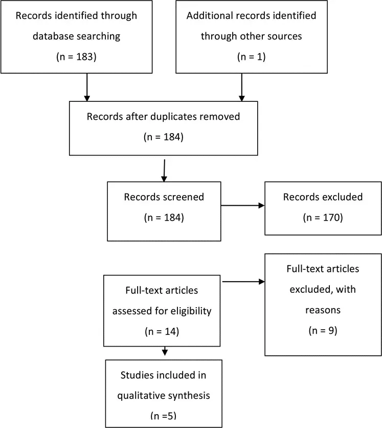
Fig. 1. Flowchart showing the process involved in identifying relevant literature.
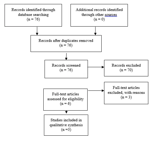
Fig. 2. Flowchart showing the process involved in identifying relevant literature for the 2020 Update. The criteria for “records excluded” and “full text articles excluded with reasons” are detailed in Part 1 of the Guidelines.
Conclusions
Recommendation: There is insufficient evidence to recommend the occipital versus frontal point of entry for the ventricular catheter; therefore, both entry points are options for the treatment of pediatric hydrocephalus. Strength of Recommendation: Level III, unclear degree of clinical certainty.
It is unclear which variable (entry point and/or catheter location) affects shunt survival. In other words, frontal versus occipital entry does not completely determine ultimate catheter position. For example, most frontally placed shunts end up in the frontal horn, but some can also end up in the body of the ventricle or the brain. Occipital placement may result in a catheter situated in the occipital horn, atrium, or frontal horn. In no study did researchers analyze patient factors such as preoperative configuration of the ventricles as a factor in the choice of entry site or shunt survival. The creation of conclusive recommendations or guidelines for the entry point or position of a ventricular catheter is impeded by the limited amount of existing evidence and, in most reports, by the lack of a multivariate analysis accounting for patient age at surgery, ventricular configuration, etiology, and other factors that might be relevant to clinical decision making. Review and evaluation of available evidence leads to the recommendation that either entry is acceptable and decisions about catheter entry site should be made based on the clinical scenario and the surgeon’s experience. The evidence would seem to support attempts to position the catheter tip so that it is surrounded by CSF and does not contact adjacent tissues. As is often the case, additional randomized controlled studies or comparative effectiveness approaches with larger data sets would provide better evidence to support a stronger recommendation. The use of technical adjuvants to achieve that goal leads to the discussion found in Part 3.
In conclusion, there is no new evidence from the 2020 Update to substantially change the original recommendations, that the frontal and occipital entry points are options for the treatment of pediatric hydrocephalus.
Acknowledgments
We acknowledge the American Association of Neurological Surgeons (AANS)/Congress of Neurological Surgeons (CNS) Joint Guidelines Committee for their review, comments, and suggestions; Laura Mitchell, Guidelines Project Manager for the CNS, for her contributions; Pamela Shaw, research librarian, for her assistance with the literature searches; Kevin Boyer for his assistance with data analysis; and Sue Ann Kawecki for her assistance with editing. We also acknowledge the following peer reviewers for their contributions to review the update to the guidelines: Jennifer Sweet, MD, Brandon Rocque, MD, Christoph Greissenauer, MD, Jeffrey Olson, MD.
Disclosure
The systematic review and evidence-based guidelines were funded exclusively by the CNS and the AANS Pediatric Section, which received no funding from outside commercial sources to sup- port the development of this document.
Conflict(s) of Interest: None. All Task Force members declared any potential conflicts of interest prior to beginning work on this evidence review.
Conflict(s) of Interest: None. All Pediatric Hydrocephalus Systematic Review and Evidence-Based Guidelines Update Task Force members declared any potential conflicts of interest prior to beginning work on this systematic review and evidence-based guidelines.
Author contributions to the study and manuscript preparation include the following. Conception and design: AANS/CNS Joint Section on Pediatrics. Acquisition of data: all authors. Analysis and interpretation of data: all authors. Drafting the article: Kemp. Criti- cally revising the article: all authors. Reviewed submitted version of manuscript: all authors. Approved the final version of the manuscript on behalf of all authors: Flannery. Administrative/technical/material support: all authors. Study supervision: Flannery.
Table 1. Effect of Ventricular Catheter Position Evidence Table
| Author | Study Description | Data Class, Quality and Reasons | Results and Conclusions |
| Bierbrau et al., 1991 | Prospective, randomized by month, study of new shunt insertionJuly 1988 to October 1990,N=121, Follow up 2-30 months | Class II RCT, weak randomization, although both groups statistically similar. No assurance of blinded enrollmentChi square analysis of data, life table analysis of shunt survival | Z value 1.74 for posterior versus anterior, p=<0.05.Conclusion: longer shunt survival with posteriorly placed shunts |
| Nakahar et al., 2009 | Retrospective reviewReview of 130 charts, excluded 102 for inadequate data.Evaluated 28: 9 frontal, 119 parietal occipitalMean age: 4.7 and 4.5 monthsFollow up average: 78.6 and 93.9 monthsComputed Tomography (CT) and plain skull radiographs measured | Class IIINo statistical analysis | Mean shortening of ventricular catheter in frontal group(A) was 0.83, in P/o group b it was 0.99Burr hole displacement in a was 1.29 axial, and 1.38 lateralIn group(B) the displacement was 1.08 and 1.07Conclusion: Shortening of ventricular catheter was more pronounced in the frontal position in the age group |
| Albright, et al., 1988 | Retrospective chart review,180 records reviewed at 2 institutions from 1978-1981,114 children included, CT scans available for 83 patients.4 surgeons, plus “others” | Class IIIChi square analysis of variables, logistic regression, and life table analysis with time to first malfunction as “survival.”Statistical significance less than or equal to p value of 0.05. | Two groups were similar in age, cause of hydrocephalus, and infection.Also considered catheter position as independent variable (Figure 2).Conclusion: statistically significant better long-term survival in frontal group compared to parietal per life table analysis (Wilcoxon, p=0.0008, Savage, p=0.0015 |
| Tuli et al., 1999 | Multi-center randomized trial, secondary data analysis,344 patients randomized at 12 centers followed for 2 years, blinded review of images,Comparability of groups commented on in prior publications | Class III, secondary end points, post-hoc analysisKaplan–Meier estimated shunt survivalCox regression to evaluate variables. | Occipital entry site had the highest survival rate (HR,0.45, 95%CI,0.28-0. 74;p=0.001) versus frontal (HR,0.60;95%,CI, 039-0.91;p=0.02).Tip position surrounded by CSF also decreased risk of failure by half (HR;0.21;95%CI0.094-0.45;p= 0.0001).Conclusion: occipital catheters may have better long term survival, but catheter tip location may be more important |
| Sainte-Rose et al., 1991 | Retrospective review of 1,719 patients from 1973 to 1984, 2 institutions | Class III retrospective, uncontrolled chart review | Catheter located in the frontal horn was more likely to obstruct than catheter located in the atrium from occipital route (p<0.001). |
Table 2. New evidence included in 2020 Update
| Author | Study Description | Data Class, Quality and Reasons | Results and Conclusions |
| Whitehead et al, 2017 | This is a large, multicenter study. | III | This is a secondary analysis of information gleaned from different studies. |
| Janson et al, 2014 | This is a single center retrospective study. This is a largely adult study, although they did report the pediatric data separately. Nevertheless, again there is no control for site of entry or randomization. | III | No new evidence is available to substantially change the prior recommendation that the frontal and occipital entry points are options |
| Buster et al, 2016 | This paper was termed a prospective and retrospective cohort study but is a single center. There is no randomization or control for a site of entry in the design of the study. | III | No new evidence is available to substantially change the prior recommendation that the frontal and occipital entry points are options |
References
- Tuli S, O’Hayon B, Drake J, Clarke M, Kestle J. Change in ventricular size and effect of ventricular catheter placement in pediatric patients with shunted hydrocephalus. Neurosurgery. 1999;45(6):1329-1333; discussion 1333-1325.
- Bierbrauer KS, Storrs BB, McLone DG, Tomita T, Dauser R. A prospective, randomized study of shunt function and infections as a function of shunt placement. Pediatric neurosurgery. 1990;16(6):287-291.
- Sainte-Rose C, Piatt JH, Renier D, et al. Mechanical complications in shunts. Pediatric neurosurgery. 1991;17(1):2-9.
- Nakahara K, Shimizu S, Utsuki S, et al. Shortening of ventricular shunt catheter associated with cranial growth: effect of the frontal and parieto-occipital access route on long-term shunt patency. Child’s nervous system : ChNS : official journal of the International Society for Pediatric Neurosurgery. 2009;25(1):91-94.
- Albright ALH, S. J.; Taylor, F. H. Function of parietal and frontal shunts in childhood hydrocephalus. Journal of neurosurgery. 1988;69(88):883-886.
- Sekhar LN, Moossy J, Guthkelch AN. Malfunctioning ventriculoperitoneal shunts. Clinical and pathological features. Journal of neurosurgery. 1982;56(3):411-416.
- Whitehead WE, Riva-Cambrin J, Kulkarni AV, et al. Ventricular catheter entry site and not catheter tip location predicts shunt survival: a secondary analysis of 3 large pediatric hydrocephalus studies. Journal of neurosurgery Pediatrics. 2017;19(2):157-167.
- Janson CG, Romanova LG, Rudser KD, Haines SJ. Improvement in clinical outcomes following optimal targeting of brain ventricular catheters with intraoperative imaging. Journal of neurosurgery. 2014;120(3):684-696.
- Buster BE, Bonney PA, Cheema AA, et al. Proximal ventricular shunt malfunctions in children: Factors associated with failure. Journal of clinical neuroscience : official journal of the Neurosurgical Society of Australasia. 2016;24:94-98.
- Farahmand D, Hilmarsson H, Hogfeldt M, Tisell M. Perioperative risk factors for short term shunt revisions in adult hydrocephalus patients. Journal of neurology, neurosurgery, and psychiatry. 2009;80(11):1248-1253.
- Kestle JR, Drake JM, Cochrane DD, et al. Lack of benefit of endoscopic ventriculoperitoneal shunt insertion: a multicenter randomized trial. Journal of neurosurgery. 2003;98(2):284-290.
- Albright AL. Hydrocephalus shunt practice of experienced pediatric neurosurgeons. Child’s nervous system : ChNS : official journal of the International Society for Pediatric Neurosurgery. 2010;26(7):925-929.
- Berry JG, Hall MA, Sharma V, Goumnerova L, Slonim AD, Shah SS. A multi-institutional, 5-year analysis of initial and multiple ventricular shunt revisions in children. Neurosurgery. 2008;62(2):445-453; discussion 453-444.
- Howard MA, 3rd, Srinivasan J, Bevering CG, Winn HR, Grady MS. A guide to placement of parietooccipital ventricular catheters. Technical note. Journal of neurosurgery. 1995;82(2):300-304.
- Kast J, Duong D, Nowzari F, Chadduck WM, Schiff SJ. Time-related patterns of ventricular shunt failure. Child’s nervous system : ChNS : official journal of the International Society for Pediatric Neurosurgery. 1994;10(8):524-528.
- Robinson S, Kaufman BA, Park TS. Outcome analysis of initial neonatal shunts: does the valve make a difference? Pediatric neurosurgery. 2002;37(6):287-294.
- Sood S, Canady AI, Ham SD. Evaluation of shunt malfunction using shunt site reservoir. Pediatric neurosurgery. 2000;32(4):180-186.
- Steinbok P, Poskitt KJ, Cochrane DD, Kestle JR. Prevention of postshunting ventricular asymmetry by transseptal placement of ventricular catheters. A randomized study. Pediatric neurosurgery. 1994;21(1):59-64; discussion 65.
- Thomale UW, Hosch H, Koch A, et al. Perforation holes in ventricular catheters–is less more? Child’s nervous system : ChNS : official journal of the International Society for Pediatric Neurosurgery. 2010;26(6):781-789.
Part 10: Change in ventricle size as a measurement of effective treatment of hydrocephalus
J Neurosurg Pediatrics (Suppl) 14:77–81, 2014
AANS, 2014
(Original text of the guideline was edited to reflect the update. Please click here for the original publication.)
UPDATE
Dimitrios C. Nikas, MD1,2, Alexander F. Post, MD3, Asim F. Choudhri, MD4,5, Catherine A. Mazzola, MD6, Laura Mitchell, MA7 and Ann Marie Flannery8
1Department of Neurosurgery, University of Illinois at Chicago, Chicago, Illinois; 2Advocate Children’s Hospital, Oak Lawn, Illinois; 3Division of Pediatric Neurological Surgery, Department of Neurosciences and Pediatrics, Goryeb Children’s Hospital–Morristown Medical Center, Morristown, New Jersey; 4Departments of Radiology, Ophthalmology, and Neurosurgery, University of Tennessee Health Science Center, and 5Le Bonheur Neuroscience Institute, Le Bonheur Children’s Hospital, Memphis, Tennessee; 6Division of Pediatric Neurological Surgery, Goryeb Children’s Hospital, Morristown, New Jersey; 7Congress of Neurological Surgeons, Schaumburg, Illinois; 8Department of Neurological Surgery, Saint Louis University, St. Louis, Missouri
Object. The objective of this systematic review is to answer the following question: Does ventricle size after treatment have a predictive value in determining the effectiveness of surgical intervention in pediatric hydrocephalus? Methods. The US National Library of Medicine PubMed/MEDLINE database and the Cochrane Database of Systematic Reviews were searched using MeSH headings and key words relevant to change in ventricle size after surgical intervention for hydrocephalus in children. An evidentiary table was assembled summarizing the studies and the quality of evidence (Classes I–III).
Results. Six articles satisfied inclusion criteria for the evidentiary tables for this part of the guidelines. All were Class III retrospective studies.
Conclusions. Recommendation: There is insufficient evidence to recommend a specific change in ventricle size as a measurement of the effective treatment of hydrocephalus and as a measurement of the timing and effectiveness of treatments including ventriculoperitoneal shunts and third ventriculostomies. Strength of Recommendation: Level III, unclear clinical certainty.
(http://thejns.org/doi/abs/10.3171/2014.7.PEDS14330)
Keywords: hydrocephalus, ventricle size, evidence-based guidelines, practice guidelines
Abbreviations used in this paper: AANS = American Association of Neurological Surgeons; CNS = Congress of Neurological Sur- geons; CPC = choroid plexus cauterization; ETV = endoscopic third ventriculostomy; FOR = frontal and occipital horn ratio; VP = ventriculoperitoneal.
The decision to treat hydrocephalus with an external shunt or endoscopic third ventriculostomy (ETV) is based on a variety of factors. The determination of successful outcome rests on a multitude of clinical and imaging correlates. Imaging indicators that have been examined include ventricle size, presence of a flow void in the ETV site, amount of CSF over the cerebral hemispheres, and the degree of periventricular edema. The objective of this particular systematic review is to answer the question: Does ventricle size after treatment have a predictive value for effectiveness of surgical intervention in pediatric hydrocephalus?
Ventricle size before and after intervention is a readily available measurement that has been used to assess success or failure of treatment. Particular attention has been given to changes in ventricle size after ETV.1-3
Correlation with neurodevelopmental sequelae as well as correlation with other imaging parameters, such as presence of flow voids after ETV, have been suggested as indications of successful interventions.4-7 The evaluation of the effectiveness of treatment has therefore been limited because developmental outcomes are most applicable in infants and younger children and flow voids observed on MR images are applicable only to ETV treatment. This review, therefore, focuses on ventricle size as a tool, albeit a limited one, in the evaluation of the effectiveness of treatment. Ventricle size is an outcome that could be used to assess patients of all ages as well as both forms of intervention—the ventriculoperitoneal (VP) shunt and the ETV. The purpose of this evidence-based review is to critically examine data from the literature pertaining to change of ventricle size as a predictor of the success of surgical intervention.
Methods
We searched the US National Library of Medicine (PubMed/MEDLINE) and the Cochrane Database of Systematic Reviews for the period January 1966 through March 2012 using strategies listed below. The inclusion and exclusion criteria adhered to the protocol outlined in the methods section, Part 1, of these evidence-based guidelines.8
Search Terms
PubMed/MEDLINE
- “Cerebrospinal Fluid Shunts”[MeSH] AND “Hy- drocephalus”[Majr:noexp]
- 1 AND (“Magnetic Resonance Imaging”[MeSH] OR “Tomography, X-Ray Computed”[MeSH] OR Ultra- sonography[MeSH] OR imaging[tiab])
- 2 AND (“ventricular size”[TIAB] OR “ventricular dilation”[tiab] OR ventricle[tiab] OR ventricles[tiab])
- Limit to Child (0–18 years)
- Limit to English and Humans
- Limit 3 to Child (0–18 years)
- Limit to English and Humans Number = 81
Cochrane Database
- MeSH descriptor Child
- MeSH descriptor Infant
- MeSH descriptor Hydrocephalus
- MeSH descriptor Cerebrospinal Fluid Shunts
- (MeSH descriptor Magnetic Resonance Imaging) or (MeSH descriptor Ultrasonography) or (MeSH de- scriptor Tomography, X-Ray Computed) or imaging
- (1 or 2) and 3 and 4 and 5
- 3 and 4 and 5
Search Strategy
An evidentiary table was constructed to facilitate data review and analysis by the Pediatric Hydrocephalus Systematic Review and Evidence-Based Guidelines Task Force.
For each article included in the evidentiary table, the study type, summary findings, and major conclusions were recorded, and a preliminary data class was assigned. The Task Force met to discuss the ranking of the evidence and the classification of data. Recommendations then were made based on the strength of the data in the evidentiary table. In these discussions, if a disagreement was encountered among members, a blinded vote was held and a consensus or majority opinion was reached.
Authors performed an updated literature search (in PubMed and Cochrane Central) for this guideline chapter through a medical librarian at the Congress of Neurological Surgeons Guidelines office using the below-mentioned existing search terms to update the original search through November 30, 2019.
Search Results
A total of 81 abstracts were screened and 18 full-text articles listed in the databases were retrieved for review (Fig. 1). The selection for review was based on the determination of evidence data relevant to the question of the effect of treatment on ventricle size. An examination of the reference lists of these 18 full-text articles yielded 4 additional articles that warranted full-text review. All 22 articles were read and reviewed in detail by the full Task Force. Sixteen articles were excluded based on predefined criteria, which are described in Part 18 of the Guidelines. Six articles satisfied the inclusion criteria and form the basis for the evidentiary tables in this recommendation.
An additional 3 studies out of the 70 yielded by the 2020 update met inclusion criteria from the original guideline and were included (Figure 2).
Results
The 6 articles that met the inclusion criteria and were selected for final review were all Class III retrospective studies (Table 1).
In 2000, Kulkarni et al2 published the results of a retrospective, blinded observational study of a group of 29 children who had undergone ETV and whose ventricle size was assessed by 4 independent observers using the FOR (frontal and occipital horn ratio) both preoperatively and postoperatively (Table 1). Postoperatively, the mean reduction in ventricle size was 7% in cases that were deemed treatment failures (8 patients in whom symptoms either recurred or never resolved) and 16% in cases that were clinically successful (21 patients)—a result that was statistically significant (p = 0.03, t-test). The authors concluded that ventricle size appeared to be somewhat reduced in both groups of patients; however, the reduction was significantly greater among the clinically successful cases. The authors also assessed imaging correlates; they found that the presence of a flow void seemed to correlate with clinical success and its absence with clinical failure. The most significant limitation in this study is its retrospective nature. Surgeon bias, the linear measurement of ventricle size, and the variation in time when postoperative imaging was conducted were confounding factors.
In another retrospective study, published in 2009, Warf et al8 described neurocognitive outcomes and ventricle volumes in infants with myelomeningoceles and hydrocephalus (Table 1). The same method was used to compare ventricle size in 55 children treated by ETV with choroid plexus cauterization (CPC), 19 children who received a VP shunt, and another 19 children who required no intervention. The mean FOR was similar among groups, with no significant difference between the untreated group and either the VP shunt or ETV with CPC group before treatment. The groups also were compared for neurocognitive outcomes, and no significant difference was identified. The FOR did not correlate with neurocognitive performance. Bias existed with respect to the VP shunt group because most patients had already experienced ETV with CPC failure. This is another non- randomized study with the potential for strong confounding factors within treatment groups. A valid conclusion was that stable mild-to-moderate ventriculomegaly alone should not trigger intervention in an asymptomatic infant with a myelomeningocele.
Another Class III single-center retrospective study in the myelomeningocele population was published by Chakraborty et al9 in 2008 (Table 1). These authors studied 28 patients who were selected from a group of 54 who satisfied the determined inclusion criteria. Overall, using a stringent shunt placement policy, about half (51.9%) of their patients required a VP shunt. The authors suggest that a more critical evaluation and tolerance of ventriculomegaly may decrease the need for shunt placement and will reduce shunt dependency in children with myelomeningocele, without worsening outcome.
In a study by St. George et al., limited by its small number of subjects, the authors examined 13 patients with multiple diagnoses who had undergone ETV (Table 1).3
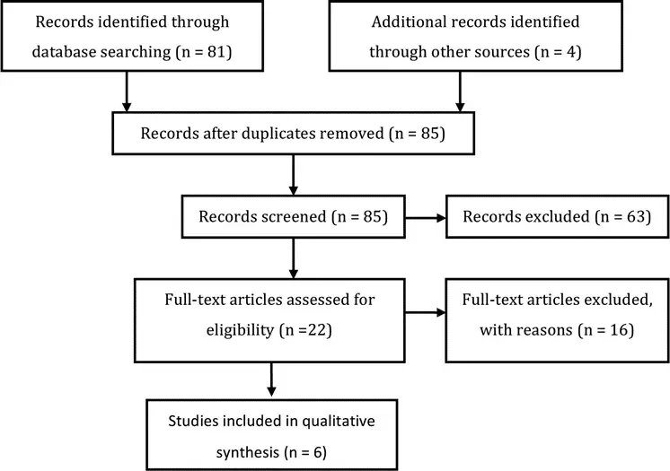
Fig. 1. Flowchart showing the process involved in identifying relevant literature.
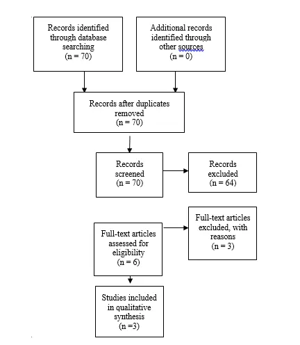
Fig. 2. Flowchart showing the process involved in identifying relevant literature for the 2020 Update The criteria for “records excluded” and “full text articles excluded with reasons” are detailed in Part 1 of the Guidelines.
The comparison of preoperative and postoperative ventricular volume was confined to patients in this treatment group. The authors found postoperative measurements of ventricular volume to be lower than measurements obtained preoperatively but higher than normalized values for patient ages and sexes. The pattern of change in ventricle size varied between a large ventricular volume group, which demonstrated a significant decrease at the 3- to 6-month postoperative time point, and a small volume group, in which there was a much less steep reduction in ventricle size in the first 3–6 months. After that time period, the volume appeared to stabilize or fall slightly.
Two additional papers reported that ventricle size was not necessarily a predictor of outcome (Table 1). These 2 studies looked at ETV alone and found that in both infants and older children, reduction in ventricle size was not necessary to have a clinically effective treatment of hydrocephalus. Like the previously cited studies, these studies were retrospective, and both enrolled a small number of patients (Buxton et al.: n = 27; Kim et al.: n = 29 in whom neuroimaging studies were available).4,10
2020 Update
Di Rocco et al11 presented Class III data indicating that all successful ETV cases had a progressive reduction in ventricular volume (as well as increase in volume of subarachnoid spaces). Pindrik et al12, in a class III study, applied measurements of 3rd ventricle (width and mid-sagittal cross-sectional area) and report that these respond more to a successful ETV than lateral ventricular measurements. Romeo et al13 report reduction in ventricular size in patients with tectal plate gliomas whose hydrocephalus was treated with ETV – with the most significant ventricular size reduction observed in the 1-year follow-up (class III). Because of the small cohort of patients (22) in combination with the retrospective design and the specific etiology of hydrocephalus, these studies cannot substantially change the original recommendations.
Excluded Evidence
The evidence reviewed was predominantly Class III. Those papers not used in the analysis were excluded for multiple reasons including the following: reports combining adult and pediatric patient populations without reporting pediatric patients separately;4,5,14-17 a review of other studies;18 and a technical report.19 Jain et al20 reported the effect of valve design on repeated operation, not on effectiveness of treatment. Another paper described how to measure ventricles.21 Kombogiorgas et al22 confined their evaluation to the question of ventricle size predicting the need for shunting after tumor resection in patients with posterior fossa tumors. The large study by Shankaran and colleagues23 looked at the impact of vetriculomegaly on outcome, without expressly addressing the shunt status of the patients and was thus excluded. One article could not be retrieved for full-text review and was therefore excluded.24 The paper by Horbar et al25 did not meet inclusion criteria due to enrollment of fewer than 10 patients. One by Choudhury26 was excluded because it did not report ventricle size as an outcome of interest and therefore did not report data to answer the clinical questions addressed in this section. Goumnerova and Frim5 reported a study including adults and children, but did not analyze the pediatric patients separately.
Conclusions
Recommendation: There is insufficient evidence to recommend a specific change in ventricle size as a measurement of effective treatment of hydrocephalus and as a measurement of the timing and effectiveness of treatments including ventriculoperitoneal shunts and third ventriculostomies. Strength of Recommendation: Level III, unclear clinical certainty.
The purpose of hydrocephalus treatment is to return the CSF and/or pressure within the brain of an affected infant or child to as normal a condition as possible. Resources to effect these changes are currently limited primarily to the use of a ventriculoperitoneal (VP) shunt or endoscopic third ventriculostomy (ETV). Intracranial pressure and its effect on brain function cannot easily be measured, and thus an alternate way of judging treatment effectiveness is necessary. The size of ventricles revealed by a number of imaging modalities, including ultrasonography, CT, and MR imaging, is frequently used as a measure of the effectiveness of intervention. Our evaluation of the evidence reveals that reliance on ventricle size alone, as a demonstration of treatment effectiveness, is not supported by the available evidence. Unfortunately, there are no other direct methods currently in general use. Certainly, developmental progress is worth monitoring, but this is more difficult to accomplish for routine post- surgical evaluations.
This systematic review and evidence-based guideline demonstrates that the objective measurement of ventricle size by simple methods such as the frontal and occipital horn ratio (FOR) has not proven to be a reliable bench- mark of effectiveness. Clinical outcome ascertained by the neurosurgeon and team is still the most accepted and useful evaluation, surpassing other more “objective” standards.
Acknowledgments
We acknowledge the American Association of Neurological Surgeons (AANS)/Congress of Neurological Surgeons (CNS) Joint Guidelines Committee for the members’ reviews, comments, and suggestions; Laura Mitchell, Guidelines Project Manager for the CNS, for her contributions; Pamela Shaw, research librarian, for her assistance with the literature searches; Kevin Boyer for his assistance with data analysis; and Sue Ann Kawecki for her assistance with editing. We also acknowledge the following peer reviewers for their contributions to review the update to the guidelines: Jennifer Sweet, MD, Brandon Rocque, MD, Christoph Greissenauer, MD, Jeffrey Olson, MD.
Disclosure
The systematic review and evidence-based guidelines were funded exclusively by the CNS and AANS Pediatric Section, which received no funding from outside commercial sources to support the development of this document.
Conflict(s) of Interest: None. All Task Force members declared any potential conflicts of interest prior to beginning work on this evidence review.
Conflict(s) of Interest: None. All Pediatric Hydrocephalus Systematic Review and Evidence-Based Guidelines Update Task Force members declared any potential conflicts of interest prior to beginning work on this systematic review and evidence-based guidelines.
Author contributions to the study and manuscript preparation include the following. Conception and design: AANS/CNS Joint Section on Pediatrics. Acquisition of data: all authors. Analysis and interpretation of data: all authors. Drafting the article: all authors. Critically revising the article: all authors. Reviewed submitted version of manuscript: all authors. Approved the final version of the manu- script on behalf of all authors: Flannery. Administrative/technical/ material support: all authors. Study supervision: Flannery.
Table 1. Ventricular Size Evidence Table
| Author | Study Description | Data Class, Quality and Reasons | Results and Conclusions |
| Chakraborty et al., 2008 | Single center, 10 year retrospective review of MMC pts, excluded patients closed at OSH. Evaluated shunt insertion, complications, and clinical outcomes. Shunt placed for symptomatic hydrocephalus or severe hydrocephalus, or progression of ventriculomegaly after closure. 54 cases included, 28 (52%) required a shunt | Class III, single center, retrospective, single cohort, “Increasing ventriculomegaly assessed by a neuroradiologist with the neurosurgeron. No specific imaging measurement was used to define hydrocephalus” | Shunt rates in MMC lower than previously published results (and similar to that of in-utero closure) when allowing mild ventriculomegaly |
| Kim et al., 2000 | 32 children with ETV. Mean follow-up: 19 months. 28 had neuroimagingTwo groups: Good (21) and poor (8) outcome. Ventricular size, edema, widening of SA space, surgical changes in the IIIv. Floor, cine-MR findings studied between the 2 groups. | Class III: Retrospective | Good outcomes group:11/16 patients had decrease in ventricular size one month postop.5/16 patients minimal changes onlyVentricular size tended to decrease with timeChanges in ventricular size could not predict surgical outcome completely in themselves. No correlation |
| St George et al., 2004 | Single center, 13 consecutive hydrocephalic patients undergoing ETV studied, MRI scans reviewed and ventricular volume calculated. Preop volume 207cc, one week 120 cc, three months 104cc, six months 119 cc, 12 months 146 cc, 24 months 185 cc (skewed because they didn’t have as many 24 month follow-ups so it was overly influenced by increase in patients 13 who had big vents to start with). Decrease in size was more rapid in those with larger vents | Class III, single center, no comparison group (didn’t compare using volumetric evaluation of shunt patients) | Patients with moderate ventriculomegaly had a less steep decrease in vent size in first 6 months after ETV than those with large preop vents. Steady-state volumes were larger than normal |
| Warf et al., 2009 | 93 patients, spina bifidaDevelopmental assessment, and vent size non-random, non-controlled55 ETV CPC19 VPS, 19 no Tx | Class III: retrospective | No Tx better development than treated, better receptive communication in ETV c/w VPS(p=0.02)No difference in vent volume between ETV and VPS |
| Buxton et al., 1998 | Outcome and reasons for failure in ETVs performed in children <1year old: 27 total patients.Postop ventricular size and flow through stoma documented. Comparison was performed between successes and failures (21 patients -77%). | Class III: Retrospective review of Prospectively acquired data | Postop size (table 2) was not an indicator of success or failure. Size does not matter |
| Kulkarni et al., 2000 | Retrospective FOR (measure of ventricular size) change post ETV comparing successes and failures | Class III: Retrospective case seriesImages evaluated by two observers in terms of ventricular size, flow void, periventricular edema, and CSF in SA space.Comparison with two-tailed t-test | ETV failures; mean postop reduction in ventricular size: 7%ETV clinically successful: 16%(reduction significantly greater)All had decreased FOR, successes slightly more but not to normal (vents do not return to normal even after successful ETV) |
Table 2. New evidence included in 2020 Update
| Author | Study Description | Data Class, Quality and Reasons | Results and Conclusions |
| Di Rocco et al, 2012 | Prospective study that analyzed the changes in cerebrospinal fluid (CSF) distribution after endoscopic third ventriculocisternostomy (ETV). | III | Results indicate that all successful ETV cases had a progressive reduction in ventricular volume (as well as increase in volume of subarachnoid spaces). |
| Pindrik et al, 2013 | Retrospectively reviewed applied measurements of 3rd ventricle (width and mid-sagittal cross-sectional area). | III | This study showed that these respond more to a successful ETV than lateral ventricular measurements. |
| Romeo et al, 2013 | Retrospective study of change in ventricular size after ETV for TPGs. | III | This study reported reduction in ventricular size in patients with tectal plate gliomas whose hydrocephalus was treated with ETV – with the most significant ventricular size reduction observed in the 1-year follow-up. |
References
- Cinalli G, Spennato P, Ruggiero C, et al. Intracranial pressure monitoring and lumbar puncture after endoscopic third ventriculostomy in children. Neurosurgery. 2006;58(1):126-136; discussion 126-136.
- Kulkarni AV, Drake JM, Armstrong DC, Dirks PB. Imaging correlates of successful endoscopic third ventriculostomy. Journal of neurosurgery. 2000;92(6):915-919.
- St George E, Natarajan K, Sgouros S. Changes in ventricular volume in hydrocephalic children following successful endoscopic third ventriculostomy. Child’s nervous system : ChNS : official journal of the International Society for Pediatric Neurosurgery. 2004;20(11-12):834-838.
- Buxton N, Turner B, Ramli N, Vloeberghs M. Changes in third ventricular size with neuroendoscopic third ventriculostomy: a blinded study. Journal of Neurology, Neurosurgery and Psychiatry. 2002;72(3):385-387.
- Goumnerova LC, Frim DM. Treatment of hydrocephalus with third ventriculocisternostomy: outcome and CSF flow patterns. Pediatric neurosurgery. 1997;27(3):149-152.
- Jack CR, Jr., Kelly PJ. Stereotactic third ventriculostomy: assessment of patency with MR imaging. AJNR American journal of neuroradiology. 1989;10(3):515-522.
- Wilcock DJ, Jaspan T, Worthington BS, Punt J. Neuro-endoscopic third ventriculostomy: evaluation with magnetic resonance imaging. Clinical radiology. 1997;52(1):50-54.
- Warf B, Ondoma S, Kulkarni A, et al. Neurocognitive outcome and ventricular volume in children with myelomeningocele treated for hydrocephalus in Uganda. Journal of neurosurgery Pediatrics. 2009;4(6):564-570.
- Chakraborty A, Crimmins D, Hayward R, Thompson D. Toward reducing shunt placement rates in patients with myelomeningocele. Journal of neurosurgery Pediatrics. 2008;1(5):361-365.
- Kim SK, Wang KC, Cho BK. Surgical outcome of pediatric hydrocephalus treated by endoscopic III ventriculostomy: prognostic factors and interpretation of postoperative neuroimaging. Child’s nervous system : ChNS : official journal of the International Society for Pediatric Neurosurgery. 2000;16(3):161-168; discussion 169.
- Di Rocco F, Grevent D, Drake JM, et al. Changes in intracranial CSF distribution after ETV. Child’s nervous system : ChNS : official journal of the International Society for Pediatric Neurosurgery. 2012;28(7):997-1002.
- Pindrik J, Jallo GI, Ahn ES. Changes in third ventricular size in pediatric patients undergoing endoscopic third ventriculostomy. Child’s nervous system : ChNS : official journal of the International Society for Pediatric Neurosurgery. 2013;29(11):2027-2034.
- Romeo A, Naftel RP, Griessenauer CJ, et al. Long-term change in ventricular size following endoscopic third ventriculostomy for hydrocephalus due to tectal plate gliomas. Journal of neurosurgery Pediatrics. 2013;11(1):20-25.
- Feng H, Huang G, Liao X, et al. Endoscopic third ventriculostomy in the management of obstructive hydrocephalus: an outcome analysis. Journal of neurosurgery. 2004;100(4):626-633.
- Fukuhara T, Vorster SJ, Luciano MG. Risk factors for failure of endoscopic third ventriculostomy for obstructive hydrocephalus. Neurosurgery. 2000;46(5):1100-1109; discussion 1109-1111.
- Hommet C, Cottier JP, Billard C, et al. MRI morphometric study and correlation with cognitive functions in young adults shunted for congenital hydrocephalus related to spina bifida. European neurology. 2002;47(3):169-174.
- Jindal A, Mahapatra AK. Correlation of ventricular size and transcranial Doppler findings before and after ventricular peritoneal shunt in patients with hydrocephalus: prospective study of 35 patients. Journal of Neurology, Neurosurgery and Psychiatry. 1998;65(2):269-271.
- Rekate HL. To shunt or not to shunt: hydrocephalus and dysraphism. Clinical Neurosurgery. 1985;32:593-607.
- Schroeder HW, Oertel J, Gaab MR. Endoscopic aqueductoplasty in the treatment of aqueductal stenosis. Child’s nervous system : ChNS : official journal of the International Society for Pediatric Neurosurgery. 2004;20(11-12):821-827.
- Jain H, Natarajan K, Sgouros S. Influence of the shunt type in the difference in reduction of volume between the two lateral ventricles in shunted hydrocephalic children. Child’s nervous system : ChNS : official journal of the International Society for Pediatric Neurosurgery. 2005;21(7):552-558.
- O’Hayon BB, Drake JM, Ossip MG, Tuli S, Clarke M. Frontal and occipital horn ratio: A linear estimate of ventricular size for multiple imaging modalities in pediatric hydrocephalus. Pediatric neurosurgery. 1998;29(5):245-249.
- Kombogiorgas D, Natarajan K, Sgouros S. Predictive value of preoperative ventricular volume on the need for permanent hydrocephalus treatment immediately after resection of posterior fossa medulloblastomas in children. Journal of neurosurgery Pediatrics. 2008;1(6):451-455.
- Shankaran S, Koepke T, Woldt E, et al. Outcome after posthemorrhagic ventriculomegaly in comparison with mild hemorrhage without ventriculomegaly. The Journal of pediatrics. 1989;114(1):109-114.
- Bajpai M, Kataria R, Bhatnagar V, et al. Management of hydrocephalus. Indian journal of pediatrics. 1997;64(6 Suppl):48-56.
- Horbar JD, Walters CL, Philip AG, Lucey JF. Ultrasound detection of changing ventricular size in posthemorrhagic hydrocephalus. Pediatrics. 1980;66(5):674-678.
- Choudhury AR. Avoidable factors that contribute to the complications of ventriculoperitoneal shunt in childhood hydrocephalus. Child’s nervous system : ChNS : official journal of the International Society for Pediatric Neurosurgery. 1990;6(6):346-349.