





1. Introduction And Methods: Update
Sponsored by: Congress of Neurological Surgeons (CNS) and the Section on Tumors
Endorsement: Reviewed for evidence-based integrity and endorsed by the American Association of Neurological Surgeons (AANS) and Congress of Neurological Surgeons
(CNS)
Author: Jeffrey J. Olson, MD1
Departmental and institutional affiliations:
- Department of Neurosurgery, Emory University School of Medicine, Atlanta, Georgia, USA
Corresponding Author contact information:
Keywords: vestibular schwannoma, acoustic neuroma, guideline
Abbreviations: AANS: American Association of Neurological Surgeons, Akt: protein kinase B, CNS: Congress of Neurological Surgeons, DNA: deoxyribonucleic acid, ErbB: erythroblastic leukemia viral oncogene, ERK: extracellular signal regulated kinase, MEK: mitogen-activated extracellular signal-regulated kinase, NF2: neurofibromatosis type 2, mTORC1: mammalian target of rapamycin complex 1, PDGFR: platelet derived growth factor receptor, PI3K: phosphoinositide 3-kinase , Raf: rapidly accelerated fibrosarcoma, Ras: rat sarcoma, RNA: ribonucleic acid, VS: vestibular schwannoma
ABSTRACT
Background: The management of vestibular schwannomas is an important facet of physicians practicing neurosurgery, neuro-otology, and an important diagnosis to keep in mind for audiologists, radiologists, and pathologists.
Objective: This is an update of the evidence-based guidelines for management of vestibular schwannomas published by the Congress of Neurological Surgeons in 2018.
Methods: The questions from the previously published guideline were updated to PICO format. The medical literature from 1/1/2015 through 5/20/2022 was searched with these reformatted questions serving as guidance to determine if information was available to update, modify or create new recommendations in the context of the prior guidelines related to audiologic screening, imaging, surgery, intraoperative cranial nerve monitoring, hearing preservation and radiation therapy.
Results: The writing group utilized the information from the updated literature search to modify old recommendations or formulate new recommendations based on this evidence. Across all six sections of the guideline, 31 questions were utilized to direct a search of the literature ultimately yielding 27 recommendations.
Conclusion: This series of guideline documents provides an update and addition to the information and recommendations provided in the 2018 version. It now serves as this organization’s most up to date and current recommendations on the management of vestibular schwannoma. It also sets a point of reference for treatment development and topics for research going forward to improve management of this difficult disease.
INTRODUCTION
Background and Rationale
The guidelines for the management of vestibular schwannomas was sponsored by the Congress of Neurological Surgeons (CNS) were published in 2018.[1-9] As suggested by the Institute of Medicine, now the National Academy of Medicine, guidelines should be updated in the range of every 5 years.[10] That interval allows for a reasonable time to assess new methods of diagnosis and treatment including audiologic screening, imaging, surgery, intraoperative cranial nerve monitoring, hearing preservation and radiation therapy that have developed since the time of the publication in 2018.[11-20] A set of guideline documents representing each of these management topics have been produced. They were produced using the evidence-based methodology supported by the Joint Guidelines Review Committee of the American Association of Neurological Surgeons (AANS) and Congress of Neurological Surgeons (CNS) which can be found at https://www.cns.org/guidelines/guideline-development-methodology.
Objectives and Guideline Panel Development
The objectives of these guidelines are to update the evidence-based management of vestibular schwannomas published in 2018. The writing group was recruited from those who had written the original version of this guideline and new writers from the neurosurgical, neuro-otology and radiation oncology communities. The authors were screened for professional conflicts, and this was documented as outlined under the Conflicts of Interest section below.
Topic Range of this Systematic Review and Evidence-Based Clinical Practice Guideline
The subjects of this guideline update include initial otologic evaluation, imaging diagnosis, use of surgical techniques, intraoperative monitoring, and the administration of radiation therapy. The initial set of publications in 2018 included sections on pathology and emerging therapies. However, the ability to make strong recommendations regarding those topics was limited. Upon review of the literature published since the close of the searches for the initial publication new publications provided negative or contradictory class III information at best and did not warrant an update in those areas.
To elaborate on why the pathology and emerging therapies have no updates, targeted therapy for either sporadic vestibular schwannomas or those related to neurofibromatosis type 2 (NF2) certainly is an area of interest. However, the following data regarding these interventions are both positive and negative, precluding development of a coherent recommendation. In a small study of everolimus in ten NF2 subjects, no reduction in vestibular schwannoma volume was noted over 12 months of therapy, though the rate of tumor growth was observed in four individuals.[21] In a later phase 0 study looking at the effect of everolimus on its commonly understood targets in theses tumors, it was observed that the drug incompletely inhibited mTORC1 and its downstream signaling, thus explaining this lack of substantial tumor controlling effect.[22] In a small carefully monitored 14 subject cohort of NF2 associated VS, treated with bevacizumab for nearly a year, 35% of subjects had measurable hearing improvement and 43% had a partial radiographic response.[23] Contrary to that experience, in another study, assessing 9 individuals with 16 surgically debulked tumors, bevacizumab induced no evidence of tumor growth control or measurable improvements in speech discrimination scores or pure tone audiometry.[24] In a prospective study of fourteen individuals with NF2 related vestibular schwannomas, half the subjects reported self-assessed reductions in hearing difficulty with over a 48 week course of bevacizumab , 7.5 mg/kg every three weeks. This improvement correlated with objective word recognition scores but not with pure tone audiometry.[25] When used, higher doses of bevacizumab to not seem to increase rates of hearing response or radiographic response in the short term.[26] In the studies using bevacizumab, varying proportions of subjects, usually in the range of 25%, had to stop or substantially modify therapy because of toxicity, generally manifest as fatigue, hypertension, proteinuria, skin and mucous membrane dryness, and infection.[24, 27] Based on these findings, the authors chose against making a recommendation for bevacizumab at this time. Interestingly, and in spite of these reported experiences from the interval of the search for this guideline update not being uniformly positive, the use of bevacizumab has been adopted an established treatment option for NF2 associated schwannomas based on previous experience and publications.[1-8]
METHODOLOGY
As first step the guidelines task force reviewed the questions from the 2018 publications and modified them to PICO (patient/intervention/comparison/outcome) format. With these as guidance the guidelines task force initiated a systematic review of the literature and evidence-based guideline relevant to the treatment of patients with vestibular schwannomas. Through objective evaluation of the evidence and transparency in the process of making recommendations, this evidence-based clinical practice guideline update was developed for the diagnosis and treatment of adult patients with vestibular schwannomas. These guidelines are developed for educational purposes to assist practitioners in their clinical decision-making processes. Additional information about the methods utilized in this systematic review is provided below.
Literature Examination Approach
A thorough literature search strategy was undertaken to identify all citations relevant to the management of vestibular schwannomas in regard to the set of chosen topics. The PubMed and Embase electronic databases were searched, with guidance of a reference librarian, from January 1, 2015, through May 20, 2022 using the search strategies provided in Appendix I. The search strategies used a combination of controlled vocabulary and text words. The specifics of the searches for a given topic are outlined in each respective guideline section. The writing groups used the citations from these databases that were pertinent to answering the questions they had developed. In addition, important articles from before this interval were reviewed and included if deemed to be critical evidence by the writing group. For completeness, reference lists of the publications chosen for full-text review were also screened for potentially relevant studies.
Study Selection, Quality Assessment and Statistical Methods
The searches of the bibliographic databases identified possibly relevant citations for a given topic, and often these were large in number. The eligibility (inclusion/exclusion) criteria to screen the citations for each of the questions were determined ahead of time for each section by the respective writing group. These are documented in the individual clinical practice guideline sections in this series to assist the reader in understanding the development process. At least two authors evaluated the titles and abstracts using the inclusion and exclusion criteria. Cases of disagreement about pertinence were resolved by a third author when needed. Full text articles of the selected abstracts were then collected and the same process of applying the eligibility criteria was carried out again with the more in depth information available. Articles that met the eligibility criteria were grouped according to the questions they addressed and used to create the evidence tables and scientific foundation sections. Reasons for exclusion for papers were also documented so as to be able to discuss pertinent problem citations in the scientific foundation as needed.
The method by which evidence was assessed and reported is expanded upon in the CNS Guideline Development Methodology document (https://www.cns.org/guidelines/guideline-development-methodology). The qualifying evidence derived from the searches was then collected and compared to the evidence already present in the 2018 publications, i.e., through the end of 2014, to determine if the recommendations could remain unchanged, needed updating, or if whole new recommendations were warranted. Internal drafts of the tables and manuscripts were developed by sharing them between writers electronically, by telephone, and in face-to-face meetings. Summary and conclusion statements were included for each section, with comments on key issues for future investigation being added where pertinent.
No question resulted in a collection of studies that warranted a meta-analysis.
Rating Quality of Evidence
The quality of evidence was rated using an evidence hierarchy for each of four different study types; therapeutic, prognostic, diagnostic, and decision modeling. These hierarchies are shown in Appendix II: Rating Evidence Quality. Additional information regarding the hierarchy classification of evidence can be located here: https://www.cns.org/guidelines/guideline-procedures-policies/guideline-development-methodology.
Revision Plans
In accordance with the National Academy of Medicine’s standards for developing clinical practice guidelines and criteria specified by the former National Guideline Clearinghouse, the task force will monitor related publications following the release of this document and will revise the entire document and/or specific sections “if new evidence shows that a recommended intervention causes previously unknown substantial harm; that a new intervention is significantly superior to a previously recommended intervention from an efficacy or harms perspective; or that a recommendation can be applied to new populations.[28]” In addition, the task force will confirm within five years from the date of publication that the content reflects current clinical practice and the available technologies for the evaluation and treatment for patients with vestibular schwannomas.
SUMMARY
This series of guidelines assessed new literature from 2015 to 2022 in the context of the data already abstracted for the prior version to update and make current and clinically relevant evidence for management of vestibular schwannomas. They serve to set a point of reference for patient care while also highlighting important key areas for future research. This will allow design of future investigations in a manner that overcomes prior weaknesses noted in these guidelines. Secondarily, the suggestions provided are set forth for conscientious use by the practicing physician who must account for all the unique individual conditions in the therapy of a given person during his or her illness.
Fortunately, new research is constantly underway. This includes clinical trials currently pursuing the potential value of systemic brigatinib, aspirin, losartan, axitinib and local doxycycline.[29]
Application of advanced RNA profiling and DNA methylation analysis of vestibular schwannomas that have been banked for analysis by Landry et al resulted in identification of two main subgroups of tumor, they term “immunogenic” and “proliferative”. The immune checkpoint pathway and MEK pathway integrity is very different in these subgroups may provide an avenue to application of gene network analysis and computational drug repurposing.[30] In another analysis of banked NF2 vestibular schwannomas Amit et al correlated rate of tumor progession after resection with immune cell profiles showing a distinct difference from those that recur more slowly.[31] In respect to the possibility of using immunologically directed therapies, a clinical trial using aVEGFR-1/2 peptide vaccine was also conducted in patients with progressive NF2-derived tumors, showing hearing improvement and tumor volume reduction.[32]
Merlin, the protein lost in NF2 related schwannomas, and many sporadic vestibular schwannomas inhibits the signaling of various tyrosine kinases and their downstream proliferative signals. This naturally leads to the consideration of several protein kinase inhibitors that can prevent tumor progression by inhibtion of these tyrosine kinases or members of their downstream pathways. This includes the including retrovirus-associated DNA sequences(Ras)/rapidly accelerated fibrosarcoma (Raf)/mitogen extracellular signal-regulated kinase(MEK)/extracellular-signal-regulated kinases (ERK) pathways and mammalian target of rapamycin complex 1 (mTORC1)/phosphoinositide 3-kinase (PI3K)/Akt pathways.[33, 34 Though of scientific interest clinical trials of inhibitors of ErbB and PDGFR pathway function in vestibular schwannomas have been without positive effect.[35, 36] Although a phase II study has shown that everolimus is ineffective in progressive NF2-related VS patients, another study has shown that everolimus reduced the tumor volume in 55.6% of patients with NF2-related VS.[21, 37, 38]. Thus, the effect of everolimus is still debatable and warrants additional study.
Wider afield is the preclinical development of therapy with dietary supplements such as sulforane from broccoli, bacteriotherapy (direct injection of Salmonella typhimurium followed by administration of enrofloxacin, and adeno-associated virus mediated gene therapy to restore the NF2 gene to merlin deficient cells.[29, 39, 40, 41] The result of this work on immunologic, targeted and alternative therapies will eventually result in the modification of these guidelines as updates are published.
The data analyzed for this set of guidelines has been collected through May 20, 2022. It is estimated that the updated iteration of this guideline overall will be written in approximately five years with modification of this time-line dependent on emergence of important scientific and therapeutic advances.
Conflicts of Interest
All Guideline Task Force members were required to disclose all potential COIs prior to beginning work on the guideline, using the COI disclosure form of the AANS/CNS Joint Guidelines Review Committee. The CNS Guidelines Committee and Guideline Task Force Chair reviewed the disclosures and either approved or disapproved the nomination and participation on the task force. The CNS Guidelines Committee and Guideline Task Force Chair may approve nominations of task force members with possible conflicts and restrict the writing, reviewing, and/or voting privileges of that person to topics that are unrelated to the possible COIs. See Appendix III for a complete list of disclosures.
Disclosure of Funding
These evidence-based clinical practice guidelines were funded exclusively by the Congress of Neurological Surgeons, which received no funding from outside commercial sources to support the development of this document.
Disclaimer of Liability
This clinical systematic review and evidence-based guideline was developed by a physician volunteer task force as an educational tool that reflects the current state of knowledge at the time of completion. Each chapter is designed to provide an accurate review of the subject matter covered. This guideline is disseminated with the understanding that the recommendations by the authors and consultants who have collaborated in their development are not meant to replace the individualized care and treatment advice from a patient’s physician(s). If medical advice or assistance is required, the services of a competent physician should be sought. The proposals contained in these guidelines may not be suitable for use in all circumstances. The choice to implement any particular recommendation contained in these guidelines must be made by a managing physician in light of the situation in each particular patient and on the basis of existing resources.
Acknowledgments:
The guidelines task force would like to acknowledge the CNS Guidelines Committee for their contributions throughout the development of the guideline, the AANS/CNS Joint Guidelines Review Committee, as well as the contributions Trish Rehring, MPH, Director for Evidence-Based Practice Initiatives for the CNS, and Janet Waters, MLS, BSN, RN, for assistance with the literature searches. Throughout the review process, the reviewers and authors were blinded from one another. At this time the guidelines task force would like to acknowledge the following individual peer reviewers for their contributions: Patti Raksin, Tjoumakaris, Andrew Carlson, Neil Majmundar, Jeff Mullin and Koji Ebersole.
REFERENCES
- Olson JJ; Kalkanis SN; Ryken TC. Congress of Neurological Surgeons Systematic Review and Evidence-Based Guidelines on the Treatment of Adults with Vestibular Schwannomas: Executive Summary. Neurosurgery. 82(2):129-134, 2018 Feb 01.
- Sweeney AD; Carlson ML; Shepard NT; McCracken DJ; Vivas EX; Neff BA; Olson JJ. Congress of Neurological Surgeons Systematic Review and Evidence-Based Guidelines on Otologic and Audiologic Screening for Patients with Vestibular Schwannomas. Neurosurgery. 82(2):E29-E31, 2018 Feb 01.
- Dunn IF; Bi WL; Mukundan S; Delman BN; Parish J; Atkins T; Asher AL; Olson JJ. Congress of Neurological Surgeons Systematic Review and Evidence-Based Guidelines on the Role of Imaging in the Diagnosis and Management of Patients with Vestibular Schwannomas. Neurosurgery. 82(2):E32-E34, 2018 Feb 01.
- Carlson ML; Vivas EX; McCracken DJ; Sweeney AD; Neff BA; Shepard NT; Olson JJ. Congress of Neurological Surgeons Systematic Review and Evidence-Based Guidelines on Hearing Preservation Outcomes in Patients with Sporadic Vestibular Schwannomas. Neurosurgery. 82(2):E35-E39, 2018 Feb 01.
- Hadjipanayis CG; Carlson ML; Link MJ; Rayan TA; Parish J; Atkins T; Asher AL; Dunn IF; Corrales CE; Van Gompel JJ; Sughrue M; Olson JJ. Congress of Neurological Surgeons Systematic Review and Evidence-Based Guidelines on Surgical Resection for the Treatment of Patients with Vestibular Schwannomas. Neurosurgery. 82(2):E40-E43, 2018 Feb 01.
- Sughrue ME; Fung KM; Van Gompel JJ; Peterson JEG; Olson JJ. Congress of Neurological Surgeons Systematic Review and Evidence-Based Guidelines on Pathological Methods and Prognostic Factors in Vestibular Schwannomas. Neurosurgery. 82(2):E47-E48, 2018 Feb 01.
- Vivas EX; Carlson ML; Neff BA; Shepard NT; McCracken DJ; Sweeney AD; Olson JJ. Congress of Neurological Surgeons Systematic Review and Evidence-Based Guidelines on Intraoperative Cranial Nerve Monitoring in Vestibular Schwannoma Surgery. Neurosurgery. 82(2):E44-E46, 2018 Feb 01.
- Germano IM; Sheehan J; Parish J; Atkins T; Asher A; Hadjipanayis CG; Burri SH; Green S; Olson JJ. Congress of Neurological Surgeons Systematic Review and Evidence-Based Guidelines on the Role of Radiosurgery and Radiation Therapy in the Management of Patients with Vestibular Schwannomas. Neurosurgery. 82(2):E49-E51, 2018 Feb 01.
- Van Gompel JJ; Agazzi S; Carlson ML; Adewumi DA; Hadjipanayis CG; Uhm JH; Olson JJ. Congress of Neurological Surgeons Systematic Review and Evidence-Based Guidelines on Emerging Therapies for the Treatment of Patients with Vestibular Schwannomas. Neurosurgery. 82(2):E52-E54, 2018 Feb 01.
- Greenfield S, Steinberg EP, Auerbach A, Avorn J, Galvin R, Gibbons R, et al. Clinical practice guidelines we can trust. Washington, DC: Institute of Medicine; 2011. Available from: www.iom.edu/Reports/2011/Clinical-PracticeGuidelines-We-Can-Trust.aspx.
- Conley M, Diaz RC. Asymmetric sensorineural hearing loss and vestibular schwannoma: when to image? Current Opinion in Otolaryngology & Head & Neck Surgery. 28(5):335-339, 2020 Oct. UI: 32841960
- Carlson ML, Link MJ. Vestibular Schwannomas. New England Journal of Medicine. 384(14):1335-1348, 2021 04 08. UI: 33826821
- Buss EJ, Wang TJC, Sisti MB. Stereotactic radiosurgery for management of vestibular schwannoma: a short review. Neurosurgical Review. 44(2):901-904, 2021 Apr. UI: 32170501
- Soltys SG, Milano MT, Xue J, Tome WA, Yorke E, Sheehan J, Ding GX, Kirkpatrick JP, Ma L, Sahgal A, Solberg T, Adler J, Grimm J, El Naqa I. Stereotactic Radiosurgery for Vestibular Schwannomas: Tumor Control Probability Analyses and Recommended Reporting Standards. International Journal of Radiation Oncology, Biology, Physics. 110(1):100-111, 2021 05 01. UI: 33375955
- Weiss NM, Grosmann W, Schraven SP, Oberhoffner T, Mlynski R. Neuromonitoring of the cochlear nerve during vestibular schwannoma resection and simultaneous cochlear implantation. HNO. 69(Suppl 2):82-87, 2021 Aug. UI: 34019139
- Wu X, Song G, Wang X, Li M, Chen G, Guo H, Bao Y, Liang J. Comparison of surgical outcomes in cystic and solid vestibular schwannomas: a systematic review and meta-analysis. Neurosurgical Review. 44(4):1889-1902, 2021 Aug. UI: 33009643
- Connor SEJ. Imaging of the Vestibular Schwannoma: Diagnosis, Monitoring, and Treatment Planning. Neuroimaging Clinics of North America. 31(4):451-471, 2021 Nov. UI: 34689927
- Thai NLB, Mai NY, Vuong NL, Tin NM, Karam D, Refaey MA, Shahin KM, Soliman AL, Al Khudari R, Thuan TM, Sabbah GM, El-Qushayri AE, Karimzadeh S, Hirayama K, Huy NT . Treatment for vestibular schwannoma: Systematic review and single arm meta-analysis. American Journal of Otolaryngology. 43(2):103337, 2022 Mar-Apr. UI: 34973662
- Ong V, Zhang AB, Wilson B, Brown NJ, Lien BV, Shahrestani S, Yang I. The 100 Most Highly Cited Publications on Hearing Preservation for Vestibular Schwannomas. World Neurosurgery. 165:115-130, 2022 Sep. UI: 35779753
- Perera MB, Janjua N, Swaminathan R, Apthorp C, Al-Deerawi HB. Magnetic resonance imaging of the internal auditory meatus for vestibular schwannoma in ENT practice: a retrospective analysis with literature and guidelines review. Journal of Laryngology & Otology. 136(10):888-891, 2022 Oct. UI: 34666846
- Goutagny, S.; Raymond, E.; Esposito-Farese, M.; Trunet, S.; Mawrin, C.; Bernardeschi, D.; Larroque, B.; Sterkers, O.; Giovannini, M.; Kalamarides, M. Phase II study of mTORC1 inhibition by everolimus in neurofibromatosis type 2 patients with growing vestibular schwannomas. J. Neuro-Oncol. 2015, 122, 313–320.y
- Karajannis, M.A.; Legault, G.; Hagiwara, M.; Giancotti, F.G.; Filatov, A.; Derman, A.; Hochman, T.; Goldberg, J.D.; Vega, E.; Wisoff, J.H.; et al. Phase II study of everolimus in children and adults with neurofibromatosis type 2 and progressive vestibular schwannomas. Neuro-Oncology 2014, 16, 292–297.
- Blakeley JO, Ye X, Duda DG, et al. Efficacy and biomarker study of bevacizumab for hearing loss resulting from neurofibromatosis type 2-associated vestibular schwannomas. J Clin Oncol 2016;34(14):1669–75.
- Gugel I, Kluwe L, Zipfel J, Teuber C, Tatagiba M, Mautner VF, Schuhmann MU, Grimm F: Minimal Effect of Bevacizumab Treatment on Residual Vestibular Schwannomas after Partial Resection in Young Neurofibromatosis Type 2 Patients. Cancers 2019, 11, 1862; doi:10.3390/cancers11121862
- Huang 16. Huang V, Bergner AL, Halpin C, et al. Improvement in patient-reported hearing after treatment with bevacizumab in people with neurofibromatosis type 2. Otol Neurotol 2018;39(5):632–8.
- Plotkin, S.R.; Duda, D.G.; Muzikansky, A.; Allen, J.; Blakeley, J.; Rosser, T.; Campian, J.L.; Clapp, D.W.; Fisher, M.J.; Tonsgard, J.; et al. Multicenter, Prospective, Phase II and Biomarker Study of High-Dose Bevacizumab as Induction Therapy in Patients with Neurofibromatosis Type 2 and Progressive Vestibular Schwannoma. J. Clin. Oncol. 2019, 37, 3446–3454.
- Morris, K.A.; Golding, J.F.; Axon, P.R.; Afridi, S.; Blesing, C.; Ferner, R.E.; Halliday, D.; Jena, R.; Pretorius, P.M.; Evans, G.; et al. Bevacizumab in neurofibromatosis type 2 (NF2) related vestibular schwannomas: A nationally coordinated approach to delivery and prospective evaluation. Neuro-Oncol. Pract. 2016, 3, 281–289.
- Ransohoff, D.F., M. Pignone, and H.C. Sox, How to decide whether a clinical practice guideline is trustworthy. JAMA, 2013. 309(2): p. 139-40.
- Welling DB: Targeted Therapies in the Treatment of Vestibular Schwannomas: Current State and New Horizons. Otolaryngol Clin N Am 56 (2023) 543–556. https://doi.org/10.1016/j.otc.2023.02.0130030-6665/23
- Landry AP Wang JZ, Suppiah S, Gelareh Zadeh G: Multiplatform molecular analysis of vestibular schwannoma reveals two robust subgroups with distinct microenvironment. Journal of Neuro-Oncology (2023) 161:491–499 https://doi.org/10.1007/s11060-022-04221-2
- Amit M, Xie T, Gleber‑Netto FO, Hunt PJ, Mehta GU, Bell D, Silverman DA, Yaman I, Ye Y,Burks JK, Fuller GN, Gidley PW, Nader ME, Raza SM, DeMonte F: Distinct immune signature predicts progression of vestibular schwannoma and unveils a possible viral etiology. J Exp Clin Cancer Res (2022) 41:292. https://doi.org/10.1186/s13046-022-02473-4
- Tamura R, Fujioka M, Morimoto Y, et al. A VEGF receptor vaccine demonstrates preliminary efficacy in neurofibromatosis type 2. Nat Commun 2019;10(1): 5758. https://doi.org/10.1038/s41467-019-13640, published correction appears in Nat Commun. 2020 Apr 21;11(1):2028.
- Pe´cina-Šlaus, N. Merlin, the NF2 Gene Product. Pathol. Oncol. Res. 2013, 19, 365–373.
- Santarpia, L.; Lippman, S.M.; El-Naggar, A.K. Targeting the MAPK-RAS-RAF signaling pathway in cancer therapy. Expert Opin. Ther. Targets 2012, 16, 103–119.
- Ammoun, S.; Cunliffe, C.H.; Allen, J.; Chiriboga, L.; Giancotti, F.G.; Zagzag, D.; Hanemann, C.O.; Karajannis, M.A. ErbB/HER receptor activation and preclinical efficacy of lapatinib in vestibular schwannoma. Neuro-Oncology 2010, 12, 834–843.
- Tamura 2022 Tamura R, Masahiro T: A Critical Overview of Targeted Therapies for Vestibular Schwannoma. Int. J. Mol. Sci. 2022, 23, 5462. https://doi.org/10.3390/ijms23105462.
- Lane, H.A.;Wood, J.M.; McSheehy, P.M.; Allegrini, P.R.; Boulay, A.; Brueggen, J.; Littlewood-Evans, A.; Maira, S.-M.; Martiny-Baron, G.; Schnell, C.R.; et al. mTOR Inhibitor RAD001 (Everolimus) Has Antiangiogenic/Vascular Properties Distinct from a VEGFR Tyrosine Kinase Inhibitor. Clin. Cancer Res. 2009, 15, 1612–1622.
- Karajannis, M.A.; Legault, G.; Hagiwara, M.; Giancotti, F.G.; Filatov, A.; Derman, A.; Hochman, T.; Goldberg, J.D.; Vega, E.; Wisoff, J.H.; et al. Phase II study of everolimus in children and adults with neurofibromatosis type 2 and progressive vestibular schwannomas. Neuro-Oncology 2014, 16, 292–297.
- Kim BG, Fujita T, Stankovic KM, et al. Sulforaphane, a natural component of broccoli, inhibits vestibular schwannoma growth in vitro and in vivo. Sci Rep 2016;6:36215. https://doi.org/10.1038/srep36215
- Ahmed SG, Brenner GJ. Effect of antibiotic treatment on attenuated salmonella typhimurium VNP20009 mediated schwannoma growth control. Anticancer Res 2023;43(1):1–6.
- Prabhakar S, Beauchamp RL, Cheah PS, et al. Gene replacement therapy in a schwannoma mouse model of neurofibromatosis type 2. Mol Ther Methods Clin Dev 2022;26:169–80.
Appendix I: Literature Searches
See individual chapters
Appendix II: Rating Evidence Quality
Classification of Evidence on Therapeutic Effectiveness and Levels of Recommendation
| Class I Evidence Level I (or A) Recommendation | Evidence from one or more well-designed, randomized controlled clinical trial, including overviews of such trials. |
| Class II Evidence Level II (or B) Recommendation | Evidence from one or more well-designed comparative clinical studies, such as non-randomized cohort studies, case-control studies, and other comparable studies, including less well-designed randomized controlled trials. |
| Class III Evidence Level III (or C) Recommendation | Evidence from case series, comparative studies with historical controls, case reports, and expert opinion, as well as significantly flawed randomized controlled trials. |
Classification of Evidence on Prognosis and Levels of Recommendation
| Class I Evidence Level I (or A) Recommendation | All 5 technical criteria above are satisfied. |
| Class II Evidence Level II (or B) Recommendation | Four of five technical criteria are satisfied. |
| Class III Evidence Level III (or C) Recommendation | Everything else. |
Classification of Evidence on Diagnosis and Levels of Recommendation
| Class I Evidence Level I (or A) Recommendation | Evidence provided by one or more well-designed clinical studies of a diverse population using a “gold standard” reference test in a blinded evaluation appropriate for the diagnostic applications and enabling the assessment of sensitivity, specificity, positive and negative predictive values, and, where applicable, likelihood ratios. |
| Class II Evidence Level II (or B) Recommendation | Evidence provided by one or more well-designed clinical studies of a restricted population using a “gold standard” reference test in a blinded evaluation appropriate for the diagnostic applications and enabling the assessment of sensitivity, specificity, positive and negative predictive values, and, where applicable, likelihood ratios. |
| Class III Evidence Level III (or C) Recommendation | Evidence provided by expert opinion or studies that do not meet the criteria for the delineation of sensitivity, specificity, positive and negative predictive values, and, where applicable, likelihood ratios. |
Classification of Evidence on Clinical Assessment and Levels of Recommendation
| Class I Evidence Level I (or A) Recommendation | Evidence provided by one or more well-designed clinical studies in which interobserver and/or intraobserver reliability is represented by a Kappa statistic > 0.60. |
| Class II Evidence Level II (or B) Recommendation | Evidence provided by one or more well-designed clinical studies in which interobserver and/or intraobserver reliability is represented by a Kappa statistic > 0.40. |
| Class III Evidence Level III (or C) Recommendation | Evidence provided by one or more well-designed clinical studies in which interobserver and/or intraobserver reliability is represented by a Kappa statistic < 0.40. |
Appendix III. Conflicts of Interest
| Task Force Member | Disclosure |
| Julie Honaker PhD, AuD | Nothing to Disclose |
| Ben Allen Strickland, MD | Nothing to Disclose |
| Eric J. Lehrer, MD | Servier Pharmaceuticals, Novocure Inc. |
| Sheryl Green, MBBCh | Nothing to Disclose |
| John P. Marinelli MD | Medtronic |
| Christopher S. Graffeo MD, MS | Nothing to Disclose |
| Isabelle M. Germano, MD, MBA | Brianlab |
| Mateo Ziu, MD | Omniscient Neurotechnology America Ltd; GT Medical Technologies, Inc |
| Walavan Sivakumar, MD | Stryker Corporation |
| Sherwin Tavakol, MD | IRRAS USA, Inc.; Globus Medical, Inc. |
| Lucas Paul Carlstrom, MD, PhD | Kuros Biosciences USA, Inc |
| Jamie J. Van Gompel, MD | Medtronic, Cadence |
| Ian Dunn, MD | Nothing to Disclose |
| Jeffrey J. Olson, MD | Verastem, Inc., Research Grant American Cancer Society, Editorial Consultant; Azurity Pharmaceuticals, Inc. |
| Ghazal S. Daher MD | Nothing to Disclose |
| Matthew L. Carlson, MD | Cochlear Americas, Advanced Bionics, Stryker Corporation; iotaMotion, Inc.; Stryker Corporation |
| Neil S. Patel, MD | Cochlear Americas, Zeiss, Viridian Therapeutics, IotaMotion, Inc. |
| Michael Sughrue, MD | Omniscient Neurotechnology America Ltd |
| Constantinos G. Hadjipanayis, MD, PhD | Stryker Corporation; Integra LifeSciences Corporation; Omniscient Neurotechnology America Ltd |
| Jeffrey Jacob, MD | Stryker Corporation; KLS; Synthes |
2. The Role Of Audiologic Screening in the Diagnosis and Management of Patients with Vestibular Schwannomas
Sponsored by: Congress of Neurological Surgeons (CNS) and the Section on Tumors
Endorsement: Reviewed for evidence-based integrity and endorsed by the American Association of Neurological Surgeons (AANS) and CNS
Authors: Ben Allen Strickland, MD1, Julie Honaker PhD, AuD2 and Jeffrey J. Olson, MD3
Departmental and institutional affiliations:
- MUSC Health Neurosurgery at Hollings Cancer Center, Columbia, SC
- Integrated Surgical Institute, Head and Neck Department, Cleveland Clinic, Cleveland, OH
- Department of Neurosurgery, Emory University School of Medicine, Atlanta, GA
Corresponding Author contact information:
Ben Allen Strickland, MD
86 Jonathan Lucas St
2nd Floor, 3rd Floor
Charleston, SC 29425
Phone: 843-792-7700
Email: bastrick720@gmail.com
No part of this article has been published or submitted for publication elsewhere.
Keywords: Acoustic neuroma, Vestibular schwannoma, skull base surgery, audiologic screening, otologic screening
Running Title: Audiologic Screening in the Diagnosis and Management of Patients with Vestibular Schwannomas
Abbreviations: Vestibular schwannoma (VS); computed tomography (CT); magnetic resonance imaging (MRI); Electronystagmography (ENG); American Association of Neurologic Surgeons (AANS); Congress of Neurologic Surgeons (CNS); Sensorineural hearing loss (SNHL); Cerebellopontine angle (CPA)
No part of this manuscript has been published or submitted for publication elsewhere.
ABSTRACT
Background:Vestibular schwannoma(VS)represents a benign tumor of the vestibulocochlear nerve that presents with otologic dysfunction. Although magnetic resonance imaging (MRI) remains the most common technique for imaging diagnosis of VS, there are no unifying guidelines to suggest when a practitioner should obtain a screening MRI for new otologic complaints to rule out VS.
Objective We aim to assess the diagnostic yield of MRI in the detection of VS in patients presenting with asymmetric SNHL, unilateral tinnitus, and sudden SNHL.
Methods: The questions from the previously published guideline were updated to PICO format. A comprehensive literature search from 1/1/2015 to 5/20/2022 was carried out to answer preconceived research questions drafted by the joint tumor task force. A systematic review of the existing body of evidence was conducted based on predefined inclusion criteria to determine the diagnostic yield of MRI for the diagnosis of VS in patients presenting with (1) asymmetric SNHL (2) unilateral tinnitus and (3) sudden SNHL.
Results: Of the 704 articles initially reviewed, 15 individual publications incorporating 13,733 patients met inclusion criteria. When considering non-redundant data sets, the diagnostic yield of MRI for VS remains low in patients presenting with SNHL (1.68%), unilateral tinnitus (1.56%), and sudden SNHL (3.66%).
Conclusion This document serves as CNS’s’s most up to date and current recommendations on the audiometric screening of VS expanding upon the previous 2018 version. Patients presenting with otologic complaints of asymmetric SNHL, tinnitus, or sudden SNHL have an estimated 1-3% chance of a VS being the causative lesion. Current screening protocols have an approximate 15% rate of abnormal MRI leading to a diagnosis other than VS meaning 85% of patients presenting with asymmetric SNHL, tinnitus, and sudden SNHL will have no structural cause on imaging studies.
UPDATED RECCOMMENDATIONS
Question 1: In adult patients with asymmetric sensorineural hearing loss (SNHL) on audiometric testing, is MRI or other diagnostic tests (such as CT, Electronystagmography [ENG]) more effective in the diagnosis of VS?
Patient Population: These recommendations apply to adults presenting with asymmetric SNHL on audiometric testing.
Recommendation
Level 3: MRI remains the most effective diagnostic measure that can differentiate VS from other labyrinthine conditions (e.g., labyrinthine hemorrhage), and is suggested when patients present with asymmetric hearing loss especially in the setting of abnormal auditory brainstem response test results.
Question 2: In adult patients with subjective complaints of asymmetric tinnitus, is MRI or other diagnostic tests (such as CT, ENG) more effective in the diagnosis of VS?
Patient Population: These recommendations apply to adults presenting with asymmetric tinnitus.
Recommendation:
Level 3: MRI screening is suggested for patients presenting with asymmetric tinnitus to minimize the incidence of undiagnosed VS, although the diagnostic yield is low.
Question 3: In adult patients with verified sudden SNHL on audiometric testing, is MRI or other diagnostic tests (such as CT, ENG) more effective in the diagnosis of VS?
Patient Population: These recommendations apply to adults presenting with verified sudden SNHL on audiometric testing.
Recommendation:
Level 3: MRI is suggested for the diagnosis of VS in the setting of sudden SNHL.
INTRODUCTION
Rationale:
While our understanding of VS has improved since the initial publication of these guidelines due to investigations of genetic dysregulation, observational studies of natural history, and an ever increasing body of surgical series and radiosurgical series, the optimal screening protocols for tumor diagnosis remain unclear.1 Contrast-enhanced high resolution MRI remains the gold standard imaging screening modality for the diagnosis of VS given its high sensitivity.2,3 The challenge is to identify which patients presenting with otologic complaints in the absence of neurologic deficits are best suited to undergo screening MRIs to rule out VS. Given the widespread increase of healthcare costs and the ever-limited resources of health care systems, it is neither practical nor cost effective to screen all patients with otologic complaints with MRI. This necessitates the development of evidence based guidelines to assist the practitioner in identifying which patients are most likely to benefit from an MRI in the diagnosis of a suspected VS.4,5 The previous evidence based guidelines were constructed from pertinent data published at the time of its creation, however, sufficient time has passed to warrant an update to these guidelines.
Objectives:
The task force aims to analyze the presenting otologic symptomatology as they relate to the diagnosis of VS. As the approach to the VS varies widely by institution and practitioner, there remains a wide variety of screening protocols for the detection of this tumor. Presenting symptoms often associated with VS such as SNHL or tinnitus are vague and nonspecific, complicating the creation of meaningful yet cost-effective MRI screening protocols. The ideal protocol would minimize the probability of either a missed tumor diagnosis (false negative screen) or an unremarkable scan (false positive screen). To achieve these objectives, the following questions were addressed:
- In adult patients with asymmetric SNHL on audiometric testing, is MRI or other diagnostic tests (such as CT, ENG) more effective in the diagnosis of VS?
- In adult patients with subjective complaints of asymmetric tinnitus, is MRI or other diagnostic tests (such as CT, ENG) more effective in the diagnosis of VS?
- In adult patients with verified sudden SNHL on audiometric testing, is MRI or other diagnostic tests (such as CT, ENG) more effective in the diagnosis of VS?
Methodology
Writing Group and Question Establishment:
The Joint Tumor Section of the AANS and the CNS identified the need for a unifying set of guidelines for the management of VS. The first step of the guidelines task force was reviewing the questions from the 2018 publications and modifying them to PICO (patient/intervention/comparison/outcome) format. Members of the Tumor Section, as well as other neurosurgeons and otolaryngologists, involved in the management of VSs were appointed to form the VS Evidence-Based Practice Guidelines Task Force. Once relevant topic sections were identified, a team of writers were divided and tasked with the development of pertinent questions for their topics. These questions were then circulated for approval by the task force at large. With the scope of questions clearly identified, the subsequent literature searches were carried out. Further information regarding the literature search and review methodology can be found in the Introduction and Methodology Chapter. The qualifying evidence derived from the searches was then collected and compared to the evidence reported in the 2018 publication which incorporated evidence until 12/31/2014. This guideline was then developed using multiple iterations of written review conducted by the authors, then by members of the task force, and finally approved by the AANS/CNS Joint Guideline Review Committee.
Literature Search Method
Search Method:
The task force, in conjunction with medical librarians, conducted a search for articles published between January 1, 2015 to May 20, 2021. Two electronic databases were searched (Ovid Medline, EMBASE). Strategies for searching electronic databases were constructed by the taskforce members and the medical librarians using previously published search strategies to identify relevant studies (Appendix I). The exact search parameters for each electronic database are identified below.
Study Selection and Eligibility
Seven hundred and four citations were manually reviewed by the task force with specific inclusion and exclusion criteria as outlined below. These criteria were also applied to articles provided by guideline task force members who supplemented the electronic database searches with articles from their own files. To reduce bias, these criteria were specified before conducting the literature searches. Two independent reviewers reviewed and abstracted full-text data for articles meeting initial screening criteria. Inconsistencies regarding inclusion/exclusion were refereed by a third reviewer. Citations that considered the audiologic symptom profile of patients with VSs were considered.
Articles that do not meet the following criteria were, for the purposes of this evidence-based clinical practice guideline, were excluded. To be included as evidence in the guideline, an article had to be a report of a study that:
- Investigated patients with VSs.
- Was a full article report of a clinical study.
- Appeared in a peer-reviewed publication or a registry report.
- Enrolled a minimum of 30 patients.
- Was of humans.
- Was published between January 1, 2015, and May 20, 2021.
- Quantitatively presented results.
- Was published in English.
- Diagnosis was made radiographically or by pathology:
- Enhanced MRI or heavily weighted T2 sequence (i.e., FIESTA sequences)
- Histopathologic confirmation of VS
- Verified pure tone thresholds and word recognition with formal audiometry
Articles were excluded if it was determined they:
- were an in vitro study.
- were performed on cadavers.
- were medical records reviews, meeting abstracts, historical articles, editorial letters, or a commentary.
- Were a systematic review, meta-analysis, or guideline developed by others.
- Involved a distinct analysis of VS patients in reviews that included various pathologies of the IAC and Cerebellopontine angle (CPA)
Systematic reviews or meta-analyses conducted by others, or guidelines developed by others were not included as evidence to support this review due to the differences in article inclusion/exclusion criteria specified compared to the criteria specified by the Guidelines Task Force.
Assessment for Risk of Bias
Query of largely retrospective reviews of screening paradigms from multiple tertiary care centers is undoubtedly subject to a degree of selection bias given differing institutional protocols. It is likely that some VS patients were not effectively captured by screening protocols, and not all patients that met inclusion criteria were actually enrolled and able to complete an MRI.6,7 This would imply that the resulting data set does not represent the true number of VS cases screened by the tertiary centers. As the majority of citations meeting criteria came from institutions with dedicated otology/audiology centers, these centers will encounter a patient population with a higher incidence of hearing loss compared to the general population effectively overestimating rates of audiology complaints and thus diagnosis of VS.8
Rating Quality of Evidence and Linking It to Recommendations
All included citations were retrospective in nature. All evidence incorporated into the current guidelines originate from mostly retrospective review of the screening protocols implemented by tertiary referral centers. Quality of evidence was classified using the Congress of Neurological Surgeons Guidelines Development Methodology (https://www.cns.org/guidelines/guideline-development-methodology). In summary, for diagnostic testing as is being addressed in this document, class I evidence is evidence provided by one or more well-designed clinical studies of a diverse population using a “gold standard” reference test in a blinded evaluation appropriate for the diagnostic applications and enabling the assessment of sensitivity, specificity, positive and negative predictive values, and, where applicable, likelihood ratios. Class II evidence is similar to class I evidence, but with the study being conducted in restricted population. Class III evidence is evidence provided by expert opinion or studies that do not meet the criteria for the delineation of sensitivity, specificity, positive and negative predictive values, and, where applicable, likelihood ratios. Class I evidence is used to support recommendations of the strongest type, defined as Level I recommendations, indicating a high degree of clinical certainty. Studies with less strength and designated as Class II evidence are used to support recommendations defined as Level II, reflecting a moderate degree of clinical certainty. Other publications including expert opinion, and studies with flaws that do not statistical calculations are considered Class III evidence and support Level III recommendations, reflecting unclear clinical certainty.
Revision Plans
In accordance with the National Academy of Medicine’s standards for developing clinical practice guidelines, the task force will monitor related publications following the release of this document and will revise the entire document and/or specific sections “if new evidence shows that a recommended intervention causes previously unknown substantial harm; that a new intervention is significantly superior to a previously recommended intervention from an efficacy or harms perspective; or that a recommendation can be applied to new populations.”9 In addition, the task force will confirm within five years from the date of publication that the content reflects current clinical practice and the available technologies for the evaluation and treatment for patients with VS.
SUMMARY OF PREVIOUS GUIDELINE
The first version of these guidelines1 addressed the same three study questions, yielding class III recommendations. With regard to patients presenting with asymmetric hearing loss with >10 dB of interaural difference at 2 or more contiguous frequencies or ≥ 15 dB at one frequency, MRI should be pursued to confirm or exclude the diagnosis of VS. In patients presenting with unilateral tinnitus, MRI again is recommended for the diagnosis of VS, though the yield is low (<1%). Lastly, with patients presenting with sudden onset of SNHL, an MRI again is recommended to minimize the incidence of undiagnosed VS, though again the yield is low (<1%). Since the publication of these results, there have not been any significant difference in institutional protocols for the screening of VS, and therefore the conclusions of the current guidelines remain similar to previous conclusions.
RESULTS
The literature search yielded a total of 704 abstracts from the electronic databases, with 704 remaining after removing duplicates. The authors reviewed all abstracts yielded from the literature search and identified the literature for full text review and extraction, addressing the clinical questions, in accordance with the literature search strategy. Task force members identified the published evidence meeting the stated inclusion criteria to answer the targeted clinical questions.
The task force selected 48 full-text articles for full text review. Of these, 34 were rejected for not meeting inclusion criteria or for being off-topic. Fifteen were selected for evidence table creation, text development and ultimately recommendation creation (Figure PRISMA). Assessment of this literature resulted in an update in the recommendations for each of the questions.
QUESTION 1
In adult patients with asymmetric SNHL on audiometric testing, is MRI or other diagnostic tests (such as CT, ENG) more effective in the diagnosis of VS?
Recommendation(s)
Level 3: MRI remains the most effective diagnostic measure that can differentiate VS from other labyrinthine conditions (e.g., labyrinthine hemorrhage), and is suggested when patients present with asymmetric hearing loss especially in the setting of abnormal auditory brainstem response test results.
STUDY SELECTION
704 abstracts were identified during the literature search process. The task force members first reviewed all abstracts from the targeted literature review search and then placed those articles meeting the inclusion criteria. Studies needed to have included formal audiometry, including verified audiometric thresholds and word recognition. After applying these criteria, the task force selected 48 full-text articles for full-text review. The task force pulled the papers for full-text extraction. Thirty-four were rejected for failing to meet inclusion criteria or for being off topic.
RESULTS OF INDIVIDUAL STUDIES
Seven articles met the inclusion criteria for this question.10-16 All studies were classified as III based on the single-center retrospective nature of the study design. In addition, one study was a single institutional cross-sectional study over two-time frames12, and another was a single institution prospective series spanning five years.10 A summary of the study type, objective, methodology, summary, and the conclusion is included in the evidence table.
Asymmetric SNHL is a common, early symptom of retrocochlear pathologies, and thus audiometry could serve as an early, cost-effective marker of VS compared to MRI.17,18 However, there are many labyrinthine conditions that can present with similar symptom complaints including labyrinthitis, vascular conditions, and even labyrinthine hemorrhage to name a few. Often MRI is the only means to differentiate between retrocochlear and cochlear conditions. Kim et al. investigated MRI findings in patients with labyrinthine hemorrhage as a source of sudden asymmetrical SNHL.15 The retrospective review included 59 patients who met the inclusion screening (1512 initially screened), with 19 presenting with abnormal MRI results. Only six patients had high labyrinthine signals on pre-enhanced T1-weight and 3D FLAIR VISTA MRI with no contrast suggesting labyrinthine hemorrhage, and two patients had VS. The remaining patients (n=11) were diagnosed with labyrinthitis. The work highlights the importance of MRI in diagnosing the cause of asymmetrical SNHL.
Aguilar and colleagues conducted a cross-sectional study from 2009-2010 and 2016-2018 to determine the best audiometric pattern that predicts VS in patients with asymmetric SNHL.12 A total of 107 patients with asymmetric SNHL documented with pure tone audiometry were included in the retrospective review, nine of whom had confirmed VS. Asymmetric SNHL was defined as a difference of 15 dB in one or more frequencies between both ears. In addition, patients with abnormal audiology screening underwent MRI with contrast. Clinical decision analysis techniques (sensitivity, specificity, positive and negative predictive values, and accuracy) were conducted based on audiometric patterns. The patients were grouped based on the presence of VS (as determined by MRI). Interestingly, there was a significant difference in auditory brainstem response testing between the two groups (p < 0.001); however, no significant differences were observed based on audiometric patterns. No particular audiometric pattern demonstrated a high diagnostic yield for VS. The best clinical performance was indicated for greater than 20dB at 4000Hz with a sensitivity and specificity of 77.78% and 30.61%, respectively. However, when considering the area under the receive operating curve (AUC) of only 0.542, this audiometric variable’s diagnostic utility for VS approaches simple chance performance. The authors concluded that the difference of >20dB at 4000Hz is a useful screening metric though is not a sufficient substitute for MRI.
As stated above, MRI is the gold standard diagnostic tool for identifying VS; however, patients presenting with asymmetrical SNHL often first present for audiometric testing to confirm reported symptoms. The use of audiometry to screen for VS presents many cost-saving values over MRI. Abbas et al.10 conducted a prospective study of 1126 patients first seen in audiology with reports of asymmetric SNHL or unilateral tinnitus to determine if this is a safe and cost-effective first or only mean to screen for VS. MRI was suggested when audiometric thresholds demonstrated a difference in ear thresholds of 15 dB or > at two or more adjacent frequencies. Out of 1126 patients, only 25 (2.22%) were diagnosed with VS. The authors stated that patients with asymmetrical SNHL can be screened by audiologic measures as a first and sometimes only means of evaluation. However, it is essential to note that the tumor size identified in this study ranged from 3mm to 20mm. Audiometric behavioral and objective screenings can miss identifying tumors of smaller sizes. Therefore, even with a known decrease in sensitivity with small tumors, audiometric screening presents a cost-effective first step to evaluate for VS. Bielinka et al.14 argued that patients with asymmetrical SNHL should have auditory brainstem response (ABR) testing in addition to comprehensive audiometric testing. The authors reviewed patient charts from 2011-2016 to demonstrate that VS is responsible for inner ear dysfunction. Of 3,456 patients, 252 had abnormal ABRs (7.3%) and were referred for gadolinium-enhanced MRIs. Of these patients, 13 were diagnosed with VS. The ABR criteria warranted MRIs included the following: 1) waves I-II >2.55 ms, 2) waves III –V > 2.35 ms, and 3) waves I-V > 4.6 ms. The authors concluded that prolonged ABR waveform patterns should trigger further referral for neuroimaging (MRI of IAC with contrast). However, the sensitivity of ABR decreases with smaller tumor sizes: ~85% for smaller tumors compared to ~96% for more extensive tumors (Koors et al., 2013).
Kim et al.13 also evaluated the patterns of audiometric testing in 171 patients identified with CPA tumors (115 VS tumors) from 2001 to 2013. All patients completed pure tone and speech audiometry and ABRs. Audiometric results varied based on tumor size and location. Audiogram patterns were classified into the following groups: descending high-tone hearing loss type based on if the hearing threshold was lower at a high-tone than at a low-tone frequency, ascending low-tone hearing loss type if a hearing threshold was lower at a low tone than at a high-tone frequency, flat hearing loss type when similar thresholds were observed at all tested frequencies, concave hearing loss type when the primary frequency lowered was mid-tone, convex hearing loss type with preserved mid-frequency ranged hearing. VS tended to follow the descending pattern of hearing loss with increased pure-tone thresholds and symptoms of tinnitus as compared to non-VS-type tumors. Pure tone averages were significantly higher in patients with VS than in non-VS tumors of 11–25mm in size (p<0.05); tumors outside of this size range did not reach statistical significance. ABR testing was abnormal in 89.7% of the VS group and 81.8% in the non-VS group but it is unclear if this was statistically significant. Interestingly, there was no biopsy confirmation of tumors after surgery.
Earlier diagnosis of VS is essential to preserve facial nerve and hearing outcomes. Lee et al.19 explored audiometric test findings (pure-tone and speech audiometry), ABR results, and symptom characteristics in 114 patients prior to MRI. Asymmetric hearing loss with associated tinnitus was most frequently reported in the patients. Hearing loss is commonly reported in tumors ≤ 25 mm in size. In tumors greater than 25mm, tumors often present with dizziness. Like Kim et al. (2016), this study also found that descending hearing loss patterns are most common in VS. Lower speech discrimination scores correlated with severe to profound SNHL. Abnormal ABR patterns (prolonged latencies of waves I, III, and V or absent waves) were observed in 30-52% of the patients, and abnormal interaural latencies differences (waves I-III and III-V) in 68.1 to 40.9% of patients, respectively. However, there was no observed significant correlation between tumor size or site with a degree of hearing loss or speech discrimination score. The authors concluded that MRIs are necessary to confirm the suspicion of VS when patients have asymmetrical SNHL, tinnitus, abnormal ABR patterns, and poor speech discrimination scores. MRI is the only measure to confirm the size and site of the lesion.
Krane et al.11 conducted a retrospective review from 1980-2013 to determine the clinical utility of comprehensive neurotologic testing in patients with primary symptoms of hearing loss, tinnitus, and dizziness. The retrospective review included 1170 patients who underwent various testing (audiometric, ABR, ENG, and MRI). Hearing loss was the most common symptom (n =762/1170 patients) with 91% (n = 1059) with documented hearing loss and of those 82% (n = 870) had SNHL. ABR was performed in 911 patients, with 26% (n= 234) demonstrating abnormal results. ENG testing was completed in 1010 patients, and 34% (n= 344) had central findings and 35% (n= 350) with peripheral findings, and 31% with both central and peripheral findings. MRI was obtained in 1120 patients and identified 560 cases with abnormalities, yet only 68 (6%) were stemming from VS. The authors omitted to indicate the criterion used to recommend MRI screening, as all but 50 patients received neuroimaging. The study highlighted that a comprehensive neurotological approach to care could yield diagnoses that may go unrecognized otherwise.
Synthesis: MRI remains the gold standard diagnostic measure that can differentiate VS from other labyrinthine conditions (e.g., labyrinthine hemorrhage) and should be ordered when patients present with asymmetric hearing loss and abnormal auditory brainstem response test results.
QUESTION 2
In adult patients with subjective complaints of asymmetric tinnitus, is MRI or other diagnostic tests (such as CT, ENG) more effective in the diagnosis of VS?
Recommendation(s):
Level 3: MRI screening is suggested for patients presenting with asymmetric tinnitus to minimize the incidence of undiagnosed VS, although the diagnostic yield is low.
STUDY SELECTION
A total of 704 studies were screened and evaluated for eligibility under the previously defined criteria, and 15 publications were included for the final review.10-14,19-21 The objective of this recommendation was to evaluate the presenting symptom of asymmetric tinnitus in the general population as a screening tool for the diagnosis of VS on MRI. Further, we sought to identify the frequency at which VS patients reported asymmetric tinnitus at the time of their presentation. To fulfill the objectives of this recommendation, we only considered studies reporting the presence of subjective, asymmetric tinnitus as either a solitary symptom or as part of a symptom profile in a patient screened for or diagnosed with VS. With the application of this exclusion criteria, 8 studies were included.10-14,19-21 Data extraction included study design, study objective, methodology, level of evidence, number of patients, number of tumors found in the setting of asymmetric tinnitus, and individual study conclusions (Appendix IV: Evidence Table). There were no included institutions with multiple papers, therefore there was no risk of duplicate reporting of data.
RESULTS OF INDIVIDUAL STUDIES
Of the 8 studies meeting inclusion criteria and addressing this question, only one encompassed a cohort of patients with asymmetric tinnitus as the sole presenting symptom without any form of hearing loss.20 The remaining 7 studies were comprised of patients with asymmetric tinnitus in conjunction with some other form of otologic complaint, most often with asymmetric SNHL.10-14,19,21 All studies represent class III data due to lack of blinded assessment and absence of a validation set. Specific data from each citation can be found in Appendix IV, the Evidence Table.
Saxby et al.20 employed MRI in the investigation of patients with unilateral non-pulsatile tinnitus without asymmetrical hearing loss over a 5-year period. In their retrospective analysis, 566 MRIs were conducted which ultimately led to the diagnosis of a VS in 3 patients (0.53%). Two of these three patients had tinnitus ipsilateral to their tumor, measuring between 3-5mm. The third patient was found to have a 4mm VS contralateral to the tinnitus. With such a low diagnostic yield for VS, the authors call into question the utility of MRI for patients presenting with asymmetric tinnitus without associated SNHL.
Lee at al.19 retrospectively investigated the presenting symptoms of clinical manifestations of known VS patients to determine factors associated with VS before an MRI is performed. The study involved 113 patients from a tertiary referral center over a 12-year period. The main presenting symptoms was asymmetric SNHL, present in 64.6% of patients, with accompanying symptom of tinnitus in 48% of patients. Interestingly, the rates of tinnitus at time of presentation correlated with increasing tumor size up to 30 mm in maximum diameter, then tapered off thereafter. The authors ultimately conclude that an MRI is warranted in patients presenting with asymmetric SNHL, especially if accompanied by tinnitus, though do not comment on the diagnostic yield of MRI for VS in patients with tinnitus alone.
The remaining 6 citations focused on all otologic presenting symptoms of patients referred to tertiary referral centers that eventually led to the diagnosis of VS.10-14,21 Presenting symptoms in these publications included SNHL, tinnitus, gait instability, and other signs of mass effect caused by VS, though the objective of each citation differed. Kim et al.13 examined two cohorts of patients, one being known VS patients while the other representing non-VS lesions of the CPA, to investigate any potential differences in presenting otologic complaints. Of the 171 patients included, 116 were diagnosed histologically with VS while the remaining 5 had some other form of CPA tumor. The most common presenting symptom in each cohort was expectedly SNHL, however VS tended to present with tinnitus as an additional symptom while non-VS tumors had a higher incidence of gait instability and dizziness. Tinnitus as the chief complaint was equally represented amongst cohorts (7.2% versus 7.9%), though tinnitus was recorded as a presenting symptom in the VS cohort in nearly double the rates of the non-VS cohort (40% versus 22.2%).
Abbas et al10 describe the development of a protocol resulting from investigating whether an audiologist is sufficient to screen patients presenting with a variety of otologic complaints without the need for further otolaryngology assessment. The authors conducted a prospective cost-analysis study at a tertiary referral center screening 1126 patients in an audiology clinic with asymmetric SNHL and/or unilateral tinnitus to identify the diagnostic yield of MRI for VS prompting additional otolaryngology referral. All presenting patients underwent MRI for screening purposes which diagnosed a VS in 2.22% (n=25/1126) of the screened patients. However, this study is limited in that there is no mention of rates of SNHL or tinnitus in the individual patients diagnosed with VS. Therefore, it is not possible to draw meaningful conclusions other than to acknowledge that the majority of patients presenting for complaints typically associated with VS are often not actually caused by VS and will not require referral to otolaryngology.
The remaining four studies investigated the prevalence of VS as a causative lesion for first signs of hearing dysfunction or tinnitus.11,14,21 While the primary focus of Yang et al21 was to describe the rate of VS being the causative lesion for sudden SNHL, the authors’ cohort did also specify the existence of tinnitus as an additional presenting symptom. Amongst the 1249 patients undergoing screening MRI for a chief complaint of sudden SNHL, 1.12% (n=14/1249) were ultimately diagnosed with VS. Of the VS patients, 78.6% (n=11/14) had co-existing tinnitus at time of diagnosis. However, this study is limited in that the rates of tinnitus in the non-VS population is not reported. Krane et al11 aimed to determine the clinical efficacy of comprehensive neurotologic testing in patients presenting with complaints of hearing loss, tinnitus and/or dizziness in the ultimate diagnosis of VS. 1170 patients were assessed over a 33 year span, all undergoing screening MRI. Of the 1170 patients, 56% (n=657/1170) complained of tinnitus at time of presentation, with an abnormal MRI in only 30% of tinnitus patients. VS was diagnosed in 6% (n=48/1170) of cases, although the MRI was diagnostic for some form of causative lesion in 48% (n=536/1170) of cases. The authors conclude that tinnitus is not a reliable screening symptom for VS. Similarly, Bielinskha et al14 investigated the rates of VS as a causative lesion of initial signs of inner ear dysfunction. 3456 patients presented to the authors’ tertiary care center for otologic complaints though only 13 (0.4%) were ultimately diagnosed with VS. The VS cohort of patients reported tinnitus as a presenting symptom in 92% of cases, although this matched the rate of tinnitus in non-VS patients also at 92%. The authors conclude concur with previous findings in that tinnitus is often present with a new diagnosis of VS but is not specific to the pathology. Aguilar et al12 also investigated the rate of VS diagnosis with new otologic complaints with screening MRI conducted in 107 patients. Of the 107 patients, only 8.4% (n=9/107) were diagnosed with VS. Again, the rates of tinnitus at time of presentation was similar between the VS cohort (100%) and non-VS cohort (94.8%) making tinnitus non-specific to VS.
Synthesis: These 8 studies analyzed the examined the association of asymmetric tinnitus with the diagnosis of VS. The diagnosis of VS was made based upon MRI and confirmed pathologically in the majority of studies. A total of 9459 MRIs performed for the presenting symptom of asymmetric tinnitus, the diagnostic yield for VS was approximately 1.56%, though most were in conjunction with other otologic complaints such as SNHL. In total, only 2066 MRIs were performed for the sole complaint of asymmetric tinnitus, with a diagnostic yield of less than 1% (n=3) or VS. However, of the VS patients included in the cohort, a majority of patients did report asymmetric tinnitus at time of diagnosis. In this regard, it appears asymmetric tinnitus correlates more with asymmetric hearing loss as opposed with VS. Although the incidence of VS in the asymmetric tinnitus population remains low, it is suggested the practitioner still screens for IAC pathologies with MRI.
QUESTION 3
In adult patients with verified sudden SNHL on audiometric testing, is MRI or other diagnostic tests (such as CT, ENG) more effective in the diagnosis of VS?
Recommendation
Level 3: MRI is suggested for the diagnosis of VS in the setting of sudden SNHL.
STUDY SELECTION
Seven hundred and four abstracts were identified during the literature search process. The task force members reviewed all abstracts against inclusion criteria and selected those appropriate to address clinical question 3. Inclusion criteria pertained to 1) studies with at least 30 patients and diagnosis of VS either radiographically or histopathologically, and 2) formal audiometry, including verified audiometric thresholds and word recognition. The diagnostic effectiveness of MRI vs. alternative diagnostic tests in identifying VS for patients with sudden SNHL was the basis for this review. After applying these criteria, the task force selected 48 full-text articles for full-text review. The papers were pulled for full-text extraction (see PRISMA diagram) and summarized to answer the clinical question. Seven retrospective review articles (class III data) met the inclusion criteria for question 3.16,21-26
RESULTS OF INDIVIDUAL STUDIES
VS typically presents with progressive hearing loss, tinnitus, and balance concerns; however, cases of sudden SNHL can occur with VS. However, the prevalence of sudden SNHL arising from VS is relatively low. In a recent retrospective review, Fujita et al.23 analyzed MRI findings in 499 patients with sudden SNHL. Only 15 patients (prevalence 3.0 %) were identified as having VS, with tumor sizes ranging from eight grade 1 (intracanalicular tumor), six grade II (up to 2 cm) to one grade III tumor (up to 3 cm). No grade IV tumors were found. The authors stated that additional screening beyond audiometric testing for documenting hearing loss is necessary to screen for VS. This work also found that non-contrast, high-resolution, three-dimensional T2WI or T2*WI MRI is a cost-effective alternative to contrast MRI for VS screening.
Sudden SNHL is defined as sensorineural hearing decrease > 30 dB over at least three consecutive frequencies in pure tone audiometry, occurring within 72 hours.27 While there is a clear definition for sudden SNHL, there is a lack of consensus on the clinical features that can distinguish VS from other causes of sudden SNHL.24 Labyrinthitis, labyrinthine hemorrhage, and vascular causes may all present as sudden SNHL.28 Moreover, the recovery and audiometric patterns are similar between VS and other non-tumorous causes of sudden SNHL. According to the American Academy of Otolaryngology-Head and Neck Surgery (AAO-HNS) guidelines, MRI or ABR should be used for the retrocochlear pathology evaluation of patients with sudden SNHL.28 Jeong et al.24 examined the value of MRI of the internal auditory canal (IAC) within ten days of symptom onset to determine the cause of sudden SNHL. A review of 291 patients revealed that only 4.5% (13 of 291) presenting with sudden SNHL had an abnormal MRI of the IAC, and the most common abnormality detected in these patients was VS (9/13 or 69.2%). However, tumor sizes were small to medium, and tumors were not verified with histopathologic examination. A strength of this work is that MRI-identified VS in patients with sudden SNHL further supports the inclusion of neuroimaging when patients present with symptoms of sudden SNHL. Cho et al.25 also investigated the frequency of IAC lesions in patients with sudden SNHL. The ten-year retrospective review included 200 consecutive patients with sudden SNHL who had an MRI within three days of symptom onset and received 60 mg of prednisone over seven days. Twenty-five patients (12.5% of the sample) had abnormal MRI, with VS identified in 9/25 (36%) patients in this group, which was 4.5% of the total (n=200). Interestingly, patients with IAC tumors had poorer hearing recovery following prednisone therapy than the idiopathic sudden SNHL group. This study also examined the presence of spontaneous nystagmus, headshake nystagmus, and canal paresis via a videonystagmography system. There were no significant differences in canal paresis between tumorous and non-tumorous groups. Nevertheless, statistically significant differences between spontaneous and headshake nystagmus were observed between groups, with more nystagmus findings observed in the non-tumorous groups. Additionally, more patients in the non-tumorous groups presented with secondary vestibular disorders (benign paroxysmal positional vertigo [BPPV]). Non-tumorous causes should be considered when patients present with vascular risk factors, BPPV, canal paresis on VNG, and symptoms of acute vertigo. Larger scale studies are necessary due to the small sample of patients identified with IAC lesions.
Cho et al.25 suggested that patients with retrocochlear pathologies such as VS have poorer hearing recovery outcomes after steroid treatment. It stands to reason that documenting the hearing improvement with steroids may be a sensitive predictor of the presence of VS. In a large sample (n= 420) retrospective review of patients presenting with sudden SNHL, Ungar et al.22 conducted a comparative analysis for those patients with and without VS responding to 5 days of oral prednisone. Twenty patients were identified as having VS (4.76%), and 80% (16/20) of those with VS had documented improved hearing after the course of steroids. In contrast, only 60% (240) of patients without VS had improvement in audiometric testing after treatment. Based on these findings, two clinical indicators are needed to recommend MRI scanning: 1) documentation of improvement in hearing within one week of steroid treatment and 2) a low speech discrimination score.
Additional work by Lee et al.16 reviewed 31 patient charts to determine the value of MRI in cases of isolated acute audiovestibular loss. Thirty-one patients presenting with isolated acute audiovestubular loss were retrospectively reviewed. All 31 patients had positive findings on a three-step bedside examination of head impulse, nystagmus type, test of skew (HINT). Fifteen patients demonstrated increased labyrinthine FLAIR signal on MRI: 11 had negative MRIs, two had AICA territory strokes, and three had confirmed VS (9% prevalence). All of the VS were confined to the internal auditory canal.
Additionally, there was no significant difference between pure tone audiometry and recovery rates amongst pathologies. Isolated acute audiovestibular loss is most commonly due to labyrinthitis as opposed to AICA stroke or VS. However, the authors recommended an abbreviated MRI protocol to identify the exact etiology. The work presents some limitations most notably a small sample size limiting generalizability to a larger population.
Ren and colleagues examined clinical predictors in 120 patients with sudden SNHL who completed audiometric testing (pure-tone testing, acoustic immittance, and ABR) in addition to MRI.26 Of the 120 reviewed patients, 42 (35%) had abnormal MRI results, yet only three (2.5%) were identified with VS. All of the patients completed MRI after lack of improvement of hearing loss after a course of steroids. Of the three patients with identified VS, two presented with no response on ABR testing and had severe SNHL, while the third patient (with moderate sudden SNHL) had normal ABR results. The authors recommended MRI when patients with sudden SNHL demonstrating severe SNHL and no response to ABR. The authors further recommend MRI for patients with sudden SNHL with ineffective response after one week of steroid treatment. The authors surmised that when patients with sudden SNHL present with severe SNHL and no response to ABR tests, MRI imaging is clinically beneficial, especially for patients with profound sudden SNHL. One noted concern with this summary statement is that ABR is often absent when hearing loss is in the severe SNHL range, particularly for high-frequency hearing loss, and is not necessarily a marker for retrocochlear pathology. This summary adds further support for MRI scanning to rule out retrocochlear pathology rather than alternative measures when patients present with clinical characteristics of sudden SNHL, unilateral tinnitus, and vestibular symptoms. However, it also underscores the lack of consensus regarding patient response to high dose steroid administration in the VS population presenting with sudden SNHL. The conclusions of Ren.26 and Cho25 suggested both suggest that sudden SNHL in the VS population does not improve after steroid administration. However, Ungar 22 concludes that symptoms of sudden SNHL do improve following steroid administration. Given the evidence presented, it is not possible to discern if VS patients presenting with sudden SNHL are more likely to respond to steroids. It is also important to realize several pathologies more common than VS also present with sudden SNHL that are improved by rapid steroid treatment. It is often the case that patients will have received steroids prior to being able to undergo MRI to diagnose a potential VS.
Auditory brainstem response testing may have a high diagnostic yield as an initial screener, but sensitivity is dependent on tumor size, and as aforementioned, the ability to complete the test is contingent on hearing loss status. Yang et al.21 conducted a review from 2009-2019 in southern China to determine the prevalence and clinical features of VS in patients with sudden SNHL. A large sample (n = 1249) of patients who underwent ABRs and MRI were included in the study. One thousand two hundred forty-nine patients were included in the review, with only 14 (1.12%) diagnosed with VS. ABRs were abnormal in 12/14 of the patients identified with VS, yielding a sensitivity of 85.7%. However, the majority of the patients had small tumors (grade 1, or < 10mm), which significantly reduced the sensitivity (71.4%) for ABR to identify VS.
Synthesis: Sudden SNHL may be secondary to labyrinthine and retrocochlear causes. The overall prevalence of VS causing sudden SNHL is relatively low. MRI scanning remains the gold standard for identification although the expected diagnostic yield is low. Clinical features such as poor word recognition scores, profound SNHL, and abnormal or absent auditory brainstem response testing may also trigger the necessity for performing MRI. The patient response to high dose steroid administration for sudden SNHL should not guide the need for an MRI. Audiometric testing, both behavioral and objective (e.g., ABR), is inefficient in identifying VS in cases of sudden SNHL; only MRI can identify the site and size of the tumor and help to rule out other etiologies presenting with sudden SNHL.
DISCUSSION
The first rendition of guidelines on audiologic and otologic screening for patients with VS ultimately favored the use of MRI for patients with (1) SNHL >10 dB of interaural difference at 2 or more contiguous frequencies or ≥ 15 dB at one frequency, (2) sudden SNHL regardless of how profound, and (3) unilateral tinnitus. The recommendations were based on level 3 evidence. With respect to tinnitus and sudden SNHL, the recommendations identified that the diagnostic yield of MRI for VS in these cohorts approached 1%, though ultimately advocated for image screening in an effort to minimize undiagnosed tumors. Since the initial publication of these guidelines, no major shifts in the field or development of screening protocols have been made that dramatically affect the conclusions in the current work. While the recent data sets support a slight increase in the incidence of VS in patients presenting with otologic complaints up to 1-3%, this is likely reflective of more liberal screening protocols.
It is clear that an MRI offers a high degree of sensitivity such that an existing VS will be detected in virtually all scenarios.29-31 If the goal of screening protocols were to simply detect all existing VS at time of symptom onset, then the role of the physician would be quite simple: any patient complaining of SNHL >10dB at 2 or more contiguous frequencies, sudden SNHL, or any degree of asymmetric tinnitus would require an MRI which would in most certainty detect the causative lesion. However, we exist in a world of limited resources, and as such must operate under somewhat restrained conditions. Rather, the more accurate goal is to maximize the number of VS detected based on objective audiologic and otologic data in order to minimize patient morbidity from such tumors going undetected, all the while under the confinement of limited resources of the healthcare system. As the overall cost of an undetected tumor outweighs the risk of a negative MRI in a patient with otologic complaints, we allow for generous screening protocols leading to MRIs with low diagnostic yields given the presenting symptoms. As imaging technologies advance, the question is no longer which imaging platform has the highest sensitivity for the detection of a VS, but which patient is most appropriate to undergo an MRI?
With respect to unilateral tinnitus or sudden SNHL, our cumulative body of evidence is clear: in isolation these presenting symptoms have an exceedingly low chance of being caused by a VS (1.56% and 3.66%, respectively). Similarly, the chance of VS as a causative lesion in a patient presenting with SNHL>10 dB of interaural difference at 2 or more contiguous frequencies or ≥ 15 dB at one frequency is merely 1.66%, though in conjunction with tinnitus the risk profile does mildly increase. While it can be debated that certain factors can increase the likelihood that these symptoms are VS-derived (i.e. improvement of sudden SNHL with steroids)32,33, the end result of obtaining an MRI is not always evidenced-based as seen by the wide variety of adopted screening protocols at high volume tertiary centers included in these update guidelines. Practitioners continue to screen patients with these symptoms due to high risk aversion for an undetected tumor. For instance, current evidence suggests that the overwhelming majority of sudden SNHL presentations are due to infectious or vascular causes which is logical given the acuity of symptom onset.34,35 As VS are slow growing, it should reasonably follow that such an acute onset of symptoms is unlikely to be caused by the progressive slow growth of an otherwise benign tumor. Regardless, we continue to observe screening protocols advocating for MRI on the 3.66% chance we detect VS. Similarly, in the patients presenting purely with tinnitus not coinciding with SNHL the diagnostic yield of MRI was 0.3%.20 Thus, it is unlikely that foregoing an MRI in the setting of sudden SNHL will miss a VS. Furthermore, of the pure tinnitus cohort, the VS was detected at less than 1cm making the clinical urgency to treat non-existent.
With the diagnostic yield of MRI for diagnosis of VS hovers between 1.56-3.66% amongst the current dataset, it is important to realize this diagnosis rate is not too different from other more widespread imaging screening protocols. For example, the diagnostic yield of a mammogram to diagnose breast cancer in a high risk population for breast cancer is estimated to be 1.2%.36 While we do observe low diagnostic yields of specifically for VS on MRI for patients presenting with SNHL, tinnitus, or sudden SNHL, we cannot dismiss the fact that an abnormal MRI leading to a different clinical diagnosis is made in approximately 15% of cases. The MRI with contrast would serve as a useful screen to determine if an MRV is necessary. Incorporating the vascular imaging component upfront would shift the narrative away from tinnitus and onto a dissertation on causes of sinus stenosis. If we believe the goal of screening protocols is to effectively utilize resources to make a diagnosis while minimizing rates of negative MRI scans, then the current screening protocols do seem to approach competency for that criterion.
KEY ISSUES FOR FUTURE RESEARCH AND CONCLUSIONS
VS remain the most common tumor of the CPA, often presenting with some form of SNHL and/or tinnitus. The screening protocols employed to detect VS vary considerably across institutions given economic restraints of their given healthcare system. While an MRI is able to detect existing tumors with exceedingly high sensitivity and sensitivity, the true challenge is deciphering which patient’s symptoms are most reasonably caused by a VS warranting an MRI in the first place. Current screening protocols continue to rely upon objective data gathered by less resource intensive modalities such as audiology screening, with a threshold of SNHL required before an MRI is considered judicious. This reliance on audiology screening harbors the inherent risk of missing the diagnosis of some VS as evidenced by some tumors present without hitting the required threshold to prompt follow up MRI. Similarly, despite the low diagnostic yield of unilateral tinnitus or sudden SNHL, we currently lack more eloquent methods of objectively assessing these presenting symptoms as they relate to VS and therefore opt to screen all such patients.
Future screening protocols will likely need to extend past the current metrics to warrant MRI. As the natural history of these tumors are often slow growing, perhaps multiple audiologic screenings over time are a better use of resources compared to the current threshold necessary to trigger an MRI. Similarly, the incorporation of other factors unrelated to SNHL or tinnitus such as readily available genetic data or social history (noise damage) might offer additional insights to improve the diagnostic yield of MRI for VS. Nonetheless, in current practice the conclusions of these guidelines closely mirror those of the initial publication due to the lack of significant changes to screening protocols during the time interval and continued risk aversion of practitioners that accommodate negative MRIs over a delayed diagnosis of VS.
Conflicts of Interest
All Guideline Task Force members were required to disclose all potential COIs prior to beginning work on the guideline, using the COI disclosure form of the AANS/CNS Joint Guidelines Review Committee. The CNS Guidelines Committee and Guideline Task Force Chair reviewed the disclosures and either approved or disapproved the nomination and participation on the task force. The CNS Guidelines Committee and Guideline Task Force Chair may approve nominations of task force members with possible conflicts and restrict the writing, reviewing, and/or voting privileges of that person to topics that are unrelated to the possible COIs.
Disclosure of Funding
These evidence-based clinical practice guidelines were funded exclusively by the Congress of Neurological Surgeons, which received no funding from outside commercial sources to support the development of this document.
Disclaimer of Liability
This clinical systematic review and evidence-based guideline was developed by a physician volunteer task force as an educational tool that reflects the current state of knowledge at the time of completion. Each chapter is designed to provide an accurate review of the subject matter covered. This guideline is disseminated with the understanding that the recommendations by the authors and consultants who have collaborated in their development are not meant to replace the individualized care and treatment advice from a patient’s physician(s). If medical advice or assistance is required, the services of a competent physician should be sought. The proposals contained in these guidelines may not be suitable for use in all circumstances. The choice to implement any particular recommendation contained in these guidelines must be made by a managing physician in light of the situation in each particular patient and on the basis of existing resources.
Acknowledgments:
The guidelines task force would like to acknowledge the CNS Guidelines Committee for their contributions throughout the development of the guideline, the AANS/CNS Joint Guidelines Review Committee, as well as the contributions Trish Rehring, MPH, Associate Director for Evidence-Based Practice Initiatives for the CNS, and Janet Waters, MLS, BSN, RN, for assistance with the literature searches. Throughout the review process, the reviewers and authors were blinded from one another. At this time the guidelines task force would like to acknowledge the following individual peer reviewers for their contributions: Patti Raksin, Tjoumakaris, Andrew Carlson, Neil Majmundar, Jeff Mullin and Koji Ebersole.
References
- Sweeney AD, Carlson ML, Shepard NT, et al. Congress of Neurological Surgeons Systematic Review and Evidence-Based Guidelines on Otologic and Audiologic Screening for Patients With Vestibular Schwannomas. Neurosurgery. 2018;82(2):E29-E31.
- Dunn IF, Bi WL, Mukundan S, et al. Congress of Neurological Surgeons Systematic Review and Evidence-Based Guidelines on the Role of Imaging in the Diagnosis and Management of Patients With Vestibular Schwannomas. Neurosurgery. 2018;82(2):E32-E34.
- Dang L, Tu NC, Chan EY. Current imaging tools for vestibular schwannoma. Curr Opin Otolaryngol Head Neck Surg. 2020;28(5):302-307.
- Stangerup SE, Tos M, Thomsen J, Caye-Thomasen P. True incidence of vestibular schwannoma? Neurosurgery. 2010;67(5):1335-1340; discussion 1340.
- Verma S, Anthony R, Tsai V, Taplin M, Rutka J. Evaluation of cost effectiveness for conservative and active management strategies for acoustic neuroma. Clin Otolaryngol. 2009;34(5):438-446.
- Marinelli JP, Nassiri AM, Habermann EB, Lohse CM, Holton SJ, Carlson ML. Underreporting of Vestibular Schwannoma Incidence Within National Brain Tumor and Cancer Registries in the United States. Otol Neurotol. 2021;42(6):e758-e763.
- Hentschel M, Scholte M, Steens S, Kunst H, Rovers M. The diagnostic accuracy of non-imaging screening protocols for vestibular schwannoma in patients with asymmetrical hearing loss and/or unilateral audiovestibular dysfunction: a diagnostic review and meta-analysis. Clin Otolaryngol. 2017;42(4):815-823.
- Saba ES, Marinelli JP, Lohse CM, Link MJ, Carlson ML. Quantifying Tertiary Referral Center Bias in Vestibular Schwannoma Research. Otol Neurotol. 2020;41(2):258-264.
- Ransohoff DF, Pignone M, Sox HC. How to decide whether a clinical practice guideline is trustworthy. JAMA. 2013;309(2):139-140.
- Abbas Y, Smith G, Trinidade A. Audiologist-led screening of acoustic neuromas in patients with asymmetrical sensorineural hearing loss and/or unilateral tinnitus: our experience in 1126 patients. J Laryngol Otol. 2018;132(9):786-789.
- Krane NA, McKinnon B, White M, et al. The clinical value of a thorough diagnostic evaluation for neurotologic complaints. Am J Otolaryngol. 2019;40(1):16-21.
- Celis-Aguilar E, Obeso-Pereda A, Castro-Borquez KM, Dehesa-Lopez E, Vega-Alarcon A, Coutinho-De Toledo H. Multiple Audiometric Analysis in the Screening of Vestibular Schwannoma. Cureus. 2022;14(1):e21492.
- Kim SH, Lee SH, Choi SK, Lim YJ, Na SY, Yeo SG. Audiologic evaluation of vestibular schwannoma and other cerebellopontine angle tumors. Acta Otolaryngol. 2016;136(2):149-153.
- Bielinska M, Owczarek K, Nowosielska-Grygiel J, Olszewski J, Pietkiewicz P. Acoustic neuroma as first sign of inner ear functional disorders. Otolaryngol Pol. 2016;70(5):19-25.
- Kim DS, Park DW, Kim TY, et al. Characteristic MR findings suggesting presumed labyrinthine hemorrhage. Acta Otolaryngol. 2017;137(12):1226-1232.
- Lee SJ, Lee SA, Kim BG, Hong HS, Lee JY, Lee JD. Feasibility of magnetic resonance imaging in the differential diagnosis of isolated acute audiovestibular loss. J Vestib Res. 2018;28(5-6):385-391.
- Htun HM, Mui SL, Williams C, Hans PS. Incidental findings on magnetic resonance imaging of the internal auditory meatus performed to investigate audiovestibular symptoms. J Laryngol Otol. 2017;131(1):32-36.
- Hall AE, Brandenburg C, Ward EC, et al. Evaluation of health service outcomes for an audiology first point of contact retrocochlear clinic: a 6-year retrospective cohort study. Int J Audiol. 2022:1-9.
- Lee SH, Choi SK, Lim YJ, et al. Otologic manifestations of acoustic neuroma. Acta Otolaryngol. 2015;135(2):140-146.
- Saxby C, Koumpa F, Mohamed S, Singh A. The use of magnetic resonance imaging in the investigation of patients with unilateral non-pulsatile tinnitus without asymmetrical hearing loss. J Laryngol Otol. 2021;135(8):680-683.
- Yang W, Mei X, Li X, et al. The prevalence and clinical characteristics of vestibular schwannoma among patients treated as sudden sensorineural hearing loss: A 10-year retrospective study in southern China. Am J Otolaryngol. 2020;41(4):102452.
- Ungar OJ, Wengier A, Cavel O, Handzel O, Oron Y. Hearing Improvement after Sudden Sensorineural Hearing Loss as a Predictor of Vestibular Schwannoma. ORL J Otorhinolaryngol Relat Spec. 2020;82(1):53-58.
- Fujita T, Saito K, Kashiwagi N, Sato M, Seo T, Doi K. The prevalence of vestibular schwannoma among patients treated as sudden sensorineural hearing loss. Auris Nasus Larynx. 2019;46(1):78-82.
- Jeong KH, Choi JW, Shin JE, Kim CH. Abnormal Magnetic Resonance Imaging Findings in Patients With Sudden Sensorineural Hearing Loss: Vestibular Schwannoma as the Most Common Cause of MRI Abnormality. Medicine (Baltimore). 2016;95(17):e3557.
- Cho J, Cheon H, Park JH, et al. Sudden sensorineural hearing loss associated with inner ear lesions detected by magnetic resonance imaging. PLoS One. 2017;12(10):e0186038.
- Hongmiao Ren LW, Kaitian Chen, Xuan Wu. Functional magnetic resonance imaging evidence in patients with sudden sensorineural hearing loss. Int J Clin Exp Med. 2017;10(4):6613-6621.
- Stachler RJ, Chandrasekhar SS, Archer SM, et al. Clinical practice guideline: sudden hearing loss. Otolaryngol Head Neck Surg. 2012;146(3 Suppl):S1-35.
- Chandrasekhar SS, Tsai Do BS, Schwartz SR, et al. Clinical Practice Guideline: Sudden Hearing Loss (Update) Executive Summary. Otolaryngol Head Neck Surg. 2019;161(2):195-210.
- Lin EP, Crane BT. The Management and Imaging of Vestibular Schwannomas. AJNR Am J Neuroradiol. 2017;38(11):2034-2043.
- Ramaswamy AT, Golub JS. Management of Vestibular Schwannomas for the Radiologist. Neuroimaging Clin N Am. 2019;29(1):173-182.
- Mishra A, Thomas B, Kapilamoorthy TR. Susceptibility weighted imaging – a problem-solving tool in differentiation of cerebellopontine angle schwannomas and meningiomas. Neuroradiol J. 2017;30(3):253-258.
- Lee SA, Kim SY, Lee Y, Lee JD. Efficacy of steroid treatment for sudden sensorineural hearing loss in patients with vestibular schwannoma. Acta Otolaryngol. 2022;142(5):402-405.
- Nakamura Y, Kurioka T, Sano H, Furuki S, Yamashita T. Clinical Characteristics and Corticosteroid Responses of Acoustic Neuroma Treated as Idiopathic Sudden Sensorineural Hearing Loss. J Int Adv Otol. 2023;19(1):5-9.
- Song M, Wang D, Li J, et al. Sudden sensorineural hearing loss as the initial symptom in patients with acoustic neuroma. Front Neurol. 2022;13:953265.
- Puccinelli C, Carlson ML. Improvement or Recovery From Sudden Sensorineural Hearing Loss With Steroid Therapy Does Not Preclude the Need for MRI to Rule Out Vestibular Schwannoma. Otol Neurotol. 2019;40(5):674-680.
- Lehman CD, Isaacs C, Schnall MD, et al. Cancer yield of mammography, MR, and US in high-risk women: prospective multi-institution breast cancer screening study. Radiology. 2007;244(2):381-388.
Appendix I: Literature Searches
Ovid MEDLINE(R)
- exp Hearing Loss/ or HEARING.mp. 154256
- (hypoacuses or hypoacusis or deafness*).ti,ab,kw. 24135
- AUDITORY DEFECT*.ti,ab,kw. 66
- Tinnitus/ 8968
- tinnitus*.ti,ab,kw. 13248
- or/1-5 165821
- DIAGNOS*.mp. 5610758
- exp Magnetic Resonance Imaging/ 511451
- (magnetic resonance imag*or chemical shift imaging* or mr tomograph* or magnetization transfer contrast imaging or nmr imaging or nmr tomograph* or proton spin tomograph* or spin echo imaging or spin echo imagings or zeugmatograph* or fmri or magnetic resonance tomograph* or mr imaging* or MRI or MRIs).ti,ab,kw. 366096
- tomography, x-ray computed/ or exp four-dimensional computed tomography/ or exp positron emission tomography computed tomography/ or exp single photon emission computed tomography computed tomography/ 426211
- (ct x ray* or cine ct or cine-ct or computed x ray tomograph* or computed x-ray tomograph* or electron beam computed tomograph* or electron beam tomograph* or tomodensitometr* or transmission computed tomograph* or x ray computer assisted tomograph* or x ray computerized axial tomograph* or x ray computerized tomograph* or x-ray computed tomograph* or x-ray computer assisted tomograph* or x-ray computerized axial tomograph* or x-ray computerized tomograph* or xray computed tomograph*).ti,ab,kw. 8082
- Electronystagmography/ 3093
- Electronystagmograph*.ti,ab,kw. 1684
- (electric nystagmogram* or electric nystagmograph* or electrical nystagmogram* or electrical nystagmograph* or electro nystagmogram* or electro nystagmograph* or electronystagmogram* or photoelectronystagmograph*).ti,ab,kw. 211
- (cat scanning or cat scan or cat scans or CT scan or CT scans or computed tomograph* or computer tomograph* or computerised axial tomograph* or computerised tomograph* or computerized axial tomograph* or computerized tomograph*).ti,ab,kw. 407502
- or/7-15 5951743
- 6 and 16 52885
- exp Neuroma, Acoustic/ 8763
- ((vestib* or acoustic) adj3 (neuroma* or neurilemmoma* or neurilemoma* or neurinoma* or tumor* or tumour* or schwannoma*)).mp. [mp=title, abstract, original title, name of substance word, subject heading word, floating sub-heading word, keyword heading word, organism supplementary concept word, protocol supplementary concept word, rare disease supplementary concept word, unique identifier, synonyms] 11046
- (acoustic nerve cancer* or acoustic neurofibroma* or acusticus neurinoma* or auditory nerve neurinoma* or ear schwannoma* or angle tumor* or angle tumour* or cerebellopontine angle tumor* or neurinoma of the acoustic nerve or neurosensory deafness* or sensoryneural deafness* or sensory neural deafness*).ti,ab,kw. 1211
- 18 or 19 or 20 12489
- limit 21 to english language 10469
- Animals/ not Humans/ 4974929
- 22 not 23 10374
- comment/ or editorial/ or letter/ or review/ or systematic review/ 5102112
- 24 not 25 8685
- exp adolescent/ or exp child/ or exp infant/ 3849849
- exp Adult/ 7797507
- 27 not 28 2052582
- 26 not 29 8366
- limit 30 to dt=20150101-20220520 2297
- in vitro techniques/ 387712
- Culture Techniques/ 47809
- Drug Evaluation, Preclinical/ 54481
- Disease Models, Animal/ 383220
- Xenograft Model Antitumor Assays/ 44247
- 31 not (32 or 33 or 34 or 35 or 36) 2275
- 17 and 37 488
Embase
(‘hearing impairment’/exp OR ‘hearing impairment’:ti,ab,kw OR ‘hearing loss’:ti,ab,kw OR ‘hearing losses’:ti,ab,kw OR ‘auditory defect’:ti,ab,kw OR deaf:ti,ab,kw OR deafness:ti,ab,kw OR ‘hearing damage’:ti,ab,kw OR ‘hearing defect’:ti,ab,kw OR ‘hearing difficulty’:ti,ab,kw OR hypacusia:ti,ab,kw OR hypacusis:ti,ab,kw OR hypoacousia:ti,ab,kw OR hypoacusis:ti,ab,kw OR ‘impaired hearing’:ti,ab,kw OR ‘hearing’/exp OR ‘hearing’:ti,ab,kw,de OR audition:ti,ab,kw OR ‘auditory function’:ti,ab,kw OR ‘auditory perception’:ti,ab,kw OR ‘noise perception’:ti,ab,kw OR ‘sound perception’:ti,ab,kw OR ‘tinnitus’/exp OR ‘tinnitus’:ti,ab,kw) AND (‘diagnosis’/de OR diagnos*:ti,ab,kw,de OR ‘nuclear magnetic resonance imaging’/exp OR ‘magnetic resonance imaging’:ti,ab,kw,de OR ‘magnetic resonance tomography’:ti,ab,kw OR ‘magnetization transfer imaging’:ti,ab,kw OR ‘mr imaging’:ti,ab,kw OR mri:ti,ab,kw OR ‘nmr imaging’:ti,ab,kw OR mris:ti,ab,kw OR ‘chemical shift imaging’:ti,ab,kw OR ‘mr tomography’:ti,ab,kw OR ‘magnetic resonance image’:ti,ab,kw OR ‘magnetic resonance images’:ti,ab,kw OR ‘magnetization transfer contrast imaging’:ti,ab,kw OR ‘nmr tomography’:ti,ab,kw OR ‘proton spin tomography’:ti,ab,kw OR ‘spin echo imaging’:ti,ab,kw OR ‘spin echo imagings’:ti,ab,kw OR zeugmatograph*:ti,ab,kw OR fmri:ti,ab,kw OR ‘x-ray computed tomography’/exp OR ‘x-ray computed tomography’:ti,ab,kw OR ‘x-ray tomography’/exp OR ‘ct scan’:ti,ab,kw OR ‘ct scanning’:ti,ab,kw OR ‘ct scans’:ti,ab,kw OR ‘four dimensional computed tomography’/exp OR ‘four dimensional computed tomography’:ti,ab,kw OR ‘4-dimensional computed tomography’:ti,ab,kw OR ‘4-dimensional ct’:ti,ab,kw OR ‘4d computed tomography’:ti,ab,kw OR ‘4d ct’:ti,ab,kw OR ‘4dct’:ti,ab,kw OR ‘four dimensional ct’:ti,ab,kw OR ‘positron emission tomography’/exp OR ‘positron emission tomography’:ti,ab,kw OR ‘pet scan’:ti,ab,kw OR ‘pet scans’:ti,ab,kw OR ‘pet scanning’:ti,ab,kw OR ‘computer assisted tomography’/exp OR ‘computer assisted tomography’:ti,ab,kw OR ‘cat scan’:ti,ab,kw OR ‘cat scanning’:ti,ab,kw OR ‘cat scans’:ti,ab,kw OR ‘computed axial tomography’:ti,ab,kw OR ‘computed tomographic scan’:ti,ab,kw OR ‘computed tomography’:ti,ab,kw OR ‘computer tomography’:ti,ab,kw OR ‘computerised axial tomography’:ti,ab,kw OR ‘computerised tomography’:ti,ab,kw OR ‘computerized axial tomography’:ti,ab,kw OR ‘computerized tomography’:ti,ab,kw OR ‘ct x ray’:ti,ab,kw OR ‘cine ct’:ti,ab,kw OR ‘cine-ct’:ti,ab,kw OR ‘electronystagmography’/exp OR electronystagmograph*:ti,ab,kw OR ‘electric nystagmogram’:ti,ab,kw OR ‘electric nystagmography’:ti,ab,kw OR ‘electrical nystagmogram’:ti,ab,kw OR ‘electrical nystagmography’:ti,ab,kw OR ‘electro nystagmogram’:ti,ab,kw OR ‘electro nystagmography’:ti,ab,kw OR electronystagmogram:ti,ab,kw OR photoelectronystagmograph*:ti,ab,kw) AND (‘acoustic nerve cancer’:ti,ab,kw OR ‘acoustic nerve neurinoma’:ti,ab,kw OR ‘acoustic nerve tumor’:ti,ab,kw OR ‘acoustic nerve tumour’:ti,ab,kw OR ‘acoustic neurofibroma’:ti,ab,kw OR ‘acusticus neurinoma’:ti,ab,kw OR ‘auditory nerve neurinoma’:ti,ab,kw OR ‘ear schwannoma’:ti,ab,kw OR ‘angle tumor’:ti,ab,kw OR ‘angle tumour’:ti,ab,kw OR ‘neurinoma of the acoustic nerve’:ti,ab,kw OR ‘neurosensory deafness’:ti,ab,kw OR ‘sensoryneural deafness’:ti,ab,kw OR ‘sensory neural deafness’:ti,ab,kw OR ((vestib* OR acoustic) NEAR/3 (neuroma* OR neurilemmoma* OR neurilemoma* OR neurinoma* OR tumor* OR tumour* OR schwannoma*))) AND [english]/lim NOT (‘animal’/exp NOT ‘human’/exp) NOT (‘juvenile’/exp NOT ‘adult’/exp) NOT (‘letter’/exp OR ‘editorial’/exp OR ‘conference paper’/exp OR ‘review’/exp) NOT (‘case report’/exp NOT ‘case control study’/exp) NOT ((‘acoustic nerve cancer’:ti,ab,kw OR ‘acoustic nerve neurinoma’:ti,ab,kw OR ‘acoustic nerve tumor’:ti,ab,kw OR ‘acoustic nerve tumour’:ti,ab,kw OR ‘acoustic neurofibroma’:ti,ab,kw OR ‘acusticus neurinoma’:ti,ab,kw OR ‘auditory nerve neurinoma’:ti,ab,kw OR ‘ear schwannoma’:ti,ab,kw OR ‘angle tumor’:ti,ab,kw OR ‘angle tumour’:ti,ab,kw OR ‘neurinoma of the acoustic nerve’:ti,ab,kw OR ‘neurosensory deafness’:ti,ab,kw OR ‘sensoryneural deafness’:ti,ab,kw OR ‘sensory neural deafness’:ti,ab,kw OR ((vestib* OR acoustic) NEAR/3 (neuroma* OR neurilemmoma* OR neurilemoma* OR neurinoma* OR tumor* OR tumour* OR schwannoma*))) AND [english]/lim NOT (‘animal’/exp NOT ‘human’/exp) NOT (‘juvenile’/exp NOT ‘adult’/exp) NOT (‘letter’/exp OR ‘editorial’/exp OR ‘conference paper’/exp OR ‘review’/exp) NOT (‘case report’/exp NOT ‘case control study’/exp) AND ‘conference abstract’/it) AND [01-01-2015]/sd NOT (‘preclinical study’/exp OR ‘animal experiment’/de OR ‘in vitro study’/exp)
Appendix II: Rating Evidence Quality
Classification of Evidence on Therapeutic Effectiveness and Levels of Recommendation
| Class I Evidence Level I (or A) Recommendation | Evidence from one or more well-designed, randomized controlled clinical trial, including overviews of such trials. |
| Class II Evidence Level II (or B) Recommendation | Evidence from one or more well-designed comparative clinical studies, such as non-randomized cohort studies, case-control studies, and other comparable studies, including less well-designed randomized controlled trials. |
| Class III Evidence Level III (or C) Recommendation | Evidence from case series, comparative studies with historical controls, case reports, and expert opinion, as well as significantly flawed randomized controlled trials. |
Classification of Evidence on Prognosis and Levels of Recommendation
| Class I Evidence Level I (or A) Recommendation | All 5 technical criteria above are satisfied. |
| Class II Evidence Level II (or B) Recommendation | Four of five technical criteria are satisfied. |
| Class III Evidence Level III (or C) Recommendation | Everything else. |
Classification of Evidence on Diagnosis and Levels of Recommendation
| Class I Evidence Level I (or A) Recommendation | Evidence provided by one or more well-designed clinical studies of a diverse population using a “gold standard” reference test in a blinded evaluation appropriate for the diagnostic applications and enabling the assessment of sensitivity, specificity, positive and negative predictive values, and, where applicable, likelihood ratios. |
| Class II Evidence Level II (or B) Recommendation | Evidence provided by one or more well-designed clinical studies of a restricted population using a “gold standard” reference test in a blinded evaluation appropriate for the diagnostic applications and enabling the assessment of sensitivity, specificity, positive and negative predictive values, and, where applicable, likelihood ratios. |
| Class III Evidence Level III (or C) Recommendation | Evidence provided by expert opinion or studies that do not meet the criteria for the delineation of sensitivity, specificity, positive and negative predictive values, and, where applicable, likelihood ratios. |
Classification of Evidence on Clinical Assessment and Levels of Recommendation
| Class I Evidence Level I (or A) Recommendation | Evidence provided by one or more well-designed clinical studies in which interobserver and/or intraobserver reliability is represented by a Kappa statistic > 0.60. |
| Class II Evidence Level II (or B) Recommendation | Evidence provided by one or more well-designed clinical studies in which interobserver and/or intraobserver reliability is represented by a Kappa statistic > 0.40. |
| Class III Evidence Level III (or C) Recommendation | Evidence provided by one or more well-designed clinical studies in which interobserver and/or intraobserver reliability is represented by a Kappa statistic < 0.40. |
Appendix III: PRISMA Flow Diagram
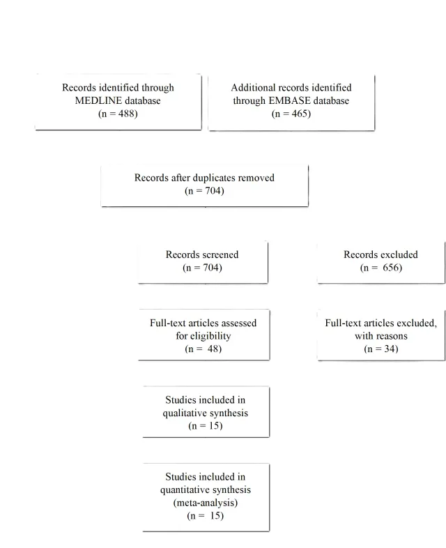
Appendix IV. Evidence Table
| Author/year | Study Description | Data Class | Conclusion | ||
| Aguilar et al. 2022 | Study Type: Single institutional cross-sectional study from 2009-2010 and then 2016-2018. Objective: To identify the audiometric pattern that best serves as a predictor for VS in patients with asymmetric SNHL Methodology: Retrospective analysis of 107 patients presenting with asymmetric SNHL on pure tone audiology. Hearing loss was defined by a difference of 15 dB in one or more frequencies between both ears. Patients with abnormal audiology screening underwent MRI with contrast. The sensitivity, specificity, positive predictive value (PPV), negative predictive value (NPV) and accuracy of different audiometric patterns were analyzed. | III | Summary: Of the 107 patients with asymmetric SNHL analyzed, only 8.4% (n=9) were determined to have VS while the remaning 91.6% (n=98) had no discernable CPA lesion. No significant difference in audiometric patterns were found in patients with and without VS. A difference of >20dB at 4000Hz was the best audiometric screening test with sensitivity 77.78%, specificity 30.61%, PPV 8.33%, and NPV 93.75%. Conclusions: While a difference of >20dB at 4000Hz is a reasonable screening test for VS in patients with asymmetric SNHL, however, this is not sufficient to forego an MRI. Commentary: This manuscript is retrospective in nature and therefore yields class III data. | ||
| Saxby et al. 2021 | Study Type: single institution retrospective review from 2014 to 2019 Objective: To evaluate MRI in the investigation of ptaients with unilateral tinnitus without associated SNHL Methodology: Retrospective review of 2066 paient undergoing RI of the internal auditory canal for sypmtoms of unilateral tinnitus without associated SNHL. | III | Summary: Of the 2066 patient MRIs reviewed, only 0.3% (n=3) VS were diagnosed, while 134 other incidental findings were discovered. Conclusions: The authors question the utility of MRI in the diagnosis of VS in patients presenting with unilateral tinnitus without associated SNHL. Commentary: No pure tone audiology or speech testing were given in this manuscript as no patients complained of, or were demonstrated to have, SNHL at tim of MRI. | ||
| Yang et al. 2020 | Study Type: Single-institution retrospective review from 2009-2019 Objective: To review the prevalence and clinical characteristics of vestibular schwannoma (VS) in patients with sudden SNHL in southern China. Methodology: Patients presenting with sudden SNHL undergoing audiology screening and subsequent MRI were eligable for inclusion. Sudden SNHL was defined as sensorineural hearing decrease of ≥30 dB over at least 3 consecutive frequencies in pure tone audiometry, which developed within a period of 72 hours. | III | Summary: 1249 patients met inclusion criteria, of which 14 (1.12%) were diagnosed with VS. Of the VS, the majority were Koos grade I (n=7), followed by grade II (n=4), and fewer grade III (n=3). Abnormal ABRs were noted in 12/14 patients, with hearing recovery observed in 3/14. Conclusions: VS were diagnosed in 1.12% of patients presenting with sudden SNHL. Predicting the presence of a VS with audiology screening and ABRs is insufficienct, and this patient population should undergo MRI to rule out VS. Commentary: This manuscript is retrospective in nature and therefore yields class III data. | ||
| Fujita et al. 2019 | Study Type: Single-institution retrospective review from 2008-2017 Objective: To assess the prevalence of vestibular schwannoma (VS) in patients with sudden SNHL. Methodology: Retrospective review of 499 patients presenting with sudden SNHL as defined by loss of 30 dB over at least 3 continuous frequencies in pure tone audiometry that develops within a period of 72 hours. MRIs of screened patients were reviewed for presence of VS. | III | Summary: Of the 499 patients meeting inclusion criteria, 15 were found to have VS. Amongst the VS, the majority were Koos grade 1 (n=8), fewer grade II (n=6), and one grade III. There were no grade IV tumors. Conclusions: The prevalence of VS in patients with sudden SNHL was 3.0%; considering this high prevalence, clinicians should consider detailed examinations in addition to audiometry for patients with sudden SNHL Commentary: This manuscript is retrospective in nature and therefore yields class III data. | ||
| Krane et al. 2019 | Study Type: Single institution retrospective review from 1980-2013 Objective: Determine the clinical efficacy of comprehensive neurotologic testing in patients presenting with complaints of hearing loss, tinnitus and/or dizziness Methodology: Retrospective review of 1170 patients presenting with neurotologic complaints. A variety of patient variables were evaluated, including audiometric screening, ABR, and MRI data. Hearing loss was not defined. | III | Summary: Of the 1170 evaluated patients, 762 (65%) presented with complaints of subjective hearing loss, 575 (49%) with vertigo, and 657 (56%) with tinnitus. Patients were determined to have an abnormal MRI in 48% (536/1120) of cases, with 6% (n=68) resulting from VS. Conclusions: Comprehensive neurotological workup results in diagnoses that would go unrecognized otherwise, allowing patients to receive prompt treatment for medically important conditions, some of which may be causally related to their neurotologic complaints. Commentary: This manuscript is retrospective in nature and therefore yields class III data. The authors make no recommendations with regard to which patient should undergo MRI, rather they simply report the prevelance of VS in the patients screened in their study. | ||
| Ungar et al. 2019 | Study Type: Single institution retrospective review from 2013-2017 Objective: Investigate whether there is a different pattern of steroidal treatment response after sudden SNHL in patients with and without a VS in order to determine whether rapid hearing improvement can serve as a predictor of the presence of VS Methodology: Retrospective analysis of 420 patients presenting with sudden SNHL were reviewed. Sudden SNHL was defined by sensorineural hearing decrease of ≥30 dB over at least 3 consecutive frequencies in pure tone audiometry, which developed within a period of 72 hours. Patients were administered 5 days of oral prednisone at 1mg/kg/day. A comparative analysis was performed between patients responding to treatment and the presence or abscence of a VS. | III | Summary: Of the 420 patients enrolled, 20 (4.76%) were found to have a VS. Audiometric testing 7 days after steroidal treatment initiation revealed that the pure-tone average of 240 patients (60%) without VS improved, and that of the 16 (80%) patients with VS improved by the same audiometric criteria. Conclusions: Improvement of hearing within 1 week after steroidal treatment initiation in patients with sudden SNHLs may suggest the presence of a VS Commentary: This manuscript is retrospective in nature and therefore yields class III data. | ||
| Abbas et al. 2018 | Study Type: Single institution prospective series from 2013-2017 Objective: To determine whether patients within an ENT department presenting with asymmetrical SNHL and/or unilateral tinnitus can be safely and cost-efficiently screened for VS by audiologists as a first or only point of contact. Methodology: Prospective study of 1126 patients presenting to an audiologist initially for complaints of asymmetric SNHL or unilateral tinnitus undergoing workup inclusive of audiometric screening and MRI. An MRI was indicated if the audiogram shows a difference in the left and right thresholds of 15 dB or greater at two or more neighbouring frequencies. All patients with unilateral tinnitus underwent an MRI. | III | Summary: Of the 1126 patients meeting inclusion criteria, only 2.22% (n=25) were diagnosed with a VS.These patients were referred to ENT for further treatment guidance. Tumor sized ranged from 3mm to 20mm. Conclusions: Patients with asymmetrical SNHL and/or unilateral tinnitus can be safely screened for VS and independently managed by audiologists as a first or only point of contact Commentary: This manuscript is retrospective in nature and therefore yields class III data. | ||
| Lee S et al. 2018 | Study Type: Single institutional retrospective review from 2007-2017 Objective: to identify the value of MRI for the evaluation of isolated acute audiovestibular loss Methodology: 31 patients’ charts were retrospectively reviewed presenting with isolated acute audiovestibular loss. Acute hearing loss was defined as above 30 dB at three consecutive frequencies on the affected side occuring less than 1 week prior to first evaluation. | III | Summary: Of the 31 enrolled patients, 15 patients demonstrated increased labrynthine FLAIR signal on MRI while 11 MRIs were negative, 2 patients had AICA territory strokes, and 3 had VS. There was no significant difference between pure tone audiometry and recovery rates amongst pathologies. Conclusions: Isolated acute audiovestibular loss is most commonly due to labrynthitis as opposed to AICA stroke or VS, however, an abbreviated MRI is recommended to identify the exact etiology. Commentary: This manuscript is retrospective in nature and therefore yields class III data. | ||
| Cho et al. 2017 | Study Type: Single institution retrosective study from 2006 to 2016 Objective: To investigate the frequency of IAC lesion in patients presenting with sudden SNHL Methodology: Retrospective review of the MRIs of 200 consecutive patients presenting with sudden SNHL. . The audiometric criteria for sudden SNHL were a rapid decrease in hearing of more than 30 dB affecting at least three consecutive frequencies within 3 days. All MRI were obtained within 3 days of symptom onset.Patients received 60mg prednisolone for 7 days. | III | Summary: Of the 200 enrolled patients presenting with sudden SNHL, 12.5% (n=25) had an abnormal finding on the MRI believed to be causitive for symptom onset. VS were diagnosed in 36% of the abnormal MRI group (n=9/25), or 4.5% of all patients enrolled presenting with sudden SNHL. Patients with a lesion of the IAC had significantly poorer hearing recovery following steroids compared to the iatrogenic sudden SNHL cohort. Conclusions: Sudden SNHL is often caused by non-tumerous causes, the majority of which are not apparent on an MRI. Commentary: This manuscript is retrospective in nature and therefore yields class III data. | ||
| Kim D. et al. 2017 | Study Type: Single-center retrospective study from 2010-2015 Objective: To evaluate the magnetic resonance imaging (MRI) findings of labyrinthine hemorrhage as a cause of sudden SNHL Methodology: 1512 patients presenting with acute SNHL underwent audiometric screening and MRI. Hearing loss was classified as mild ( 26–50 dB), moderate (50–70 dB), severe (70–90 dB), profound (90–119 dB), or anacusis (120 dB). | III | Summary: Of the 1512 screened patients, 59 met inclusion critiera. Amongst the included cohort, 40/59 (67.8%) patients had no abnormalities on MRI while 19/59 (32.3%) had an abnormal MRI .High labrynthine signals on a pre-enhanced T1-weight and 3D FLAIR VISTA MRI with no contrast on 6/19 (31.6%) of patients, consistent with hemorrhage. A diagnosis of VS was made in 2/19 (10.5%), and the remaining 11/19 (57.9%) had labrynthitis. Conclusions: MRI using pre-enhanced T1-weighted, 3D FLAIR VISTA, and post-enhanced T1-weighted, 4-h delayed enhanced FLAIR VISTA images is able to identify labyrinthine hemorrhage as the cause of sudden SNHL. Commentary: This manuscript is retrospective in nature and therefore yields class III data. | ||
| Ren et al. 2017 | Study Type: Single institution retrospective review from January 2015 to October 2015 Objective: To investigate the clinical predictors of MRI in the diagnosis and treatment of sudden SNHL Methodology: 120 patients with sudden SNHL underwent auditory evaluations and MRI examinations. Audiometric and clinical variables were compared to MRI results. sudden SNHL was defined as sensorineural hearing decrease of ≥30 dB over at least 3 consecutive frequencies in pure tone audiometry, which developed within a period of 72 hours. | III | Summary: Of the 120 evaluated patients, 42 (35%) had an abnormal MRI result. Three (2.5%) of screened patients were ultimately found to have a VS; all 3 patients harboring a VS underwent MRI after failing to improve following a week of steroid administration. Conclusions: When patients with sudden SNHL have severe or worse SNHL and no response for ABR tests, MRI imaging seems to be a useful examination, especially for patients with profound sudden SNHL. Commentary: This manuscript is retrospective in nature and therefore yields class III data. | ||
| Bielinska et al. 2016 | Study Type: Single institutional retrospective review from 2011-2016 Objective: To determine the incidence of vestibular schwannoma being responsible for the onset of inner ear dysfunction. Methodology: Patients being screened for SNHL (range not defined) or tinnitus underwent audiology screening and ABR. Prolonged ABR (I-III above 2.55 ms, III-V above 2.35 ms, I-V above 4.6 ms) underwent MRI with contrast | III | Summary: Of 3456 patient being screened, abnormal ABRs were identified in 252 (7.3%) cases of which 13 (5.16%) had MRI diagnosis of VS. Conclusions: Presentation of asymetric SNHL warrants audiology screening with ABRs. Presence of prolonged ABRs warrants further investigation with dedicated MRI of the IAC with contrast to screen for neoplasms. Commentary: This manuscript is retrospective in nature and therefore yields class III data. | ||
| Jeong et al. 2016 | Study Type: Single institution observational study from 2007-2012 Objective: To assess abnormal MRI findings in patients with sudden SNHL and evaluate the value of MRI in identifying the cause of sudden SNHL Methodology: Retrospective review of 291 patients presenting with sudden SNHL undergoing audiology screening and eventual MRI. Hearing loss was defined by loss of 30 dB or more over at least 3 contiguous frequencies in pure tone audiometry that develops within 3 days. All patients underwent a dedicated MRI of the IAC within 10 days of symptom onset. | III | Summary: Of the 291 patients enrolled, the MRI identified the cause of the sudden SNHL in 4.5% (n=13). VS was diagosed I 3.1% (n=9) of enrolled paients, or 69.2% of patients with abnormal MRI findings. Of the VS, 3 were purely intrameatal, while 6 demonstrated extrameatal extension. Conclusions: The most commonly observed MRI abnormality in patients with sudden SNHL was vestibular schwannoma, and all of the lesions were small or medium-sized tumors involving the IAC. Commentary: This manuscript is retrospective in nature and therefore yields class III data. | ||
| Kim S. et al. 2016 | Study Type: Single-center retrospective review from 2001-2013 Objective: To evalute otologic symptoms and audiometric data of VS compared to other neoplasms of the CPA. Methodology: Comparison of otologic symptoms of 171 patients diagnosed with CPA tumors (VS-116, Other-55). Factors analyzed included tumor type, size, and location and the results of audiometric examinations. Audiogram patterns were classified as high-tone hearing loss (descending) type if the hearing threshold was lower at a high-tone than at a low-tone frequency; as flat type when the thresholds of all assessed frequencies were comparable; low-tone hearing loss (ascending) type when a threshold was lower at a low-tone than at a hightone frequency; concave type when the primary frequency lowered was mid-tone; and convex type when the mid-frequency was relatively well preserved. Tinnitograms were used to measure tinnitus pitch and loudness matching. | III | Summary: The most frequent patterns of hearing loss were the descending type in patients with VS and the flat type in patients with non-VS tumors. Pure tone thresholds tended to increase more in patients with VS than non-VS tumors according to tumor size, and pure tone averages were significantly higher in patients with VS than non-VS tumors of 11–25mm in size. Conclusions:Hearing loss with tinnitus was the most common combination of symptoms in patients with VS, whereas hearing loss with dizziness was more common in patients with other types of CPA tumor. Commentary: This manuscript is retrospective in nature and therefore yields class III data. | ||
| Lee SH et al. 2015 | Study Type: Single institution retrospective review from 2001-2013 Objective: To determine the neurotologic factors associated with VS by analyzing the clinical manifestations and diagnostic test results of patients with VS before MRI scanning Methodology: 114 patients presenting with neurotologic complaints were enrolled. Multiple patient factors were retrospecively analyzed including:demographics, symptomotology, pure-tone audiometry, speech discrimination score, ABR, and MRI results. | III | Summary: Of the 114 VS patients analyzed, the most common symptom at time of presentation was asymmetric hearing loss associated iwth tinnitus. More severe deafness correlated with lower speech discimination scores. Of patients with latencies of waves I, III, and V on ABR tests, 56.1%, 92.4%, and 92.4%, had interaural latency differences ‡0.2 ms. Conclusions: MRIs should be obtained in patients presenting with asymetric hearing loss, tinnitus, low speech discrimination score, and abnormal ABRs. Commentary: This manuscript is retrospective in nature and therefore yields class III data. | ||
Appendix V. Conflicts of Interest
| Task Force Member | Disclosure |
| Julie Honaker PhD, AuD | Nothing to Disclose |
| Ben Allen Strickland, MD | Nothing to Disclose |
| Eric J. Lehrer, MD | Servier Pharmaceuticals, Novocure Inc. |
| Sheryl Green, MBBCh | Nothing to Disclose |
| John P. Marinelli MD | Medtronic |
| Christopher S. Graffeo MD, MS | Nothing to Disclose |
| Isabelle M. Germano, MD, MBA | Brianlab |
| Mateo Ziu, MD | Omniscient Neurotechnology America Ltd; GT Medical Technologies, Inc |
| Walavan Sivakumar, MD | Stryker Corporation |
| Sherwin Tavakol, MD | IRRAS USA, Inc.; Globus Medical, Inc. |
| Lucas Paul Carlstrom, MD, PhD | Kuros Biosciences USA, Inc |
| Jamie J. Van Gompel, MD | Medtronic, Cadence |
| Ian Dunn, MD | Nothing to Disclose |
| Jeffrey J. Olson, MD | Verastem, Inc., Research Grant American Cancer Society, Editorial Consultant; Azurity Pharmaceuticals, Inc. |
| Ghazal S. Daher MD | Nothing to Disclose |
| Matthew L. Carlson, MD | Cochlear Americas, Advanced Bionics, Stryker Corporation; iotaMotion, Inc.; Stryker Corporation |
| Neil S. Patel, MD | Cochlear Americas, Zeiss, Viridian Therapeutics, IotaMotion, Inc. |
| Michael Sughrue, MD | Omniscient Neurotechnology America Ltd |
| Constantinos G. Hadjipanayis, MD, PhD | Stryker Corporation; Integra LifeSciences Corporation; Omniscient Neurotechnology America Ltd |
| Jeffrey Jacob, MD | Stryker Corporation; KLS; Synthes |
3. The Role Of Intraoperative Cranial Nerve Monitoring in the Management of Patients With Vestibular Schwannoma: Update
Sponsored by: Congress of Neurological Surgeons (CNS) and the Section on Tumors
Endorsement: Reviewed for evidence-based integrity and endorsed by the American Association of Neurological Surgeons (AANS) and Congress of Neurological Surgeons
(CNS)
Authors: Neil S. Patel, MD1, Matthew L. Carlson, MD2, Michael Sughrue, MD3, Jeffrey J. Olson, MD4
Departmental and institutional affiliations:
- Department of Otolaryngology – Head and Neck Surgery, Huntsman Cancer Institute, University of Utah, Salt Lake City, UT
- Department of Otolaryngology-Head and Neck Surgery, Mayo Clinic, Rochester, MN
- Department of Neurosurgery, Columbia University, New York, NY
- Department of Neurosurgery, Emory University School of Medicine, Atlanta, GA
Corresponding Author contact information:
Neil S. Patel, MD
Huntsman Cancer Institute
University of Utah
50 Medical Dr N
Salt Lake City, UT 84132
Email: neil.patel@hsc.utah.edu
Keywords: vestibular schwannoma, acoustic neuroma, cranial nerve monitoring, facial nerve monitoring, intraoperative auditory brainstem response, functional preservation,
Running Title: Cranial Nerve Monitoring in Vestibular Schwannoma Surgery
Abbreviations:
ABR Auditory brainstem response
BMI Body mass index
BR Blink reflex
CN Cranial nerve
CNAP Cochlear nerve action potential
EMG Electromyography
FMEP Facial motor evoked potential
FN Facial nerve
HB House-Brackmann
IONM Intraoperative neuromonitoring
PICO Population, Intervention, Comparison, Outcome
NF2 Neurofibromatosis type 2-related schwannomatosis
VS Vestibular schwannoma
WRS Word recognition score
No part of this manuscript has been published or submitted for publication elsewhere.
ABSTRACT
Background
Intraoperative neuromonitoring (IONM) has become vital in the management of vestibular schwannoma (VS) with the paradigm shift from tumor eradication to functional preservation. Several facial nerve (FN) monitoring strategies have been explored over the last few decades ranging from free-running electromyography (EMG), direct nerve stimulation, continuous nerve stimulation, facial motor evoked potentials (FMEP), blink reflex (BR), and others. Hearing preservation surgery is guided primarily by far-field auditory brainstem response (ABR) and real-time cochlear nerve action potentials (CNAP). Given the heterogeneity in tumor and patient factors, it remains very difficult to accurately predict FN outcomes, regardless of monitoring strategy.
Objective
Critically appraise literature regarding IONM during VS surgery and update the previous evidence-based clinical practice guideline.
Methods
Systematic review of the literature, incorporating articles from March 2015 to May 2022. Literature published prior to 2015 that would have been included in the prior CNS guideline was not searched again in this update. In contrast to the previous CNS guideline published in 2018, the key questions are presented in the PICO format (P: population, I : intervention, C: comparison, O: outcome).
Results
FN monitoring provides better functional outcomes compared to anatomical dissection alone and may guide extent of tumor resection. While FMEP s and free-running EMG can provide continuous noninvasive FN monitoring, there are insufficient data to determine which is more strongly correlated with facial function outcome. Both electrophysiologic data and tumor size are correlated with facial function outcome. The ideal hearing monitoring strategy remains unclear as there are insufficient data comparing CNAPs to far-field ABR. All studies were graded as Class III evidence.
Conclusion
IONM should be used in all VS cases. While the optimal FN and hearing monitoring strategy remains elusive, available data support the use of a combination of strategies, including preoperative tumor size, to maximize sensitivity and specificity. There remains a significant need for high-quality comparative studies to determine which intraoperative monitoring scheme can provide intraoperative guidance and predict postoperative outcome.
RECOMMENDATIONS
FN Monitoring
Updated Questions and Recommendations
Patient Population: In patients undergoing microsurgical resection of sporadic or NF2-associated VS:
Question 1: Does the use of intraoperative FN monitoring provide superior long-term FN functional outcomes compared to anatomic dissection alone?
Recommendation: Level III: Intraoperative FN monitoring provides superior long-term FN functional outcomes compared to anatomic dissection alone.
Question 2: Is data from intraoperative FN monitoring superior to clinical and imaging information in predicting short- and long-term FN functional outcomes?
Recommendation: Level III: Electrophysiologic measures are more predictive of long-term FN functional outcomes than clinical information alone. There is insufficient evidence to determine whether electrophysiologic data is superior to clinical information in predicting short-term FN functional outcomes.
New Question and Recommendation
Question 3: Is the use of transcranial FN motor evoked potentials or BR testing superior to free-running EMG and direct FN stimulation in predicting short- and/or long-term FN functional outcomes?
Recommendation(s): There is insufficient evidence to determine whether transcranial FMEP or BR is superior to EMG-based monitoring schema in predicting FN functional outcomes.
Notable Updates to 2018 Guideline: This represents a new research question that was not addressed in the previous CNS guideline.
Cochlear Nerve Monitoring
Updated Question and Unchanged Recommendation
Question 4: Is intraoperative cochlear nerve monitoring superior to ABR monitoring in predicting short- and long-term hearing preservation outcomes?
Recommendation(s): There is insufficient data to determine whether CNAP is superior to far-field ABR in monitoring hearing function in hearing preservation surgery for VS.
New Question and Recommendation
Question 5: Does the monitoring of adjacent cranial nerves (cranial nerves [CN] V, IX, X, XI, and XII) provide for better preservation of their function than carrying out surgery without these CN s being monitored?
Recommendation(s): There is insufficient evidence to determine whether monitoring of other regional CN s affects functional preservation.
INTRODUCTION
Rationale
In the modern era of VS treatment, FN preservation, maintenance of serviceable hearing, and tumor control are the highest priorities among patients and providers alike. While these have always been considered the “goals” of treatment, falling short in one dimension was considered acceptable because of the lack of reasonable management alternatives. Early radiosurgical series reported high rates of facial palsy and hearing loss that rendered it less attractive when compared to microsurgery, which offered definitive cure. Observation was only offered to patients with very small tumors, and little was known about the rate of hearing decline over the course of observation. At present day, patients with small- to medium-sized tumors (generally considered < 1.5 cm in maximum dimension) routinely compare the risks of microsurgical treatment with the probability of FN and hearing functional preservation with nonsurgical treatment. It is well known that the risk of FN paralysis during observation is negligible. In an intention-to-treat comparative study between 167 observed tumors and 121 equivalent size surgically managed tumors, 100% of the observed cohort had HB grade I facial function at long-term (mean 62 months) follow up1. The rates of hearing preservation over the course of observation for patients with serviceable hearing at diagnosis have also placed the risk of treatment-related hearing loss under scrutiny. Based on a recent systematic review by Khandavala et al comprising over 3,600 patients with observed VS, the estimated rate of serviceable hearing at 5 years is 60%2. A more detailed summary of hearing and FN outcomes with observation, microsurgery, and radiosurgery can be found in other areas of this Guidelines Update.
What defines tumor “management” has also evolved over time. The value of complete tumor removal is no longer high enough to warrant compromise of neurologic function. Patients are routinely willing to accept less-than-gross total tumor removal if it results in a better FN outcome or long-term hearing preservation, particularly given the knowledge that 1) the tumor remnant often does not warrant additional treatment and 2) modern radiosurgery achieves high rates of tumor control in the setting of prior sub-total resection.
These factors have changed the treatment conversation to one where patients carefully select the treatment strategy that delivers the trifecta of tumor control without the need for additional treatment, hearing preservation if hearing is serviceable at the time of diagnosis, and normal or near-normal FN function. Surgeons are therefore charged with the task of refining microsurgical techniques to match or exceed what conservative management can offer, on the basis that 1) tumor removal mitigates the risk of eventual treatment if the tumor grows and 2) tumor removal halts the progression of hearing loss from the tumor itself (regardless of growth)3,4.
Objectives
This update to the “Congress of Neurological Surgeons Systematic Review and Evidence-Based Guideline on Intraoperative Cranial Nerve Monitoring in Vestibular Schwannoma Surgery” published in 20185 aims to characterize the current state of intraoperative monitoring strategies. The goal is not to recapitulate the prior comprehensive guideline statement but rather to determine whether emerging data from 2015-2022 has shed light on the predictive ability of electrophysiologic measures with regard to FN and hearing outcomes among patients undergoing microsurgery for sporadic or NF2 associated VS.
Methodology
The co-authors of this guidelines update convened to determine the most salient questions on the topic of IONM in VS surgery. Through objective evaluation of the evidence and transparency in the process of making recommendations, this evidence-based clinical practice guideline was developed for the diagnosis and treatment of adult patients with VSs. These guidelines are developed for educational purposes to assist practitioners in their clinical decision-making processes. Additional information about the methods utilized in this systematic review is provided below.
Additional details of the systematic review are provided below and within the introduction and methodology chapter of the guideline (add link).
Literature Search
The members of the task force identified key search terms pertaining to intraoperative monitoring for VS. Databases included Ovid Medline and Embase. CN s of interest included the trigeminal, facial, vestibulocochlear, glossopharyngeal, vagus, accessory, and hypoglossal, and were thus included in the search strategy. Terms pertinent to ABR or cochlear nerve monitoring were used for the vestibulocochlear nerve. Only articles available in the English language were included for review. The full search strategy for each database is outlined in Appendix I.
Inclusion/Exclusion Criteria
Two co-authors were responsible for the independent review of abstracts and subsequently full text data for each article deemed relevant to the PICO questions under examination. Articles were retrieved and included only if they met specific inclusion/exclusion criteria. Inconsistencies were re-reviewed, and disagreements were resolved by consensus. To reduce bias, these criteria were specified before conducting the literature searches.
Articles that do not meet the following criteria were, for the purposes of this evidence-based clinical practice guideline, excluded.
To be included as evidence in the guideline, an article had to be a report of a study that:
- Investigated patients with VSs who underwent microsurgery
- Was related to CN monitoring
- Was a full article report of a clinical study
- Appeared in a peer-reviewed publication or a registry report
- Enrolled a minimum of 10 patients
- Was of humans
- Was published between 1/1/2015 and 5/20/2022
- Quantitatively presented results
Articles were excluded if it was determined that the article:
- Was a case report or expert opinion paper
- Was a technique paper only
- Was a heterogenous group of tumor types where VS data could not be reliably abstracted
- Was a systematic review, meta-analysis, or guideline developed by others
Systematic reviews or meta-analyses conducted by others, or guidelines developed by others were not included as evidence to support this review due to the differences in article inclusion/exclusion criteria specified compared to the criteria specified by the Guidelines Task Force.
Assessment for Risk of Bias
Articles on prognostication of CN function are affected by several potential sources of bias. Some sources of bias were particularly relevant to CN outcome studies. Bias due to lack or loss of information over time and attrition bias is relevant due to 1) varied follow up schedules, 2) patients who traveled to a center for care but conduct follow up at a local facility, and 3) the notion that individuals with absent hearing or FN function or completely normal hearing or FN function may not elect to undergo tests or evaluations as they are perceived unnecessary. Given that most articles that comprise this guidelines update are retrospective in design, there remains the risk of publication bias, bias of change in methods over time, or ascertainment bias. In addition, the data comprising the majority of included studies have variability due to the accuracy of the electronic medical record and subjectivity in assessing FN outcome (e.g. using the House-Brackmann grading scale versus alternative functional scales).
Evidence was rated in accordance with the previously published guideline on IONM in VS surgery. Specifically, a paradigm for prognostication was used with evidence classified in three classes. Additional information regarding the hierarchy classification of evidence can be located here: https://www.cns.org/guidelines/guideline-development-methodology.
In order to evaluate papers addressing prognosis, five technical criteria are applied:
- Was a well-defined representative sample of patients assembled at a common (usually early) point in the course of their disease?
- Was patient follow-up sufficiently long and complete?
- Were objective outcome criteria applied in a “blinded” fashion?
- If subgroups with different prognoses were identified, was there adjustment for important prognostic factors?
- If specific prognostic factors were identified, was there validation in an independent “test set” group of patients?
If all five of these criteria are satisfied, the evidence is classified as Class I. If four out of five are satisfied, the evidence is Class II, and if less than 4 are satisfied, it is Class III. Class I level translates to level I recommendations, class II evidence translates to level II recommendations, and class III evidence translates to level III recommendations.
Revision Plans
In accordance with the National Academy of Medicine’s standards for developing clinical practice guidelines, the task force will monitor related publications following the release of this document and will revise the entire document and/or specific sections “if new evidence shows that a recommended intervention causes previously unknown substantial harm; that a new intervention is significantly superior to a previously recommended intervention from an efficacy or harms perspective; or that a recommendation can be applied to new populations.”6 In addition, the task force will confirm within five years from the date of publication that the content reflects current clinical practice and the available technologies for the evaluation and treatment for patients with VS.
SUMMARY OF THE PREVIOUS GUIDELINE RECOMMENDATIONS:
FN Monitoring
Question 1: Does intraoperative FN monitoring during VS surgery lead to better long-term FN function?
Target population: This recommendation applies to adult patients undergoing VS surgery regardless of tumor characteristics.
Recommendation: Level 3: It is recommended that intraoperative FN monitoring be routinely utilized during VS surgery to improve long-term FN function.
Question 2: Can intraoperative FN monitoring be used to accurately predict favorable long-term FN function after VS surgery?
Target population: This recommendation applies to adult patients undergoing VS surgery.
Recommendation: Level 3: Intraoperative FN monitoring can be used to accurately predict favorable long-term FN function after VS surgery. Specifically, the presence of favorable testing reliably portends a good long-term FN outcome. However, the absence of favorable testing in the setting of an anatomically intact FN does not reliably predict poor long-term function and therefore cannot be used to direct decision-making regarding need for early reinnervation procedures.
Question 3: Does an anatomically intact FN with poor electromyogram electrical responses during intraoperative testing reliably predict poor long-term FN function?
Target population: This recommendation applies to adult patients undergoing VS surgery.
Recommendation: Level 3: Poor intraoperative electromyogram electrical response of the FN should not be used as a reliable predictor of poor long-term FN function.
Cochlear Nerve Monitoring
Question 4: Should intraoperative eighth CN monitoring be used during VS surgery?
Target population: This recommendation applies to adult patients undergoing VS surgery with measurable preoperative hearing levels and tumors smaller than 1.5 cm.
Recommendation: Level 3: Intraoperative eighth CN monitoring should be used during VS surgery when hearing preservation is attempted.
Question 5: Is direct monitoring of the eighth CN superior to the use of far-field auditory brain stem responses?
Target population: This recommendation applies to adult patients undergoing VS surgery with measurable preoperative hearing levels and tumors smaller than 1.5 cm.
Recommendation: Level 3: There is insufficient evidence to make a definitive recommendation.
The above questions and recommendations served as the basis for this guidelines update. There were two primary limitations worth mentioning. First, the questions posed were not in a PICO format and thus were not designed to compare groups, with the exception of question 5. Therefore wording for the questions was updated. Second, this broader guideline statement was intended to answer more fundamental questions, such as whether or not intraoperative FN or eighth nerve monitoring should be used during VS surgery.
Since the publication of this foundational guideline5, there have been several technical developments in IONM. This Guidelines Update serves to critically review the literature surrounding these novel monitoring strategies and their predictive power in characterizing VS surgery outcomes.
RESULTS
The literature search yielded 222 abstracts. The authors reviewed all abstracts obtained from the literature search and identified those meeting criteria for full text review and extraction, addressing the clinical questions, in accordance with the literature search strategy. The task force members identified the evidence available to answer the targeted clinical questions.
The task force selected 49 full-text articles for full text review. Of these, 15 were included for evidence tables and subject to systematic review7-20. This is depicted graphically in the PRISMA diagram (Figure 1).
Question 1: Does the use of intraoperative FN monitoring provide superior long-term FN functional outcomes compared to anatomic dissection alone?
Recommendation: Level III: Intraoperative FN monitoring provides superior long-term FN functional outcomes compared to anatomic dissection alone.
Of the 49 articles that resulted from abstract review, 8 were marked as pertinent to this question. To be included in this section, the study had to fall into one of two categories. The first category was strictly those articles that provided objective FN outcome data (i.e. postoperative HB score) in at least one cohort in which intraoperative FN monitoring was employed and at least one cohort where only anatomical dissection was used without continuous intraoperative monitoring. The second category was more permissive and included articles that examined whether FN outcome was related to the electrophysiologic parameter studied. While the second category does not directly answer the question, the authors felt that a conclusion regarding the value of neuromonitoring for FN outcomes could still be drawn from the data. After final review, 5 studies were included for analysis, all of which were graded as Class III evidence. Data extracted from each article included number of patients, study design, surgical approach, which electrophysiologic parameters were recorded, the time at which FN function was evaluated, and what grading system was used.
Given that intraoperative FN monitoring has become routine clinical practice for lateral skull base surgery, there is a relative paucity of articles comparing FN outcomes between monitored and unmonitored groups. In 2017, Xu et al20 reported the results of a retrospective single institution study of 53 patients who underwent microsurgical resection of large VS (mean tumor size of approximately 40 mm) via retrosigmoid approach. Patients were separated into a monitored group and control group of roughly equal size. The authors found that at short, intermediate, and long-term follow up, the monitored group has statistically significant better FN function. Rates of total or subtotal resection were similar.20 These findings were corroborated in a retrospective review of 62 patients published by Hou et al in 201813. In that study, patients were divided randomly into a monitored group (observation group), and one that had anatomical dissection only (control group). A potential limitation of the study was the relatively low rate of anatomic preservation of the FN in both groups (78.8% and 51.7% in the observation and control groups, respectively), and no specific documentation regarding tumor size. In 2016, Taddei18 and colleagues published a series of 51 consecutive patients who had microsurgical removal of a VS at a single institution. Two groups were described: group 1 only had a “facial stimulator” but no continuous monitoring while group 2 had continuous monitoring and the use of a stimulator. The authors reported a higher rate of FN preservation and HB grade 1 or 2 facial function in the continuously monitored group. A potential limitation is that up to 25% of patients in each group had some degree of facial paralysis (HB grade 2 or 3) prior to surgery, which may limit generalizability, given how rare preoperative facial paralysis is in VS patients18. In spite of the limitations noted, the data from these studies will likely not be reproduced in future publications given the essentially universal implementation of continuous neuromonitoring at high-volume skull base centers.
As an adjunct to the data analyzed above, the authors included two additional articles that provide data to estimate the efficacy of FN monitoring in improving FN outcomes. In 2014, Ashram et al21 reported the results of a prospective single institution study of 42 patients who underwent VS resection via retrosigmoid or translabyrinthine approaches. In this report, FN monitoring was conducted using continuous EMG in a 5-channel setup (mentalis, orbicularis oris, nasalis, orbicularis oculi, and frontalis). The results were compared to a hypothetical 2-channel group where the responses from mentalis, nasalis, and frontalis were omitted. The multichannel setup, in particular the inclusion of the mentalis muscle, allowed earlier and more efficient detection of mechanically elicited EMG activity. Notably, increasing EMG activity portended a poorer intermediate- and long-term FN outcome.21 A report by Bernardeschi examined the predictive value of a reduction in response to supramaximal stimulation in cases of VS surgery where the FN was considered especially adherent to the tumor. In that study, dissection was interrupted if 50% or more of the response to supramaximal stimulation at the brainstem was lost. While this did guide the decision to terminate dissection, there was no significant difference in FN outcome when comparing patients meeting or not meeting the 50% criterion.7
Synthesis: Electrophysiologic monitoring of the FN provides superior FN preservation compared to anatomic dissection alone. While less relevant for smaller tumors where the FN tends not to be splayed or displaced, it is most important for larger VS. Focusing on EMG monitoring only, continuous monitoring where attention is paid to non-reassuring EMG activity during dissection has the potential to improve FN outcomes. It remains unclear whether the response to supramaximal stimulation is predictive of FN outcome. The guideline recommendation is consistent with the prior recommendation for the use of FN monitoring, but specifies that the literature supports its superiority over anatomic dissection alone. Future studies assessing the sensitivity and specificity of various EMG findings in a large prospective cohort of patients undergoing VS surgery would be valuable in determining the predictive value of each strategy. A Level III recommendation is made as all articles constituted class III data.
Question 2: Is data from intraoperative FN monitoring superior to clinical and imaging information in predicting short- and long-term FN functional outcomes?
Recommendation: Level III: Electrophysiologic measures are more predictive of long-term FN functional outcomes than clinical information alone. There is insufficient evidence to determine whether electrophysiologic data is superior to clinical information in predicting short-term FN functional outcomes.
Of the 49 articles that resulted from abstract review, 8 were marked as pertinent to this question. To be included in this section, the study had to report short- and long-term FN outcomes following VS surgery in which the predictive power of electrophysiologic monitoring was compared to that of clinical information such as patient or tumor characteristics. After final review, 4 studies were included for analysis, all of which were graded as Class III evidence. Data extracted from each article included number of patients, study design, surgical approach, what patient or tumor data were recorded and analyzed, which electrophysiologic parameters were recorded, the time at which FN function was evaluated, and what grading system was used.
While it remains unclear whether patient demographics or surgical approach reliably correlate with FN functional outcome, extrameatal tumor size remains to be the most commonly utilized preoperative predictor in patient counseling. This was demonstrated in a study by Bloch et al of 624 patients who underwent surgery for VS, where pre-operative tumor size was the only statistically significant predictor of FN functional outcome.22 However, there remains significant heterogeneity in FN-tumor adherence, tumor consistency, and FN position that result in variable FN outcomes for a given tumor size. As these characteristics often lie on a spectrum and are difficult to categorize, electrophysiologic measures are often the only means of quantifying the surgical trauma to the FN resulting from dissection.
Liu et al in 2015 reported a series of 106 patients who underwent microsurgical removal of large VS via retrosigmoid approach. Monitoring schema included EMG and FMEP and the predictive value of tumor size, train time (a form of non-reassuring EMG activity), FMEP ratio, and maximal response amplitude ratio were analyzed. Both FMEP ratio and tumor size were highly correlated with immediate and late FN functional outcome. Notably, the level of significance was greater for FMEP ratio (p<0.001) compared to tumor size (p<0.01).15 In 2017, Hong et al performed a univariable analysis of individual predictors (tumor characteristics, patient age, surgical approach characteristics, and whether or not intraoperative electrophysiologic monitoring was used) and found that both tumor diameter and the use of intraoperative monitoring were correlated with FN functional outcome.12 However, the results of this study are limited given the lack of multivariable analysis. Ling et al in 201814 found that FMEP amplitude ratio was more highly correlated with postoperative FN function (p<0.0001) when compared to maximum tumor diameter (p=0.025). Most recently, Ren et al in 202116 performed a large retrospective review of 256 patients who underwent surgery for VS that included a broader range of clinical predictors in the statistical analysis. Small tumor size (<1.5 cm) and lower body mass index (BMI) were associated with better immediate postoperative FN outcomes. However, only achieving gross-total resection and > 100 microvolt EMG responses intraoperatively were correlated with better long-term FN function on multivariate analysis. These results suggest that those patients who had complete tumor resection likely harbored tumors with less FN-tumor adherence, perhaps indicated by higher amplitude EMG responses. The predictive value of this EMG criterion should not be used in isolation, however. According to a study by Neff et al in 2005, minimum stimulus intensity in mA and response amplitude in microvolts have the highest predictive value when used in concert than if either is used in isolation, even when using a higher threshold for response amplitude23.
Synthesis: Electrophysiologic measures, specifically FMEP amplitude ratio and final EMG response amplitude, may be more predictive of long-term FN functional outcome when compared to clinical information alone. While not the focus of this question, tumor size appears to remain the clinical predictor of greatest importance in predicting FN functional outcome. It remains unclear whether electrophysiologic responses are superior to clinical information in predicting immediate postoperative FN functional outcomes. This difference may be related to less severe FN injury (i.e. Sunderland grade 1 or 2 injuries) exacerbated by patient or surgical factors that resolves to some degree over time. A notable limitation is the time at which short-term FN function is assessed. In the study by Ren et al16, for example, immediate FN function was graded within 24 hours of surgery. In contrast, Liu et al measured immediate FN function at 3-7 days postoperatively. It is well known that patients frequently experience a decline in FN function over the first few days following VS surgery that is thought to be related primarily to neural edema or devascularization. In comparison to the prior guideline on this topic, the updated guideline statements recognize that clinical information can be used to predict FN functional outcomes and addresses the question of whether intraoperative electrophysiologic data is more or less predictive of FN functional outcomes than clinical information alone. Future multi-institutional studies would be valuable in determining reproducibility of the electrophysiologic markers of FN injury given significant heterogeneity in monitoring strategies and timing of FN grading. A Level III recommendation is made as all articles constituted class III data.
Question 3: Is the use of transcranial FN motor evoked potentials or BR testing superior to free-running EMG and direct FN stimulation in predicting short- and/or long-term FN functional outcomes?
Recommendation(s): There is insufficient evidence to determine whether transcranial FMEP or BR is superior to EMG-based monitoring schema in predicting FN functional outcomes.
Of the 49 articles that resulted from full text review, 11 were marked as pertinent to this question. To be included in this section, the study had to report an analysis of FN functional outcome that used FMEP measures and EMG-based measures as outcome predictors. While a direct comparison was not mandatory, at minimum an analysis of which electrophysiologic parameters that were most highly correlated with outcome was required. After final review, 5 studies were included for analysis, all of which were graded as Class III evidence. Data extracted from each article included number of patients, study design, surgical approach, which electrophysiologic parameters were recorded for FMEP and EMG based monitoring schema, the time at which FN function was evaluated, and what grading system was used.
FN motor evoked potentials are responses recorded from facial musculature elicited using transcranial electrical stimulation of the motor cortex. Akin to EMG-based monitoring, there are multiple ways in which FMEP responses can be analyzed and subsequently categorized for surgical guidance. The correlation between FMEP and FN functional outcome has been demonstrated in multiple studies24-26. In the study by Liu et al15 referenced above, an additional analysis comparing FMEP ratio (response at the end of the case to the maximum level at the start of the case), FMEP maximum response amplitude (MRA), and intraoperative A-train time was conducted. The authors concluded that FMEP ratio showed the highest correlation with immediate and late FN function when compared to A-train time and MRA ratio15. Tawfik et al reported a series of 82 consecutive patients, 20 of whom had FMEP recorded in addition to EMG while 62 had EMG only. There was no significant difference in FN outcome between groups, though FMEP remained highly specific for predicting postoperative FN function19. The relatively small FMEP cohort and asymmetric cohort size may have limited the ability to resolve differences between groups.
Subsequent work by Frigeni in 2020 analyzed the predictive value of EMG A-train activity, noting a sinusoidal, symmetrical high frequency waveform was considered to be predictive of FN palsy9, when compared to preoperative BR testing. On multiple logistic regression analysis, A-train activity, when correctly identified, was more predictive of FN outcome than BR testing, but FMEP was not included in this study9. Hendriks et al compared direct nerve stimulation, free running EMG, and FMEP and found that FMEP threshold change of less than 20 mA resulted in a FN outcome better than HB grade 3, but that A-trains were not correlated with FN functional outcome11. Most recently, Greve et al published a retrospective review of 60 consecutive patients who had surgery with FMEP and EMG monitoring. The authors found that using a specific FMEP-based criterion (see Table 3 for details) was more predictive than A-trains on EMG10,27.
Synthesis: While FMEP-based monitoring can be used to predict FN functional outcome, there is insufficient evidence to determine whether FMEP or BR is superior to EMG-based monitoring strategies. While several groups have reported outcomes with FMEP monitoring, the interpretation of FMEP results remains challenging. Similarly, while EMG A-train activity is considered the most non-reassuring pattern in VS surgery, correctly identifying the specific pattern is not trivial. It is likely that these limitations, in addition to relatively small sample sizes in the reports cited, preclude a determination of superiority of one strategy over the other. As well, little is known about how to employ FMEP findings to inform extent of resection and whether real-time data can be used to guide surgical maneuvers, in the manner that EMG activity is employed. Larger prospective studies at institutions with extensive experience with both monitoring strategies would be vital to compare the two and evaluate ways to incorporate data from both in predicting outcomes. A Level III recommendation is made as all articles constituted class III data.
Question 4: Is intraoperative cochlear nerve monitoring superior to ABR monitoring in predicting short- and long-term hearing preservation outcomes?
Recommendation(s): There is insufficient data to determine whether CNAP is superior to far-field ABR in monitoring hearing function in hearing preservation surgery for VS.
Of the 49 articles that resulted from full text review, 21 were marked as pertinent to this question. To be included in this section, the authors must have reported comparison of direct CNAP monitoring and far-field ABR with documented postoperative hearing results. Acceptable hearing testing descriptions included pure tone average/ Word recognition score (WRS) and AAO-HNS hearing class determined from audiometric testing. After final review, only 1 study was included for analysis, and was graded as Class III evidence. Data extracted from the article included number of patients, study design, surgical approach, the method of auditory pathway monitoring, the method of audiometric evaluation and when the audiogram was completed, and what system was used to categorize audiometric results, when applicable.
The article by Sun et al published in 201817 included a series of 126 consecutive patients operated at a single institution. All underwent a middle fossa approach for resection of a small VS. The authors concluded that an absent N1 amplitude reached 100% positive predictive value for worsening of hearing class. In contrast, the negative predictive value, suggesting maintenance of preoperative hearing, was 50% NM- Perhaps inserting a statement regarding the inability to use CNAP optimally in cases of large VSs would be useful. when the waveform was intact. Intraoperative ABR had similar positive (86%) and negative (84%) predictive value for changes in postoperative hearing17. This suggests that, when used in isolation, CNAP is less likely to resolve deleterious downstream effects due to the anatomical position (i.e. proximal 8th nerve) where the recording electrode is placed.
Synthesis: There remains insufficient data at present to determine whether CNAP is superior to far-field ABR in predicting postoperative hearing function during hearing preservation surgery for VS. Given that ABR offers complimentary information to CNAP, it is likely that the ideal method of monitoring would combine the two strategies. Direct CNAP may also be difficult or impossible in larger tumors due to tumor involvement of the proximal 8th nerve at the root entry zone. The recommendation of the present update is consistent with the previous guideline. There is a significant need for more comparative studies in this area. Considering the variability in surgical approach for hearing preservation (i.e. where some institutions strongly prefer middle fossa to retrosigmoid), a multi-institutional effort would be advisable.
Question 5: Does the monitoring of adjacent CN s (CN s V, IX, X, XI, and XII) provide for better preservation of their function than carrying out surgery without these CN s being monitored?
Recommendation(s): There is insufficient evidence to determine whether monitoring of other regional CN s affects functional preservation.
Of the 49 articles that resulted from full text review, 1 was pertinent to this question. To be included in this section, the authors must have showed a comparison of regional CN outcomes when EMG monitoring was used for CN s V, IX, X, XI, and XII and when it was not. Unfortunately, zero articles matched this criterion. The one article that resulted from the literature search, authored by Romagna et al28, summarized their experience with monitoring of the vagus nerve during CPA tumor surgery. The authors concluded that endotracheal tube surface electrodes could be reliably used to indicate CN X palsy. There was no comparison to a group without monitoring, nor were other regional CN s included.
Synthesis: No recommendation can be made as to whether monitoring of regional CN s affects functional preservation in VS surgery. Though these nerves are not directly involved by VS, they are at risk during posterior fossa surgical approaches and CPA tumor dissection. Future studies are needed to determine 1) which regional CN s can be reliably monitored during VS surgery, 2) whether continuous monitoring prevents injury, and 3) what is the additional cost of monitoring “uninvolved” CN s.
DISCUSSION
These guidelines support the routine use of IONM in all VS cases for monitoring the integrity of regional CN s. The growing body of literature on FN monitoring and outcome prediction has increased our understanding of prognostication and the relative weight of intraoperative monitoring data and preoperative tumor or patient characteristics. The optimal FN monitoring strategy likely includes a combination of free-running EMG (with active monitoring for A-trains and other potentially non-reassuring waveforms) and continuous evoked potential monitoring schema such as FMEP. While chiefly determined by tumor size and position within the internal auditory canal and cerebellopontine angle, predicting hearing preservation using intraoperative measures remains challenging. While ABR can predict preservation of hearing with near complete certainty when the waveform is unchanged or improved throughout the case, waveform averaging adds a time delay that limits its ability to be used as a dissection guide. Real-time CNAP monitoring holds promise but remains technically challenging with no widely available atraumatic electrodes to deliver the stimulus. It is unclear whether lower CN monitoring is required during VS surgery. Since monitoring requires little additional instrumentation, the authors would consider its use during surgery for very large VS or in a reoperation situation where the nerves may not separate easily from the nearby cerebellum or arachnoid, for instance.
KEY ISSUES FOR FUTURE RESEARCH AND CONCLUSIONS
As it is well established that traditional EMG and ABR-based monitoring strategies are “here to stay,” the authors recommend that research efforts focus on novel monitoring strategies. Utilizing a combination of preoperative tests such as BR or ABR and complimentary intraoperative measurements may provide better prognostic information when compared to single intraoperative measures such as EMG response NM-amplitude or stimulus threshold. Multi-institutional collaborations with consistent monitoring protocols across sites would likely sufficient data to power a comparative study. Ideally, a prospective study where patients are examined for FN function or hearing function at set time points by blinded observers would be designed to reduce bias and maximize generalizability. Ultimately, the authors acknowledge that this goal will be challenging to achieve given the complexity of electrode placement, monitoring technologies, and interpretation, in combination with variations in surgical technique or approach. It is encouraging that the body of literature on IONM in VS surgery continues to grow in parallel with the advancement of function preservation skull base surgery.
Conflicts of Interest
All Guideline Task Force members were required to disclose all potential COIs prior to beginning work on the guideline, using the COI disclosure form of the AANS/CNS Joint Guidelines Review Committee. The CNS Guidelines Committee and Guideline Task Force Chair reviewed the disclosures and either approved or disapproved the nomination and participation on the task force. The CNS Guidelines Committee and Guideline Task Force Chair may approve nominations of task force members with possible conflicts and restrict the writing, reviewing, and/or voting privileges of that person to topics that are unrelated to the possible COIs.
Disclosure of Funding
These evidence-based clinical practice guidelines were funded exclusively by the Congress of Neurological Surgeons, which received no funding from outside commercial sources to support the development of this document.
Disclaimer of Liability
This clinical systematic review and evidence-based guideline was developed by a physician volunteer task force as an educational tool that reflects the current state of knowledge at the time of completion. Each chapter is designed to provide an accurate review of the subject matter covered. This guideline is disseminated with the understanding that the recommendations by the authors and consultants who have collaborated in their development are not meant to replace the individualized care and treatment advice from a patient’s physician(s). If medical advice or assistance is required, the services of a competent physician should be sought. The proposals contained in these guidelines may not be suitable for use in all circumstances. The choice to implement any particular recommendation contained in these guidelines must be made by a managing physician in light of the situation in each particular patient and on the basis of existing resources.
Acknowledgments:
The guidelines task force would like to acknowledge the CNS Guidelines Committee for their contributions throughout the development of the guideline, the AANS/CNS Joint Guidelines Review Committee, as well as the contributions Trish Rehring, MPH, Director for Evidence-Based Practice Initiatives for the CNS, and Janet Waters, MLS, BSN, RN, for assistance with the literature searches. Throughout the review process, the reviewers and authors were blinded from one another. At this time the guidelines task force would like to acknowledge the following individual peer reviewers for their contributions: Patti Raksin, Tjoumakaris, Andrew Carlson, Neil Majmundar, Jeff Mullin and Koji Ebersole.
References
- Martin TP, Tzifa K, Kowalski C, Holder RL, Walsh R, Irving RM. Conservative versus primary surgical treatment of acoustic neuromas: a comparison of rates of facial nerve and hearing preservation. Clin Otolaryngol. 2008;33(3):228-235.
- Khandalavala KR, Saba ES, Kocharyan A, et al. Hearing Preservation in Observed Sporadic Vestibular Schwannoma: A Systematic Review. Otol Neurotol. 2022;43(6):604-610.
- Patel NS, Huang AE, Dowling EM, et al. The Influence of Vestibular Schwannoma Tumor Volume and Growth on Hearing Loss. Otolaryngol Head Neck Surg. 2020;162(4):530-537.
- Ahsan SF, Huq F, Seidman M, Taylor A. Long-term Hearing Preservation After Resection of Vestibular Schwannoma: A Systematic Review and Meta-analysis. Otol Neurotol. 2017;38(10):1505-1511.
- Vivas EX, Carlson ML, Neff BA, et al. Congress of neurological surgeons systematic review and evidence-based guidelines on intraoperative cranial nerve monitoring in vestibular schwannoma surgery. Neurosurgery. 2018;82(2):E44-E46.
- Ransohoff DF, M. Pignone, and H.C. Sox, . How to decide whether a clinical practice guideline is trustworthy. . JAMA. 2013;309(2):139-140.
- Bernardeschi D, Pyatigorskaya N, Vanier A, et al. Role of electrophysiology in guiding near-total resection for preservation of facial nerve function in the surgical treatment of large vestibular schwannomas. Journal of neurosurgery. 2018;128(3):903-910.
- Chen L-H, Zhang H-T, Sun K, Chen W-J, Xu R-X. Microsurgery for Vestibular Schwannoma via Retrosigmoid Transmeatal Approach with Intraoperative Monitoring Techniques. Balkan medical journal. 2021;38(4):212-221.
- Frigeni B, Bivona R, Foresti C, Guazzo E, Danesi G. Predictive Value of Preoperative and Intraoperative Neurophysiology in Evaluating Long-term Facial Function Outcome in Acoustic Neuroma Surgery. Otology & neurotology : official publication of the American Otological Society, American Neurotology Society [and] European Academy of Otology and Neurotology. 2020;41(4):530-536.
- Greve T, Wang L, Katzendobler S, et al. Bilateral and Optimistic Warning Paradigms Improve the Predictive Power of Intraoperative Facial Motor Evoked Potentials during Vestibular Schwannoma Surgery. Cancers. 2021;13(24).
- Hendriks T, Kunst HPM, Huppelschoten M, Doorduin J, Ter Laan M. TcMEP threshold change is superior to A-train detection when predicting facial nerve outcome in CPA tumour surgery. Acta neurochirurgica. 2020;162(5):1197-1203.
- Hong W, Cheng H, Wang X, Feng C. Influencing Factors Analysis of Facial Nerve Function after the Microsurgical Resection of Acoustic Neuroma. Journal of Korean Neurosurgical Society. 2017;60(2):165-173.
- Hou B. The medium and long-term effect of electrophysiologic monitoring on the facial nerve function in minimally invasive surgery treating acoustic neuroma. Experimental and therapeutic medicine. 2018;15(3):2347-2350.
- Ling M, Tao X, Ma S, et al. Predictive Value of Intraoperative Facial Motor Evoked Potentials in Vestibular Schwannoma Surgery Under 2 Anesthesia Protocols. World neurosurgery. 2018;111:e267-e276.
- Liu SW, Jiang W, Zhang HQ, et al. Intraoperative neuromonitoring for removal of large vestibular schwannoma: Facial nerve outcome and predictive factors. Clin Neurol Neurosurg. 2015;133:83-89.
- Ren Y, MacDonald BV, Tawfik KO, Schwartz MS, Friedman RA. Clinical Predictors of Facial Nerve Outcomes After Surgical Resection of Vestibular Schwannoma. Otolaryngology–head and neck surgery : official journal of American Academy of Otolaryngology-Head and Neck Surgery. 2021;164(5):1085-1093.
- Sun DQ, Sullivan CB, Kung RW, Asklof M, Hansen MR, Gantz BJ. How Well Does Intraoperative Audiologic Monitoring Predict Hearing Outcome During Middle Fossa Vestibular Schwannoma Resection? Otology & neurotology : official publication of the American Otological Society, American Neurotology Society [and] European Academy of Otology and Neurotology. 2018;39(7):908-915.
- Taddei G, Marrelli A, Trovarelli D, Ricci A, Galzio RJ. Facial functional outcome in monitored versus not-monitored patients in vestibular schwannomas surgery. Asian journal of neurosurgery. 2016;11(4):402-406.
- Tawfik KO, Walters ZA, Kohlberg GD, et al. Impact of Motor-Evoked Potential Monitoring on Facial Nerve Outcomes after Vestibular Schwannoma Resection. The Annals of otology, rhinology, and laryngology. 2019;128(1):56-61.
- Xu X, Liang H, Zhang X, Ma L, Zhao C, Sun L. Intraoperative neurophysiological monitoring to protect the facial nerve during microsurgery for large vestibular schwannomas. Neuro endocrinology letters. 2017;38(2):91-97.
- Ashram YA, Badr-El-Dine MM. Multichannel facial nerve monitoring: value in detection of mechanically elicited electromyographic activity and prediction of postoperative outcome. Otology & neurotology : official publication of the American Otological Society, American Neurotology Society [and] European Academy of Otology and Neurotology. 2014;35(7):1290-1297.
- Bloch O, Sughrue ME, Kaur R, et al. Factors associated with preservation of facial nerve function after surgical resection of vestibular schwannoma. J Neurooncol. 2011;102(2):281-286.
- Neff BA, Ting J, Dickinson SL, Welling DB. Facial nerve monitoring parameters as a predictor of postoperative facial nerve outcomes after vestibular schwannoma resection. Otol Neurotol. 2005;26(4):728-732.
- Hiruta R, Sato T, Itakura T, et al. Intraoperative transcranial facial motor evoked potential monitoring in surgery of cerebellopontine angle tumors predicts early and late postoperative facial nerve function. Clinical neurophysiology : official journal of the International Federation of Clinical Neurophysiology. 2021;132(4):864-871.
- Song H, Ma C, Xu D, Yu M, Feng J, Sun L. Prognostic value of transcranial facial nerve motor-evoked potentials in predicting facial nerve function following cerebellopontine angle tumorectomy. Medicine. 2018;97(40):e12576.
- Bhimrao SK, Le TN, Dong CC, et al. Role of Facial Nerve Motor-Evoked Potential Ratio in Predicting Facial Nerve Function in Vestibular Schwannoma Surgery Both Immediate and at 1 Year. Otology & neurotology : official publication of the American Otological Society, American Neurotology Society [and] European Academy of Otology and Neurotology. 2016;37(8):1162-1167.
- Greve T, Wang L, Thon N, Schichor C, Tonn JC, Szelenyi A. Prognostic value of a bilateral motor threshold criterion for facial corticobulbar MEP monitoring during cerebellopontine angle tumor resection. J Clin Monit Comput. 2020;34(6):1331-1341.
- Romagna A, Rachinger W, Schwartz C, et al. Endotracheal tube electrodes to assess vocal cord motor function during surgery in the cerebellopontine angle. Neurosurgery. 2015;77(3):471-478.
Appendix I: Literature Searches
Search Strategies
OVID MEDLINE
1 Facial Nerve/ and (monitor* or neuromonitor* or electromyogra* or event related potential* or event-related potential* or evoked potential* or n1 wave* or n2 wave* or n3 wave* or n4 wave* or p2 wave* or p50 wave* or NERVE STIMULAT*).mp. 1796
2 exp Cranial Nerves/ and (monitor* or neuromonitor* or electromyogra* or event related potential* or event-related potential* or evoked potential* or n1 wave* or n2 wave* or n3 wave* or n4 wave* or p2 wave* or p50 wave* or NERVE STIMULAT*).mp. 15138
3 ((cranial nerve* or facial nerve* or marginal mandibular branch or marginal mandibular nerve* or nerve vii or nerve viis or nerve of wrisberg or nervus faciali or nervus facialis or nervus intermedius or nervus intermedius of wrisberg or wrisberg nerve or wrisberg nervus intermedius) and (MONITOR* or neuromonitor* or electromyogra* or event related potential* or event-related potential* or evoked potential* or n1 wave* or n2 wave* or n3 wave* or n4 wave* or p2 wave* or p50 wave* or NERVE STIMULAT*)).mp. 4569
4 (facial nerve/ or cranial nerves/) and exp evoked potentials/ 514
5 Cochlear Nerve/ and (MONITOR* or NEUROMONITOR*).mp. 191
6 ((acoustic nerve* or auditory nerve* or cochlear nerve*) and (MONITOR* or NEUROMONITOR*)).mp. 430
7 Evoked Potentials, Auditory, Brain Stem/ 9163
8 (acoustic evoked brain stem potential* or acoustic evoked brainstem potential* or auditory brain stem evoked response* or auditory brain stem response* or auditory brainstem evoked response* or auditory brainstem response* or brain stem auditory evoked potential* or brainstem auditory evoked potential*).ti,ab,kw. 9621
9 exp Trigeminal Nerve/ and (MONITOR* or NEUROMONITOR*).mp. 362
10 ((nerve v or nerve vs or nervus trigeminus or trigeminal nerve*) and (MONITOR* or NEUROMONITOR*)).mp. 329
11 Glossopharyngeal Nerve/ and (monitor* or neuromonitor*).mp. 40
12 (glossopharyngeal nerve* and (monitor* or neuromonitor*)).mp. 64
13 Vagus Nerve/ and (monitor* or neuromonitor*).mp. 855
14 ((nerve x or nerve xs or nervus vagus or pneumogastric nerve or pneumogastric nerves or vagus nerve*) and (monitor* or neuromonitor*)).mp. 1252
15 Accessory Nerve/ and (monitor* or neuromonitor*).mp. 24
16 ((accessory nerve* or cranial nerve xi or eleventh cranial nerve* or nerve xi or nerve xis or nervus accessorius or spinal accessory nerve*) and (monitor* or neuromonitor*)).mp. 60
17 Hypoglossal Nerve/ and (monitor* or neuromonitor*).mp. 81
18 ((cranial nerve xii or cranial nerve xiis or hypoglossal nerve* or nerve xii or nerve xiis or nervus hypoglossus or twelfth cranial nerve*) and (monitor* or neuromonitor*)).mp. 128
19 (SURGER* or SURGICAL* or OPERATION* or RESECTION* or MICROSURG* or MICRO-SURG* or NEUROSURG* or NEURO-SURG* or INTRAOPERATIV* or INTRA-OPERATIV* or OPERATIV* or PERIOPERATIV* or PERI-OPERATIV*).mp. 3851086
20 or/1-18 29702
21 19 and 20 6555
22 exp Neuroma, Acoustic/ 8763
23 ((vestib* or acoustic) adj3 (neuroma* or neurilemmoma* or neurilemoma* or neurinoma* or tumor* or tumour* or schwannoma*)).mp. 11046
24 (acoustic nerve cancer* or acoustic neurofibroma* or acusticus neurinoma* or auditory nerve neurinoma* or ear schwannoma* or angle tumor* or angle tumour* or cerebellopontine angle tumor* or neurinoma of the acoustic nerve or neurosensory deafness* or sensoryneural deafness* or sensory neural deafness*).ti,ab,kw. 1211
25 22 or 23 or 24 12489
26 limit 25 to english language 10469
27 Animals/ not Humans/ 4974929
28 26 not 27 10374
29 comment/ or editorial/ or letter/ or review/ or systematic review/ 5102112
30 28 not 29 8685
31 exp adolescent/ or exp child/ or exp infant/ 3849849
32 exp Adult/ 7797507
33 31 not 32 2052582
34 30 not 33 8366
35 limit 34 to dt=20150101-20220522 2297
36 in vitro techniques/ 387712
37 Culture Techniques/ 47809
38 Drug Evaluation, Preclinical/ 54481
39 Disease Models, Animal/ 383220
40 Xenograft Model Antitumor Assays/ 44247
41 35 not (36 or 37 or 38 or 39 or 40) 2275
42 21 and 41 153
EMBASE.COM
((‘facial nerve’/exp OR ‘cranial nerve’/exp) AND (monitor*:ti,ab,kw,de OR ‘neuro-monitoring’:ti,ab,kw,de OR neuromonitor*:ti,ab,kw,de OR electromyogra*:ti,ab,kw,de OR ‘event related potential’:ti,ab,kw,de OR ‘evoked potential’:ti,ab,kw,de OR ‘n1 wave’:ti,ab,kw,de OR ‘n2 wave’:ti,ab,kw,de OR ‘n3 wave’:ti,ab,kw,de OR ‘n4 wave’:ti,ab,kw,de OR ‘p2 wave’:ti,ab,kw,de OR ‘p50 wave’:ti,ab,kw,de OR ‘nerve stimulation’:ti,ab,kw,de) OR ((‘cranial nerve’ OR ‘facial nerve’ OR ‘marginal mandibular branch’ OR ‘marginal mandibular nerve’ OR ‘nerve vii’ OR ‘nerve viis’ OR ‘nerve of wrisberg’ OR ‘nervus faciali’ OR ‘nervus facialis’ OR ‘nervus intermedius’ OR ‘nervus intermedius of wrisberg’ OR ‘wrisberg nerve’ OR ‘wrisberg nervus intermedius’) AND (monitor* OR neuromonitor* OR electromyogra* OR ‘event related potential’ OR ‘event-related potential’ OR ‘evoked potential’ OR ‘n1 wave’ OR ‘n2 wave’ OR ‘n3 wave’ OR ‘n4 wave’ OR ‘p2 wave’ OR ‘p50 wave’ OR ‘nerve stimulation’)) OR ((‘evoked response’/exp OR ‘evoked response’:ti,ab,kw) AND (‘facial nerve’/exp OR ‘cranial nerve’/exp)) OR ((‘cochlear nerve’/exp OR ‘acoustic nerve’ OR ‘auditory nerve’ OR ‘cochlea nerve’ OR ‘nervus acusticus’ OR ‘nervus cochlearis’ OR ‘pars acusticus nervi vestibulocochlearis’ OR ‘vestibulocochlear nerve acoustic part’ OR ‘vestibulocochlear nerve cochlear part’) AND (monitor* OR neuromonitor* OR ‘neuro-monitoring’)) OR ‘auditory evoked potential’/exp OR ‘auditory evoked potential’:ti,ab,kw OR ‘acoustic evoked brain stem potential’ OR ‘acoustic evoked brainstem potential’ OR ‘auditory brain stem evoked response’ OR ‘auditory brain stem response’ OR ‘auditory brainstem evoked response’ OR ‘auditory brainstem response’ OR ‘brain stem auditory evoked potential’ OR ‘brainstem auditory evoked potential’ OR (‘trigeminal nerve’/exp AND (monitor* OR neuromonitor* OR ‘neuro-monitoring’)) OR ((‘nerve v’ OR ‘nerve vs’ OR ‘nervus trigeminus’ OR ‘trigeminal nerve’) AND (monitor* OR neuromonitor* OR ‘neuro-monitoring’)) OR (‘glossopharyngeal nerve’/exp AND (monitor* OR neuromonitor* OR ‘neuro-monitoring’)) OR (‘glossopharyngeal nerve’ AND (monitor* OR neuromonitor* OR ‘neuro-monitoring’)) OR (‘vagus nerve’/exp AND (monitor* OR neuromonitor* OR ‘neuro-monitoring’)) OR ((‘nerve x’ OR ‘nerve xs’ OR ‘nervus vagus’ OR ‘pneumogastric nerve’ OR ‘pneumogastric nerves’ OR ‘vagus nerve’) AND (monitor* OR neuromonitor* OR ‘neuro-monitoring’)) OR (‘accessory nerve’/exp AND (monitor* OR neuromonitor* OR ‘neuro-monitering’)) OR ((‘accessory nerve’ OR ‘nerve xi’ OR ‘nerve xis’ OR ‘nervus accessorius’ OR ‘spinal accessory nerve’) AND (monitor* OR neuromonitor* OR ‘neuro-monitoring’)) OR (‘hypoglossal nerve’/exp AND (monitor* OR neuromonitor* OR ‘neuro-monitoring’)) OR ((‘hypoglossal nerve’ OR ‘nerve xii’ OR ‘nerve xiis’ OR ‘nervus hypoglossus’) AND (monitor* OR neuromonitor* OR ‘neuro-monitoring’))) AND (surger*:ti,ab,kw,de OR surgical:ti,ab,kw,de OR operation*:ti,ab,kw,de OR resection*:ti,ab,kw,de OR microsurg*:ti,ab,kw,de OR ‘micro-surgical’:ti,ab,kw,de OR ‘micro-surgery’:ti,ab,kw,de OR neurosurg*:ti,ab,kw,de OR ‘neuro-surgery’:ti,ab,kw,de OR intraoperativ*:ti,ab,kw,de OR ‘intra-operative’:ti,ab,kw,de) AND (‘acoustic nerve cancer’:ti,ab,kw OR ‘acoustic nerve neurinoma’:ti,ab,kw OR ‘acoustic nerve tumor’:ti,ab,kw OR ‘acoustic nerve tumour’:ti,ab,kw OR ‘acoustic neurofibroma’:ti,ab,kw OR ‘acusticus neurinoma’:ti,ab,kw OR ‘auditory nerve neurinoma’:ti,ab,kw OR ‘ear schwannoma’:ti,ab,kw OR ‘angle tumor’:ti,ab,kw OR ‘angle tumour’:ti,ab,kw OR ‘neurinoma of the acoustic nerve’:ti,ab,kw OR ‘neurosensory deafness’:ti,ab,kw OR ‘sensoryneural deafness’:ti,ab,kw OR ‘sensory neural deafness’:ti,ab,kw OR ((vestib* OR acoustic) NEAR/3 (neuroma* OR neurilemmoma* OR neurilemoma* OR neurinoma* OR tumor* OR tumour* OR schwannoma*))) AND [english]/lim NOT (‘animal’/exp NOT ‘human’/exp) NOT (‘juvenile’/exp NOT ‘adult’/exp) NOT (‘letter’/exp OR ‘editorial’/exp OR ‘conference paper’/exp OR ‘review’/exp) NOT (‘case report’/exp NOT ‘case control study’/exp) NOT ((‘acoustic nerve cancer’:ti,ab,kw OR ‘acoustic nerve neurinoma’:ti,ab,kw OR ‘acoustic nerve tumor’:ti,ab,kw OR ‘acoustic nerve tumour’:ti,ab,kw OR ‘acoustic neurofibroma’:ti,ab,kw OR ‘acusticus neurinoma’:ti,ab,kw OR ‘auditory nerve neurinoma’:ti,ab,kw OR ‘ear schwannoma’:ti,ab,kw OR ‘angle tumor’:ti,ab,kw OR ‘angle tumour’:ti,ab,kw OR ‘neurinoma of the acoustic nerve’:ti,ab,kw OR ‘neurosensory deafness’:ti,ab,kw OR ‘sensoryneural deafness’:ti,ab,kw OR ‘sensory neural deafness’:ti,ab,kw OR ((vestib* OR acoustic) NEAR/3 (neuroma* OR neurilemmoma* OR neurilemoma* OR neurinoma* OR tumor* OR tumour* OR schwannoma*))) AND [english]/lim NOT (‘animal’/exp NOT ‘human’/exp) NOT (‘juvenile’/exp NOT ‘adult’/exp) NOT (‘letter’/exp OR ‘editorial’/exp OR ‘conference paper’/exp OR ‘review’/exp) NOT (‘case report’/exp NOT ‘case control study’/exp) AND ‘conference abstract’/it) AND [01-01-2015]/sd NOT (‘preclinical study’/exp OR ‘animal experiment’/de OR ‘in vitro study’/exp)
Appendix II: Rating Evidence Quality
Classification of Evidence on Therapeutic Effectiveness and Levels of Recommendation
| Class I Evidence Level I (or A) Recommendation | Evidence from one or more well-designed, randomized controlled clinical trial, including overviews of such trials. |
| Class II Evidence Level II (or B) Recommendation | Evidence from one or more well-designed comparative clinical studies, such as non-randomized cohort studies, case-control studies, and other comparable studies, including less well-designed randomized controlled trials. |
| Class III Evidence Level III (or C) Recommendation | Evidence from case series, comparative studies with historical controls, case reports, and expert opinion, as well as significantly flawed randomized controlled trials. |
Classification of Evidence on Prognosis and Levels of Recommendation
| Class I Evidence Level I (or A) Recommendation | All 5 technical criteria above are satisfied. |
| Class II Evidence Level II (or B) Recommendation | Four of five technical criteria are satisfied. |
| Class III Evidence Level III (or C) Recommendation | Everything else. |
Classification of Evidence on Diagnosis and Levels of Recommendation
| Class I Evidence Level I (or A) Recommendation | Evidence provided by one or more well-designed clinical studies of a diverse population using a “gold standard” reference test in a blinded evaluation appropriate for the diagnostic applications and enabling the assessment of sensitivity, specificity, positive and negative predictive values, and, where applicable, likelihood ratios. |
| Class II Evidence Level II (or B) Recommendation | Evidence provided by one or more well-designed clinical studies of a restricted population using a “gold standard” reference test in a blinded evaluation appropriate for the diagnostic applications and enabling the assessment of sensitivity, specificity, positive and negative predictive values, and, where applicable, likelihood ratios. |
| Class III Evidence Level III (or C) Recommendation | Evidence provided by expert opinion or studies that do not meet the criteria for the delineation of sensitivity, specificity, positive and negative predictive values, and, where applicable, likelihood ratios. |
Classification of Evidence on Clinical Assessment and Levels of Recommendation
| Class I Evidence Level I (or A) Recommendation | Evidence provided by one or more well-designed clinical studies in which interobserver and/or intraobserver reliability is represented by a Kappa statistic > 0.60. |
| Class II Evidence Level II (or B) Recommendation | Evidence provided by one or more well-designed clinical studies in which interobserver and/or intraobserver reliability is represented by a Kappa statistic > 0.40. |
| Class III Evidence Level III (or C) Recommendation | Evidence provided by one or more well-designed clinical studies in which interobserver and/or intraobserver reliability is represented by a Kappa statistic < 0.40. |
Appendix III: PRISMA Flowchart
From: Moher D, Liberati A, Tetzlaff J, Altman DG, The PRISMA Group (2009). Preferred Reporting Items for Systematic Reviews and Meta-Analyses: The PRISMA Statement. PLoS Med 6(6): e1000097. doi:10.1371/journal.pmed1000097
Appendix IV. Evidence Tables
Table 1: Comparing FN outcomes with neuromonitoring versus anatomical dissection alone
| Author/year | Study Description | Data Class | Conclusion |
| Chen, 20218 | Retrospective, cross-sectional review, n=436. All patients underwent retrosigmoid approach and meatal drilling for VS removal at a single institution from 2008 to 2017. FN EMG, BAEP, ipsilateral trigeminal EMG, and motor and somatosensory evoked potentials were recorded. Patients had electrophysiological monitoring only (E), electrophysiological monitoring plus intraoperative imaging (CT or MRI) (E+I), or electrophysiological monitoring plus neuronavigation (E+N). FN function was evaluated at 3 months postoperatively. HB grading system and AAO-HNS hearing classification systems used. | Class III | Statistically significant difference in extent of tumor resection by type of monitoring strategy (E vs. E+I vs. E+N) but no difference in FN or hearing functional outcome. Authors Conclusions: Tumor size affects FN localization. Intraoperative neuronavigation and electrophysiologic monitoring may be helpful to improve the extent of resection. Comments and Conclusions: Short follow up time with some patients only followed for 3 months. Classification of evidence on prognosis class III. Did not blind outcome measure. No validation in an independent “test set” of patients. |
| Bernardeschi, 20187 | Retrospective review of prospectively maintained database, n=25. All patients underwent surgical removal of a large (mean CPA diameter 28 mm) VS via translabyrinthine or retrosigmoid approach during a 12 month period (2014). Electrophysiological monitoring of FN was employed in all cases. Dissection was interrupted if response to supramaximal stimulation (2 mA) of the FN comparing brainstem to stylomastoid foramen was reduced by approximately 50%. Immediate and 1-year postoperative FN outcomes were assessed. HB grading system was used. Excluded patients with NF2 or those with preoperative FN palsy. | Class III | Low volume of residual tumor (< 0.5 cc) when tumor removal was terminated based on 50% or greater drop in supramaximal stimulation amplitude. However, no significant difference in FN functional outcome was noted when comparing patients meeting and not meeting the 50% criterion. Author Conclusions: Electrophysiology can help guide the decision to terminate dissection with particularly adherent tumors. Conclusions and Comments: Only included patients with incomplete resection and reduced FN responses by supramaximal stimulation. Classification of evidence on prognosis class III. Did not blind outcome measure. No validation in an independent “test set” of patients. |
| Taddei, 201618 | Retrospective review, n=51 consecutive patients. All patients underwent surgical removal of a VS at a single institution from 2005-2011. Individuals with NF2 or recurrent tumors were excluded from study. Group 1 did not have continuous FN monitoring (only a facial stimulator). Group 2 had continuous FN monitoring and use of a stimulator. HB grading system was used. | Class III | Statistically significant difference between monitored and non-monitored groups. Specifically, higher rates of excellent (HB grade 1 or 2) and intermediate (HB grade 3 or 4) were noted in the continuously monitored group. Author Conclusions: Bipolar nerve stimulation and continuous EMG provided a high rate of FN preservation: Conclusions and Comments: Authors reported HB grade 2 or 3 FN function preoperatively in up to 25% of patients in each group, which raises the question of generalizability, as it is very rare to have preoperative facial weakness. Classification of evidence on prognosis class III. Did not blind outcome measure. No validation in an independent “test set” of patients. |
| Hou, 2017 | Retrospective review, n=62 patients. All patients underwent surgical removal of a VS at a single institution via retrosigmoid approach. Patients were divided randomly into two groups, 33 of whom had intraoperative neurophysiologic monitoring (observation group) and 29 who did not (control group). Rates of complete resection, FN outcome, and patient-reported outcomes measures (physiological, social, emotional, functional, and correlated symptoms) were compared. While use of the HB grading system was not explicit in the manuscript, the grades of function reported were similar. | Class III | Patients who had continuous EMG monitoring of the FN had higher rates of anatomical preservation, and a higher proportion of better FN function. In addition, outcomes were statistically significantly better across all patient reported outcomes domains. There was no difference in the rate of total resection. Author Conclusions: Intraoperative FN monitoring offered higher anatomical and functional preservation rate and improved quality of life outcomes. Conclusions and Comments: There was a low rate of anatomic preservation of the FN in both groups and no documentation regarding tumor size. Classification of evidence on prognosis class III. Did not blind outcome measure. No validation in an independent “test set” of patients. |
| Xu, 201720 | Retrospective review, n=53 patients. All patients underwent microsurgical resection using a retrosigmoid approach for resection of large VS (mean tumor size of approximately 40 mm) Intraoperative EMG and FN motor evoked potentials via transcranial stimulating eletrodes were used for FN monitoring. Patients were separated into a monitored group (n=29) and a control group (n=24). HB grading system was used. | Class III | At 2 weeks, 3 months, and 6 months after surgery, the monitored group had statistically significant better FN function. Rates of total or subtotal resection were similar between the groups. Author Conclusions: Intraoperative monitoring showed no significant difference in resection rate but did contribute to FN anatomic and functional preservation. Conclusions and Comments: Generalizable study given comparable rates of FN preservation with other published series. Classification of evidence on prognosis class III. Did not blind outcome measure. No validation in an independent “test set” of patients. |
Abbreviations: VS, vestibular schwannoma; FN, facial nerve; EMG, electromyography; BAEP, brainstem auditory evoked potentials; CT, computed tomography; MRI, magnetic resonance imaging; HB, House-Brackmann, AAO-HNS, American Academy of Otolaryngology – Head and Neck Surgery
Table 2: Predictive value of intraoperative monitoring compared to clinical information
| Author/year | Study Description | Data Class | Conclusion |
| Ren, 202116 | Retrospective review, n=256. All patients underwent microsurgery for VS using translabyrinthine, retrosigmoid, or middle fossa approaches. Potential clinical predictors included age, sex, BMI, tumor size, prior treatment, surgical approach, symptoms, preoperative HB score, extent of resection, and EMG response. HB grading system was used. | Class III | On multivariate analysis, FN EMG response over 100 uV to a low amplitude stimulus (0.05 mA) was the most predictive of HB I or II facial function. Extent of resection (gross-total resection) was correlated with better facial function as well (p=0.003). Tumor size and surgical approach were not statistically significantly correlated. While BMI was correlated with worse facial function immediately postoperatively, it was not at latest follow up and therefore not part of multivariate analysis. Author Conclusions: While small size (≤1.5 cm) and lower BMI were associated with good immediate postoperative FN outcomes, only GTR and >100 microvolt intraoperative EMG FN amplitude were significant prognostic indicators of good long term FN function on multivariate analysis. Comments and Conclusions: Only study with all three surgical approaches represented in the data. Classification of evidence on prognosis class III. Did not blind outcome measure. No validation in an independent “test set” of patients. |
| Ling, 201814 | Prospective study, n=106. All patients underwent microsurgical excision of VS via retrosigmoid approach. Predictors of FN outcome analyzed included age, gender, side, maximum extrameatal tumor diameter, preoperative HB grade, and FMEP amplitudes (at start and end), latency, stimulation intensity, FMEP ratio, and extent of resection. HB grading system was used. A “satisfactory” outcome was deemed HB grade I or II. | Class III | Final to start FMEP amplitude ratio was the most statistically significant predictor of FN functional outcome at hospital discharge and last follow-up. Other statistically significant predictors at last follow-up included: maximum extrameatal diameter, preoperative HB grade, starting FMEP amplitude, FMEP latency, and extent of resection. Author Conclusions: FMEP amplitude ratio of >57% could predict HB I or II long-term postoperative function. Comments and Conclusions: Somewhat limited as all patients had large tumors. Classification of evidence on prognosis class III. Did not blind outcome measure. No validation in an independent “test set” of patients. |
| Hong, 201712 | Retrospective review, n=105. All patients underwent retrosigmoid approach for microsurgical resection. Potential clinical predictors included age, tumor size, intraoperative electrophysiologic monitoring, whether the IAC was drilled, whether the tumor recurred, the presence of cystic degeneration, hydrocephalus, surgical approach. Statistical analysis was performed on individual predictors; multivariable analysis was not conducted. HB grading system was used. | Class III | Both tumor diameter and the use of intraoperative electrophysiologic monitoring were correlated with FN functional outcome. Tumor diameter over 5 cm was shown to have markedly poorer FN functional outcomes. Author Conclusions: Long term (≥1 year) FN function was related to immediate postoperative FN function, tumor size, and whether FN monitoring was used. Age, surgical approach, whether the IAC was drilled, the presence of cystic degeneration, eventual recurrence, duration of symptoms, or hydrocephalus were not correlated with FN outcome. Comments and Conclusions: Lack of multivariable analysis is a significant weakness to this study. Classification of evidence on prognosis class III. Did not blind outcome measure. No validation in an independent “test set” of patients. |
| Liu, 201515 | Retrospective study, n=106 patients. All patients underwent large VS (≥30 mm) resection via retrosigmoid approach at a single institution. EMG and FMEP monitoring were used for the FN. BAEP monitoring was used if hearing was present. The predictive value of tumor size, train time, FMEP ratio, and maximal response amplitude ratio were compared using Spearman correlation analysis. FN function was assessed preoperatively, at 3-7 days postoperatively, 3 months postoperatively, and 2 years postoperatively. HB grading system was used. | Class III | Tumor size (p<0.01) and FMEP ratio (p<0.001) were both highly correlated with immediate and late postoperative FN function. Author Conclusions: Indicative factors of both immediate and long-term postoperative FN function in large VSs include tumor size, intraoperative train time, start to final FMEP ratios and proximal to distal MRA ratios. Comments and Conclusions: 26.4% of patients had HB II or III FN function preoperatively, which may affect generalizability. Classification of evidence on prognosis class III. Did not blind outcome measure. No validation in an independent “test set” of patients. |
Abbreviations: VS, vestibular schwannoma; HB, House-Brackmann; BMI, body mass index; EMG, electromyography; IAC, internal auditory canal; FMEP, facial motor evoked potential; AAO-HNS, American Academy of Otolaryngology – Head and Neck Surgery
Table 3: Benefit of novel FN monitoring methods
| Author/year | Study Description | Data Class | Conclusion |
| Greve, 202110 | Retrospective review, n=60 consecutive patients with VS. All patients underwent resection via retrosigmoid approach at a single center. FMEP was elicited by TES and was compared to spontaneous EMG activity. Surgeon was alerted when FMEP amplitude decreased or if EMG showed A-train activity (monomorphic high-frequency EMG activity patterns). The bilateral motor threshold criterion was defined as the ratio of the final to baseline ipsilateral motor threshold minus the contralateral final to baseline ratio. A cutoff value of ≥ 20% was set to assess sensitivity and specificity. HB grading system was used. | Class III | Using the bilateral motor threshold criterion (difference in stimulation threshold level from dural opening to end of tumor resection) of over 20% in combination with an optimistic approach (warning issues only if all facial muscles deteriorated on the affected side) was superior to intraoperative warnings such as A-trains. Prior work (Greve 2020), the 20% cutoff showed the highest sensitivity (76%) to predict HB deterioration ≥ 2 compared to other predictive criteria, such as using only the ipsilateral motor threshold or an amplitude ratio. Author Conclusions: The optimistic approach combined with the above defined FMEP criteria may guide safer VS surgery by reducing false positive and false negative warnings. Comments and Conclusions: Optimistic and traditional warning approaches only applied to 75% of patients as the remaining fraction did not have FMEP responses in all facial muscles. Classification of evidence on prognosis class III. Did not blind outcome measure. No validation in an independent “test set” of patients. |
| Hendriks, 202011 | Retrospective review, n=43 patients. All patients underwent resection via retrosigmoid approach at a single center. Direct nerve stimulation, free-running EMG, and FMEP were monitored, in addition to BAEP when hearing was present. FN function was assessed at 6 weeks, 6 months, and 1 year after surgery. HB grading system was used. | Class III | FMEP threshold change of less than 20 mA had a good (HB < 3) FN outcome. Author Conclusions: The correlation between FMEP and FN outcome was statistically significant at all time points, while A-train time was not. Comments and Conclusions: Correlation between threshold increase and HB score was present at 6 weeks, 6 months, and 1 year after surgery. Data set comprised of large tumors only which may limit generalizability. Classification of evidence on prognosis class III. Did not blind outcome measure. No validation in an independent “test set” of patients. |
| Frigeni, 20209 | Retrospective review, n=62 patients included for analysis. All patients underwent translabyrinthine approach for resection of VS. Patients underwent preoperative BR, ENoG, and EMG preoperatively and intraoperative direct nerve stimulation, FN mean action potential (first finding of FN, brainstem, and at IAC meatus), and free-running EMG. FN function was assessed on the day of discharge, 7 days postoperatively, and 1 year postoperatively. HB grading system was used. | Class III | BR testing was the only preoperative indicator of poor FN outcome. Multiple logistic regression revealed that the odds ratio for A-train activity was 9.81-11.72 compared to 5.54-8.20 for BR testing, suggesting that correctly identified A-train activity is more predictive of FN outcome. Author Conclusions: Preoperative pathologic BR testing and A-train activity were correlated with poor long term (1 year) FN outcomes. Comments and Conclusions: FMEP was not used. Classification of evidence on prognosis class III. Did not blind outcome measure. No validation in an independent “test set” of patients. |
| Tawfik, 201919 | Retrospective review, n=82 consecutive patients. All patients underwent surgical resection of VS via translabyrinthine, retrosigmoid, middle fossa, or combined approaches. FMEP was recorded in addition to facial EMG in 20 patients (the post-FMEP group), while 62 underwent EMG monitoring only (the pre-FMEP group). FN function was assessed at the time of hospital discharge and at > 9 months postoperatively. HB grading system was used. | Class III | No significant difference in FN outcome was noted between pre- and post-FMEP groups. Author Conclusions: Intraoperative FMEP is highly specific and moderately sensitive in predicting postoperative FN function, but its use may not improve outcomes overall. Comments and Conclusions: Asymmetric cohort size for pre- and post-FMEP implementation groups. Classification of evidence on prognosis class III. Did not blind outcome measure. No validation in an independent “test set” of patients. |
| Liu, 201515 | Retrospective study, n=106 patients. All patients underwent large VS (≥30 mm) resection via retrosigmoid approach at a single institution. EMG and FMEP monitoring were used for the FN. BAEP monitoring was used if hearing was present. The predictive value of tumor size, train time, FMEP ratio (end of case to maximum level at start of case), and maximal response amplitude (MRA) ratio were compared using Spearman correlation analysis. FN function was assessed preoperatively, at 3-7 days postoperatively, 3 months postoperatively, and 2 years postoperatively. HB grading system was used. | Class III | FMEP ratio showed the highest correlation with immediate and late postoperative facial function when compared to train time and MRA ratio. Author Conclusions: Tumor size, intraoperative A-train time, start to final FMEP ratios, and proximal to distal MRA ratios were correlated with immediate and long-term FN function. Comments and Conclusions: Only large tumors included which may limit generalizability. Classification of evidence on prognosis class III. Did not blind outcome measure. No validation in an independent “test set” of patients. |
Abbreviations: VS, vestibular schwannoma; FMEP, facial motor evoked potential; TES, transcranial electrical stimulation; BAEP, brainstem auditory evoked potential; HB, House-Brackmann; EMG, electromyography; BR, blink reflex; ENoG, electroneuronography
Table 4: Predictive value of CNAP monitoring compared to ABR.
| Author/year | Study Description | Data Class | Conclusion |
| Sun, 201817 | Retrospective review, n=126 consecutive patients. All patients underwent middle fossa approach for resection of VS. Audiologic monitoring was performed using far-field ABR and near-field CNAP. Audiometric testing was performed preoperatively and approximately 1 month postoperatively. 54.8% of patients had one subsequent postoperative audiogram that occurred at a mean 22 months after surgery. ABR and CNAP results were evaluated independently and in combination to estimate sensitivity and specificity. Hearing outcome was categorized using AAO-HNS Committee on Hearing and Equilibrium reporting guidelines and WRS class. | Class III | Positive predictive value of absent N1 amplitude (suggesting worsening of postoperative hearing class) reached 100% for CNAP, while negative predictive value of intact waveform (suggesting maintenance of postoperative hearing class) was 50%. In contrast, positive predictive value of absent ABR waveform for worse postoperative WRS class was 86% while negative predictive value was 84%. Classification of evidence on prognosis class III. Did not blind outcome measure. No validation in an independent “test set” of patients. |
Abbreviations: VS, vestibular schwannoma; ABR, auditory brainstem response; CNAP, cochlear nerve action potential; AAO-HNS, American Academy of Otolaryngology – Head and Neck Surgery; WRS, word recognition score
Appendix V. Conflicts of Interest
| Task Force Member | Disclosure |
| Julie Honaker PhD, AuD | Nothing to Disclose |
| Ben Allen Strickland, MD | Nothing to Disclose |
| Eric J. Lehrer, MD | Servier Pharmaceuticals, Novocure Inc. |
| Sheryl Green, MBBCh | Nothing to Disclose |
| John P. Marinelli MD | Medtronic |
| Christopher S. Graffeo MD, MS | Nothing to Disclose |
| Isabelle M. Germano, MD, MBA | Brianlab |
| Mateo Ziu, MD | Omniscient Neurotechnology America Ltd; GT Medical Technologies, Inc |
| Walavan Sivakumar, MD | Stryker Corporation |
| Sherwin Tavakol, MD | IRRAS USA, Inc.; Globus Medical, Inc. |
| Lucas Paul Carlstrom, MD, PhD | Kuros Biosciences USA, Inc |
| Jamie J. Van Gompel, MD | Medtronic, Cadence |
| Ian Dunn, MD | Nothing to Disclose |
| Jeffrey J. Olson, MD | Verastem, Inc., Research Grant American Cancer Society, Editorial Consultant; Azurity Pharmaceuticals, Inc. |
| Ghazal S. Daher MD | Nothing to Disclose |
| Matthew L. Carlson, MD | Cochlear Americas, Advanced Bionics, Stryker Corporation; iotaMotion, Inc.; Stryker Corporation |
| Neil S. Patel, MD | Cochlear Americas, Zeiss, Viridian Therapeutics, IotaMotion, Inc. |
| Michael Sughrue, MD | Omniscient Neurotechnology America Ltd |
| Constantinos G. Hadjipanayis, MD, PhD | Stryker Corporation; Integra LifeSciences Corporation; Omniscient Neurotechnology America Ltd |
| Jeffrey Jacob, MD | Stryker Corporation; KLS; Synthes |
4. Hearing Preservation Outcomes In Patients With Sporadic Vestibular Schwannoma: Update
Sponsored by:Congress of Neurological Surgeons (CNS) and the Section on Tumors
Endorsement: Reviewed for evidence-based integrity and endorsed by the American Association of Neurological Surgeons (AANS) and Congress of Neurological Surgeons
(CNS)
Authors:Ghazal S. Daher MD1, John P. Marinelli MD1, Jamie J. Van Gompel, MD1,2, Neil S. Patel MD3, Jeffrey J. Olson, MD4, Matthew L. Carlson, MD1,2
Departmental and institutional affiliations:
- Department of Otorhinolaryngology, Mayo Clinic School of Medicine, Rochester, Minnesota, USA
- Department of Neurologic Surgery, Mayo Clinic, Rochester, Minnesota, USA
- Department of Otorhinolaryngology, University of Utah Hospital, Salt Lake City, UT, USA
- Department of Neurosurgery, Emory University School of Medicine, Atlanta, Georgia, USA
Corresponding author:
Matthew L Carlson, MD
Professor in Otolaryngology and Neurosurgery
Chair, Division of Neurotology and Skull Base Surgery
Program Director, Neurotology and Skull Base Fellowship
200 First St. SW | Mayo Clinic | Rochester, MN 55905
Carlson.matthew@mayo.edu | Secretary: 507-284-8532 | Fax: 507-284-8855
Keywords: vestibular schwannoma; acoustic neuroma; hearing loss; hearing preservation; radiosurgery; skull base surgery
Running Title:Hearing Preservation in Sporadic Vestibular Schwannoma
No part of this manuscript has been published or submitted for publication elsewhere
ABSTRACT
Background: Given the increasing prevalence and shifting disease demographic of vestibular schwannoma toward smaller tumors in people with less advanced symptoms, increasing emphasis has been placed on functional hearing preservation.
Objective: To provide an update to the 2018 CNS Guideline on hearing preservation outcomes in patients with sporadic vestibular schwannoma.
Methods: Systematic review and clinical practice guideline summarizing data pertaining to hearing preservation outcomes within the first 10 years after contemporary stereotactic radiation, microsurgery, or observation with serial imaging. The analysis incorporates studies included in the 2018 guideline as well as additional literature published up to 2022, providing a comprehensive up-to-date review of published clinical outcome data over time.
Results: Pooled estimated rates of serviceable hearing preservation are presented for observation, radiosurgery, and microsurgery for adults with sporadic vestibular schwannoma who have documented serviceable hearing in the ipsilateral ear at the time of diagnosis. Overall estimated rates of maintaining serviceable hearing are 78% at 2 years, 59% at 5 years, and 47% at 10 years during observation; 71% at 2 years, 59% at 5 years, and 38% at 10 years after radiosurgery; and 48% at 2 years, 40% at 5 years, and 32% at 10 years after microsurgery. Additionally, features portending hearing outcome among management modalities are reported to guide accurate patient counseling.
Conclusion: Regardless of treatment modality, fewer than half of patients with sporadic vestibular schwannoma who present with serviceable hearing will maintain useful hearing by 10 years. Across all studies, microsurgery and radiosurgery appear to accelerate this decline over the natural history, although further research is needed given limitations of available evidence.
UPDATED QUESTIONS AND RECOMMENDATIONS
Question 1: Among patients with sporadic vestibular schwannoma and baseline serviceable hearing who are managed with a conservative wait-and-scan strategy, what factors are associated with accelerated progression to non-serviceable hearing <5 years following initial diagnosis? The following factors were explored: tumor size ≤1 cm vs >1 cm in maximum CPA diameter, tumor growth vs no growth, presence vs absence of fundal CSF fluid cap, presence vs absence of radiographic labyrinthine fluid change, baseline AAO-HNS class A vs B hearing (GR grade I vs II) or baseline word recognition score of 100% vs <100%.
Patient Population
These recommendations apply to adults with sporadic vestibular schwannoma who have documented serviceable hearing in the ipsilateral ear at the time of diagnosis.
Recommendations
Level 3: It is suggested that individuals with serviceable hearing at time of sporadic vestibular schwannoma diagnosis, who proceed with an initial wait-and-scan strategy, be informed that there is a high rate (>75% to 100%) of hearing preservation at 2 years, moderately high rate (>50% to 75%) of hearing preservation at 5 years, and moderately low rate (>25% to 50%) of hearing preservation at 10 years.
Level 3: It is suggested that individuals with serviceable hearing at time of sporadic vestibular schwannoma diagnosis, who proceed with an initial wait-and-scan strategy, be informed about the likelihood of maintaining serviceable hearing based on predictive factors including better baseline hearing level (AAO-HNS class A or GR I status) and absence of tumor growth during observation.
Question 2: Among patients with sporadic vestibular schwannoma and baseline serviceable hearing who are treated with single fraction stereotactic radiosurgery, what factors are associated with accelerated progression to non-serviceable hearing <5 years following radiosurgical treatment? The following factors were explored: tumor size ≤2 cm vs >2 cm in maximum CPA diameter, marginal dose ≤13 Gy vs >13 Gy, cochlear dose ≤4 Gy vs >4 Gy, presence vs absence of radiographic labyrinthine fluid change, presence vs absence of fundal CSF fluid cap, baseline AAO-HNS class A vs B hearing (GR grade I vs II) or baseline word recognition score of 100% vs <100%.
Target Population
These recommendations apply to adults with sporadic vestibular schwannoma who have documented serviceable hearing in the ipsilateral ear at the time of diagnosis.
Recommendations
Level 3: It is suggested that individuals with serviceable hearing prior to stereotactic radiosurgery be informed that after radiosurgery there is a moderately high rate (>50% to 75%) of hearing preservation at 2 years, moderately high rate (>50% to 75%) of hearing preservation at 5 five years, and moderately low rate (>25% to 50%) of hearing preservation at 10 years.
Level 3: It is suggested that individuals with serviceable hearing prior to stereotactic radiosurgery should be informed about the likelihood of maintaining serviceable hearing after treatment based on predictive factors including cochlear dose ≤4 Gy, marginal dose ≤13 Gy, and better baseline hearing level (AAO-HNS class A or GR grade I status).
Question 3: Among patients with sporadic vestibular schwannoma and baseline serviceable hearing who are treated with attempted hearing preservation microsurgery, what factors are associated with an elevated risk of acquiring non-serviceable hearing <6 months following surgery? The following factors were explored: tumor size ≤1 cm vs >1 cm in maximum CPA diameter, presence vs absence of fundal CSF fluid cap, retrosigmoid approach vs middle cranial fossa approach, presence vs absence of radiographic labyrinthine fluid change, baseline AAO-HNS class A vs B hearing (GR grade I vs II) or baseline word recognition score of 100% vs <100%.
Target Population
These recommendations apply to adults with sporadic vestibular schwannoma who have documented serviceable hearing in the ipsilateral ear at the time of diagnosis.
Recommendations
Level 3: It is suggested that individuals with serviceable hearing prior to microsurgery be informed that after surgery there is a moderately low rate (>25% to 50%) of hearing preservation immediately following surgery, moderately low rate (>25% to 50%) of hearing preservation at 2 years, moderately low rate (>25% to 50%) of hearing preservation at 5 years, and moderately low rate (>25% to 50%) of hearing preservation at 10 years.
Level 3: It is suggested that individuals with serviceable hearing prior to microsurgery should be informed about the likelihood of maintaining serviceable hearing after treatment based on predictive factors comprising better baseline hearing level (AAO-HNS class A and GR grade I status), smaller tumor size, and presence of a fundal CSF fluid cap.
INTRODUCTION
Rationale
The management of sporadic vestibular schwannoma has undergone a dramatic evolution over the last century.1 Whereas skull base surgeons of the early-20th century were hailed for bringing perioperative mortality below 20%,2,3 the early-21st century is being increasingly characterized by “chronic disease management” aimed at preserving neurologic function over achieving radical cure.1,4-6 Paralleled by the advances in radiosurgical and microsurgical technique over the past half-century, disease detection underwent a similar reform with the advent of magnetic resonance imaging (MRI) and subsequent widespread adoption of screening protocols for asymmetrical sensorineural hearing loss. Consequently, the incidence of sporadic vestibular schwannoma has climbed nearly 5-fold over the past 30 years, with disease lifetime prevalence estimates exceeding 1 per 500 persons.7,8 Across international population-based studies, improved detection of vestibular schwannoma has led to most patients being diagnosed in their 6th decade of life with less advanced symptoms and oftentimes with tumors that are confined to the internal auditory canal.7,9,10
Stemming from this epidemiological shift, the primary symptom bringing patients with sporadic vestibular schwannoma to medical attention is asymmetric hearing loss.1,9 As post-treatment facial nerve preservation has become the mainstay, attention has been increasingly directed to hearing preservation in recent years.11 Perhaps unsuspectingly, significant controversy exists regarding the optimal treatment approach for patients wishing to maximize their long-term hearing.11 Upfront radiosurgery, upfront microsurgery, and active surveillance have all been defended as reliable options for long-term hearing preservation.11 To this end, studies have demonstrated that patients with similar tumors may receive very different recommendations and ultimately undergo varying treatments depending on the geographic location in which they seek care.12,13
The 2022 update revisits the methodology of the 2018 guideline with refined PICO questions tailored to recent advancements in vestibular schwannoma management. Notably, the topics of pathology and emerging therapies, included in the 2018 guideline, were excluded in this update. This decision was made based on a focused scope intended to concentrate on the most impactful and recent evidence influencing clinical practice directly.1 Over the past 5 years, the topic of hearing preservation in management of sporadic vestibular schwannoma has continued to receive considerable attention with multiple international publications during the intervening period.2 For this reason, an updated systematic review and evidence-based guideline was pursued with the primary aim of describing modern rates of hearing preservation across the three management options for patients with sporadic vestibular schwannoma.
Objectives
This systematic review and clinical practice guideline aims to provide an updated summary of the rate of hearing preservation within the first 10 years after contemporary stereotactic radiation, microsurgery, or observation with serial imaging. Similar to the original review,1 candidate prognostic features, such as tumor size and location, patient age, pretreatment hearing status, and others are explored for their potential contribution to long-term hearing preservation. Additionally, similar to the prior published guideline,1 the current review focuses on patient and tumor-related factors, while detailed analyses of radiosurgical planning parameters, cochlear shielding strategies, comparison of microsurgical approaches, and methods of eighth nerve monitoring are deferred as they are reviewed in other updated guidelines.
Methodology
Literature Search
In updating the guideline, the task force conducted a comprehensive literature search for articles published from January 1, 2015, to May 20, 2022. This new evidence was evaluated de novo and then integrated with the abstracted data from prior to 2015. The integration of new studies was approached as a continuation of the prior abstracted data to maintain a cohesive evidence chain, ensuring that new insights were evaluated within the context of established knowledge. This method allowed us to systematically assess whether new evidence substantively shifted or reinforced existing recommendations. Each new study was weighed against the established criteria from 2018, with adjustments made to recommendations based on a synthesis of cumulative evidence, ensuring both rigor and continuity in our guidelines. Two electronic databases were searched: Ovid Medline and EMBASE. Strategies for searching electronic databases were constructed by the evidence-based clinical practice guideline task force members and the medical librarian using previously published search strategies to identify relevant studies (Appendix I).
The authors supplemented searches of electronic databases with manual screening of the bibliographies of all retrieved publications. The authors also searched the bibliographies of recent systematic reviews and other review articles for potentially relevant citations. All articles identified were subject to the study selection criteria listed above. As noted above, the guideline committee also examined lists of included and excluded studies for errors and omissions. The authors went to great lengths to obtain a complete set of relevant articles. Having a complete set ensures that the guideline is not based on a biased subset of articles.
Study Selection and Eligibility
Articles were retrieved and included only if they met specific inclusion/exclusion criteria. These criteria were also applied to articles provided by guideline task force members who supplemented the electronic database searches with articles from their own files. To reduce bias, these criteria were specified before conducting the literature searches.
Articles that did not meet the following criteria were, for the purposes of this evidence-based clinical practice guideline, excluded. To be included as evidence in the guideline, an article had to be a report of a study that:
- Investigated patients with sporadic vestibular schwannoma.
- Was a full article report of a clinical study.
- Appeared in a peer-reviewed publication or a registry report.
- Enrolled a minimum of 20 patients.
- Was of humans.
- Was published between January 1, 2015, and May 20, 2022.
- Quantitatively reported serviceable hearing or raw data was available.
- Was published in English.
Articles were excluded if it was determined they:
- Were an in vitro study.
- Were performed on cadavers.
- Were medical records reviews, meeting abstracts, historical articles, editorial letters, or a commentary.
- Were a systematic review, meta-analysis, or guideline developed by others.
Systematic reviews or meta-analyses conducted by others, or guidelines developed by others were not included as evidence to support this review due to the differences in article inclusion/exclusion criteria specified compared to the criteria specified by the Guidelines Task Force.
In the update of this guideline, the methodology employed remains consistent with that used in the 2018 edition. Table 4 presents side-by-side comparison of PICO elements for the 2018 and 2022 guidelines. The nine questions in the 2018 guidelines have been condensed to three questions (consolidating the 3 observation questions into one question, the 3 stereotactic radiation questions into one question and the 3 surgical questions into one question) for the current version. The literature review process, including the inclusion and exclusion criteria, continued to adhere to the established protocols previously set forth. This ensures continuity and comparability of the findings across different editions of the guidelines.
Assessment for Risk of Bias
All publications included in this review were retrospective or non-randomized prospective studies, which indicates a risk of treatment selection bias. Treatment modalities are often selected based on tumor size at presentation. For example, certain institutions choose to observe small tumors and only intervene on medium to large sized schwannomas, while other institutions prefer upfront intervention with radiosurgery or microsurgery in an attempt to preserve hearing. Most studies include a single treatment arm that hinders the ability to differentiate the effect of radiation on hearing loss from the natural history inherent to having a vestibular schwannoma. Lastly, the retrospective nature of most included studies poses risk of variability related to problems with erroneous data entry or incomplete data.
Evidence tables for radiation treatment, microsurgery, and observation were constructed using key study parameters as outlined above. In addition, the percentages of patients who maintained serviceable hearing at time points between 1 and 10 years and who had serviceable hearing at baseline were recorded according to data available in each study. “Serviceable hearing” or “useful hearing” was defined by a word recognition score (WRS) of ≥50% and a pure tone average (PTA) or speech response threshold of ≤50 dB HL, which is equivalent to American Academy of Otolaryngology-Head and Neck Surgery (AAO-HNS) class A-B and Gardner-Robertson (GR) grade I-II. The aggregate data obtained from individual studies were summarized via a weighted average to determine the overall percentage of patients with useful hearing at years 1 through 10 for each treatment modality. To accommodate a range of outcomes between studies, ordinal categories of rates were devised for the purpose of guideline formulation: “high rate” of hearing preservation defined by >75% to 100%, “moderately high rate” defined by >50% to 75%, “moderately low rate” defined by >25% to 50%, and “low rate” defined by 0% to 25%.
Rating Quality of Evidence
The American Medical Association and many specialty societies, including the AANS, the CNS, and the American Academy of Neurology formalized the concept of linking evidence to recommendations by designating specific relationships between the strength of evidence and the strength of recommendations to avoid ambiguity. In the paradigm for prognostication used in this guideline, evidence is classified into I of III tiers based upon the degree to which the study fulfills 5 technical criteria as outlined below:
- Was a well-defined representative sample of patients assembled at a common (usually early) point in the course of their disease?
- Was patient follow-up sufficiently long and complete?
- Were objective outcome criteria applied in a “blinded” fashion?
- If subgroups with different prognoses were identified, was there adjustment for important prognostic factors?
- If specific prognostic factors were identified, was there validation in an independent “test set” group of patients?
Class I evidence is used to support recommendations of the strongest type, defined as Level 1 recommendations, and require that all 5 technical criteria are satisfied. Class II evidence supports intermediate strength recommendations, defined as level 2 recommendations, and require that 4 of the 5 technical criteria be met. Finally, Class III evidence supports level 3 recommendations, comprising all remaining studies that satisfy 3 or fewer of the 5 technical criteria. A basis for these guidelines and the hierarchy classification of evidence can be located here: https://www.cns.org/guidelines/guideline-development-methodology.
Revision Plans
In accordance with the National Academy of Medicine’s standards for developing clinical practice guidelines, the Guidelines Task Force will monitor related publications following the release of this document and will revise the entire document and/or specific sections “if new evidence shows that a recommended intervention causes previously unknown substantial harm; that a new intervention is significantly superior to a previously recommended intervention from an efficacy or harms perspective; or that a recommendation can be applied to new populations”3. In addition, the Guidelines Task Force will confirm within 5 years from the date of publication that the content reflects current clinical practice and the available technologies for the evaluation and treatment for patients with sporadic vestibular schwannoma. Given that the guideline development process is comprehensive and time-intensive, not all sections will be updated immediately to reflect newly published studies unless they are truly practice changing. Instead, newly available evidence is systematically reviewed and incorporated in scheduled updates rather than ad hoc revisions.
SUMMARY OF PREVIOUS GUIDELINE
The first version of the guideline summarized existent evidence at the time regarding rates of hearing preservation at different time points following treatment. Most patients eventually developed non-serviceable hearing as a result of disease or treatment. Given the class III and limited class II evidence, there was no clear advantage of one modality over another with regard to long-term hearing preservation. At 10 years following treatment, more than half of patients with baseline serviceable hearing will decline to non-useful hearing levels regardless of treatment modality.
RESULTS
The literature search yielded 439 abstracts. The authors reviewed all abstracts from the literature search and identified the articles for full-text review and extraction, addressing the clinical questions, in accordance with the literature search strategy. Task force members identified the best research evidence available to answer the targeted clinical questions.
The task force selected 114 full-text articles for full-text review. Of these, 85 were rejected for not meeting inclusion criteria or for being off topic. Twenty-nine were selected for systematic review (Figure 1 of the full online report). The authors supplemented searches from the electronic data bases with manual screening of the bibliographies of all retrieved publications. Additionally, bibliographies of systematic reviews and other review articles were screened for relevant citations. All articles identified were subject to the study selection criteria. Table 5 of the full report presents a side-by-side comparison of recommendations in the 2018 vs 2022 guidelines.
Observation
Question 1: Among patients with sporadic vestibular schwannoma and baseline serviceable hearing who are managed with a conservative wait-and-scan strategy, what factors are associated with accelerated progression to non-serviceable hearing <5 years following initial diagnosis? The following factors were explored: tumor size ≤1 cm vs >1 cm in maximum CPA diameter, tumor growth vs no growth, presence vs absence of fundal CSF fluid cap, presence vs absence of radiographic labyrinthine fluid change, baseline AAO-HNS class A vs B hearing (GR grade I vs II) or baseline word recognition score of 100% vs <100%.
Recommendations
Level 3: It is suggested that individuals with serviceable hearing at time of sporadic vestibular schwannoma diagnosis, who proceed with an initial wait-and-scan strategy, be informed that there is a high rate (>75% to 100%) of hearing preservation at 2 years, moderately high rate (>50% to 75%) of hearing preservation at 5 years, and moderately low rate (>25% to 50%) of hearing preservation at 10 years.
Level 3: It is suggested that individuals with serviceable hearing at time of sporadic vestibular schwannoma diagnosis, who proceed with an initial wait-and-scan strategy, be informed about the likelihood of maintaining serviceable hearing based on predictive factors including better baseline hearing level, including AAO-HNS class A or GR I status and absence of tumor growth during observation.
Seven observational studies were included in the final review.15-21 Only studies evaluating outcomes of hearing preservation following conservative observation with serial imaging, a minimum of 20 patients, and a medium or mean of at least 2 years of follow-up were included. The key results of initial studies are outlined in Table 1 and are summarized within the guideline recommendations. There was 1 study that included both an irradiation cohort and an observation control arm. All included articles were retrospective and class III evidence.
In 2022, Gurewitz et al. reported outcomes on hearing preservation with the conservative approach.16 The study included 107 patients with AAO-HNS hearing class A or B and reported a 77.6% (83/107) hearing preservation rate at the last follow-up (median of 24 months). Patients with class A hearing retained serviceable hearing at a rate of 69.4% (50/72). There was no statistically significant association between time to non-serviceable hearing and age. Time to non-serviceable hearing was significantly associated with initial tumor volume and tumor growth rate. However, hearing deterioration was noted even in patients with slow growing tumors.
A retrospective study encompassing 213 patients with baseline serviceable hearing by Patel et al. reported rates of preservation of 88%, 74%, 67%, 65%, and 49% at 2-, 4-, 6-, 8-, and 10-years following diagnosis, respectively.21 Larger tumor volume at diagnosis was associated with increased PTA and decreased WRS. Tumor growth was not found to be significantly associated with time to non-serviceable hearing. Similarly, a study by Hunter et al. reported 76.8% (358/466) of patients retained serviceable hearing at a median of 2.4 years post diagnosis.17 The study quantified the risk of developing non-serviceable hearing based on patient’s presenting audiometric measures. Each 10% decrease in WRS and 10 dB increase in PTA was associated with a 1.5-fold and a 2-fold increased risk of developing non-serviceable hearing, respectively. In another retrospective study by Milner et al., hearing outcomes were assessed in vestibular schwannoma patients managed conservatively or treated with stereotactic radiosurgery (SRS).20 Overall serviceable hearing at a mean of 6 years of follow-up was found to be 53% in the conservative management group versus 26% in the SRS group.
Kirchmann et al. reported long-term hearing outcomes in 156 patients with intracanalicular vestibular schwannoma.19 At 10 years, 34.2% (25/73) patients retained serviceable hearing. Risk of hearing loss was 82.7% (24/29) in patients with class A hearing at diagnosis. Another study by Elliott et al. compared serviceable hearing preservation between conservative management and stereotactic radiotherapy (SRT) in patients with sporadic vestibular schwannoma.15 Ninety-eight patients underwent conservative observation and 25 underwent fractionated SRT. At a median follow-up of 46 months, serviceable hearing was preserved in 54% and 40% for conservative observation and SRT, respectively. A multivariable model revealed both tumor growth rate and baseline hearing class were predictors of hearing preservation at the end of follow-up. Jethanamest et al. described hearing outcomes of 37 patients undergoing serial observation.18 Overall, 75.7% (28/37) of patients retained serviceable hearing at a median of 34.8 months. For 50% of patients, the median time to hearing loss worsening to non-serviceable hearing was 76 months.
Synthesis: Class III evidence supports the conclusion that risk of progression to non-serviceable hearing continually increases with time during conservative observation. When evaluating all patients with sporadic vestibular schwannoma and serviceable hearing at the initiation of an observation approach, 78% maintained serviceable hearing at 2 years, 59% at 5 years and 47% at 10 years.
Radiosurgery
Question 2: Among patients with sporadic vestibular schwannoma and baseline serviceable hearing who are treated with single fraction stereotactic radiosurgery, what factors are associated with accelerated progression to non-serviceable hearing <5 years following radiosurgical treatment? The following factors were explored: tumor size ≤2 cm vs >2 cm in maximum CPA diameter, marginal dose ≤13 Gy vs >13 Gy, cochlear dose ≤4 Gy vs >4 Gy, presence vs absence of radiographic labyrinthine fluid change, presence vs absence of fundal CSF fluid cap, baseline AAO-HNS class A vs B hearing (GR grade I vs II) or baseline word recognition score of 100% vs <100%.
Recommendations
Level 3: It is suggested that individuals with serviceable hearing prior to stereotactic radiosurgery be informed that after radiosurgery there is a moderately high rate (>50% to 75%) of hearing preservation at 2 years, moderately high rate (>50% to 75%) of hearing preservation at 5 five years, and moderately low rate (>25% to 50%) of hearing preservation at 10 years.
Level 3: It is suggested that individuals with serviceable hearing prior to stereotactic radiosurgery should be informed about the likelihood of maintaining serviceable hearing after treatment based on predictive factors including cochlear dose ≤4 Gy, median marginal dose ≤13 Gy, and better baseline hearing level, including AAO-HNS class A or GR grade I status.
Nineteen radiosurgery studies were included in the final review.20,22-39 Only studies evaluating single-fraction stereotactic radiosurgery using a contemporary low-dose treatment paradigm including a median dose of ≤13 Gy to the tumor margin, with a minimum of 20 patients, and a medium or mean of at least 2 years of follow-up were included. The key results of initial studies are outlined in Table 2 and are summarized within the guideline recommendations. There was 1 study that included both a radiosurgery cohort and an observation control arm20 and two publications that included both radiation and microsurgery cohorts25,26. All included articles reported class III evidence.
In 2022, Teyateeti et al. evaluated outcomes of gamma knife radiosurgery (GKS) in 21 patients with serviceable hearing over a median of 114 months. 36 They reported estimated rates of hearing preservation at 2-, 5-, and 10-years post treatment of 84%, 72% and 39%, respectively. The study included two patient groups, one treated with 50% isodose line (IDL50) and the other with 40% isodose line (IDL40). The average time to loss of serviceable hearing for the IDL50 group was 82 months versus a more prolonged time to loss of hearing preservation of 128 months for the IDL40 group. Maksimoski et al. reported serviceable hearing preservation rates of 27%, 24%, 13%, and 4.1% at 3-, 5-, 7-, and 10-years post radiosurgery in 133 vestibular schwannoma patients with serviceable hearing at time of treatment.30 The more rapid decline in hearing over time seen in this study may have been attributed to reported high cochlear dose range: median of 9.7 Gy and maximum range 3.7-19.9 Gy. Van Linge et al. evaluated change in hearing after stereotactic radiosurgery and radiotherapy (FRST) in 57 vestibular schwannoma patients with serviceable hearing. 37One year after treatment, 84% of SRS and 71% of FSRT had preservation of serviceable hearing. Rates were dramatically lower at the 3 year mark, at 27% for SRS and 50% for FSRT. Hearing preservation was not statistically different between the two treatment groups. Multivariable regression revealed that restricted cochlear V90 (>5.3 Gy) and worse pretreatment PTA were associated with progression to non-serviceable hearing.
Three studies were published by Ogino et al. in 2021 investigating the effects of radiosurgery on rates of hearing deterioration.32-34 The first study evaluated outcomes in 100 patients with small-to-medium sized tumors.32 All 100 patients were initially observed for 17 months and then treated and followed for a median of 4.4 years. At last followup, 63% (42/67) of patients who started with GR grade I maintained serviceable hearing in comparison to 24% (8/33) of those who started with GR grade II. Overall estimated hearing preservation rates were 65.9%, 52.5%, and 40.5% at 3-, 5-, and 10-years. Younger age (<55 years) and GR grade I were significantly associated with improved hearing preservation. The second study evaluated hearing preservation rates post SRS in 120 patients with serviceable hearing and intracanalicular tumors over a 10-year period.34 Preservation rates of serviceable hearing at 3, 5, and 10 years were 77%, 64%, and 27%. Better hearing at the time of SRS and smaller tumor volume were associated with improved hearing preservation. The third study by Ogino et al. reported hearing preservation outcomes in 42 vestibular schwannoma patients with Koos grade IV tumors and serviceable hearing.33 Hearing preservations rates were found to be 58%, 50%, and 36% at 3-, 5-, and 7-years post SRS. Furthermore, younger age and initial GR grade I were associated with better hearing outcomes. The authors concluded that SRS can be a reasonable option for large vestibular schwannoma in elderly patients and those with many medical comorbidities.
Wage et al. evaluated long-term hearing outcomes in 53 patients with serviceable hearing treated with GKS.38 Serviceable hearing was maintained in 40% (21/53) of patients at 5 years. Median time to loss of serviceable hearing was 19 months. Rates of hearing loss in this study correlated with maximum cochlear modiolus dose. In a 2020 study by Han et al., 51 patients with small-to-medium sized tumors and serviceable hearing were treated with either GKS or microsurgery.26 At 6 years of follow-up, hearing was preserved in 16/30 (53%) patients in the GKS cohort vs 15/21 (71%) in the microsurgery cohort, which was not noted to be significantly different.
Johnson et al. reported long-term outcomes of SRS in 326 patients with serviceable hearing and Koos I – VI tumor sizes.28 Serviceable hearing preservation rates were 77%, 63%, and 51% at 3, 7, and 10 years. Pre-SRS GR grade I hearing, absence of subjective vestibulopathy, and younger age portended better rates of hearing preservation. Prabhuraj et al. reported treating 77 patients with small-to-medium sized vestibular schwannoma using a median marginal dose of 12 Gy.35 The reported rate of serviceable hearing preservation was 79% at last follow-up, a median of 30 months following treatment.35 Tumor volume was not found to affect hearing preservation. However, on multivariable analysis, pretreatment PTA <30, GR grade I, and age >40 were predictors of better hearing preservation.
Similarly, Frischer et al. reported outcomes in 132 patients with baseline serviceable hearing treated with GKS.24 Hearing was preserved in 53%, 34%, and 34% of patients at 5-, 10-, and 15-years following treatment. In agreement with studies described above, the authors noted that lower GR hearing grade prior to GKS and smaller median dose to the cochlea were independent predictors of the GR grade at follow-up. Bowden et al. reported hearing preservation of 82% and 62% at 2- and 5-years post SRS in 111 patients with SH.23 All patients starting with 100% WRS maintained serviceable hearing at 5 years following SRS. No significant differences in auditory outcomes were noted between the macrocystic and microcystic tumors. Watanabe et al. also found that older age (>65 years), large tumor volume (>8 cm3), and higher cochlear dose (>4.2 Gy) are unfavorable factors for hearing preservation following SRS.39 Their study included 66 patients with serviceable hearing who underwent SRS with 12 Gy marginal dose (range 8.8-15.5 Gy) and median cochlear dose of 4.1 Gy (range 2.3-5.7 Gy). Hearing preservation rates were found to be 49%, 24%, and 12% at 5-, 10-, and 15-years post SRS.
In a study by Horiba et al., dose to the brainstem, cranial nerves, and cochlea were kept below 14 Gy, 12 Gy, and 4 Gy, respectively, with mean marginal dose of 11.9 Gy (range of 11-12 Gy).27 Serviceable hearing was preserved in 57% (28/49) of patients at the last follow-up at a median of 56 months following treatment. Although not statistically significant, hearing preservation rates for tumors with and without extension of the intrameatal part of the neoplasm up to the fundus were 40% and 60%. Klijn et al. reported comparable hearing preservation rates of 64% and 42% at 3- and 5- years post treatment.29 Their cohort consisted of 71 patients with serviceable hearing.
Three studies only included patients with baseline GR grade I.22,25,31 Akpinar et al. reported a serviceable hearing preservation rate of 88% at 5 years when patients were treated within 2 years of diagnosis vs 55% when patients were treated greater than 2 years after diagnosis.22 However, the late treatment group had a significantly higher pre-SRS PTA and slightly lower WRS, which likely biased study results. Golfinos et al. demonstrated delayed loss of preoperative hearing in the SRS cohort when compared to an age- and tumor size-matched surgical cohort (21 patients in each group).25 In their study, 86% of patients retained serviceable hearing at 3 years post SRS compared to 43% of microsurgery patients. However, preservation of serviceable hearing converged for the two cohorts by month 60. Lastly, Mousavi et al. reported hearing outcomes in patients having baseline GR grade I who underwent GKS.31 Their cohort was further divided into patients with and without subjective hearing loss. Patients with no subjective hearing loss retained 100% serviceable hearing at 3-year follow-up compared to 55% of those who had subjective hearing loss. Their overall hearing preservation rate was 72% (48/68) at 3 years post GKS. Patients with PTA <15 dB before GKS had significantly higher rates of serviceable hearing preservation.
Synthesis: Class III evidence supports the conclusion that the risk of losing serviceable hearing with radiosurgery increases with time at a slightly accelerated rate compared to observation. When evaluating all patients with serviceable hearing at baseline, approximately 71% maintain serviceable hearing at 2 years, 56% at 5 years, and 38% at 10 years.
Microsurgery
Question 3: Among patients with sporadic vestibular schwannoma and baseline serviceable hearing who are treated with attempted hearing preservation microsurgery, what factors are associated with an elevated risk of acquiring non-serviceable hearing <6 months following surgery? The following factors were explored: tumor size ≤1 cm vs >1 cm in maximum CPA diameter, presence vs absence of fundal CSF fluid cap, retrosigmoid approach vs middle cranial fossa approach, presence vs absence of radiographic labyrinthine fluid change, baseline AAO-HNS class A vs B hearing (GR grade I vs II) or baseline word recognition score of 100% vs <100%.
Recommendations
Level 3: It is suggested that individuals with serviceable hearing prior to microsurgery be informed that after surgery there is a moderately low rate (>25% to 50%) of hearing preservation immediately following surgery, moderately low rate (>25% to 50%) of hearing preservation at 2 years, moderately low rate (>25% to 50%) of hearing preservation at 5 years, and moderately low rate (>25% to 50%) of hearing preservation at 10 years.
Level 3: It is suggested that individuals with serviceable hearing prior to microsurgery should be informed about the likelihood of maintaining serviceable hearing after treatment based on predictive factors comprising better baseline hearing level, including AAO-HNS class A and GR grade I status, smaller tumor size, and presence of a fundal CSF fluid cap.
Six microsurgery studies were included in the final review.25,26,40-43 Only studies evaluating outcomes with intent of hearing preservation using the middle cranial fossa or retrosigmoid/suboccipital craniotomy, a minimum of 20 patients, and a median or mean of at least 2 years of follow-up were included. The key results of initial studies are outlined in Table 3 and are summarized within the guideline recommendations. There were 2 studies that included both an SRS cohort and a microsurgery control arm.25,26 All included articles reported class III evidence.
In 2020, Han et al. reported long-term outcomes in 51 patients with small-to-medium sized vestibular schwannoma receiving GKS or microsurgery with baseline serviceable hearing.26 In total, 71% (15/21) of patients treated with microsurgery maintained serviceable hearing at the last follow-up, 5 years after treatment. This rate was higher than the reported 53% serviceable hearing preservation for GKS patients, although this difference did not achieve statistical significance. Similarly, Ichimasu et al. reported hearing preservation rates of 74.9% and 64.6% at 8 and 10 years, respectively.42 Dowling et al. evaluated hearing outcomes in patients with tumors confined to the IAC or with 1 cm or less of CPA extension.41 In patients who had serviceable hearing preservation postoperatively (i.e., not including patients who lost serviceable hearing during microsurgical resection), the rate of hearing preservation at 5 years was 81%. For the same vestibular schwannoma size, another study by Zhu et al. reported 47% (33/70) rate of hearing preservation at 4 years of follow-up.43 In the study, postoperative rates for preservation of serviceable hearing were 85.7% among those with preoperative class A hearing versus 46.5% among those with class B hearing. Similarly, Ahmed et al. reported that preoperative AAO-HNS class A hearing had significantly higher rates of successful serviceable hearing preservation at all postoperative intervals.40 Preservation of serviceable hearing was 82% at 3-5 years, 67% at 6-8 years, 68% at 9-11 years, and 18% at 12 or more years. The study by Golfinos et al. only included patients with class A hearing, and preservation of serviceable hearing was found to be 43% (9/21) in the microsurgery group opposed to 86% (18/21) in the SRS group.25 Despite the difference in reported outcomes, estimated rates of hearing preservation converged by month 60 for both groups. Two studies report data on 7 patients’ hearing status at 10 years following treatment, 4 of whom demonstrated serviceable hearing.26,41
Synthesis: When evaluating all patients with preoperative serviceable hearing who are considered candidates for hearing preservation surgery, approximately 43% maintain serviceable hearing immediately postoperatively, 48% maintain serviceable hearing at 2 years, 40% at 5 years, and 32% at 10 years. Studies consistently indicate rates of serviceable hearing preservation are higher with better preoperative hearing. Although maintenance of serviceable hearing can be durable postoperatively, studies indicate a progressive decline over time following treatment for many patients.
Additional Comments:
Observation
Published rates of maintaining serviceable hearing during observation from 2015 to 2022 are consistent with the previously published guideline.11 Overall, at 10 years following diagnosis, approximately 45% of patients can be expected to maintain serviceable hearing if observation is continued. These data are comparable to a recent large systematic review and meta-analysis that chronicled approximately 1,700 patients with serviceable hearing at the time of diagnosis, suggesting that the approximate rate of maintaining serviceable hearing was 75% at 3 years, 60% at 5 years, and 40% at 10 years.44
Paralleling the radiosurgery and microsurgery data, better hearing at time of diagnosis is consistently associated with better maintenance of serviceable hearing long-term. Research has demonstrated a step-wise progression in risk of losing serviceable hearing during observation per 10% decrease in WRS and per 10 dB increase in PTA.17 For this reason, patient counseling must be tailored to the individual patient. For instance, a patient with an intracanalicular tumor who has 100% word recognition and a pure-tone average of 10 dB in the affected ear does not have the same 10-year risk of losing serviceable hearing as another patient with the same tumor who has a WRS of 65% and a pure-tone average of 35 dB – despite both having serviceable hearing. In this scenario, although this systematic review demonstrates that the overall chance of maintaining serviceable hearing during observation approximates 45% at 10 years, it is likely that the first patient has a much greater than a 45% chance of having serviceable hearing at 10 years whereas the latter patient likely has less than a 45% likelihood of this outcome. In any case, existing data exceeding 5 years of observation is limited, and further research into the broad array of patients who present with serviceable hearing is necessary.
Radiosurgery
The overall probability of maintaining serviceable hearing following radiosurgery was found to be consistent with the previously published CNS guideline.11 In the short-term, patients are likely to maintain serviceable hearing following low-dose radiation therapy. However, protracted or delayed hearing loss is expected in most cases. Independent factors portending serviceable hearing preservation were marginal dose, cochlear dose, and baseline hearing at diagnosis. Cochlear dose continues to be an independent predictor of hearing preservation post radiosurgery treatment in many studies. 24,30,37,39 In the study by Frischer et al., patients receiving a median cochlear dose of >6 Gy showed a significantly higher rate of acquiring non-serviceable hearing at last follow-up than patients receiving a lower dose.24 This finding was supported by other authors verifying results at even lower cochlear doses.30,37,39 Watanabe et al. noted a mean cochlear dose greater than 4.2 Gy was an unfavorable factor for hearing preservation.39 Serviceable hearing preservation rates were notably poorer in studies that included high cochlear doses. Maksimoski et al. reported 27% hearing preservation rate at 3 years of follow-up with a median cochlear dose of 9.7 Gy.30 Patients who started with GR grade I or a AAO-HNS class A when undergoing radiosurgery with medial marginal dose of <12.5 Gy were found to have significantly improved serviceable hearing preservation rates.28,32,35 Further, Mousavi et al. showed that serviceable hearing in patients with GR grade I was preserved at a higher rate in people who reported no subjective hearing loss.31 Age at treatment was associated with improved serviceable hearing in some studies21,28,39 and worse in others.35 Therefore, a conclusion cannot be drawn regarding this factor considering these inconsistent outcomes.
Microsurgery
The previous CNS guideline and other publications indicate that if hearing can be successfully preserved immediately following surgery, 65-100% of patients maintain serviceable hearing long-term.11,41 This finding is consistent with the included studies in this updated guideline. Reported outcomes on microsurgical resection and hearing preservation suggest that proactive resection for small tumors with good preoperative hearing can be a way to preserve preoperative hearing levels.41 Similar to the radiosurgery group, postoperative hearing preservation rates were higher in patients with preoperative AAO-HNS class A compared to class B.43 Additionally, success rates of serviceable hearing preservation following microsurgery are strongly associated with tumor size.25 A recent study by Macielak et al. showed that the probability of incurring less optimal microsurgical outcomes begins to steeply increase at 14-20 mm of CPA extension.45 Overall maintenance of hearing preservation at 2, 5, and 10 years were comparable in the studies included here to the previously 2018 published guideline with rates of 47%, 45%, and 43%, respectively.11
DISCUSSION
The updated guideline continues to reflect the complexity of managing sporadic vestibular schwannoma, particularly concerning the preservation of serviceable hearing. As established in our 2018 guideline, most patients are likely to experience hearing degradation over time, irrespective of the treatment modality chosen. This ongoing risk underscores the importance of realistic patient counseling about the long-term prospects of hearing preservation as highlighted in our previous discussion and also recognizes the critical need to develop new and innovative strategies to improve hearing preservation and hearing rehabilitation in this population1. The current systematic review integrates these foundational insights with new evidence from 2015 to 2022, maintaining consistency in our analytical framework while updating our recommendations based on the latest research findings.
Published following completion of the current systematic review, Schnurman et al. recently reported a study using propensity score matching to match 99 patients with serviceable hearing treated with GKRS to 99 patients also with serviceable hearing who underwent observation alone, with over half of the cohort having AAO-HNS class A hearing at time of diagnosis.46 Interestingly, the rate of losing class A hearing did not statistically or clinically significantly differ between those undergoing radiosurgery compared with observation alone (median time of 27.2 months versus 29.2 months; p=0.88). Among the larger cohort inclusive of those with class B hearing, the median time to loss of serviceable hearing also did not significantly differ (37.7 months versus 48.8; p=0.18), although the difference may represent a clinically significant difference of almost one year. Among those with class A hearing, increasing mean cochlear dose was not significantly associated with loss of class A hearing. Taken together with the current systematic review, these data highlight that the long-term difference in maintenance of serviceable hearing, and particularly class A hearing, require further study.
On systematic review of the literature from 2015 to 2022, the most notable limitation that confound conclusions drawn from existing published data surrounds follow-up duration and selection bias. Very limited data exist beyond 5 years after diagnosis, and attrition in most studies is often significant by 3 years. As a result, published estimates of maintaining serviceable hearing are skewed by small numbers. This limitation was present across all three treatment modalities in the current review where less than 10 published cases inform the rate of maintaining serviceable hearing after microsurgery, 20 for radiosurgery, and 10 for observation. What is more, published rates of maintaining serviceable hearing may be susceptible to informative censoring, where patients in one arm are disproportionally censored for reasons directly related to the study – therefore violating one of the key assumptions of survival analysis.47,48 As an illustrative example, this scenario would occur whenever a patient may experience a poor outcome and seek care elsewhere. They are therefore censored and “lost to follow-up” but actually experienced the endpoint of interest.
Perhaps the most critical issue surrounding hearing preservation in sporadic vestibular schwannoma is the ongoing controversy regarding the optimal management approach, particularly for patients with small tumors and useful hearing. Individual centers have published very promising reports regarding upfront microsurgery or upfront radiosurgery to achieve long-term tumor control while also preserving serviceable hearing at a rate comparable to observation long-term. Unfortunately, these results do not appear to be consistently reproducible across all publications, as evidenced by the current review and the previously published guideline.11 To this end, the current review demonstrates that most patients with serviceable hearing, regardless of treatment modality, will ultimately lose serviceable hearing by 10 years of follow-up. When looking at recently published data, the difference among the three treatment modalities appears less than historically considered. Nonetheless, the level of evidence supporting these claims remains low secondary to pragmatic limitations surrounding the prospective randomized study of a relatively rare benign tumor where significant individual patient and provider preferences drive management. As prior research suggests similar patients may receive differing recommendations and ultimately undergo different treatment depending on where they seek care across the United States,12,13 the pursuit of evidence-based practice should be paramount.
Extending from the observation that, regardless of treatment modality, most patients will ultimately lose serviceable hearing by 10 years following diagnosis, an emphasis on rehabilitation of single-sided deafness and asymmetrical sensorineural hearing loss is also critical. Recent studies have shown that about one third of patients with unilateral vestibular schwannoma ultimately use hearing assistive devices long-term.49,50 This observation suggests that most patients either sufficiently adjust to unilateral hearing loss or are unsatisfied with the benefits achieved with current device options. Since the publication of the prior guideline in 2018,11 one of the significant developments surrounds the United States Food and Drug Administration’s approval in 2019 of single-sided deafness and asymmetrical sensorineural hearing loss as an indication for unilateral cochlear implantation among patients 5 years and older. The literature investigating the utility of simultaneous or sequential cochlear implantation in unilateral vestibular schwannoma is growing rapidly, with roughly 150 related publications on the topic over the past 5 years. Recent investigations have employed cochlear implants in patients undergoing active surveillance,51 radiosurgery,52 and microsurgery,53 all of which report promising results. Further research will be required to determine the ultimate utility of cochlear implantation in this patient population relative to tumor treatment modality.
CONCLUSIONS
Reflecting on both the current and past discussions, it is clear that the management of vestibular schwannoma is dynamic and requires a nuanced understanding of both the disease process and the evolving landscape of treatment options. Our guideline is committed to providing the most current, evidence-based recommendations to aid clinicians in delivering optimal care to their patients. Modern management of sporadic vestibular schwannoma has progressively shifted towards maximizing quality of life through preservation of neurologic function over “cure.” As a natural consequence of pragmatic limitations surrounding performing robust prospective randomized studies across the three management modalities, the currently available quality of evidence is low. Significant provider biases thus influence modern management, with studies demonstrating notably variant practice patterns for similar patients across the United States4. In this light, it is worth emphasizing that regardless of treatment modality, less than half of patients with sporadic vestibular schwannoma who present with serviceable hearing will maintain this useful hearing by 10 years. Across all studies, microsurgery and radiosurgery appear to accelerate this decline, although further research is needed given limitations of available evidence.
Conflicts of Interest
All Guideline Task Force members were required to disclose all potential COIs prior to beginning work on the guideline, using the COI disclosure form of the AANS/CNS Joint Guidelines Review Committee. The CNS Guidelines Committee and Guideline Task Force Chair reviewed the disclosures and either approved or disapproved the nomination and participation on the task force. The CNS Guidelines Committee and Guideline Task Force Chair may approve nominations of task force members with possible conflicts and restrict the writing, reviewing, and/or voting privileges of that person to topics that are unrelated to the possible COIs.
Disclosure of Funding
This evidence-based clinical practice guideline was funded exclusively by the Congress of Neurological Surgeons, which received no funding from outside commercial sources to support the development of this document.
Disclaimer of Liability
This clinical systematic review and evidence-based guideline was developed by a physician volunteer task force as an educational tool that reflects the current state of knowledge at the time of completion. Each chapter is designed to provide an accurate review of the subject matter covered. This guideline is disseminated with the understanding that the recommendations by the authors and consultants who have collaborated in their development are not meant to replace the individualized care and treatment advice from a patient’s physician(s). If medical advice or assistance is required, the services of a competent physician should be sought. The proposals contained in this guideline may not be suitable for use in all circumstances. The choice to implement any recommendation contained in this guideline must be made by a managing physician in light of the situation in each particular patient and on the basis of existing resources.
Acknowledgments
The Guidelines Task Force would like to acknowledge the CNS Guidelines Committee for their contributions throughout the development of the guideline, the AANS/CNS Joint Guidelines Review Committee, as well as the contributions Trish Rehring, MPH, Director for Evidence-Based Practice Initiatives for the CNS, and Janet Waters, MLS, BSN, RN, for assistance with the literature searches. Throughout the review process, the reviewers and authors were blinded from one another. At this time the Guidelines Task Force would like to acknowledge the following individual peer reviewers for their contributions: Patti Raksin, Tjoumakaris, Andrew Carlson, Neil Majmundar, Jeff Mullin and Koji Ebersole.
References
- Carlson ML, Vivas EX, McCracken DJ, Sweeney AD, Neff BA, Shepard NT, Olson JJ. Congress of Neurological Surgeons Systematic Review and Evidence-Based Guidelines on Hearing Preservation Outcomes in Patients With Sporadic Vestibular Schwannomas. Neurosurgery. 2018;82(2):E35-E9. doi: 10.1093/neuros/nyx511. PubMed PMID: 29309683.
- Carlson ML, Link MJ. Vestibular Schwannomas. N Engl J Med. 2021;384(14):1335-48. Epub 2021/04/08. doi: 10.1056/NEJMra2020394. PubMed PMID: 33826821.
- Ransohoff DF, Pignone M, Sox HC. How to decide whether a clinical practice guideline is trustworthy. JAMA. 2013;309(2):139-40. doi: 10.1001/jama.2012.156703. PubMed PMID: 23299601.
- Carlson ML, Glasgow AE, Grossardt BR, Habermann EB, Link MJ. Does where you live influence how your vestibular schwannoma is managed? Examining geographical differences in vestibular schwannoma treatment across the United States. J Neurooncol. 2016;129(2):269-79. Epub 20160622. doi: 10.1007/s11060-016-2170-5. PubMed PMID: 27334903.
Appendix I: Literature Searches
Search Strategies
| MEDLINE |
| Ovid MEDLINE(R) 1 Radiosurgery/ 18595 2 (RADIOSURG* or RADIO-SURG*).mp. 24199 3 ((stereotactic or stereotaxic) and (radiation* or radiotherap* or RADIO-THERAP* or RADIOGRAPH*)).mp. [mp=title, abstract, original title, name of substance word, subject heading word, floating sub-heading word, keyword heading word, organism supplementary concept word, protocol supplementary concept word, rare disease supplementary concept word, unique identifier, synonyms] 19400 4 Radiotherapy/ and (stereotactic or stereotaxic).mp. [mp=title, abstract, original title, name of substance word, subject heading word, floating sub-heading word, keyword heading word, organism supplementary concept word, protocol supplementary concept word, rare disease supplementary concept word, unique identifier, synonyms] 693 5 Microsurgery/ or (micro-surger* or microscale surger* or microscopic surger* or microsurgical*).ti,ab,kw. 39078 6 Watchful Waiting/ 4898 7 (active surveillance or expectant management or watchful waiting).ti,ab,kw. 14821 8 Conservative Treatment/ 4588 9 (conservative management* or conservative therap* or conservative treatment* or CONSERVATIVE APPROACH*).ti,ab,kw. 63508 10 (‘WAIT and SCAN’).ti,ab,kw. 393 11 (WAIT adj2 SCAN*).mp. [mp=title, abstract, original title, name of substance word, subject heading word, floating sub-heading word, keyword heading word, organism supplementary concept word, protocol supplementary concept word, rare disease supplementary concept word, unique identifier, synonyms] 164 12 or/1-11 150465 13 exp Hearing/ 26179 14 HEARING.mp. 140534 15 (auditory function or auditory perception or noise perception or sound perception or AUDITION).ti,ab,kw. 7121 16 or/13-15 146032 17 12 and 16 2003 18 exp Neuroma, Acoustic/ 8763 19 ((vestib* or acoustic) adj3 (neuroma* or neurilemmoma* or neurilemoma* or neurinoma* or tumor* or tumour* or schwannoma*)).mp. [mp=title, abstract, original title, name of substance word, subject heading word, floating sub-heading word, keyword heading word, organism supplementary concept word, protocol supplementary concept word, rare disease supplementary concept word, unique identifier, synonyms] 11046 20 (acoustic nerve cancer* or acoustic neurofibroma* or acusticus neurinoma* or auditory nerve neurinoma* or ear schwannoma* or angle tumor* or angle tumour* or cerebellopontine angle tumor* or neurinoma of the acoustic nerve or neurosensory deafness* or sensoryneural deafness* or sensory neural deafness*).ti,ab,kw. 1211 21 18 or 19 or 20 12489 22 limit 21 to english language 10469 23 Animals/ not Humans/ 4974929 24 22 not 23 10374 25 comment/ or editorial/ or letter/ or review/ or systematic review/ 5102112 26 24 not 25 8685 27 exp adolescent/ or exp child/ or exp infant/ 3849849 28 exp Adult/ 7797507 29 27 not 28 2052582 30 26 not 29 8366 31 limit 30 to dt=20150101-20220522 2297 32 in vitro techniques/ 387712 33 Culture Techniques/ 47809 34 Drug Evaluation, Preclinical/ 54481 35 Disease Models, Animal/ 383220 36 Xenograft Model Antitumor Assays/ 44247 37 31 not (32 or 33 or 34 or 35 or 36) 2275 38 17 and 37 315 |
| EMBASE |
| (‘radiosurgery’/exp OR radiosurg*:ti,ab,kw,de OR ‘radio-surgery’:ti,ab,kw OR (‘stereotactic procedure’/exp AND (radiation* OR radiotherap* OR ‘radio therapy’ OR radiograph*)) OR ((stereotactic* OR stereotaxic*) AND (radiation* OR radiotherap* OR ‘radio-therapy’ OR radiograph*)) OR (‘radiotherapy’/exp AND (stereotaxic OR stereotactic)) OR ‘microsurgery’/exp OR microsurg*:ti,ab,kw OR ‘micro-surgery’:ti,ab,kw OR ‘microscale surgery’:ti,ab,kw OR ‘microscopic surgery’:ti,ab,kw OR ‘watchful waiting’/exp OR ‘watchful waiting’:ti,ab,kw OR ‘active surveillance’/exp OR ‘active surveillance’:ti,ab,kw OR ‘expectant management’/exp OR ‘expectant management’:ti,ab,kw OR ‘conservative treatment’/de OR ‘conservative treatment’:ti,ab,kw OR ‘conservative management’ OR ‘conservative therapy’ OR ‘conservative approach’ OR (wait NEAR/2 scan*) OR ‘wait and scan’:ti,ab,kw) AND (‘hearing’/exp OR hearing:ti,ab,kw,de OR ‘auditory function’:ti,ab,kw OR ‘auditory perception’:ti,ab,kw OR ‘noise perception’:ti,ab,kw OR ‘sound perception’:ti,ab,kw) AND (‘acoustic nerve cancer’:ti,ab,kw OR ‘acoustic nerve neurinoma’:ti,ab,kw OR ‘acoustic nerve tumor’:ti,ab,kw OR ‘acoustic nerve tumour’:ti,ab,kw OR ‘acoustic neurofibroma’:ti,ab,kw OR ‘acusticus neurinoma’:ti,ab,kw OR ‘auditory nerve neurinoma’:ti,ab,kw OR ‘ear schwannoma’:ti,ab,kw OR ‘angle tumor’:ti,ab,kw OR ‘angle tumour’:ti,ab,kw OR ‘neurinoma of the acoustic nerve’:ti,ab,kw OR ‘neurosensory deafness’:ti,ab,kw OR ‘sensoryneural deafness’:ti,ab,kw OR ‘sensory neural deafness’:ti,ab,kw OR ((vestib* OR acoustic) NEAR/3 (neuroma* OR neurilemmoma* OR neurilemoma* OR neurinoma* OR tumor* OR tumour* OR schwannoma*))) AND [english]/lim NOT (‘animal’/exp NOT ‘human’/exp) NOT (‘juvenile’/exp NOT ‘adult’/exp) NOT (‘letter’/exp OR ‘editorial’/exp OR ‘conference paper’/exp OR ‘review’/exp) NOT (‘case report’/exp NOT ‘case control study’/exp) NOT ((‘acoustic nerve cancer’:ti,ab,kw OR ‘acoustic nerve neurinoma’:ti,ab,kw OR ‘acoustic nerve tumor’:ti,ab,kw OR ‘acoustic nerve tumour’:ti,ab,kw OR ‘acoustic neurofibroma’:ti,ab,kw OR ‘acusticus neurinoma’:ti,ab,kw OR ‘auditory nerve neurinoma’:ti,ab,kw OR ‘ear schwannoma’:ti,ab,kw OR ‘angle tumor’:ti,ab,kw OR ‘angle tumour’:ti,ab,kw OR ‘neurinoma of the acoustic nerve’:ti,ab,kw OR ‘neurosensory deafness’:ti,ab,kw OR ‘sensoryneural deafness’:ti,ab,kw OR ‘sensory neural deafness’:ti,ab,kw OR ((vestib* OR acoustic) NEAR/3 (neuroma* OR neurilemmoma* OR neurilemoma* OR neurinoma* OR tumor* OR tumour* OR schwannoma*))) AND [english]/lim NOT (‘animal’/exp NOT ‘human’/exp) NOT (‘juvenile’/exp NOT ‘adult’/exp) NOT (‘letter’/exp OR ‘editorial’/exp OR ‘conference paper’/exp OR ‘review’/exp) NOT (‘case report’/exp NOT ‘case control study’/exp) AND ‘conference abstract’/it) AND [01-01-2015]/sd NOT (‘preclinical study’/exp OR ‘animal experiment’/de OR ‘in vitro study’/exp) |
| Summary of Primary Search Combined from 2 database searches, total of 674 candidate articles Deleted all duplicate articles Total number of candidate articles after primary search = 439 |
Appendix II: Rating Evidence Quality
Classification of Evidence on Therapeutic Effectiveness and Levels of Recommendation
| Class I Evidence Level I (or A) Recommendation | Evidence from one or more well-designed, randomized controlled clinical trial, including overviews of such trials. |
| Class II Evidence Level II (or B) Recommendation | Evidence from one or more well-designed comparative clinical studies, such as non-randomized cohort studies, case-control studies, and other comparable studies, including less well-designed randomized controlled trials. |
| Class III Evidence Level III (or C) Recommendation | Evidence from case series, comparative studies with historical controls, case reports, and expert opinion, as well as significantly flawed randomized controlled trials. |
Classification of Evidence on Prognosis and Levels of Recommendation
| Class I Evidence Level I (or A) Recommendation | All 5 technical criteria above are satisfied. |
| Class II Evidence Level II (or B) Recommendation | Four of five technical criteria are satisfied. |
| Class III Evidence Level III (or C) Recommendation | Everything else. |
Classification of Evidence on Diagnosis and Levels of Recommendation
| Class I Evidence Level I (or A) Recommendation | Evidence provided by one or more well-designed clinical studies of a diverse population using a “gold standard” reference test in a blinded evaluation appropriate for the diagnostic applications and enabling the assessment of sensitivity, specificity, positive and negative predictive values, and, where applicable, likelihood ratios. |
| Class II Evidence Level II (or B) Recommendation | Evidence provided by one or more well-designed clinical studies of a restricted population using a “gold standard” reference test in a blinded evaluation appropriate for the diagnostic applications and enabling the assessment of sensitivity, specificity, positive and negative predictive values, and, where applicable, likelihood ratios. |
| Class III Evidence Level III (or C) Recommendation | Evidence provided by expert opinion or studies that do not meet the criteria for the delineation of sensitivity, specificity, positive and negative predictive values, and, where applicable, likelihood ratios. |
Classification of Evidence on Clinical Assessment and Levels of Recommendation
| Class I Evidence Level I (or A) Recommendation | Evidence provided by one or more well-designed clinical studies in which interobserver and/or intraobserver reliability is represented by a Kappa statistic > 0.60. |
| Class II Evidence Level II (or B) Recommendation | Evidence provided by one or more well-designed clinical studies in which interobserver and/or intraobserver reliability is represented by a Kappa statistic > 0.40. |
| Class III Evidence Level III (or C) Recommendation | Evidence provided by one or more well-designed clinical studies in which interobserver and/or intraobserver reliability is represented by a Kappa statistic < 0.40. |
Appendix III: PRISMA Flowchart
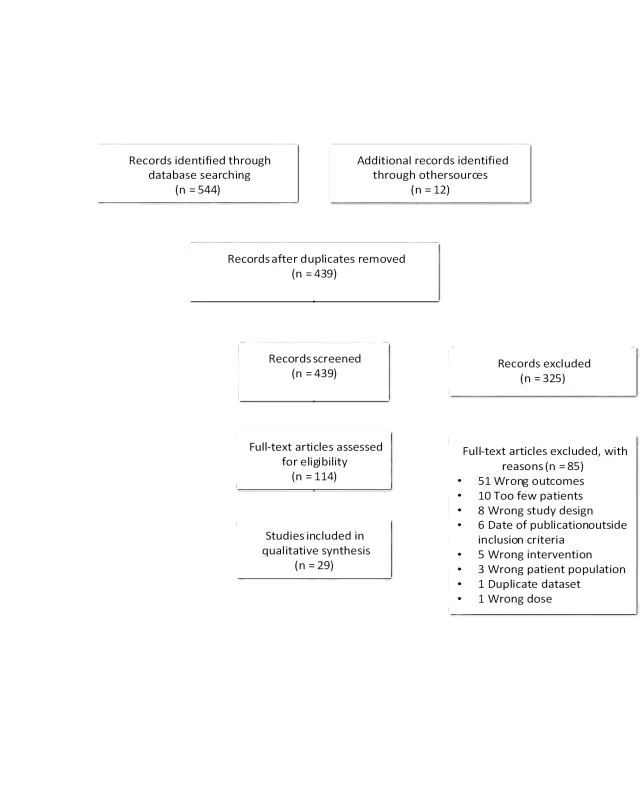
Appendix IV. Evidence Tables
Table 1: Hearing preservation with wait and watch
| Author/Year | Study Description | Data Class | Results and Conclusion |
| Gurewitz et al, 2022 | Objective: To investigate the relationship between hearing loss and tumor volumetric growth rates in patient with unilateral VS. Design: Retrospective chart review. Number of patients: 107 patients with unilateral VS serviceable hearing; 72 were AAO-HNS hearing class A. Follow-up: Median of 24.3 months (IQR 8.5-48.8 months). | III | Results: 77.6% (83/107) retained serviceable hearing at last followup, 69.4% (50/72) of those with hearing class A retained it. No significant association between baseline time to non-serviceable hearing and age. Time to non-serviceable hearing was associated with initial tumor volume and tumor growth rate. Authors Conclusion: Larger initial tumors and faster growth rates are associated with elevated risk of losing serviceable hearing. Comments and Conclusions: The retrospective nature of this study yields class III data. Many patients with stable or slow growing tumors demonstrated a decline in hearing function thus stratifying patients based on hearing loss risk is problematic. |
| Patel et al, 2020 | Objective: To ascertain relationship among VS tumor volume, growth and hearing loss. Design: Retrospective chart review, single center. Number of patients: 213 patients with serviceable hearing (AAO-HNS class A or B) and audiometric data were included. Follow-up: Median of 3 years. | III | Results: Rates of maintaining serviceable hearing at 2-, 4-, 6-, 8-, and 10-years following diagnosis were 88%, 74%, 67%, 65%, and 49%, respectively. Larger volume at diagnosis was associated with increased PTA and decreased WRS. Tumor growth was not significantly associated with time to non-serviceable hearing. Authors Conclusion: Larger initial tumor volume was associated with poorer hearing at baseline and with the development of non-serviceable hearing during observation. Comments and Conclusion: The retrospective nature of this study yields class III data. |
| Hunter et al, 2018 | Objective: To characterize risk of progression to non-serviceable hearing in patient with VS undergoing observation. Design: Retrospective case series in two tertiary care centers. Number of patients: 466 patients with serviceable hearing with AAO-HNS hearing class A or B. Follow-up: Median of 2.3 years | III | Results: 76.8% (358/466) patients retained serviceable hearing at median of 2.4 years (IQR 1.0-4.2). 23.2% (108/466) developed non-serviceable hearing at a median of 1.9 years following diagnosis. Each 10% decrease in WRS and 10 dB increase in PTA was associated with a 1.5-fold and 2-fold increased risk of developing non-serviceable hearing, respectively. Authors Conclusion: Good baseline PTA and WRS are associated with maintenance of serviceable hearing. Comments and Conclusions: The retrospective nature of this study yields class III data. Objectively quantified the risk of developing non-serviceable hearing based on patient’s presenting audiometric measures. |
| Kirchmann et al, 2017 | Objective: To report long-term occurrence of hearing loss in VS patients. Design: Retrospective chart review. Number of patients: 156 patients with intracanalicular VS managed conservatively. 73 of those has serviceable hearing class A or B, AAO-HNS. Follow-up: 9.5 years (range, 1-25 years). | III | Results: 34.2% (25/73) retained serviceable hearing at last followup. Tumor growth to extrameatal extension occurred in 23% of patients, with the majority occurring in the first years after diagnosis (4.6 years). Authors Conclusion: Serviceable hearing was preserved in 34% according to AAO-HNS and in 58% according to the WRS. Risk of hearing loss was small in patients with class A hearing at diagnosis. Comments and Conclusion: The retrospective nature of this study yields class III data. Also, there is potential selection bias with the selected tumor size population. |
| Milner et al, 2017 | Objective: To assess audiological outcomes in patient with growing VS managed conservatively or treated with SRS (12-13 Gy marginal dose, single fraction gamma knife). Design: Retrospective chart review, single center Number of patients: 15 patients treated conservatively and 27 treated with SRS with serviceable hearing. Follow-up: Mean 69.6 months for SRS and 71.7 for months conservative management | III | Results: No significant difference in deterioration of AAO-HNS and GR between the two groups. 15/42 patient overall maintained serviceable hearing at last followup: 53% (8/15) in the conservative management cohort and 26% (7/27) in the SRS group. Authors Conclusion: Similar audiological outcomes and hearing preservation for SRS and conservative management of VS. Comments and Conclusion: The retrospective nature of this study yields class III data. Rate of progression to loss of functional hearing was not significantly different between SRS and conservative management. |
| Elliot et al, 2015 | Objective: To compare hearing preservation between conservative treatment and SRT for patient with unilateral VS. Design: Retrospective case series, single institution. Number of patients: 123 patients with serviceable hearing, AAO-HNS hearing class A or B (98 underwent conservative therapy, 25 underwent SRT). SRT via LINAC of 31.25 Gy in 5 fractions or in one case 62.5 Gy in 25 fractions. Follow-up: median of 42.5 months for observation group and median of 44 months for SRT group. | III | Results: Hearing was preserved for median of 46 months at 51% (63/123) for both modalities with preservation of 54% (53/98) and 40% (10/25) for conservative observation and SRT, respectively. Multivariant model revealed that tumor growth status was an independent predictor for treatment modality with SRT. Median followup and hearing class were predictors of hearing preservation at the end of follow up. Authors Conclusion: No significant difference between the hearing survival of conservative versus SRT patients. Comments and Conclusion: The retrospective nature of this study yields class III data. Onset class A conferred significant hearing survival advantage over class B (62% vs 39%) at median of 46 months, irrespective of treatment modality. |
| Jethanamest et al, 2015 | Objective: To describe clinical outcomes of VS patients undergoing serial observation. Design: Retrospective chart review. Number of patients: 94 patients managed conservatively. 37 of those has serviceable hearing class A or B, AAO-HNS. Follow-up: 34.8 months median | III | Results: 75.7% (28/37) patients retained serviceable hearing at a median of 34.8 months. Median time to hearing loss worsening to non-serviceable level for 50% of patients was 76 months. Median time to hearing loss worsening to non-serviceable level for 50% of patients was 76 months. Authors Conclusion: Serial observation is a viable treatment strategy for select VS. Comments and Conclusion: The retrospective nature of this study yields class III data. |
| AAO-HNS, American Academy of Otolaryngology-Head and Neck Surgery; dB, decibel; IQR, interquartile; GR, Gardner-Robertson hearing classification; LINAC, linear accelerator; PTA, pure tone average; SRS, stereotactic radiosurgery; SRT, stereotactic radiotherapy; VS, vestibular schwannoma; WRS, word recognition score. | |||
Table 2: Hearing preservation with stereotactic radiosurgery
| Author/year | Study Description | Data Class | Results and Conclusion |
| Teyateeti et al, 2022 | Objective: To evaluate the outcomes of gamma knife SRS with 50% isodose line (IDL50) vs isodose 40% (IDL40). Design: Single institution SRS registry. Number of patients: 21 patients with serviceable hearing prior to SRS (8 in the IDL40 group and 13 in the IDL 50 group). Follow-up: 114 months. | III | Results: Hearing preservation rates overall were 84%, 72% and 39% at 2, 5, and 10 years, respectively. IDL40 treatment was significantly associated with higher hearing preservation rate with a longer hearing preservation time, 128 months, versus 82 months for IDL50. Authors Conclusion: Dose prescription at IDL40 or IDL50 provide excellent long-term tumor control and toxicity profile, however, IDL40 may be associated with improved long-term hearing preservation. Comments and Conclusion: The retrospective nature of this study yields class III data. Small sample size. Prescription dose of 12-14 Gy with IDL40 may have a more favorable outcome with improved hearing preservation. |
| Maksimoski et al, 2021 | Objective: To describe long-term hearing outcomes with modern SRS techniques for VS treatment. Design: Retrospective chart review, single center. Number of patients: 133 patients with serviceable hearing underwent SRS (11-16 Gy at the 50% isodose line). Follow-up: No mean or median available. Half of study had data beyond 5 years. | III | Results: Serviceable hearing was maintained in 27%, 24%, 13% and 4.1% of patients at 3-, 5-, 7- and 10- year follow up. Authors Conclusion: Stereotactic radiosurgery treatments for VS showed a continued reduction over time in serviceable hearing. Comments and Conclusion: The retrospective nature of this study yields class III data. Ranges for maximum cochlear dose were 3.7-19.9 Gy with a median of 9.7 Gy. |
| Ogino et al, 2021 | Objective: To evaluate whether hearing deterioration during observation reduced serviceable hearing after SRS (Median marginal dose of 12.5 Gy, range of 11-15 Gy) in VS patients with useful hearing. Design: Retrospective chart review, single institution. Number of patients: 100 patients with GR I were observed for median of 17.4 months. 67 patients remained GR I and 33 deteriorated to GR II. All then underwent SRS. Follow-up: Median of 4.4 years. | III | Results: At the last followup, 63% (42/67) of patients that started with GR I maintained serviceable hearing compared to 24% (8/33) of those who started with GR II. The overall serviceable hearing preservation rate was 65.9% at 3 years, 52.5% at 5 years and 40.5% at 10 years. In multivariate analysis, younger age (<55 years) and GR grade I were significantly associated with improved serviceable hearing preservation rates. Authors Conclusion: SRS before hearing deterioration was recommended for hearing preservation. Comments and Conclusion: The retrospective nature of this study yields class III data. Stereotactic radiosurgery has more favorable hearing preservation outcomes long term in younger patients with hearing maintained at GR I at time of treatment. |
| Ogino et al, 2021 | Objective: To evaluate outcomes of SRS for intracanalicular VS. Design: retrospective chart review, single center. Number of patients: 120 patients with serviceable hearing (GR I or II) with a median marginal dose of 12.5 Gy (range 11-25 Gy) and median cochlear dose 3.2 Gy (range 0.8-6.9 Gy). Follow-up: 43 months (range 6-232 months). | III | Results: 77%, 64%, and 27% patient had preservation of serviceable hearing at 3, 5 and 10 years. Better hearing and smaller tumor volume at time of SRS were significantly associated with improved hearing preservation. Authors Conclusion: Patient with initially better hearing and smaller VSs has enhanced serviceable hearing preservation during long-term followup of up to 10 years post SRS. Comments and Conclusion: The retrospective nature of this study yields class III data. Selection bias for tumor size. |
| Ogino et al, 2021 | Objective: To report role of SRS as primary management option for Koos grade IV VS. Design: Retrospective chart review, single center. Number of patients: 170 patients in total. 42 patients with serviceable hearing and available data (GR grade I or II) were treated with median margin dose of 12.5 Gy (range 10.5-22 Gy). Follow-up: 75.8 months median. | III | Results: Preservation of serviceable hearing was 58%, 50% and 36% at 3, 5 and 7 years. Younger age (<60 years) and initial GR grade I were associated with improved serviceable hearing preservation rate. Authors Conclusion: Single session SRS prevented need for delayed resection even in large volume VS with lower serviceable hearing preservation rates. Comments and Conclusion: The retrospective nature of this study yields class III data. SRS in large VS is a reasonable primary management for elderly patients with many medical comorbidities. |
| Wage et al, 2021 | Objective: To evaluate long-term outcomes of VS patients treated with GKS. Design: Retrospective chart review, single center study. Number of patients: 53 patients had serviceable hearing (GR I and II) at treatment time (median marginal dose of 12.5 Gy to the 50% isodose line. Follow-up: No mean or median available. | III | Results: 40% (21/53) maintain serviceable hearing at last followup with median time to hearing loss at 19 months (range 3-158). Actuarial median time to loss of serviceable hearing 3.4 years with actuarial hearing preservation at 2, 5, and 10 years of 66.5%, 43.1%, and 37.6%, with rate of hearing loss correlating with maximum cochlea and modiolus doses. Authors Conclusion: Modern GKS is a safe and effective treatment for VS on long-term follow-up. Comments and Conclusion: The retrospective nature of this study yields class III data. |
| Han et al, 2020 | Objective: To report long-term outcomes of patients with small to medium sized VS with serviceable hearing undergoing SRS or MS and evaluate potential prognostic factors for hearing preservation. Design: Retrospective chart review, single institution Number of patients: 51 patients total with serviceable hearing and a tumor <25 mm in greatest dimension; 21 patients underwent MS and 30 patients underwent GKS. Follow-up: 5 years. | III | Results: 53% (16/30) of patients treated with GKS and 71% (15/21) of patient treated with MS maintained serviceable hearing at last followup. Difference was not found to be statistically significant. Authors Conclusion: MS is more suitable for younger healthy patients with serviceable hearing preoperatively and medial type VS. SRS was more suitable for elderly patients with poor physical status and preoperative hearing AAO-HNS class A. Comments and Conclusion: The retrospective nature of this study yields class III data. Number of included patients was small. |
| Johnson et al, 2019 | Objective: To report long-term outcomes of SRS as a treatment modality for VS. Design: Retrospective chart review, single institution. Number of patients: 326 patients with serviceable hearing (GR I or II) that received SRS (median marginal dose of 13 Gy, range 12-25 Gy). Follow-up: Median 5.2 years (range 1-25 years). | III | Results: The last audiological examination demonstrated that 196/326 (60.1%) retained SH. Serviceable hearing preservation rates were 90% at 1 year, 77% at 3 years, 68% at 5 years, 63% at 7 years, and 51% at 10 years. Younger age, GR grade 1 at SRS, and absence of subjective vestibulopathy were associated with improved serviceable hearing preservation. Authors Conclusion: Serviceable hearing at 10 years was maintained in 76% of patients younger than 45 years of age and tumors smaller than 0.56 cc in volume at the time of the SRS. Comments and Conclusion: The retrospective nature of this study yields class III data. Study included an equal distribution of all 4 Koos classes. Analysis vestibulopathy as a separate predictor of hearing outcome added an interesting finding. |
| Prabhuraj et al, 2019 | Objective: To find hearing preservation and tumor control rates for small and medium sized VS treated with GKS. Design: Single center, retrospective cohort study. Number of patients: 77 patients with GR grade I or II who underwent GKS (median marginal dose of 12 Gy, range 11.5-14). Follow-up: 30 months. | III | Results: Hearing preservation rate was 79.2% (61/77). This was not affected by tumor volume. On multivariate analysis, age >40, pre-GKS PTA <30 dB and GR grade I were independent predictors of better hearing preservation. Authors Conclusion: GKS is an effective treatment for patients with small VSs with retained serviceable hearing and good tumor control. Comments and Conclusion: The retrospective nature of this study yields class III data. Well-conducted multivariate data analysis. Mean cochlear dose ranged from 2.3-6.5 Gy with a range of 3.75 Gy. |
| Frischer et al, 2018 | Objective: To present long-term follow up data on VS patients treated with GKS. Design: Single center retrospective chart review. Number of patients: 132 patients had GR grade I or II serviceable hearing at time of GKS. Follow-up: Median 5.1 years. | III | Results: At the last follow-up, functional hearing was preserved in 55% of patients who had been classified. Hearing preservation rates were 53%, 34%, and 34% at 5, 10, and 15 years after GKS. On multivariate regression analysis, GR hearing grade prior to GKRS and the median dose to the cochlea were independent predictors of the GR grade at follow-up. Authors Conclusion: The retrospective nature of this study yields class III data. GKS should be a primary treatment for early stage small to medium sized VS. Authors do not recommend undertreating intracanalicular tumors in favor of low cochlear doses. Comments and Conclusion: No tumor size selection bias. Patients who received a median cochlear dose > 6 Gy showed a significantly higher rate of nonserviceable hearing at the last follow-up than that in patients in whom the median cochlear dose was <6 Gy. |
| Van Linge et al, 2018 | Objective: To assess change in hearing loss after SRT and identify prognostic factors affecting hearing preservation. Design: Retrospective chart review, single center. Number of patients: 37 patient received single fraction of 12 Gy and 20 patients received FSRT with 30 fraction of 1.8 Gy (all with GR grade I or II) Follow-up: 36 months for SRS group and 63 months for FSRT group. | III | Results: One year after treatment, 84% of SRS and 71% of FRST has preservation of serviceable hearing. At 3 years, hearing was preserved at 27% for SRS and 50% for FSRT. Preservation of hearing did not differ significantly between the two groups. On multivariate regression, restricted cochlear V90 and worse pretreatment PTA were associated with progression of serviceable hearing loss. Authors Conclusion: Hearing deteriorates after SRT for VS. Most rapid decline occurs shortly after treatment without significant difference between SRS and FSRT. Comments and Conclusion: The retrospective nature of this study yields class III data. Better baseline PTA does not protect against changes in PTA however it provides and protective effect for overall preservation of serviceable hearing. |
| Bowden et al, 2017 | Objective: To correlate radiographic appearance of VS before SRS with delayed volumetric response. Design: Single center retrospective chart review. Number of patients: 111 patients with serviceable hearing (GR grade I or II) at SRS (median dose 12.5 Gy, range 11-13 Gy). Follow-up: Median of 37 months. | III | Results: Serviceable hearing was maintained in 82.2% and 61.5% patients at 2- and 5-years post SRS. 85% (29/34) of patients with 100% SDS at SRS maintained serviceable hearing at 5 years. There was no significant difference in auditory outcomes between the macrocystic and microcystic groups. Six patients (21.4%) who initially tested as GR grade 3 increased by >10% and moved into the serviceable hearing range at last follow-up. Authors Conclusion: SRS provided VS tumor control in >95% of patients, regardless of radiographic characteristics. Comments and Conclusion: The retrospective nature of this study yields class III data. Selection bias for cystic tumors. No statistical difference in hearing preservation rates regardless of the radiological characteristics of the VS. |
| Akpinar et al, 2016 | Objective: To evaluate effect of SRS on long-term hearing preservation in VS patients. Design: Retrospective chart review, single center. Number of patients: 88 patients with GR grade I hearing at time of SRS: 57 underwent SRS early (≤2 years from diagnosis) and 31 underwent late SRS (>2 years after diagnosis). Follow-up: median of 75 months | III | Results: At 5 years after SRS, 88% of early treatment group retained serviceable hearing and 77% retained normal hearing compared with 55% with serviceable hearing and 33% with normal hearing in the late treatment group. Authors Conclusion: SRS within two years after VS diagnosis in normal hearing patients resulted in improved retention of all hearing measures compared with later SRS. Comments and Conclusion: Late treatment group has pre-SRS significantly higher PTA and slightly lower SDS which may have biased the results. |
| Golfinos et al, 2016 | Objective: To conduct matched cohort analysis to evaluate hearing outcomes in patients treated with SRS or MS. Design: Retrospective chart review, single center. Number of patients: 21 patients in SRS group (maximum marginal and cochlear dose of 14 Gy and 6.5 Gy) and 21 patients in MS group (matched by age and tumor size), all with class A hearing, AAO-HNS. Follow-up: 43.7 months for MS and 30.3 months for SRS. | III | Results: 43% (9/21) in the MS group had serviceable hearing at last followup vs 86% (18/21) in the SRS group. Preservation of preoperative Class A hearing status was also better after SRS than MS (14.3% for MS vs 42.9% for SRS). Authors Conclusion: VS resection or radiosurgery for tumors <2.8 cm in diameter was associated with low overall morbidity. SRS was associated with improved hearing rates but followup period was shorter. Comments and Conclusion: The retrospective nature of this study yields class III data. Survival curve analysis shows the preservation of preoperative hearing status converges by month 60. Loss of postoperative hearing was delayed in the SRS cohort. |
| Klijn et al, 2016 | Objective: To report long-term outcomes of GKS for VS patients. Design: Retrospective chart review, single center. Number of patients: 420 included in the study with 71 patients with serviceable hearing underwent GKS (11 Gy marginal dose). Follow-up: Median of 5.1 years. | III | Results: Serviceable hearing was maintained in 64% and 42% of patients at 3- and 5- year follow up. Authors Conclusion: Rates of hearing preservation were comparable to those in the literature. Comments and Conclusion: The retrospective nature of this study yields class III data. |
| Watanabe et al, 2016 | Objective: to report long-term outcomes of SRS for VS. Design: Multicenter, retrospective chart review. Number of patients: 183 patients included, 66 of which has serviceable hearing (GR I or II) and available audiometric data pretreatment with SRS (median marginal dose 12 Gy, range 8.8-15.5 Gy; median cochlear dose was 4.1 Gy and range 2.3-5.7 Gy). Follow-up: 59 months. | III | Results: At last follow-up, 35% (23/66) of patients with serviceable hearing before SRS had preserved hearing. Actuarial serviceable hearing preservation rates were 49%, 24%, and 12% at 5-, 10-, and 15- years post-SRS month, respectively. Older age (≥65 years), larger tumor volume (≥8 cm3) and higher cochlear dose (mean cochlear dose >4.2 Gy) are unfavorable factors for hearing preservation. Authors Conclusion: Hearing preservation was not a satisfactory long-term outcome of SRS. Comments and Conclusion: The retrospective nature of this study yields class III data. PTA was only audiometric follow-up method, without tracking SDS thus the results may be overestimated. |
| Horiba et al, 2015 | Objective: To evaluate factors associated with hearing preservation after low dose GKS. Mean marginal dose of 11.9 Gy (range, 11-12 Gy). The doses for brain stem, cranial nerves, and cochlea were kept below 14 Gy, 12 Gy, and 4 Gy, respectively. Design: Retrospective chart review, single institution. Number of patients: 49 patients with GR grade I (22 cases) and II (27 cases). Follow-up: Median and mean of 56 and 55 months. | III | Results: Serviceable hearing preserved at 57% (28/49) at the last followup. Patients who lost serviceable hearing after GKS experienced the loss within 1 (6/21, 29%), 2 (14/21, 67%), and 3 (18/21, 86%) posttreatment years. In cases with and without extension of the intrameatal part up to fundus, preservation of the serviceable hearing was at 40% and 60% of cases, respectively, however the difference in not statistically significant (odds ratio, 2.6; 95% confidence interval, 0.5–12.4). Authors Conclusion: Low-dose GKS of vestibular schwannoma preserves serviceable hearing in more than half of the patients at the 3-year followup. Comments and Conclusion: The retrospective nature of this study yields class III data. No multivariate analysis presented. |
| Mousavi et al, 2015 | Objective: To evaluate hearing outcomes at 2 and 3 years in GR grade I patients who underwent GKS for VS. Design: Retrospective chart review, single institution. Number of patients: 68 patients total with GR grade I hearing divided into two groups. Group A: 25 patients with no subjective hearing loss (median PTA was 12 dB, and median SDS was 100). Group B: 43 patients all of whom reported subjective hearing dysfunction (median PTA 22 dB and SDS 90%). All underwent SRS with marginal dose of 12.5 Gy. Follow-up: 33 months for group A and 35 months for group B. | III | Results: All 25 patients (100%) in group A retained serviceable hearing at the last follow-up, only 24/43 group B patients (55%) maintained serviceable hearing. Thus, 72.1% (49/68) of hearing was preserved at 3 years in both groups combined. Serviceable hearing retention were 100% for group A compared with 81% at 1 year, 60% at 2 years, and 57% at 3 years after GKS for group B patients. Group A patients had significantly higher rates of hearing preservation in either GR class I or GR class II. Patients with a PTA <15 dB before GKS had significantly higher rates of serviceable hearing preservation. Authors Conclusion: Modification of the GR I hearing classification into 2 groups (group A, those with no subjective hearing loss and a PTA <15 dB; and group B, those with subjective hearing loss and a PTA >15 dB) may be useful to help predict hearing preservation rates at 2 to 3 years after GKS. Comments and Conclusion: The retrospective nature of this study yields class III data. No meaningful long-term data beyond 3 years of followup. Median cochlear dose (4.0-4.15 Gy) had no statistical relationship to hearing status at last followup. |
| AAO-HNS, American Academy of Otolaryngology-Head and Neck Surgery; dB, decibel; FSRT, fractionated stereotactic radiotherapy; IQR, interquartile; GKS, gamma knife surgery; GR, Gardner–Robertson hearing classification; IDL50, 50% isodose line; IDL40, 40% isodose line; LINAC, linear accelerator; MS, microsurgery; PTA, pure tone average; SRS, stereotactic radiosurgery; SRT, stereotactic radiotherapy; SDS, speech discrimination score; VS, vestibular schwannoma. | |||
Table 3: Hearing preservation with microsurgery
| Author/Year | Study Description | Data Class | Results and Conclusion |
| Ichimasu et al, 2020 | Objective: To report long-term hearing preservation in patient with serviceable hearing immediately after VS surgery and to identify prognostic of hearing function. Design: Retrospective chart review, multicenter. Number of patients: 91 patients with class A or B hearing (AAO-HNS). Follow-up: 63 months mean. | III | Results: At the last followup, 87% (79/91) patient maintained serviceable hearing. 61% (39/64) patient who were in class A deteriorated to class B. Rate of maintenance of useful earing at 1, 3, 5, 8 and 10 years was 98.9%, 98.9%, 94.1%, 74.9% and 64.6%, respectively. Authors Conclusion: Useful hearing was lost in 13% of patients and hearing class deteriorated in 55% during followup without signs of tumor recurrence. Comments and Conclusion: The retrospective nature of this study yields class III data. Selection bias is present; however, this article provides an estimate of hearing preservation in the long-term follow up in patient with preserved hearing post surgically. |
| Dowling et al, 2019 | Objective: To evaluate hearing outcomes in patients with serviceable hearing following microsurgical resection in tumors confined to the IAC or with ≤ 1 cm of CPA extension. Design: Retrospective cohort, tertiary academic referral center. Number of patients: 43 patients with unilateral vestibular schwannoma and AAO-HNS class A or B. Follow-up: Median 3.3 years | III | Results: 19% (8/43) patients developed non-serviceable hearing at median 4.1 years followup. Rate of maintaining serviceable hearing at 5 years was 81%. Tumor control was achieved in 95% (41/43) patients. Authors Conclusion: Microsurgical resection provides durable tumor control and long-term hearing in patients with AAO-HNS class A or B hearing postoperatively. Comments and Conclusion: The retrospective nature of this study yields class III data. Proactive microsurgical resection for small tumors with good hearing can be a way to preserve preoperative hearing levels however a selection bias is present here. |
| Zhu et al, 2018 | Objective: To report long-term hearing outcomes after retrosigmoid tumor removal for small VS in the IAC or <15 mm CPA tumor. Design: Retrospective chart review, single center. Number of patients: 70 patients with class A or B hearing (AAO-HNS) underwent surgery. Follow-up: 4 years mean follow up for surgery group. | III | Results: 47% (33/70) of patient with serviceable hearing at time of surgery achieved hearing preservation at 4-year followup. In all surgery patients (n = 110), the postoperative rate for preservation of serviceable hearing was 85.7% among those with preoperative Class A hearing, 46.5% among those with Class B hearing. Authors Conclusion: Better preoperative hearing predicted a higher rate of postoperative hearing preservation. Comments and Conclusion: The retrospective nature of this study yields class III data. Authors defined serviceable hearing as AAO-HNS classes A, B and C but they provided all data for the surgical subgroup so adjustment to include only classes A and B was possible. |
| Ahmed et al, 2017 | Objective: to analyze postoperative long-term hearing outcome data in VS patients undergoing MCF approach. Design: Retrospective chart review, single center. Number of patients: 71 patients with serviceable hearing (AAO-HNS, class A or B) and long-term data. Follow-up: 7 years mean. | III | Results: Hearing preservation was 82% at 3-5 years, 67% at 6-8 years, 68% at 9-11 years and 18% at 12+ years. The rate of WRS preservation was 98% at 3-5 years. Patients with preoperative class A hearing had significantly higher rates of successful hearing preservation in all postoperative intervals. Authors Conclusion: In patients with preserved serviceable hearing immediately postoperatively, hearing loss occurs at different rates over time. Comments and Conclusion: The retrospective nature of this study yields class III data. |
| AAO-HNS, American Academy of Otolaryngology-Head and Neck Surgery; CPA, cerebropontine angle; ICA, intracanalicular; VS, vestibular schwannoma. | |||
Table 4. Comparison of PICO Elements in 2018 and 2022 Guidelines
| PICO Element | 2018 Guideline | 2022 Guideline |
| Population (P) | Adults with sporadic vestibular schwannomas who have serviceable hearing. No specific age limit mentioned, generally referring to adult patients. | Adults with sporadic vestibular schwannomas who have serviceable hearing at the time of diagnosis. Like the 2018 guideline, there is no specific age cut-off, but the focus remains on the adult population. |
| Intervention (I) | – Radiation Therapy: Single-fraction radiation using ≤ 13 Gy to the tumor margin. – Microsurgery: Microsurgical resection via middle cranial fossa or retrosigmoid approach for small to medium-sized tumors (< 2 cm). – Observation: Monitoring small to medium-sized tumors with serial imaging without immediate intervention. | – Radiation Therapy: Single-fraction stereotactic radiosurgery with specified dosages, including ≤ 13 Gy marginal dose and a recommended ≤ 4 Gy cochlear dose when possible. – Microsurgery: Criteria for selecting surgical approach based on tumor size and location, with emphasis on surgical techniques aimed at hearing preservation. – Observation: Emphasized watchful waiting with a focus on baseline characteristics like tumor size and initial hearing status. |
| Comparison (C) | Comparing outcomes of different treatment modalities (radiosurgery, microsurgery, observation), particularly focusing on hearing preservation across adult patients. | More detailed comparisons involving prognostic factors and treatment specifics, such as radiation dosages and surgical approaches, aimed at refining treatment efficacy predictions for adults. |
| Outcome (O) | Probability of maintaining serviceable hearing at various time intervals post-treatment (2, 5, and 10 years). | Provides detailed statistical probabilities of maintaining serviceable hearing post-treatment, integrating predictive factors for a more nuanced evaluation. |
Table 5. Comparison of Recommendations: 2018 vs. 2022 Guidelines
| Modality/Aspect | 2018 Recommendations | 2022 Recommendations |
| Radiosurgery | – Moderately high rate (>50% to 75%) of hearing preservation at 2 and 5 years, moderately low rate (>25% to 50%) at 10 years. – For AAO-HNS class A/GR I hearing: High rate (>75% to 100%) of hearing preservation at 2 years, moderately high (>50% to 75%) at 5 years, and moderately low (>25% to 50%) at 10 years. – Predictive factors for maintaining serviceable hearing comprise better preoperative word recognition/pure tone thresholds, smaller tumor size, marginal dose ≤12 Gy, cochlear dose ≤4 Gy. | – Moderately high rate (>50% to 75%) of hearing preservation at 2 and 5 years, moderately low rate (>25% to 50%) at 10 years. – Overall estimated rates of maintaining serviceable hearing after radiosurgery are 71% at 2 years, 59% at 5 years, 38% at 10 years. – Predictive factors for maintaining serviceable hearing comprise cochlear dose ≤ 4 Gy, marginal dose ≤ 13 Gy, and better baseline hearing level including AAO-HNS class A or GR I status. |
| Microsurgery | – Moderately low rate (>25% to 50%) of hearing preservation immediately after surgery and at 2, 5, and 10 years. – For AAO-HNS class A/GR I hearing: Moderately high rate (>50% to 75%) immediately, at 2 and 5 years, and moderately low (>25% to 50%) at 10 years. – Predictive factors for maintaining serviceable hearing comprise good preoperative word recognition/pure tone thresholds, smaller tumor size commonly <1 cm. | – Moderately low rate of hearing preservation post-surgery (>25% to 50%): 43% immediately, 48% at 2 years, 40% at 5 years, and 32% at 10 years. – Predictive factors for maintaining serviceable hearing comprise better baseline hearing level, including AAO-HNS class A and GR I status, smaller tumor size, and presence of a fundal CSF fluid cap. |
| Observation | – High rate (>75% to 100%) of hearing preservation at 2 years, moderately high (>50% to 75%) at 5 years, and moderately low (>25% to 50%) at 10 years. – For AAO-HNS class A/GR I hearing: High rate at 2 years (>75% to 100%), and moderately high at 5 years (>50% to 75%). Insufficient data for 10 years. | – High rate of hearing preservation at 2 years (>75% to 100%), moderately high rate at 5 years (>50% to 75%), and moderately low rate at 10 years (>25% to 50%). – Overall estimated rates of maintaining serviceable hearing with observation are 78% at 2 years, 59% at 5 years, 47% at 10 years. – Predictive factors for maintaining serviceable hearing comprise better baseline hearing level, including AAO-HNS class A or GR I status, and absence of tumor growth during observation. |
Appendix V. Conflicts of Interest
| Task Force Member | Disclosure |
| Julie Honaker PhD, AuD | Nothing to Disclose |
| Ben Allen Strickland, MD | Nothing to Disclose |
| Eric J. Lehrer, MD | Servier Pharmaceuticals, Novocure Inc. |
| Sheryl Green, MBBCh | Nothing to Disclose |
| John P. Marinelli MD | Medtronic |
| Christopher S. Graffeo MD, MS | Nothing to Disclose |
| Isabelle M. Germano, MD, MBA | Brianlab |
| Mateo Ziu, MD | Omniscient Neurotechnology America Ltd; GT Medical Technologies, Inc |
| Walavan Sivakumar, MD | Stryker Corporation |
| Sherwin Tavakol, MD | IRRAS USA, Inc.; Globus Medical, Inc. |
| Lucas Paul Carlstrom, MD, PhD | Kuros Biosciences USA, Inc |
| Jamie J. Van Gompel, MD | Medtronic, Cadence |
| Ian Dunn, MD | Nothing to Disclose |
| Jeffrey J. Olson, MD | Verastem, Inc., Research Grant American Cancer Society, Editorial Consultant; Azurity Pharmaceuticals, Inc. |
| Ghazal S. Daher MD | Nothing to Disclose |
| Matthew L. Carlson, MD | Cochlear Americas, Advanced Bionics, Stryker Corporation; iotaMotion, Inc.; Stryker Corporation |
| Neil S. Patel, MD | Cochlear Americas, Zeiss, Viridian Therapeutics, IotaMotion, Inc. |
| Michael Sughrue, MD | Omniscient Neurotechnology America Ltd |
| Constantinos G. Hadjipanayis, MD, PhD | Stryker Corporation; Integra LifeSciences Corporation; Omniscient Neurotechnology America Ltd |
| Jeffrey Jacob, MD | Stryker Corporation; KLS; Synthes |
5. The Role of Imaging in the Diagnosis and Management of Patients with Vestibular Schwannomas
Sponsored by: Congress of Neurological Surgeons (CNS) and the AANS/CNS Tumor Section
Endorsed by: Joint Guidelines Committee of the American Association of Neurological Surgeons (AANS) and the Congress of Neurological Surgeons (CNS)
Authors:
Ian F. Dunn, MD1, Wenya Linda Bi, MD, PhD1, Srinivasan Mukundan, MD, PhD2, Bradley N. Delman, MD3, Jonathan Parish, MD4, Tyler Atkins, MD5, Anthony L. Asher, MD4, Jeffrey J. Olson, MD5
1. Department of Neurosurgery, Brigham and Women’s Hospital, Harvard Medical School, Boston, Massachusetts, USA
2. Division of Neuroradiology, Brigham and Women’s Hospital, Boston, Massachusetts, USA
3. Department of Radiology (Neuroradiology), Icahn School of Medicine at Mount Sinai, New York, New York, USA
4. Carolinas Medical Center, Charlotte, North Carolina, USA
5. Carolina Neurosurgery & Spine Associates, Charlotte, North Carolina, USA
6. Department of Neurosurgery, Emory University School of Medicine, Atlanta, Georgia, USA
Correspondence:
Ian F. Dunn, MD
Department of Neurosurgery
Brigham and Women’s Hospital
Boston, Massachusetts, USA
IDUNN@PARTNERS.ORG
Keywords: Acoustic neuroma, advanced imaging, cystic, growth rate, facial nerve, MRI, vestibular schwannoma
No part of this manuscript has been published or submitted for publication elsewhere.
Abbreviations
AN: Acoustic neuroma
AAO-HNS: American Academy of Otolaryngology-Head and Neck Surgery
CISS: Constructive interference in steady state
CPA: Cerebellopontine angle
CSF: Cerebrospinal fluid
CT: Computed tomography
DTI: Diffusion tensor imaging
DTT: Diffusion tensor tractography
DWI: Diffusion weighted imaging
EMG: Electromyography
FFE: Fast-field echo
FIESTA: Fast imaging employing steady-state acquisition
FLAIR: Fluid attenuated inversion recovery
FN: Facial nerve
FSE: Fast spin echo
GRE: Gradient echo
GTR: Gross total resection
HB: House–Brackmann
HP: Hearing preservation
IAC: Internal auditory canal
MF: Middle fossa
MPRAGE: Magnetization prepared rapid acquisition gradient echo
MRI: Magnetic resonance imaging
NEA: Nonenhancing area
NF2: Neurofibromatosis type 2
NGR: No growth rate
NTR: Near-total resection
PRESTO: Principles of echo-shifting with a train of observations
RS: Retrosigmoid
SIMCAST: Segment-interleaved motion-compensated acquisition in steady state
SRS: Stereotactic radiosurgery
SSHL: Sudden sensorineural hearing loss
STR: Subtotal resection
TSE: Turbo spin echo
TL: Translabyrinthine
VDE: Velocity of diametric expansion
VDT: Volume doubling time
VS: Vestibular schwannoma
VSTR: Vestibular schwannoma tumor remnant
Abstract
Question 1
What sequences should be obtained on MRI to evaluate vestibular schwannomas before and after surgery?
Target Population
Adults with vestibular schwannomas
Recommendation
Initial Preoperative Evaluation
Level 3: Imaging used to detect vestibular schwannomas should use high-resolution T2-weighted and contrast-enhanced T1-weighted MRI.
Level 3: Standard T1, T2, FLAIR, and DWI MR sequences obtained in axial, coronal, and sagittal plane may be used for detection of vestibular schwannomas.
Preoperative Surveillance
Level 3: Preoperative surveillance for growth of a vestibular schwannoma should be followed with either contrast-enhanced 3D T1 MPRAGE or high-resolution T2 (including CISS or FIESTA sequences) MR imaging.
Postoperative Evaluation
Level 2: Postoperative evaluation should be performed with post-contrast 3D T1 MPRAGE, with nodular enhancement considered suspicious for recurrence.
Question 2
Is there a role for advanced imaging for facial nerve detection preoperatively (eg, CISS/FIESTA or DTI imaging)?
Target Population
Adults with proven or suspected vestibular schwannomas by imaging
Recommendation
Level 3: T2-weighted MRI may be used to augment visualization of the facial nerve course as part of preoperative evaluation.
Question 3
What is the expected growth rate of vestibular schwannomas on MRI, and how often should they be imaged if a “watch and wait” philosophy is pursued?
Target Population
Adults with suspected vestibular schwannomas by imaging
Recommendation
Level 3: MRIs should be obtained annually for 5 years, with interval lengthening thereafter with tumor stability.
Question 4
Do cystic vestibular schwannomas behave differently than their solid counterparts?
Target Population
Adults with vestibular schwannomas with cystic components
Recommendation
Level 3: Adults with cystic vestibular schwannomas should be counseled that their tumors may more often be associated with rapid growth, lower rates of complete resection, and facial nerve outcomes that may be inferior in the immediate postoperative period but similar to non-cystic schwannomas over time.
Question 5
Should the extent of lateral internal auditory canal (IAC) involvement be considered by treating physicians?
Target Population
Adult patients with a vestibular schwannomas
Recommendation
Level 3: The degree of lateral IAC involvement by tumor adversely affects facial nerve and hearing outcomes and should be emphasized when interpreting imaging for preoperative planning.
Question 6
How should patients with neurofibromatosis type 2 (NF2) and vestibular schwannoma be imaged and over what follow-up period?
Target Population
Adult patients with neurofibromatosis type 2 and vestibular schwannomas
Recommendation
Level 3: In general, vestibular schwannomas associated with NF2 should be imaged (similar to sporadic schwannomas) with the following caveats:
1. More frequent imaging may be adopted in NF2 patients because of a more variable growth rate for vestibular schwannomas, and annual imaging may ensue once the growth rate is established.
2. In NF2 patients with bilateral vestibular schwannomas, growth rate of a vestibular schwannoma may increase after resection of the contralateral tumor, and therefore, more frequent imaging may be indicated, based on the non-operated tumor’s historical rate of growth.
3. Careful consideration should be given to whether contrast is necessary in follow-up studies or if high-resolution T2 (including CISS or FIESTA-type sequences) MRI may adequately characterize changes in lesion size instead.
Question 7
How long should vestibular schwannomas be imaged after surgery, including after gross total, near total, and subtotal resection?
Target population
Adult patients with vestibular schwannomas followed after surgery
Recommendation
Level 3: For patients receiving gross total resection, a postoperative MRI may be considered to document the surgical impression and may occur as late as 1 year after surgery. For patients not receiving gross total resection, more frequent surveillance scans are suggested; annual MRI scans may be reasonable for 5 years. Imaging follow-up should be adjusted accordingly for continued surveillance if any change in nodular enhancement is demonstrated.
Introduction
Vestibular schwannomas (VSs) are the most common intracranial nerve sheath tumor, arising from the vestibular division of the vestibulocochlear (VIIIth) nerve. VSs are usually sporadic in origin but may also arise in the context of neurofibromatosis type 2 (NF2) and present with a familiar constellation of symptoms including but not limited to, hearing loss, dizziness, vertigo, and, in larger tumors, symptoms related to cerebellar or brainstem compression. Management of VSs has classically included watchful waiting with surveillance imaging, treatment with microsurgical resection, or radiotherapy in one of several forms. Symptoms, tumor size, and specific patient and surgeon characteristics and preferences influence the choice of which treatment is adopted. While these broad themes are considered from center to center, considerable variation in treatment patterns exists among practitioners.
The detection of VSs is usually done using MRI, which provides unparalleled radiographic analysis and confers opportunities to establish an understanding of the natural history in cases where conservative management is pursued. The growth rates of surgical remnants may also be followed reliably using MRI. Moreover, specific details of tumor makeup (including the extent of intracanalicular involvement and the presence of cystic components, among others) can be discerned, and novel MRI sequences may also be applied to their radiographic assessment.
The following review was performed to provide a set of evidence-based recommendations for the use of imaging in the management of patients with VSs.
Objectives
This article aims to critically analyze the primary literature regarding the role of imaging in the management of VSs based on the following questions:
1. What sequences should be obtained on MRI to evaluate vestibular schwannomas before and after surgery?
2. Is there a role for advanced imaging for facial nerve detection preoperatively? (eg, CISS/FIESTA or DTI imaging)
3. What is the expected growth rate of vestibular schwannomas on MRI, and how often should they be imaged if a “watch and wait” philosophy is pursued?
4. Do cystic vestibular schwannomas behave differently than their solid counterparts?
5. Should the extent of lateral internal auditory canal (IAC) involvement be considered by treating physicians?
6. How should patients with neurofibromatosis type 2 (NF2) and vestibular schwannomas be imaged and over what follow-up period?
7. How long should vestibular schwannomas be imaged after surgery, including after gross total, near total, and subtotal resection?
Methods
Process Overview
The evidence-based clinical practice guideline taskforce members and the Joint Tumor Section of the American Association of Neurological Surgeons (AANS) and the Congress of Neurological Surgeons (CNS) conducted a systematic review of the literature relevant to the management of VSs. Additional details of the systematic review are provided below and within the introduction and methodology chapter of the guideline (here).
During the development process, the panel participated in a series of conference calls and meetings. Multiple iterations of written review were conducted by the individuals of the panel and the AANS/CNS Joint Guidelines Committee prior to approval. A list of members of the guideline task force can be found in the guideline introductory publication.
Study Selection and Eligibility Criteria
A total of 2070 citations were manually reviewed. Two independent reviewers evaluated and abstracted full-text data for each article. Citations focused on the imaging of VSs in adult patients largely in the MRI era (January 1, 1990 to December 31, 2014), published in English, were considered.
- Investigated patients suspected of having VSs
- Patients ≥18 years of age
- Was of humans
- Published between January 1, 1946 and December 31, 2014
- Quantitatively presented results
- Was not an in vitro study (for novel molecular markers, in vitro studies were included on patient samples)
- Was not a biomechanical study
- Was not performed on cadavers
- Was published in English
- Was not a, meeting abstract, editorial, letter, or commentary
- Studies may include mixed pathology, however the data pertaining to acoustic neuromas (ANs)/VSs was abstractable from the paper.
- >5 patients or patient samples
Systematic reviews, guidelines, or meta-analyses conducted by other authors were not included in this guideline creation. These documents were developed using different inclusion criteria than those specified in this guideline. Therefore, they may have included studies that do not meet the inclusion criteria stated above.
Search Strategies
The task force collaborated with a medical librarian to search for articles published between January 1, 1990 and December 31, 2014. The following electronic databases were searched: PubMed and Cochrane Central. Strategies for searching electronic databases were constructed by the evidence-based clinical practice guideline taskforce members and the medical librarian using previously published search strategies to identify relevant studies (Figure 1; Table 1). The guideline committee also examined lists of included and excluded studies for errors and omissions.
Data Collection Process and Assessment of Bias
Abstracts that met the inclusion criteria were retrieved in full text form and evaluated for confirmation that they met criteria as suggested by prior abstract review. The information was then used for construction of the evidence tables.
The possibility of systematic bias in results was addressed by first stratifying the evidence based on the class of evidence quality, which highlights the limitations in this literature. Given the dearth of evidence for many of these questions, formal methods for studying publication bias, such as funnel plots were not feasible.
In addition, one obvious bias inherent to these studies is selection bias. For a patient to be in an imaging study, that patient, by definition, underwent imaging for a clinical reason, which may bias results toward larger and possibly more aggressive tumors than would be seen in a cohort of all VSs. However, it is important to note that this bias is uniform across all studies of this type. Therefore, while individual practitioners may have skewed results by differences in case selection, there is no clear mechanism by which these biases are systematically distributed.
Classification System and Recommendation Formulation
The concept of linking evidence to recommendations has been further formalized by the American Medical Association (AMA) and many specialty societies, including the AANS, CNS, and the American Academy of Neurology (AAN). This formalization involves the designation of specific relationships between the strength of evidence and the strength of recommendations to avoid ambiguity. In the paradigm for diagnostic maneuvers, evidence is classified into that which is derived from well-designed studies of a diverse population using a “gold standard” reference test in a blinded evaluation, or class I evidence. Class I evidence is used to support recommendations of the strongest type, defined as level 1 recommendations, indicating a high degree of clinical certainty. Well-designed studies of a restricted population using a “gold standard” reference test in a blinded evaluation provide class II evidence. These are used to support recommendations defined as level 2 reflecting a moderate degree of clinical certainty. Other sources of information, including expert opinions or studies that do not delineate sensitivity, specificity, positive and negative predictive values, and likelihood ratios, are considered class III evidence and SUPPORT Level 3 recommendations, reflecting unclear clinical certainty. A summary of these categories of evidence can be viewed at here
Results
Question 1
What sequences should be obtained on MRI to evaluate vestibular schwannomas before and after surgery?
Target population
Adults with vestibular schwannomas
Recommendations:
Initial Preoperative Evaluation
Level 3: Imaging used to detect vestibular schwannomas should use high-resolution T2-weighted and contrast-enhanced T1-weighted MRI.
Level 3: Standard T1, T2, FLAIR, and DWI MR sequences obtained in axial, coronal, and sagittal plane may be used for detection of vestibular schwannomas.
Preoperative Surveillance
Level 3: Preoperative surveillance for growth of a vestibular schwannoma should be followed with either contrast-enhanced 3D T1 MPRAGE or high-resolution T2 (including CISS or FIESTA sequences) MR imaging.
Postoperative Evaluation
Level 2: Post-operative evaluation should be performed with post-contrast 3D T1 MPRAGE, with nodular enhancement considered suspicious for recurrence.
Study Selection
Seventy full text articles published between 1990 and 2015 were initially reviewed. Of these 70 articles, 16 papers published before 1995 were excluded because of limited utility (eg, used older technology, retrospective or descriptive nature of the papers). Subsequently, articles were then divided into: 1) initial diagnosis (12 papers; Table 2) and 2) postoperative surveillance (10 papers; Table 3).
Risk of Bias and Limitations
Many papers were retrospective and nearly all papers were limited to individuals presenting with symptoms suggestive of VSs or other cerebellopontine angle mass lesion. In addition, there was variability in many technical parameters involving MRI. Fundamental elements including imaging slice thicknesses, acquisition plane, use of specific contrast agents, MRI field strength (3.0, 1.5, and 0.2 T were used) and method for image review (film, picture archiving and communication system, and 3D workstation) varied throughout this set of manuscripts, thereby potentially compromising both qualitative and quantitative accuracy.
Study Characteristics and Results of Studies
Initial Evaluation
Numerous studies over the last 20 years have used high-resolution MRI techniques to evaluate the presence of VSs in patients. MRI is superior to computed tomography (CT) for evaluation of VSs,1,2 although CT provides valuable information on bony anatomy for the surgeon. Initially, studies included a variety of high-resolution 2D T1 and T2 techniques,1,3,4 but quickly progressed to 3D techniques. The 2 most commonly used techniques are 3D T2 CISS and 3D T1 MPRAGE postcontrast imaging. Stuckey et al5 reported high sensitivity (94-100%) and specificity (94-98%) for the ability of CISS to detect tumor. Hermens et al6 demonstrated a high kappa for both intra- (0.93–1) and interobserver (0.83–0.84) reproducibility of the sensitivity (89–94%) and specificity (94–97%) of results.
Standard T1, T2, DWI, and FLAIR imaging also provides high sensitivity (96–100%) and specificity (88–93%).7,8 The role of FLAIR imaging as an adjunct technique has been raised by many studies as a means of identifying abnormal signal in the affected side in the setting of VSs.9 None of these studies demonstrated that this is an essential component of diagnosis.
Similarly, one study raises the use of T2 sequences as a means of identifying microhemorrhage as an adjunctive sign for the presence of a VS rather than other cerebellopontine angle (CPA) pathologies.10 However, this is adjunctive and not of primary diagnostic concern. The 3D T2 techniques may also help play a role in identifying the nerve of origin of masses and the extent of involvement of the IAC, but are not of primary diagnostic concern.11
Preoperative Surveillance
High-resolution T2 CISS imaging demonstrates equal characterization of tumor size as postcontrast T1-weighted imaging.11,12 However, T2 CISS imaging does not appear to supplant postcontrast T1-weighted imaging for identifying regions of necrosis and understanding internal tumor architecture.13 Of note, these apparent changes in internal architecture suggestive of necrosis may sometimes reflect artifacts produced by different temporal phases of imaging after contrast administration.14
At the time of publication, the evolving concerns around gadolinium retention within the brain and nephrogenic systemic sclerosis raise the consideration of avoiding contrast altogether if the overarching goal of routine surveillance is to identify lesion growth.15–17 If there is significant change in tumor size or clinical presentation, the patient could obtain postcontrast imaging at that time.
Posttreatment Surveillance
Several studies demonstrate that almost all postsurgical beds initially demonstrate enhancement that is typically thin and non-nodular.18–21 Such linear enhancement may persist for several years, but typically diminishes in avidity over time.22 In addition, the use of fibrin and muscular tissue or fat grafts for reconstruction may produce a nodular enhancement as early as 3 days after surgery and should prompt postoperative imaging within the first 2 days23,24 and include fat suppression sequences. In comparison, the development of nodular enhancement is highly correlated with tumor recurrence.18–20,25
The precise algorithm for surveillance is not clear, but most reports suggested that there might be a role for postoperative imaging at 1 and 5 years. Interval surveillance regimens varied, but annual imaging as remote as 10 years postoperatively were used in some studies. In comparison, after the initial diagnosis of a VS, a MRI at 6 months to identify tumors likely to grow followed by annual MRIs for 5 years is recommended.
Synthesis of Results
Class II evidence demonstrates that excellent preoperative identification of VSs can be achieved using 3D T2 CISS or postcontrast 3D T1 MPRAGE MRI. When these methods are unavailable, high-quality results may be obtained using T1, T2, FLAIR, and DWI images in the three main orthogonal planes. The relative equivalence in evaluating lesion size between T2 CISS and postcontrast 3D T1 MPRAGE imaging suggests that non-contrast imaging may be useful for monitoring lesion size. The development of nodular enhancement on postcontrast imaging is the hallmark of recurrent lesions.
Question 2
Is there a role for advanced imaging for facial nerve detection preoperatively (eg, CISS/FIESTA or DTI imaging)?
Target population
Adults with proven or suspected vestibular schwannomas by imaging
Recommendation
Level 3: T2-weighted MRI may be used to augment visualization of the facial nerve course as part of preoperative evaluation.
Study Selection
Twenty-two full-text articles published between 1990 and 2015 were reviewed, and 13 articles were included in this discussion (Table 4). Nine publications were excluded that did not address the role of advance imaging modalities or the detection of FN course.
Risk of Bias and Limitations
Many studies in this analysis were retrospective and therefore have biases inherent to that study method. Technical variations in image acquisition and tractography reconstruction may also influence visualization of the FN across studies.
Study Characteristics and Results of Studies
Awareness of the course of the FN is crucial during surgery for VSs. Since the advent of MRI, various studies have explored the optimal imaging sequence to enhance visualization of the FN as it courses through CSF in the cisternal segment into the canalicular segment, where it might be deflected and deformed by pathology. These include T1-weighted with contrast MR sequences, as well as specialized T2-weighted sequences, which highlight fluid–tissue interfaces, such as CISS MRI, and more recently, DTI-based tractography.
Across studies, T2-based MRI sequences are more suited to delineating the course of the FN, especially when displaced by a tumor, than T1-weighted imaging, with a sensitivity of 63% to 90%.51–53 Schmalbrock et al52 could distinguish the facial or vestibulocochlear nerve branches in 63% of 27 ears with VSs, ranging from 0.06 to 3 cm3 in size, using axial T2-weighted segment-interleaved motion-compensated acquisition in steady state (SIMCAST) imaging. SIMCAST allowed for clearer differentiation of the facial-vestibulocochlear nerve complex compared to T1 contrast-enhanced techniques, while both imaging modalities were consistent in demarcating tumor size. Satoretti-Schefer et al53 corroborated the superiority of T2-weighted fast spin echo (FSE) imaging over T1-weighted contrast-enhanced sequences in discerning the FN adjacent to tumor in the CPA and IAC in 86% of 22 cases with VS. They further observed that visualization of the course of the FN diminishes with larger sized tumors, and were unable to distinguish the FN in tumors >25 mm in diameter because of either nerve thinning or obliteration of anatomic landmarks.
Other authors integrate data from both contrast-enhanced T1- and T2-weighted sequences to extrapolate the likely position of the FN based on the appearance of the intrameatal and extrameatal portions of the tumor, even when the nerve itself may not be visualized on imaging. Jung et al51 applied this strategy to 19 extra-large VSs (mean size 50 mm in diameter, range 41–70 mm) and accurately predicted the direction of FN displacement in 80% of cases, as validated by intraoperative observation. The authors attributed the few cases of false prediction to a near absence of intracanalicular tumor mass or severe destruction of the IAC, preventing an estimation of the likely direction of displacement.
Another contrast-enhanced technique used by Nakai et al54 applied gadolinium-enhanced fast-field echo (FFE) MRI to identify the FN in 46.3% of 82 patients with VSs, of which 74% (28/38) demonstrated congruence between preoperative predicted course and intraoperative observation. The authors observed that the FN was more likely to be visualized in smaller tumors, with a solid consistency.
Appreciation of the sensitivity of T2-weighted sequences to detect the FN has led to the investigation of several newer MRI modalities to further enhance the visualization of its course on preoperative imaging. In 1 study of 48 healthy subjects and 8 patients with a facial or vestibulocochlear pathology, CISS imaging successfully identified the cisternal and canalicular segments of the facial and vestibulocochlear nerves, as well as structures within the membranous labyrinth in all cases.55 Traditional T2-weighted turbo spin echo (TSE) sequences could distinguish similar segments of the FN, but had lower sensitivity in detecting individual cochlear, superior, and inferior vestibular nerves. Comparison of the spatial resolution offered by these two T2-based sequences suggested that CISS was significantly superior to TSE for visualizing canalicular segments of facial and vestibulocochlear nerves and slightly better for the cisternal segments of facial and vestibulocochlear nerves.55 Across a different cohort of 50 normal subjects and 10 patients with inner ear pathologies, the FN could be identified in the IAC in 90% of normal ears on both axial and coronal CISS sequences, most easily in the cisternal and horizontal segments, and least reliably around the posterior genu and vertical segment, where sparse CSF surrounds the nerve.56 The addition of contrast to CISS increased the ability to identify facial and vestibulocochlear nerves, as well as the discrimination between nerve and enhancing tumor in 9 patients harboring 11 VSs.57 Although contrast is not routinely added to CISS because of the hyperintense fluid signals of this T2-weighted sequence, contrast-enhanced CISS imaging was helpful in improving the distinction between nerve and immediately adjacent solid tumor, which could not be easily distinguished on precontrast CISS. In comparison, a nerve that lies against a cystic portion of a VS could be identified on noncontrast CISS.
The challenge of visualizing thinned or splayed facial-vestibulocochlear nerve fibers on standard MRI sequences when distorted by a tumor has motivated investigation into 3D fiber tractography to augment the visualization of nerves adjacent to a VS. In a proof of concept study on 3 patients with VSs, Chen et al58 reconstructed the course of the facial, trigeminal, abducens, and vestibulocochlear nerves using DTI superimposed on 3D contoured tumor volumes. The path of the facial-vestibulocochlear complex could be reconstructed in all cases, but individual contributions of the facial versus vestibular nerves within the complex could not be distinguished, nor could cisternal segment fibers in 1 case of a smaller tumor.
The sensitivity and specificity of preoperative imaging analysis of FN location has been validated by a number of studies that demonstrate excellent congruence between imaging-based prediction of FN course and intraoperative findings. In a prospective study of 11 patients with VSs, Choi et al59 correlated the FN course on preoperative diffusion tensor tractography (DTT) with intraoperative findings in all cases, and further confirmed preservation of the FN after tumor resection on postoperative tractography. The study authors achieved gross total resection in all cases. However, 91% (10/11) of patients experienced a decline in FN function early postoperatively, with 80% of those patients improving to a House–Brackmann (HB) grade II (6/10) or grade III (2/10) at 1-year follow-up.
Taoka et al60 reported a slightly lower concordance rate of 71.4% between FN course as determined on preoperative tractography when compared to intraoperative observation of the nerve trajectory in 8 patients undergoing resection of VSs. The cause of incongruence was attributed to the cystic nature of 1 schwannoma, through which the constructed tract penetrated, and the large size of another tumor, which impeded intraoperative identification of the FN. Of note, the authors were unable to reconstruct a tract that represented the FN course in a case with a smaller tumor (18 mm diameter), which was the only case where preoperative T2-weighted magnetic resonance cisternography could identify the FN. In another series of 22 patients with large VSs and normal baseline facial function, Gerganov et al61 correlated the prospective prediction of the course of the cisternal segment of the FN, in relation to the tumor, using preoperative DTT, as well as CISS imaging with intraoperative observations in 90.9% (20/22) of cases. They further correlated the morphology of the FN, defined as flat or compact, with DTI fiber patterns, but found no relation.
Ultimately, the question remains as to whether an enhanced awareness of the FN course on preoperative imaging impacts the overall clinical outcome and postoperative facial function. Kocaoglu et al62 prospectively identified the facial and cochlear nerve course in 22 patients with small VSs undergoing hearing preservation operations using both contrast-enhanced T1-weighted and CISS MRI sequences. The spatial relationship of the FN and the tumor could be determined in 82% (18/22) of cases on CISS images, but not on any of the contrast-enhanced T1-weighted sequences. The authors did not observe a correlation between the direction of the FN displacement and postoperative facial palsy or hearing loss. In comparison, Zhang et al63 identified the FN in 87.5% of 8 cases with VSs using DTT, with intraoperative concordance in all cases. They reported on the anatomic preservation of the FN with postoperative HB grade I or II function in all cases. However, operative technique and continuous intraoperative neuromonitoring also influenced the functional outcome in these cases.
Synthesis of Results
Class III evidence supports that the course of the FN may be determined on preoperative MRI, especially with T2-weighted sequences and with tractography reconstruction. However, overall functional outcome remains influenced by operative technique, philosophy, the use of neuromonitoring, and the biologic characteristics of the VS itself.
Question 3
What is the expected growth rate of vestibular schwannomas on MRI, and how often should they be imaged if a “watch and wait” philosophy is pursued?
Target population
Adults with suspected vestibular schwannomas by imaging
Recommendation
Level 3: MRIs should be obtained annually for 5 years, with interval lengthening thereafter with tumor stability.
Study Selection
For this section, 25 full-text articles were reviewed after an initial analysis of 88 abstracts. Of these, 15 were included as evidence (Table 5). Articles were excluded because of the predominant use of CT, small numbers of patients, and a lack of focus on growth or the assessment of growth after treatment.
Risk of Bias and Limitations
The majority of studies are retrospective. The accurate comparison of studies is confounded by the variable definition of growth, the inclusion of both static and growing tumors, patient selection within cohorts, and the method of tumor measurement.
Study Characteristics and Results of Studies
A significant amount of literature has documented the natural history of untreated sporadic VSs, with average rates of growth cited as 1.2 to 1.9 mm/year in systematic literature reviews.64,65
Stangerup et al66 published the largest study on the growth rate of sporadic VSs; these are especially unique because all the patients with VSs were evaluated in a single center in Denmark in a prospective fashion. Of 552 patients who had ≥2 MRI scans since 1989, the mean observation period was 3.6 years. Their group, in general, conservatively manages intrameatal tumors and extrameatal tumors <2 cm. The authors defined the growth of intrameatal tumors as growth to extrameatal extension, and of extrameatal tumors by an increase in size of ≥2 mm.
In intrameatal tumors, 83% of the tumors remained in the meatus during the observation period. Thirty-nine tumors (17%) fulfilled the criteria for growth by growing to the extrameatal extension. During the first year of observation, growth was observed in 25 of 39 (64%) patients, with an average growth rate in these tumors of 10.3 mm/year. Fewer tumors were detected to be enlarging in successive years, and no tumors initiated tumor growth after the fourth year. Growth rates were highest if growth was detected in the first year.
In extrameatal tumors, 70.2% of tumors were unchanged in size, 28.9% increased in size, and 0.9% decreased in size. A similar trend was observed in these tumors compared to intrameatal schwannomas. In 62% of growing tumors, the growth was detected during the first year, and these tumors had faster growth rates (4.9 mm/year). In the second year, growth was determined in 26% with a mean growth rate of 2.79 mm/year; in the third year, growth was determined in 10% with a mean growth rate of 1.15 mm/year; and in the fourth year, growth was noted in 2% with a mean rate of 0.75 mm/year. As in intrameatal tumors, no growth was observed after the fourth year of observation.
Overall, 29% of extrameatal tumors fulfilled the criteria of growth compared with 17% of intrameatal tumors. The authors generally recommend yearly MRI for 5 years, followed by MRI every other year for 4 years, followed by MRI after 5 years, after which the observation is terminated provided that no growth has occurred.
Flint et al67 followed 100 patients for a median of 25.5 months with tumor size <24 mm. Of these, 62% showed no growth. Of growing tumors, 80% grew within the first year; of those that grew initially, 66% continued to grow. Interestingly, 20% of patients whose tumors grew did not grow initially had a latency period of between 8 to 60 months before growth, suggesting a need for continued vigilance. Growing tumors enlarged on average 2.68 mm/year. Initial size did not predict future growth. This was similar to findings from Hoistad et al,68 who reviewed 102 patients followed conservatively for a mean of 28.5 months with ≥2 MRI scans; 44% of patients showed growth (average 2.17 mm), and the presenting tumor size was not predictive of future growth, similar to findings from Bozorg-Grayeli et al.69
Other groups with large cohorts of patients have also reported their observations, with some making recommendations on scanning intervals. Moffat et al,70 in a cohort of 381 patients with small- to medium-sized sporadic VSs managed conservatively with ≥2 MRI scans, defined growth as the mediolateral diameter changing by ≥2 mm on successive scans. Overall, 59.3% of tumors did not change in size, 32.5% of tumors increased in size, and 8% regressed. While the average growth was 0.7 mm/year overall, growing tumors on average enlarged 2.3 mm/year. 23.5% of intrameatal tumors extended to the CPA on follow-up.70 In general, most tumors displaying a growth phenotype showed growth within 3 years of presentation; however, 7% of tumors showed growth after 5 years. Growth rates slowed over time. Patients tended to follow differing growth patterns. For patients who experienced growth then quiescence, the mean duration of initial growth was 15.6 months. For those tumors that did not grow initially but then started to grow, the mean time until growth was demonstrated was 27.4 months. Approximately 13% of these tumors started growing after 5 years of no growth, one third of which started growing after 9 years. The authors recommend an MRI 6 months after initial diagnosis followed by annual scans, at which point scans can be done every 2 years for 6 years, and then every 3 years if growth is not documented. Similarly, in Martin et al’s study,71 which followed 276 patients with ≥1 follow-up MRI, 78% were quiescent, and tumor growth occurred within 3 years. Rapidly growing tumors did show evidence of growth at 6 months.
Suryanarayanan et al72 reported similar findings in a cohort of 240 patients for whom ≥2 MRI scans were available with a mean follow-up of 3.6 years. Overall, 68% of tumors did not grow (their study used a more stringent growth criterion of change in diameter of 1 mm). Thirty percent grew, and 2% regressed. Intrameatal tumors were less likely to grow, and tumors with a cisternal size of >15 mm were more likely to continue growing. No specific scan interval recommendations were provided. Similar rates of tumor quiescence have been reported by others. Fucci et al73 followed a cohort of 119 patients with a mean size of 1.0 cm for an average of 2.5 years with ≥2 MRI scans, noting that 66% of patients did not meet growth criteria (>2 mm). The average growth rate overall was 1.2 mm, but growing tumors grew at a rate of 3.8 mm/year. Tumor size portended growth in this cohort; 71% of tumors >20 mm grew. Only size at initial presentation predicted future growth. The authors recommend an interval scan 6 months after the initial scan to identify growing tumors. In their follow-up study of this cohort of patients,74 37.7% of tumors had grown at 5 years, with a mean growth rate of 3.1 mm/year. Overall, 51.7% of patients whose tumors had grown demonstrated growth at 1 year after initial MRI; 22% of growing tumors showed growth at 6 months. After 2 years of no growth, only 12% of tumors grew thereafter; after 5 years of no growth, 4% of tumors showed growth. Monitoring by MRI is typically scheduled at 6 months after initial visit, at 1 and 2 years after that, at 5 years, and then only if symptoms change. Tumor growth is very unlikely (~4%) if no growth has been observed by the 5-year follow-up.
A similar 5-year no-growth rate (NGR) was reported by Solares et al,75 who reviewed 110 patients who had been managed conservatively with ≥2 MRIs over a mean follow-up period of 31.4 months. Overall, the 5-year NGR was 70.6%; intracanalicular tumors had a no-growth rate of 89.8%. Smaller tumors (≤10 mm of extrameatal component) had a NGR of 73.9%, and grade II or larger, 45.2%. Therefore, larger tumors were more likely to grow in their series. The authors offer conservative treatment to patients with extracanalicular tumors <15 mm.
Bakkouri et al76 reviewed a cohort of 325 patients in whom ≥2 serial MRIs had been performed. The first MRI study was performed 1 year after diagnosis, with successive scans at 1- or 2-year intervals.76 Twelve percent of tumors grew >3 mm in 1 year and were treated, so 286 patients were available for further study. The overall growth rate was 1.15 mm/year; 57.8% showed no growth overall and 87.8% had either no growth or growth <3 mm. Intra- and extrameatal growth rates were similar. Of 174 intrameatal tumors, 39% remained intrameatal at 3 years. Shorter duration of symptoms was associated with a failure of conservative management. Tumor growth was hard to predict. Even at the 7-year follow-up, 3 of 21 (14%) patients showed tumor growth; at 9 years, 1 of 8 patients (12.5%) showed growth. The duration of symptoms was also observed to predict growth in other reports. Tschudi et al,77 in their cohort followed for a mean of 35 months, noted a 68.9% NGR, but added that growth in the first year was significantly predictive of future growth. Moreover, in their series, patients with progressive hearing loss were associated with slower growing tumors.
Ferri et al78 followed 123 patients conservatively per their treatment algorithm, scanning each patient 6 months after their initial scan. Overall, 64.5% of tumors showed no increase in size growth (defined by changed >2 mm); 6% showed growth reduction. Of growing tumors, 45.4% grew within the first year and 22.7% grew at least 3 years after the initial scan. No growth occurred after 6 years. Growing tumors grew 1.2 mm/year. Intracanalicular tumors were less likely to grow. Symptoms >10 years portended no growth, but tinnitus as an initial symptom was predictive of growth. Other authors have correlated symptoms at presentation with likelihood of growth. Artz et al79 reviewed a prospectively collected group of 234 patients with sporadic unilateral VSs, with those managed conservatively having ≥2 MRI scans followed over a mean of 28 months with a view towards developing a risk profile for predictors of growth. Growth was defined as change in axial diameter of ≥1 mm. The authors suggest that initial symptoms can assist in predicting risk of growth. Risk factors for growth included extrameatal location, and among symptoms, tinnitus, unsteadiness/vertigo, no sudden sensorineural hearing loss (SSHL), and short duration of hearing loss (1–24 months). In their model, “high risk” tumors were either extrameatal with short duration of hearing loss and either unsteadiness/vertigo or no SSHL, or were intrameatal with short duration of hearing loss, unsteadiness/vertigo, and no SSHL. In this group, the risk of growth was 36.9% in the first year and 64.6% in 2 years. Low risk tumors were extrameatal with no other risk factors or intrameatal with at most 1 other risk factor. In this group of tumors, the risk of growth was 2.5% in the first year and 12.7% within the first 2 years.
Varughese et al80 suggested an alternative means of predicting growth by assessing volume doubling time (VDT) rather than linear measurements. Their cohort consisted of 178 patients followed prospectively with a mean follow-up of 35 months and an average of 3 scans per patient to establish growth rates.80 A VDT of 5.22 years was highly predictive of discrimination between growing and nongrowing tumors.
Synthesis of Results
Class III evidence support the conclusion that about two-thirds of patients with VSs may not exhibit measurable growth, while one-third demonstrate growth, defined variably as either any increase in size or a change in diameter > mm. Intrameatal tumors are less likely to grow. While large literature surveys suggest average growth rates of 1.2 to 1.9 mm/year, separate analysis of actively growing tumors reveal faster rates. Early growth may predict future growth; however, late growth after 5 years of quiescence may occur. An MRI 6 months after tumor discovery may identify tumors likely to continue growing; otherwise, scans may be obtained annually for 5 years, and scan intervals should be lengthened if no growth is detected.
Question 4
Do cystic vestibular schwannomas behave differently than their solid counterparts?
Target population
Adults with vestibular schwannomas with cystic components
Recommendation
Level 3: Adults with cystic vestibular schwannomas should be counseled that their tumors may more often be associated with rapid growth, lower rates of complete resection, and facial nerve outcomes that may be inferior in the immediate postoperative period but similar to non-cystic schwannomas over time.
Study Selection
Thirty-four articles were initially identified for analysis of cystic VSs in the MRI era. Of these, 19 were chosen for final discussion based on relevance (Table 6).
Risk of Bias and Limitations
Studies were limited by their retrospective nature, a variable definition of what constitutes a “cystic schwannoma,” and variable follow-up.
Study Characteristics and Results of Studies
VSs with cystic radiographic features are perceived to represent a more formidable variant than their solid counterparts. Cystic VSs are often thought to grow faster, be more adherent to the FN, and are associated with worse outcome. The authors sought to investigate this question in the literature.
While the specific definition of cystic tumor differs among reports, cystic schwannomas account for 4% to 24% of tumors in the literature.26–32 Some groups have attempted to classify the cystic appearance and delineate cystic configurations that may confer greater operative difficulty and worse outcome. Benech et al33 noted anteriorly placed cysts to be more challenging. Metwali et al34 classified cysts as either multiple large thin-walled cysts, multiple small thick-walled cysts, single large thin-walled cyst, large central thick-walled cyst, or a mixed pattern of small and large cysts. They noted medially located thin-walled cystic tumors to be the most difficult to handle. Piccirillo et al35 classified cysts as to whether they were central and thick-walled (type A) or peripheral and thin-walled (type B), concluding that type B cysts presented a greater clinical challenge. When examining the volume burden contributed by the cystic portion, Mehrotra et al36 noted the increasing difficulty of FN dissection if the tumor was ≥90% cystic.
Cystic schwannomas tend to be large. Metwali et al34 reported that their 37 cases were all Hanover T4 tumors. Larger medial VSs were nearly all cystic in another report,37 and additional studies have reported average diameters of 2.5 to 6.2 cm.26,27,30,31,35 Their growth rates and symptomatic manifestations may also accelerate more rapidly than solid tumors as reported variably by Charabi et al,26 Benech et al,33 Sinha et al,38 and Mehrotra et al.36 The duration of symptoms at the time of discovery was shorter in some reports,33,38 while Mehrotra et al36 and Benech et al33 reported a 27% and 19% rate, respectively, of significant clinical worsening in patients with cystic tumors.
The extent of resection has been reported as similar to lower for cystic schwannomas compared to similarly sized solid schwannomas. Piccirillo et al35 observed a similar rate of complete resection between cystic and solid tumors (82% vs 84%). Others have corroborated comparable rates of resection,27,31,33 while some reported lower rates of complete resection when compared to solid tumors (76% vs 90.2%).36,38
Cystic schwannomas are associated with equivalent or worse FN outcomes than solid tumors, with outcomes dependent on the duration of follow-up after surgery. In a landmark series of 1000 patients, Samii et al39 noted a lower rate of FN preservation in cystic tumors. Metwali et al34 reported HB grade I to III FN function in 62.1% of cystic schwannomas compared to 82.5% of solid tumors of similar size early postoperatively, with equilibration to a nearly identical incidence at 1 year after surgery (91.8% vs 93.8%). Zaouche et al32 also noted a higher rate of FN palsy in cystic schwannomas in the early postoperative period, and Mehrotra et al36 report a lower rate of grade I to II function in a similar timeframe. Fundova et al,27 however, reported inferior FN outcomes at 1 year in patients with cystic tumors when compared to patients with solid tumors, noting a statistically significant increase in grade VI function (P = .04).40 Sinha et al38 also reported superior FN outcomes in patients with solid tumors at 6 months, with grade I to III in 67.9% of patients with cystic tumors versus compared to 82.7% with solid tumors. Piccirillo et al,35 in comparing surgical results of 57 cystic schwannomas to solid schwannomas at 1 year, reported an 81% rate of grade I to III function (with a trend towards grade III function), which was similar to patients who had solid tumors.
A near equivalence in facial function at longer follow-up in patients with cystic tumors has also been reported in other series. Jones et al41 note that function at 2 years is not statistically different in a matched cohort of 70 patients, and Benech et al33 note similar rates of grade I to III function at 1 year. In the former study, however, more patients have grade VI function who had cystic tumors, and fewer patients with cystic tumors had grade I function. In summary, FN outcomes may be worse in the early postoperative period. With longer-term follow-up, these results sometimes equilibrate.
A higher incidence of postoperative hemorrhage and hydrocephalus is associated with cystic schwannomas in some studies. Metwali et al34 reported an 8.1% rate of postoperative hematoma compared to 1.7% in solid tumors, and a higher rate of hydrocephalus, with other groups reporting higher rates of complication rates as well.27,42
Lastly, an emerging literature highlights the behavior of cystic tumors after irradiation. While some have reported the need for surgery because of symptomatic enlargement of the cystic component in a small number of cases after radiosurgery,43 other groups have reported good control rates, albeit with fractionation. In a group of 65 tumors, 20 of which were cystic with a mean size of 2.1 cm, Shirato et al44 reported a 3-year tumor reduction rate of 31% for solid tumors and 93% for cystic tumors, despite an early increase in the size of cystic tumors within the first 2 years of treatment.
Synthesis of Results
Cystic VSs are variably defined and represent between 4% and 24% of tumors in most series. Class III evidence supports the conclusions that they may demonstrate rapid growth, symptomatic deterioration, and be associated with lower rates of complete resection, worse short-term FN outcomes, and unpredictable response to radiation. Long-term FN outcomes may be equivalent to solid VSs.
Question 5
Should the extent of lateral internal auditory canal (IAC) involvement be considered by treating physicians?
Target population
Adult patients with a vestibular schwannomas
Recommendation
Level 3: The degree of lateral IAC involvement by tumor adversely affects facial nerve and hearing outcomes and should be emphasized when interpreting imaging for preoperative planning.
Study Selection
Nineteen full-text articles were reviewed, and 13 were excluded (7 did not address lateral extent of IAC involvement, 4 addressed imaging of lateral IAC but did not discuss decision-making based on imaging, and the data pertinent to the lateral extent of VSs were not extractable in 2 studies; Table 7).
Risk of Bias and Limitations
All studies that were included in this analysis were retrospective and therefore had biases inherent to that study method. Bias has the potential to be amplified across these analyses, as individual surgeons’ technical skills and experience are of particular importance in facial and cochlear nerve preservation after VS surgery.
Study Characteristics and Results of Studies
Gerganov et al45 retrospectively reviewed 99 consecutive VSs and evaluated the impact of preoperative tumor volume, width, and length on postoperative FN function. Increasing tumor volume, extrameatal tumor volume, and increasing Hannover stage all correlated with worse postoperative HB scores (P < .05). Intrameatal tumor length/width, as well as tumor-fundus distance showed no impact on postoperative HB scores. Rompaey et al46 retrospectively evaluated postoperative HB scores in 123 consecutive patients with and without fundal obliteration on preoperative MRI. In the short term, 1-month postoperative HB scores ≥3 occurred in 29.7% of patients with complete fundal obliteration compared to 13.0% with no fundal obliteration. No statistically significant difference between the groups was found at 1-year follow-up. Kobayashi et al47 retrospectively evaluated fundus distance from small ANs and normal preoperative FN function in 45 patients; distance from fundus and tumor diameter had no effect on FN function at 2 weeks or at 3 months postoperatively.
Lateral IAC involvement by tumor is hypothesized to negatively influence cochlear nerve function. Gerganov et al48 retrospectively reviewed 99 consecutive VSs (the same study population as previously discussed in this section) and showed that the degree of intrameatal tumor growth was significantly correlated with the level of preoperative hearing assessed by the Hannover scale. A shorter distance between the lateral tumor margin and the fundus was significantly correlated with worse preoperative Hanover score. Matthies et al49 retrospectively evaluated CT parameters in 202 VSs. The length of posterior auditory canal and the maximum porus width both correlated with the degree of postoperative hearing deterioration. The extent of widening of the IAC was of predictive importance for postoperative hearing preservation (P < .01). Mohr et al50 retrospectively evaluated the impact that the extent of filling of the IAC and the size of the VS had on serviceable hearing in 128 consecutive cases. Incomplete filling of the IAC and smaller size (<15 mm) proved statistically significant for serviceable hearing preservation (P = .026 and P < .001, respectively).
Synthesis of Results
Class III evidence supports the conclusion that lateral involvement correlated with decreased FN function, at least in the short-term.45,47 The extent of lateral IAC involvement appears to correlate with worse preoperative and postoperative hearing outcomes.
Question 6
How should patients with neurofibromatosis type 2 (NF2) and vestibular schwannoma be imaged and over what follow-up period?
Target population
Adult patients with neurofibromatosis type 2 and vestibular schwannomas
Recommendation
Level 3: In general, vestibular schwannomas associated with NF2 should be imaged (similar to sporadic schwannomas) with the following caveats:
- More frequent imaging may be adopted in NF2 patients because of a more variable growth rate for vestibular schwannomas, and annual imaging may ensue once the growth rate is established.
- In NF2 patients with bilateral vestibular schwannomas, growth rate of a vestibular schwannoma may increase after resection of the contralateral tumor, and therefore, more frequent imaging may be indicated, based on the non-operated tumor’s historical rate of growth.
- Careful consideration should be given to whether contrast is necessary in follow-up studies or if high-resolution T2 (including CISS or FIESTA-type sequences) MRI may adequately characterize changes in lesion size instead.
Study Selection
Twenty-one full-text articles were reviewed, and 9 were excluded (5 because of an emphasis on radiotherapy technique and success rate, 3 because of an emphasis on hearing preservation, and 1 for addressing a pediatric population; Table 8).
Risk of Bias and Limitations
All but 3 included studies were retrospective. In addition, some older studies based growth determination on thin-section imaging that did not use isotropic voxels, potentially compromising measurement accuracy.
Study Characteristics and Results of Studies
Numerous studies have characterized the rate of growth of VSs in NF2 patients. The technique used to determine growth has received some attention. When volumetric techniques are available, these are shown to have greater accuracy for detecting smaller degrees of change than simple diameter-based volume calculations.94,95 Comparisons between local radiologist and neuroradiologist measurements showed good agreement (kappa = 0.77), but neuroradiology expertise offered superior measurements for smaller tumors (<5 mm) and in postoperative imaging.96
Slattery et al97 found that VSs in NF2 increased 1.3 mm/year on average in short-term follow-up, but a minority (8%) may exhibit growth of ≥5 mm over 4 years. The pattern of growth for VSs is most frequently saltatory (47%), characterized by periods of quiescence (lasting 2.8 ± 2.2 years, range 0.4–6.9 years) punctuated with bursts of growth; exponential growth (40%) and linear growth (13%) were less common. In fact, saltatory growth is the most common pattern among all intracranial NF2-associated tumors, including meningiomas and schwannomas of both vestibular and nonvestibular origin (59% saltatory, 30% linear, 11% exponential).98
The growth rate of NF2-associated VSs differs based on the age at presentation. The natural growth rate in the pediatric population is slow but may accelerate once patients enter adulthood.99,100 A majority of elderly NF2 patients who present with VSs (8/11, 72%) exhibited no significant growth after a mean follow-up period of 8 years.101 This suggests that older patients might be reimaged less frequently than younger patients.
When one of bilateral schwannomas is resected, the rate of growth of the remaining VS may increase.92,102 One study observed a near doubling of the nonoperated tumor growth rate following resection of the contralateral tumor (4.4 ± 3.3 mm/year compared with 2.5 ± 2.2 mm/year preoperatively).92 This may lead clinicians to pursue imaging more frequently than if neither of the bilateral schwannomas has been resected.
Following radiosurgery, the vast majority of schwannomas in NF2 (≥94%) either remain stable (62%) or regress (36%) over the subsequent 3 to 5 years.103,104 Mallory et al105 found that 84% of NF2-associated VSs exhibit growth arrest at a median follow-up of 7.6 years, but also noted how marginal dose also plays a role: the median marginal dose for tumors decreasing in size was 15.5 Gy while the median marginal dose for enlarging tumors was 13 Gy.
Historically, follow-up imaging after stereotactic radiosurgery (SRS) may have been performed at 6-month intervals for ≤2 years, annually for the next 3, and biannually thereafter.97 The rationale would identify relatively faster growing lesions early, to initiate therapy, while still imaging at lower frequency later to capture slower growing lesions. Ultimately, when determining response, measurements should take into account the posttreatment enlargement that is characteristically seen within the first 1 to 2 years.
Overall, the current literature does not offer significantly different imaging recommendations between NF2 and non-NF2 patients. With any protocol, imaging should be pursued when new symptoms arise or if resection is contemplated.98 Depending upon patient age, monitoring may be eliminated or reduced in older populations if stability is established and the likelihood of lifetime growth declines.
Synthesis of Results
Class III evidence supports the conclusion that NF2-associated VSs demonstrate unique growth patterns and growth rates. Although lesions that grow linearly may enlarge only 1 to 2 mm/year, the saltatory growth pattern is often observed with periods of quiescence punctuated by periods of rapid growth. Even if some clinicians recommend tapering surveillance after periods of instability, these characteristics suggest that there should be a low threshold for imaging should a patient become symptomatic. When measuring lesions currently, an effort should be made to determine the size from volumetric analysis.95 Specific follow-up paradigms may be similar to non-NF2 tumors. However, the reported growth rate of a nonoperated tumor in cases of bilateral tumors where one has been resected may lead to more frequent imaging.92 After SRS, most (94%) schwannomas remain stable or regress over the next 3 to 5 years.
Question 7
How long should vestibular schwannomas be imaged after surgery, including after gross total, near total, and subtotal resection?
Target population
Adult patients with vestibular schwannomas followed after surgery
Recommendation
Level 3: For patients receiving gross total resection, a postoperative MRI may be considered to document the surgical impression and may occur as late as 1 year after surgery. For patients not receiving gross total resection, more frequent surveillance scans are suggested; annual MRI scans may be reasonable for 5 years. Imaging follow-up should be adjusted accordingly for continued surveillance if any change in nodular enhancement is demonstrated.
Study Selection
Twenty-eight full-text articles were reviewed, and 15 papers were included for discussion (Table 9). Excluded articles did not offer sufficient data or recommendations regarding follow-up imaging.
Risk of Bias and Limitations
The majority of these studies were retrospective and were therefore subject to the inherent bias associated with any retrospective analysis. They also largely emanated from single institutions, often from a single surgeon. The studies do not consistently include, exclude, or discuss the possible confounding factor of patients with NF2. Follow-up was variable. Finally, the conclusions given by each study reviewed were subject to the limitations present with expert opinion.
Study Characteristics and Results of Studies
Variability exists regarding the frequency of surveillance in patients undergoing treatment for VSs after complete or incomplete resection. Several groups report MRI findings in cohorts of patients with varying degrees of extent of resection. Bennett et al25 described 299 patients who underwent various approaches for surgical resection of VSs and had 1- and 5-year follow-up MRI scans. They report complete resection in all but 2 patients with NF2 that underwent near-total resection. Linear enhancement was noted in 10 patients without any noted enlargement; 3 patients had nodular enhancement with 2 undergoing enlargement and recurrence. Tysome et al81 evaluated 314 consecutively treated patients followed in a prospectively maintained database who underwent complete translabyrinthine excisions of VSs and had follow-up imaging at 2 and 5 years after surgery. All patients in whom MRI was reported to show no recurrence at 2 years (97%) also had no signs of recurrence on MRI at 5 years. All 8 patients with MRI suspicious of recurrence (linear enhancement of IAC) at 2 years had no progression on MRI at 5 to 15 years. One patient had nodular enhancement within the IAC at 2 years, with the authors concluding that patients where nodular enhancement is seen should be considered to have residual or recurrent disease. If MRI shows linear enhancement, patients should have another MRI at 5 years. If this shows no progression, further imaging may not be required. Schmerber et al82 reported on 91 patients who received gross total resection (GTR), suggesting 1 single MRI at 5 years for patients who received GTR. If any enhancement is seen, they recommend a 2-year follow-up thereafter. Carlson et al21 report 203 non-NF2 patients who underwent surgical resection and at least two follow-up MRIs (mean 2.6); 144 received GTR and 59 subtotal resection (STR; near-total resection [NTR] was not distinguished). Nodular enhancement—especially ≥15 mm in diameter—on the initial postoperative MRI was associated with a 16-fold increased risk for future recurrence when compared to linear contrast enhancement.
Arlt et al83 conducted a study of 50 patients who underwent resection that received follow-up MRIs at 3 months and then yearly. Of the 28 patients receiving STR, 9 showed progression of tumor remnant at a median time of 44 months. Of the 22 patients receiving GTR, 2 patients had recurrence at a median of 41.5 months. Fukuda et al84 conducted a review of 74 patients who underwent resection with follow-up MRIs at 3 to 6 months, 12 months, and then yearly. Of the 41 patients receiving GTR, 25 patients receiving STR, and 8 patients receiving partial resection (<90% tumor removal), 1, 13, and 5 patients recurred, respectively. Godefroy et al85 reported on 50 patients who underwent translabyrinthine resection of VSs with 13 GTR, 29 NTR, and 8 STR. Their reported follow-up protocol was first postoperative MRI at 11 ± 7 months, followed by a second surveillance MRI at 29 ± 9 months and a final MRI at 49 ± 17 months. They reported no tumor recurrences during this follow-up period. Tang et al86 discuss 88 patients with mean MRI follow-up after surgery of 3.9 years. There were 46 complete resections and 42 incomplete resections. While they do not comment explicitly on recurrence they discussed evolution of various enhancement patterns over time and make the following recommendations: obtain a baseline MRI at 6 months postoperatively; for patients with linear or no enhancement on the baseline scan, no further imaging until 5 years postoperatively unless the patient develops new symptoms; for patients with nodular enhancement on the baseline scan, they recommend obtaining annual MRIs starting at 2 years postoperatively and offering further treatment when MRI shows an increase in enhancement of 97 mm2 in maximal axial area or >133 mm3 in volume. Not all nodular enhancement portends recurrence. Carlson et al22 reported on 16 patients treated with surgery for VSs at their tertiary referral center who demonstrated unusual enhancement in the IAC lateral to preoperative tumor bed after GTR. Following their general institutional protocol of postoperative imaging (initial MRI at 3 months, and then 2, 7, and 17 years if all negative, vs annual MRI for at least 2 or 3 years [until stable] if the 3-month postoperative MRI reveals enhancement), these 16 patients were followed for an average of 39.8 months without any evidence of recurrence.
Several authors also report on MRI findings in patients with intentional or unintentional incomplete resections. Lemee et al87 conducted a review of 33 (30 non-NF) patients with a postsurgical VS tumor remnant [87]. Two non-NF patients had tumor remnant growth which occurred at 38 and 58 months after surgery. The authors make the following recommendations: if a postoperative VS remnant seen on first MRI 3 months after surgery in a non-NF2 patient, and remnant >1.5 cm3 or postoperative FN function >4 on the HB scale, then consider SRS. If these additional factors are not seen, clinical and radiologic follow-up should occur yearly for 6 years, then once every 2 years. Bloch et al88 report on 79 patients with either STR or NTR via various surgical approaches. The rate of recurrence was 3% for NTR and 32% for STR occurring at a mean time interval of 3 years postoperatively (recurrence being defined as either remnant growth on serial scans or when patients underwent additional tumor treatment following one scan). The authors describe their imaging protocol as surveillance MRI at 1 and 3 years postoperatively in GTR patients, and annual imaging in patients not receiving GTR with a gradual lengthening of interval between scans if no evidence of recurrence. Kameyama et al89 conducted a cohort study of 11 patients who were known to have received subtotal resection of VSs with known intracanalicular remnant and performed a single long-term follow-up MRI to measure the fate of these remnants. Of the 11 patients imaged, 2 had no evidence of tumor remnant, 6 had a small intracanalicular tumor, and 3 had intracanalicular tumor with slight protrusion into the intracranial compartment. None of these patients required reoperation. A similar cohort study of 14 patients known to have received subtotal resection of VSs with known “tumor capsule” remnant who underwent a single follow-up MRI90 showed enhancing tumor remnant in 7 patients. None of these patients had clinical signs suggesting tumor regrowth or required reoperation. For patients with suspected tumor remnant at time of surgery, the authors recommend a postoperative MRI at 3 months followed by another at 2 years and variable times thereafter, determined by presence or absence of enhancement as well as patient age. Kemink et al91 conducted a review of 20 patients who underwent surgery with intentional STR (n = 8) or NTR (n = 12). Follow-up protocol used included postoperative contrasted CT or MRI at 2 months, followed by a yearly scan thereafter. Mean length of follow-up was 5 years. Radiographically detectable tumor regrowth occurred in only 1 patient (in the STR group).
Peyre et al92 report on a very specific scenario with a case series of 11 NF patients with bilateral VSs who undergo an initial unilateral resection, describing 5 patients who required operation on the contralateral tumor at a median of 1.8 months because of radiographically confirmed accelerated growth compared before the initial operation. They conclude by recommending 6-month follow-up after resection one of a bilateral pair of VSs in NF patients.
Interestingly, a clinical practice survey by Lee et al93 assessing MRI acquisition patterns among 88 neurotologists (NOs) and 47 neurosurgeons (NSs) revealed that the average number of postoperative MRIs obtained by NOs was 3.6 and 5.6 for NSs, with only 2.3% of NOs obtaining an MRI on postoperative day 1 versus 23.4% of NSs. Only 21.6% of NOs obtained an MRI within the first year postoperatively vs 61.7% of NSs. 35% and 32% of NOs and NSs ended their follow-up imaging at 5 years respectively, and 16% of NOs and 11 % of NSs stopped at 10 years.
Synthesis of Results
There is striking variation among the frequency of recurrence in the various reported groups which each represented a single surgeon’s or a single institution’s experience. Class III evidence support the conclusions that a first MRI for a GTR can reasonably take place within 6 months after surgery; the first MRI for a non-GTR should certainly occur before 6 months postsurgery A final MRI for GTR patients should take place at 5 years from surgery, with optional MRI follow-up if both the 2- and 5-year scans are without nodular enhancement. If a non-GTR patient shows persistent enhancement on scans without progression, MRI should be obtained yearly with decreasing frequency towards a final MRI at approximately 10 years. Any progressive or new nodular enhancement should prompt greater surveillance frequency of a repeat MRI every 6 to 12 months until further treatment is undertaken or the enhancement stabilizes over multiple images.
Discussion
VSs are usually imaged with MRI, with contrast-enhanced scans generally considered the criterion standard for the initial evaluation and postoperative assessment of recurrence or residual tumor. The use of high-resolution T2 sequences to follow VSs, however, is supported by class II evidence. Specific imaging features with clinical application—though supported by class III evidence—include, in addition to size and the presence of hydrocephalus, the extent of lateral extension in the IAC and the presence of cystic intratumoral contents.
Growth rates of conservatively managed tumors are well established, with most tumors quiescent, particularly those confined to the IAC. Importantly, early growth appears to predict future growth, but a small percentage of patients may show initial growth after 5 years, justifying long-term surveillance for patients with VSs. Patients with NF2 require particular scrutiny. Long-term radiologic follow-up is particularly important in the event of incomplete resection or should nodular enhancement be noted on MRI.
Conclusions and Key Issues for Future Investigations
Imaging is a crucial tool in the evaluation and management of patients with VSs, with MRI supplanting CT nearly entirely. Its higher resolution and iterative use over time in conservatively managed and treated patients in large centers has allowed spatial constructions of the natural history of these tumors, arming patients and physicians alike with important information in considering treatment and monitoring. Bias as to which patients are treated conservatively and interobserver variation in the definition of growth rate may underlie differential reporting. This could be ameliorated with automated size calculations which include standard measurements of cisternal and intracanalicular components in 2D and even volumetric fashion, which larger prospective comparative studies could validate.
Higher-resolution T2 sequences and refinements in DTI may render FN identification even more reliable, providing valuable information to the surgeon—and in turn the patient—preoperatively. The contribution of reliable imaging identification of the FN course in VSs to surgical outcomes remains unclear. These sequences may become more relevant in light of nascent concerns over gadolinium retention in the brain; clarification of the clinical relevance of this deposition could lead to a more rational use of contrast administration in the MRI follow-up of patients with VSs.
Conflict of Interest (COI)
The Vestibular Schwannoma Guidelines Task Force members were required to report all possible COIs prior to beginning work on the guideline, using the COI disclosure form of the AANS/CNS Joint Guidelines Committee, including potential COIs that are unrelated to the topic of the guideline. The CNS Guidelines Committee and Guideline Task Force Chair reviewed the disclosures and either approved or disapproved the nomination. The CNS Guidelines Committee and Guideline Task Force Chair are given latitude to approve nominations of Task Force Members with possible conflicts and address this by restricting the writing and reviewing privileges of that person to topics unrelated to the possible COIs. The conflict of interest findings are provided in detail in the companion introduction and methods manuscript (here).
Disclaimer of Liability
This clinical systematic review and evidence-based guideline was developed by a multidisciplinary physician volunteer task force and serves as an educational tool designed to provide an accurate review of the subject matter covered. These guidelines are disseminated with the understanding that the recommendations by the authors and consultants who have collaborated in their development are not meant to replace the individualized care and treatment advice from a patient’s physician(s). If medical advice or assistance is required, the services of a competent physician should be sought. The proposals contained in these guidelines may not be suitable for use in all circumstances. The choice to implement any particular recommendation contained in these guidelines must be made by a managing physician in light of the situation in each particular patient and on the basis of existing resources.
Disclosures
These evidence-based clinical practice guidelines were funded exclusively by the Congress of Neurological Surgeons and the Tumor Section of the Congress of Neurological Surgeons and the American Association of Neurological Surgeons, which received no funding from outside commercial sources to support the development of this document.
Acknowledgments
The authors acknowledge the Congress of Neurological Surgeons Guidelines Committee for its contributions throughout the development of the guideline and the American Association of Neurological Surgeons/Congress of Neurological Surgeons Joint Guidelines Committee for its review, comments, and suggestions throughout peer review, as well as Trish Rehring, MPH, CHES, and Mary Bodach, MLIS, for her assistance with the literature searches. Throughout the review process, the reviewers and authors were blinded from one another. At this time, the guidelines task force would like to acknowledge the following individual peer reviewers for their contributions: Sepideh Amin-Hanjani, MD, D. Ryan Ormond, MD, Andrew P. Carlson, MD, Kimon Bekelis, MD, Stacey Quintero Wolfe, MD, Chad W. Washington, MD, Cheerag Dipakkumar Upadhyaya, MD, and Mateo Ziu, MD.
Figures
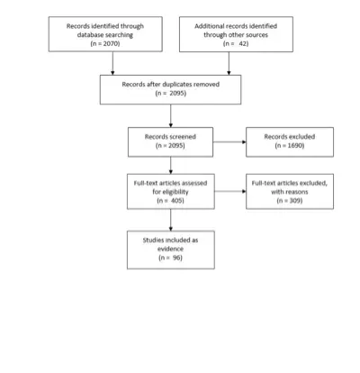
Figure 1. Article flow chart.
| PubMed (NCBI) Search |
| Step 1. Neuroma, Acoustic [MeSH] |
| Step 2. (vestibular [Title/Abstract] OR vestibulocochlear [Title/Abstract] OR acoustic [Title/Abstract]) AND (neuroma* [Title/Abstract] OR neurilemmoma* [Title/Abstract] OR neurilemoma* [Title/Abstract] OR neurinoma* [Title/Abstract] OR tumor* [Title/Abstract] OR tumour* [Title/Abstract] OR schwannoma* [Title/Abstract]) |
| Step 3. Step 1 OR Step 2 |
| Step 4. Diagnostic imaging [MeSH] OR Radiography [SH] OR Radionuclide imaging [SH] OR Ultrasonography [SH] |
| Step 5. Magnetic resonance imaging [TIAB] OR MRI [TIAB] OR Computed tomography [TIAB] OR CT [TIAB] OR Positron emission tomography [TIAB] OR PET [TIAB] OR FDG [TIAB] OR MET [TIAB] OR FET [TIAB] OR Diffusion tensor imaging [TIAB] OR DTI [TIAB] OR CISS [TIAB] OR FIESTA [TIAB] OR Spectroscop* [TIAB] OR SPECT [TIAB] OR imag* [TIAB] OR radiograph* [TIAB] |
| Step 6. Step 4 OR Step 5 |
| Step 7. Step 3 AND Step 6 |
| Step 8. Step 7 AND English [Lang] |
| Step 9. (animal [MeSH] NOT human [MeSH]) OR cadaver [MeSH] OR cadaver* [Titl] OR comment [PT] OR letter [PT] OR editorial [PT] OR addresses [PT] OR news [PT] OR “newspaper article” [PT] OR case reports [PT] |
| Step 10. Step 8 NOT Step 9 |
| Step 11. Step 10 AND (“1946/01/01” [PDAT] : “2015/01/01” [PDAT]) |
| Cochrane CENTRAL Search |
| Step 1. MeSH descriptor: [Neuroma, Acoustic] explode all trees |
| Step 2. ((vestibular or vestibulocochlear or acoustic) and (neuroma* or neurilemmoma* or neurilemoma* or neurinoma* or tumor* or schwannoma*)):ti,ab,kw |
| Step 3. Step 1 OR Step 2 |
| Step 4. MeSH descriptor: [Diagnostic Imaging] explode all trees |
| Step 5. Any MeSH descriptor with qualifier(s): [Radiography – RA] |
| Step 6. Any MeSH descriptor with qualifier(s): [Radionuclide imaging – RI] |
| Step 7. Any MeSH descriptor with qualifier(s): [Ultrasonography – US] |
| Step 8. “Magnetic resonance imaging” or MRI or “Computed tomography” or CT or “Positron emission tomography” or PET or FDG or MET or FET or “Diffusion tensor imaging” or DTI or CISS or FIESTA or Spectroscop* or SPECT or imag* or radiograph*:ti,ab,kw |
| Step 9. Step 4 or Step 5 or Step 6 or Step 7 or Step 8 |
| Step 10. Step 3 and Step 9 |
| Publication dates 1946-2014 |
| Total articles reviewed: 2,070 |
Table 2. Magnetic resonance imaging assessment of vestibular schwannoma: Initial diagnosis
| Author, Year | Description of Study | Data Class | Conclusions |
|---|---|---|---|
| Singh et al, 2015 | Prospective study of 19 patients who underwent MRI (standard protocol of T1WI, T2WI, DWI, and FLAIR images in axial, sagittal and coronal planes) for workup of VSs. | III | The sensitivity of MRI for correctly diagnosing VSs was 100% and specificity was 92.86% with a positive predictive value of 94.12% and accuracy of 96.67%. |
| Tomagane et al, 2013 | Retrospective study of the ability of PRESTO MRI sequence to distinguish between VSs (12 patients) and meningiomas (12 patients). | III | MRI with PRESTO sequence and CT were performed. PRESTO imaging showed schwannomas exhibit intratumoral spotty signal voids significantly more frequently than meningiomas, which correspond to microhemorrhages or hemosiderin deposits histologically. |
| Haque et al, 2011 | Prospective study of 42 consecutive patients referred for the evaluation of VSs. | II | After complete MRI evaluation, 61.9% were diagnosed with VSs. MRI provided 96% sensitivity, 88.2% specificity, 92.3% PPV, 93.75% NPV, and 92.86% accuracy in diagnosis of VS. (Class II achieved through comparison of MRI findings with gold standard operative histology in prospective fashion.) |
| Bhadelia et al 2008 | Retrospective study of cochlear FLAIR signal in 15 patients with VSs compared to 25 age-matched controls. | III | Patients with VSs have increased cochlear FLAIR signal intensity on the affected side compared with the unaffected side and healthy subjects. 8/15 patients also showed increased signal intensity in other portions of the membranous labyrinth, such as the semicircular canals and vestibule on the affected side. |
| Thamburaj et al 2008 | Prospective study of contribution of GRE imaging to differentiate VSs (n = 15 patients) from meningiomas (n = 5) based on presence of intratumoral microhemorrhages. | II | T2-weighted GRE MRI revealed microhemorrhages in 93.75% of VSs, a significantly higher rate than in meningiomas. T2 TSE and FLAIR imaging detected microhemorrhage in only 12.5% of VS cases. (Class II achieved through comparison of MRI findings with criterion standard operative histology in prospective fashion.) |
| Zealley et al 2000 | Prospective study comparing contrast-enhanced T1-weighted and FSE T2-weighted MRI in 1233 consecutive patients referred for exclusion of ANs. | II | Contrast-enhanced T1-weighted MRI was needed in addition to FSE T2-weighted imaging to confirm diagnosis in 44% of 33 cases identified to have VSs and 90% (9/10) of intracanalicular tumors. Identification of small intracanalicular VSs cannot rely on FSE T2-weighted imaging alone. (Class II achieved through comparison of FSE T2 findings with criterion standard contrast-enhanced MRI by independent radiologists in prospective fashion.) |
| Held et al 1999 | Retrospective study in 20 VS patients to compare T2-weighted CISS with post-contrast T1-weighted MPRAGE imaging in diagnosis of VSs. | III | All tumors were detected by both contrast-enhanced 3D MPRAGE and 3D CISS, with diameters equally well measured. 3D CISS better defined nerve of origin in lesions ≤10 mm, but with larger tumors, neither CISS nor MPRAGE yielded nerve of origin. |
| Tan 1999 | Retrospective study of FSE MRI versus CT in the diagnosis of VSs in 123 sensorineural hearing loss patients. | III | High-resolution FSE MRI is more sensitive than contrast-enhanced CT for the diagnosis of VSs. |
| Held et al 1997 | Retrospective study of 42 MRIs in 38 patients scanned with enhanced and unenhanced 3D MPRAGE and 3D CISS sequences to evaluate CPA pathologies. | III | 3D MPRAGE and 3D CISS are complementary MRI modalities. T1-weighted 3D MPRAGE is preferred to T1-weighted 2D spin echo sequences because of multiplanar reconstruction capabilities. |
| Hermans et al 1997 | Retrospective study of contrast-enhanced T1-weighted and T2-weighted CISS MRIs in 83 patients imaged to rule out VSs, with diagnosis of 18 VS tumors after review of imaging by 2 radiologists. | III | CISS and T1 postcontrast MRI offered high sensitivity (89–94%), specificity (94–97%), and accuracy (94–95%) in detecting VSs, with slight limitation for small intracanalicular and intralabyrinthine tumors. Variations in intraobserver (kappa 0.93–1) and interobserver (kappa 0.83–0.84) reproducibility were low. (Class II achieved through independent comparison of CISS MRI findings with criterion standard contrast-enhanced MRI.) |
| Soulie et al 1997 | Prospective study comparing the detection of VSs using T2-weighted FSE MRI vs contrast-enhanced T1-weighted MRI in 110 patients referred for suspected retrocochlear pathology based on sensorineural hearing loss, vertigo, or both. | II | Diagnosis of VSs can be excluded with normal 2D-FSE T2-weighted MRI with no additional gadolinium-enhanced T1 sequence. However, 6 false positives were identified relying on 2D-FSE, as confirmed by contrast-enhanced T1 MRI, resulting in an overall 100% sensitivity and 93% specificity for T2-weighted FSE imaging in ruling out VSs. (Class II achieved through comparison of FSE findings with criterion standard contrast-enhanced MRI by independent radiologists in prospective fashion.) |
| Stuckey et al 1996 | Prospective study comparing accuracy of CISS MRI with contrast-enhanced T1-weighted spin-echo MRI in detecting VSs in 125 consecutive patients after review by 2 observers. | II | Across 18 cases of CPA or IAC pathologies detected by T1-weighted MRI, CISS revealed a sensitivity of 94-100% and a specificity of 94-98% for radiographic detection of tumor. (Class II achieved through comparison of CISS findings with criterion standard contrast-enhanced MRI by independent radiologists in prospective fashion.) |
AN, acoustic neuroma; CISS, constructive interference in steady state; CPA, cerebellopontine angle; DWI, diffusion weighed imaging; FLAIR, fluid attenuated inversion recovery; GRE, gradient echo; IAC, internal auditory canal; MRI, magnetic resonance imaging; MPRAGE, magnetization prepared rapid acquisition gradient echo; PRESTO, principles of echo-shifting with a train of observations; TSE, turbo spin echo; VS, vestibular schwannoma.
Table 3. Magnetic resonance imaging assessment of vestibular schwannomas: Postoperative surveillance
| Author, Year | Description of Study | Data Class | Conclusions |
|---|---|---|---|
| Carlson et al, 2012 | Retrospective study of 203 patients undergoing microsurgical VS resection. | III | In 200/203 (98.5%) study patients, MRI demonstrated some postoperative enhancement within the preoperative tumor margin. During the course of follow-up, 49 (24.5%) patients demonstrated stable enhancement, 132 (66%) displayed lesion regression, and 7 (3.5%) had complete resolution of enhancement. 12 patients (5.9%) were diagnosed with recurrence at a mean of 3.0 years (median, 2.5; range 0.6–6.7) after surgery. |
| Carlson et al, 2011 | Retrospective study of 80 patients who underwent VS resection and had postsurgical enhancement in the distal aspect of the IAC lateral preoperative tumor margin. | III | Nodular enhancement within the fundus of the IAC lateral to the preoperative radiological tumor margin was seen in 16/80 (20%) after gross-total VS resection. Enhancement was nodular (13/16, 81%) or thin linear (3/16, 19%). |
| Rampp et al, 2011 | Prospective study of 21 patients with nontreated VSs to assess pattern of tumor enhancement on MRI at different time points after contrast administration. | III | While the appearance of overall tumor size remains stable, the interior appearance of VSs varies dependent on timing of image acquisition after contrast administration. The volume of central NEA at 11.5 minutes was only 11% of NEA at 1.5 minutes. So-called “necrotic tumor areas” may be falsely detected because of timing of image acquisition with respect to the administration of contrast medium. |
| Ozgen et al, 2009 | Retrospective study of 50 MRIs in 18 VS patients. | III | There is no difference in the detection of progression by using the CISS sequence alone compared with postcontrast sequences. However, the CISS has a low sensitivity for the detection of changes in the internal architecture, which may limit its use for the follow-up of patients after radiation treatment. |
| Bennett et al, 2008 | Retrospective study of 359 patients following resection. | III | 299 patients had MRI imaging at 1 and 5 years for analysis and a 5-year follow-up examination. Of these patients, 284 were found to have no enhancement at both one and five years. Linear enhancement was seen in 10 patients but did not enlarge in any patient. Nodular enhancement of the IAC was observed in three patients. Two patients with nodular enhancement had tumor recurrence. |
| Brors et al, 2003 | Retrospective study of postoperative MRIs of 70 patients who underwent resection of a unilateral VS. | III | At 3–6 months, all patients showed IAC enhancement from faint to high intensity; none had mass-like enhancement. At 12–24 months, 30/70 (42%) had decreasing, 35/70 (50%) stable, and 5/70 (8%) increasing enhancement; all 5 enhancers had intense nodular or mass-like pattern. At 36–48 months, 28/45 (59%) showed decreasing, 12/45 (29%) stable, and 4/45 (11%), with enhancers again nodular. At 4–6 years, 12/19 (63%) had decreased; 2/19 (11%) had no change, and 5/19 (26%) still increasing nodular or mass-like enhancement (proven in all 5 pathologically). |
| Umezu et al, 1999 | Retrospective study of 22 VS patients with 56 MRI examinations obtained between days 1 and 930 after surgery, to examine the pattern and timing of postoperative contrast enhancement after VS resection. | III | MRI should be obtained within the first 2 days after surgery, to minimize the confounding enhancement of muscle or fibrin glue packing after VS surgery. |
| Kremer et al, 1998 | Prospective study of 21 patients who underwent 3 MRI examinations after VS resection. | III | On early imaging (within 3 days postoperatively), no residual tumor but linear enhancement was seen in 16/21 (77%). On intermediate scans (6 weeks), nodular enhancement was seen in 16/18 (89%) of patients in whom muscle graft and fibrin glue were used. On late examination (6 months), nodular enhancement remained in 13/18 (72%) of implant patients, but was diminished in 10/13 compared to the intermediate scan. |
| Weissman et al, 1997 | Retrospective study of postoperative MRI patterns in 36 patients following VS resection. | III | Linear enhancement in the IAC is probably normal after surgery. Nodular and mass-like enhancement and any progressive enhancement may require close follow-up to monitor growth of residual tumor. Labyrinthine hyperintensity may reflect blood metabolites. VS patients should be followed by MRI years after surgery. |
| Mazzoni et al, 1996 | Retrospective study of postoperative MRI in 104 consecutive patients undergoing hearing preservation surgery for VSs. | III | Postoperative MRI should be performed at 1 and 3 years after surgery, and possibly at 5 and 8 years (speculative). Rounded nodular enhancing area reflected tumor, linear enhancement did not. |
CISS, constructive interference in steady state; IAC, internal auditory canal; MRI, magnetic resonance imaging; NEA, nonenhancing area; VS, vestibular schwannoma.
Table 4. Magnetic resonance imaging facial nerve assessment
| Author, Year | Description of Study | Data Class | Conclusions |
|---|---|---|---|
| Choi et al, 2014 | FN course in 11 patients with VSs was compared between preoperative DTT and intraoperative observation in prospective fashion. FN course was confirmed at 3 months postoperatively on tractography and function assessed on mean follow-up of 20 months. | III | Preoperative tractography prediction of FN course correlated with intraoperative findings in all (100%) cases. |
| Nakai et al, 2013 | FN course in 82 patients with VSs was compared between preoperative contrast-enhanced balanced fast field echo MRI and intraoperative observation in prospective fashion. | III | FN was identified in 46.3% (38/82) of patients using contrast-enhanced MRI, of which 74% (28/38) demonstrated congruence between preoperative predicted course and intraoperative observation. FN was more likely to be visualized in smaller tumors, with a solid consistency. |
| Zhang et al, 2013 | FN course was identified preoperatively using DTT in 8 patients with large (≥30-mm diameter) VSs and compared to intraoperative observation and EMG retrospectively. | III | FN could be identified in 87.5% (7/8) of cases preoperatively using DTT, and agreed to intraoperative evaluation in all of these cases. FN was anatomically preserved with HB grade I–II function in all cases. |
| Chen et al, 2011 | The relationship of the facial, trigeminal, and abducens nerves juxtaposed to tumor were reconstructed using DTI tractography superimposed on 3D tumor volumes in 3 patients with vestibular schwannoma. | III | The facial/cochlear-vestibular complex could be reconstructed in all (3/3) cases, but individual contributions of facial versus vestibular nerves within the complex could not be distinguished, nor could cisternal segment fibers in 1 case of a smaller tumor. Trigeminal nerve could be reconstructed with ease in all cases. |
| Gerganov et al, 2011 | 22 patients with large VSs and normal baseline facial function underwent preoperative DTI and CISS imaging for prospective determination of the course of the cisternal FN segment, in relation to tumor, which was correlated with intraoperative observations of the FN position. Surgeon was blinded to results of fiber tracking. The morphologic shape of FN was also recorded as flat or compact, based on intraoperative observation. | III | Tractography predicted FN cisternal (CPA) segment course and displacement by tumor in 90.9% (20/22) of cases compared to intraoperative evaluation. No DTI fiber patterns were found to be correlated with either flat or compact FN morphology. |
| Liang et al, 2010 | The ability of T2-weighted CISS versus TSE imaging to identify facial and vestibulocochlear nerves was compared in 48 volunteer subjects, as rated by 2 observers. CISS alone was performed in 8 patients with cerebellopontine angle pathology (1 bilateral VS). | III | CISS images were significantly better than TSE for visualizing canalicular segments of facial and vestibulocochlear nerves and slightly better for the cisternal segments of facial and vestibulocochlear nerves. |
| Taoka et al, 2006 | FN course in 8 patients with VSs was compared between preoperative DTI reconstruction, T2-weighted MR cisternography, and intraoperative observation. | III | FN course was reconstructed using tractography in 87.5% (7/8) of cases preoperatively and corresponded to intraoperative observation of the nerve trajectory in 71.4% (5/7). DTI tract was not obtained on the smaller tumor (18 mm diameter), which was the only case where preoperative magnetic resonance cisternography could identify the FN. |
| Kocaoglu et al, 2003 | Facial and cochlear nerve course were prospectively identified in 22 patients with small VSs undergoing hearing-preservation operations using contrast-enhanced T1-weighted and CISS MRI sequences by 2 independent radiologists. | III | Spatial relationship of FN and tumor could be determined in 82% (18/22) of cases on CISS images, but not on any of the contrast-enhanced T1-weighted sequences. No correlation between the direction of FN displacement and postoperative facial palsy or hearing loss was observed. |
| Sartoretti-Schefer et al, 2000 | Relationship of FN and tumor investigated in 22 patients with VSs using T2-weighted fast spin echo and T1-weighted MRI. | III | FN relation with the VS could be discerned in 86% of cases using T2-weighted FSE imaging but not T1-weighted sequences, with diminishing visualization of FN in larger sized tumors. |
| Schmalbrock et al, 1999 | 21 patients with 27 VSs (81% ≤1 cm3) were retrospectively assessed with T2-weighted SIMCAST and T1-weighted contrast-enhanced MRI for assessment of tumor appearance and its relation with the FN. | III | Facial or vestibulocochlear nerve branches were seen in 63% (17/27) of affected ears on axial T2-weighted SIMCAST imaging. SIMCAST delineated the relation of the 7/8 nerves and tumor with greater clarity than T1 sequence, while T1 contrast-enhanced sequence delineated the tumor-brain boundary better. Both imaging modalities were consistent in demarcating VS size. |
| Shigematsu et al, 1999 | 7th and 8th nerves and their relation to the tumor was prospectively evaluated in a phantom model and in 9 patients with 11 VSs using precontrast and contrast-enhanced CISS by 2 neuroradiologists. Imaging predictions were not compared to intraoperative observations in the 7 patients who underwent resection. | III | Addition of contrast to CISS increased the discrimination between cranial nerve and solid enhancing tumor and in identifying the 7th and 8th nerves at the IAC. |
| Jung et al, 1998 | Retrospective analysis of the correlation between preoperative prediction of FN displacement based on contrast-enhanced T1-weighted and T2-weighted MRI and intraoperative observation in 19 patients with extra-large (>40 mm in extrameatal diameter) VSs. The likely course of the FN was extrapolated from the relationship between the intrameatal and extrameatal axes of the tumor mass on MRI. | III | FN displacement was predicted in 80% of cases based on the angles formed between the intrameatal tumor and extrameatal tumor in relation to the IAC. |
| Casselman et al, 1993 | Retrospective series assessing the course of the facial, cochlear, superior vestibular, and inferior vestibular nerves in 50 normal ears and 10 ears with pathology (3 with VSs) using CISS MRI. | III | FN was identified in the IAC in 90% of normal ears in axial and coronal CISS sequences, most easily in the cisternal and horizontal segments, and least reliably around the posterior genu and vertical segment, where sparse CSF surrounds the nerve. |
HB, House–Brackmann; CISS, constructive interference in steady state; CPA, cerebellopontine angle; CSF, cerebrospinal fluid; DTI, diffusion tensor imaging; DTT, diffusion tensor tractography; EMG, electromyography; IAC, internal auditory canal; MRI, magnetic resonance imaging; MPRAGE, magnetization prepared rapid acquisition gradient echo; SIMCAST, segment-interleaved motion-compensated acquisition in steady state; TSE, turbo spin echo; VS, vestibular schwannoma.
Table 5. Use of magnetic resonance imaging for tumor surveillance
| Author, Year | Description of Study | Data Class | Conclusions |
|---|---|---|---|
| Fayad et al, 2014 | Retrospective review of 114 patients with presumed VSs undergoing conservative management. Patients were followed for a mean duration of 4.8 years and with annotation of growth, initiation of treatment, and audiologic measures. Mean tumor diameter of 10.5 mm. Growth was defined as ≥2 mm change in the maximal tumor diameter. This is a longer-term follow-up from Fucci et al, 1999. | III | 37.7% of tumors had grown at 5 years. 51.7% patients whose tumors had grown demonstrated growth at 1 year after initial MRI. After 5 years of no growth, 4% of patients showed growth. Monitoring by MRI is typically scheduled 6 months after the initial visit, at 1 and 2 years after that, at 5 years, and then if symptoms change. |
| Moffat et al, 2012 | 381 patients were included who were managed conservatively and who had at least 2 MRI scans to assess growth and associated predictive features. Mean interval between first and last scans was 4.2 years (0.5–17 years). | III | 59.3% of tumors did not grow over 5 years. Over half of growing tumors grew within the first 18 months after diagnosis. 7% of tumors grew after 5 years. The most common growth pattern was progressive growth, followed a pattern of no growth followed by growth. The authors recommend an MRI 6 months after initial diagnosis followed by annual scans, at which point scans can be done every 2 years for 6 years and then every 3 years. |
| Varughese et al, 2012 | Prospective study of 178 patients with VSs <2 cm over a mean follow-up period of 35.4 months to assess growth rate, features predictive of growth, and which measurement method was most accurate. They measured using diameter, volume, and VDT. | III | Three measurement modalities were used. Using the single diameter method, 29.2% tumors grew >1 mm. A VDT of 5.22 years strongly separated growing from nongrowing tumors. Patients are scanned at 12, 24, and 60 months for those being managed conservatively. |
| Suryanarayanan et al, 2010 | 327 patients with sporadic VSs were observed and MRI measurements taken with a mean follow-up of 3.6 years (range 1–14 years) to assess growth rate and associated predictive features. | III | Data on growth were available for 240 patients. 68% were stable. All growth began within the first 4 years of follow-up. Tumors >15 mm cisternally were likely to continue growing. The average growth rate overall was 1.1 mm/year. |
| Artz et al, 2009 | Retrospective review of a prospectively gathered database comprising 234 sporadic VSs managed conservatively for a mean of 28 months (range 4–120 months) to ascertain predictors of growth | III | Symptoms and anatomy of VSs at initial presentation help to predict future growth. “High-risk” tumors were either extrameatal with short duration of hearing loss and either unsteadiness/vertigo or no SSHL, or were intrameatal with short duration of hearing loss, unsteadiness/vertigo, and no SSHL. In this group, the risk of growth was 36.9% in the first year and 64.6% in 2 years. Low-risk tumors were extrameatal with no other risk factors or intrameatal with at most one other risk factor. In this group of tumors, the risk of growth was 2.5% in the first year and 12.7% within the first 2 years. |
| Bakkouri et al, 2009 | Retrospective review of 325 patients with VSs managed conservatively with at least 1 follow-up scan. The authors described growth rates. Follow-up ranged from 1–9 years. | III | The overall mean tumor growth rate was 1.15 mm/year. The mean percentages of cases that did not show any tumor growth, tumor growth <3 mm/year, and growth ≥3 mm/year during all observation periods were 57.8%, 28.9%, and 12%, respectively. Slowly progressive symptoms correlated with slower growth. |
| Martin et al, 2009 | Retrospective consideration of 276 patients with ≥1 follow-up scan followed over a mean period of 43 months to determine growth rate. | III | Of 276 patients, 62 (22%) demonstrated growth. Of the growing tumors, 65% grew slowly and 35% grew more rapidly; most rapidly growing tumors had grown even at 6-month MRI. Of all growing tumors, 90% were detected within 3 years. They recommend an initial MRI scan at 6 months, with annual scans for 2 years followed by a scan 2 years later, then every 5 years. |
| Ferri et al, 2008 | Retrospective review of 124 patients managed conservatively for an average follow-up period of 4.8 years to establish growth patterns. | III | 64.5% of tumors showed no increase in size (defined by change >2 mm). Of growing tumors, 45.4% grew within the first year; 22.7% grew at least 3 years after initial scan. No growth occurred after 6 years. The authors scan 6 months after initial scan, then at 12-month intervals unless growth is noted, which prompts a 6-month scan if no intervention occurs. |
| Solares et al, 2008 | Retrospective review of a prospectively collected database; 110 patients with sporadic VSs with at least 2 interval MRI scans were followed for a mean of 31 months (range 6–154 months) to assess growth patterns. | III | The 5-year no growth rate was 70.6% overall. Intracanalicular tumors were much less likely to grow (89.8% NGR vs 73.9% and 45.2% for grade I and II tumors, respectively). 10% of tumors regressed. Patients had an MRI 6 months after tumor discovery, then yearly if stable. |
| Stangerup et al, 2006 | Retrospective review of 552 patients with at least 2 MRI scans managed conservatively to determine growth rates; mean observation period was 3.6 years (range 1–15 years). Growth to extrameatal extension was the definition for growth in intrameatal tumors. In extrameatal tumors, an increase of >2 mm in the largest extrameatal diameter was defined as growth, and a decrease of >2 mm was defined as shrinkage. | III | Overall, 29% of extrameatal tumors grew. Of growing tumors, most began growth in the first year; the sooner the growth detection, the faster the growth rate. No tumor growth was initiated after the fourth year of observation. Of intrameatal tumors, 17% grew, with the majority growing in the first year, with no growth after the fourth year. The authors recommended yearly MRI for 5 years; every other year for 4 years; and a final MRI 5 years after that. |
| Bozorg Grayeli et al, 2005 | Retrospective review of 111 patients with intracanalicular or grade 2 VSs managed conservatively with serial MRI scans and mean follow-up of 33 months (range 6–111 months) with a view to establishing growth rates. | III | Growth was defined as a ≥2-mm increase in maximum AP size. 47% of tumors were stable; 47% increased in size; and 6% decreased in size. The mean growth rate was 1.1 mm/year. |
| Flint et al, 2005 | Retrospective review of 102 patients with unilateral VSs <24 mm followed for a median of 25.5 months to assess growth rates. | III | 62% of tumors showed no growth; 2 patients had tumor regression. 80% of patients with growing tumors had growth in the first year. Of these, 66% continued to grow. 20% of growing tumors had a latency period of no growth (range 8–60 months). Increase in size in first year may predict future growth. |
| Hoistad et al, 2001 | Retrospective review of 102 patients with unilateral VSs followed for a mean of 28.5 months (range 6 months–10 years) to assess tumor growth. | III | 53% of patients showed no growth; 44% grew ≥1 mm, with the mean growth rate of growing tumors estimated at 2 mm/year. Conservative management is reasonable with continued follow-up. |
| Tschudi et al, 2000 | Retrospective review of 74 patients followed for a mean of 35 months to establish growth rates. | III | 68.9% of tumors did not grow; 16% regressed. Of growing tumors, growth in the first year was predictive of future growth, as were the presence of the following symptoms: tinnitus, sudden hearing loss, or dizziness. |
| Fucci et al, 1999 | Retrospective review of 119 patients with presumed vestibular schwannomas undergoing conservative management. Patients were followed for a mean duration of 2.5 years (range 5 months–8 years) and growth, initiation of treatment, and audiologic measures were noted. At least 2 MRIs were performed per patient. | III | 66% of tumors did not grow during the study period. While the overall growth rate was 1.2 mm/year, growing tumors grew 3.8 mm/year on average. Size >20 mm at presentation predicted future growth. The authors recommend the initial follow-up scan occur at 6 months. |
MRI, magnetic resonance imaging; NGR, no growth rate; SSHL, sudden sensorineural hearing loss; VDT, volume doubling time; VS, vestibular schwannoma.
Table 6. Cystic vestibular schwannoma behavior
| Author, Year | Description of Study | Data Class | Conclusions |
|---|---|---|---|
| Dunn et al, 2014 | 52 patients with VSs with little or no extension into the IAC were retrospectively reviewed with regard to surgical detail and clinical outcome. | III | Medial VSs demonstrated a trend to increased adherence to the brainstem and FN. 71% of medial VSs were cystic. The FN outcomes were grade I/II in 87% of cases at latest follow-up, with 53% of patients experiencing transient FN palsy. |
| Metwali et al, 2014 | 37 patients with cystic VSs were reviewed and compared to similarly sized non-cystic tumors with respect to FN outcome postoperatively and at 1 year. | III | Cystic tumors tended to be large overall (T4 classification). Higher rates of incomplete resection were noted in cystic cases, as were lower rates of unfavorable FN outcome early after surgery (37.8%), when compared to solid tumors (17.5%). These rates equilibrated at 1 year (8.1% vs 6.2%). There were higher rates of postoperative hematoma and hydrocephalus in cystic cases. |
| Yashar et al, 2012 | Retrospective review of 23 patients undergoing surgery for cystic VSs. | III | Cystic schwannomas represented 12.8% of their overall series. Complete resection rates in these tumors was 48%. HB I–III function was achieved in 73% of cystic tumors in which a complete resection was achieved. |
| Piccirillo et al, 2009 | Retrospective review of morbidity and FN outcome in 57 cystic VSs with at least 1-year follow-up compared with results in patients with solid tumors. | III | Complete resection rates were similar in cystic vs solid tumors (84% vs 82%). The authors classified cysts as thick-walled and central (type A, with subtypes) or thin-walled and peripheral (type B, with subtypes). Overall, 1-year FN outcomes were similar between both groups (81% grade I-III). However, the authors recommend subtotal resection in patients with type B cystic schwannomas in their classification scheme. |
| Mehrotra et al, 2008 | Retrospective review of clinical outcomes of 22 patients with giant (>4 cm) cystic VSs with results compared to solid tumors of matched size (n = 40). | III | Patients with cystic tumors were likely to experience rapid deterioration. A large cystic component made surgery more challenging; overall, early FN outcomes were superior in patients with cystic tumors, though longer follow-up data are not presented. |
| Sinha et al, 2008 | Retrospective review of 58 patients with cystic VSs comparing surgical outcomes with patients with solid tumors. | III | Cystic schwannomas were less likely to be completely resected than solid tumors, and were more likely to have unfavorable FN outcomes. |
| Jones et al, 2007 | Retrospective review of 70 patients with cystic VSs and matched cohort of patients with solid tumors to assess effect of cystic nature on FN outcome at 2 years. | III | At 2-year follow-up, FN function is not statistically different between a group of patients with cystic and solid tumors. However, there is a trend to lower rates of grade I function and higher rates of grade VI function in the cystic tumor cohort. |
| Benech et al, 2005 | Retrospective review of 80 larger VSs, of which 26 were cystic, with a view to comparing presentation and surgical outcomes. | III | Cystic tumors were associated with shorter duration of symptoms; complete resection rates were similar. Favorable FN outcomes (HB I–III) were 58% at 1 year. |
| Wandong et al, 2005 | Retrospective review of a series of vestibular schwannoma patients to define the incidence and clinical characteristics of cystic tumors; 22 cystic schwannomas had surgery in this series | III | Cystic schwannomas constituted 7.3% of their series; the rate of FN continuity was 86%, and HB IV–VI at 2 years = 41%. They report worse outcomes in their cohort of cystic schwannoma patients. |
| Zaouche et al, 2005 | Retrospective review of 424 patients undergoing surgery for vestibular schwannomas to identify factors predicting facial palsy in the immediate postoperative period. | III | A heterogeneous and cystic appearance on preoperative MRI was associated with a statistically significant increased likelihood of HB ≥3 within 10 days of surgery |
| Fundova et al, 2000 | Retrospective review of the surgical outcomes of 44 cystic VSs (5.7% of their overall series) compared with 151 solid tumors. | III | Resection rates were similar in the cystic and solid groups. FN outcome analysis notable for a statistically significant increase in patients with grade VI facial function in cystic cases when compared to solid tumors (41% vs 27%) at 1 year. While the authors observed less structural adherence of tumors to surrounding tissue in cystic cases, the complication rates in these cases were higher when compared with patients with solid tumors (31.5% vs 20%). Average tumor size was 3.9 cm. |
| Shirato et al, 2000 | Retrospective review comparing the results and complications of SRT in 20 cystic schwannomas compared to 45 solid schwannomas. The mean follow-up period was 37 months. Overall tumor control was defined as no tumor growth of >2 mm after 2 years or no requirement of salvage surgery. | III | The 3-year tumor reduction rate was 31% for solid tumors and 93% for cystic tumors, with the difference being significant. However, cystic tumors tended to enlarge in the posttreatment period prior to size reduction. The authors report that fractionated SRT is safe in these tumors with size <3 cm, in contrast to previous reports on high-dose SRS in cystic tumors. |
| Samii et al, 1997 | Retrospective review of 1000 VS resections via the suboccipital approach to identify results and complications. | III | In cases of cystic tumor formation, the anatomic preservation rate of the FN was reduced from 93% to 88% and of the cochlear nerve from 68% to 55%. Cystic VSs were also more likely to hemorrhage postoperatively. |
| Charabi et al, 1994 | 23 cystic VSs were retrospectively reviewed to annotate imaging characteristics and growth patterns. | III | The overall incidence of cystic schwannomas was 4%. In 4 patients, growth rates between 6 months to 1 year was from 2–29 mm, all from cystic expansion. The authors concluded that patients with cystic tumors are at higher risk for symptomatic worsening given their higher growth rates, and a “wait and see” philosophy should not be adopted. |
| Charabi et al, 1994 | Retrospective review of surgical outcomes of 23 cystic schwannomas with attention to FN outcome and rates of resection. | III | Complete resection was achieved in 91% of cases, in which tumors were a mean of 4.5 cm. At 1 year, 65% of patients had unfavorable FN function (grade IV–VI). The authors recommend a lower threshold for leaving residual cyst wall if the exact anatomy is unclear. |
| Tali et al, 1993 | Retrospective review of 16 cases of cystic schwannoma to document the MRI appearance of these lesions; the incidence of this subgroup was determined in a series of 411 cases. | III | The incidence of cystic schwannomas was 19.3%. They generally displayed high signal intensity on T1 and T2 sequences, with peripheral enhancement. |
| Wallace et al, 1993 | Retrospective review of incidence of cystic tumors in 35 patients with VSs. | III | Cystic schwannomas, defined by the cystic component occupying at least 50% of the tumor mass, accounted for 24% of VSs in this series. |
FN, facial nerve; HB, House–Brackmann; IAC, internal auditory canal; MRI, magnetic resonance imaging; VS, vestibular schwannoma.
Table 7. Impact of lateral internal auditory canal involvement
| Author, Year | Description of Study | Data Class | Conclusions |
|---|---|---|---|
| Gerganov et al, 2009 | Retrospective review of 99 consecutive VS cases resected by retrosigmoid approach by single surgeon, evaluating the relationship of tumor size, intrameatal tumor extension pattern, and bony IAC involvement with preoperative hearing function. | III | The degree of intrameatal tumor growth was significantly correlated with the level of preoperative hearing. The distance between the lateral tumor end and the fundus showed significant correlation with shorter distance associated with worse Hanover score. |
| Gerganov et al, 2009 | Retrospective analysis of 99 consecutive cases resected by retrosigmoid approach by single surgeon, evaluating hearing preservation based on tumor extension pattern, volume, diameter, shape, and bony IAC involvement. | III | Increasing tumor volume, extrameatal tumor volume, and stage significantly correlated with worse postoperative HB grade. Intrameatal tumor length, intrameatal tumor width, tumor-fundus length showed no impact on immediate postoperative HB grade. |
| Mohr et al, 2005 | Retrospective study of 128 cases using intraoperative monitoring and following a retrosigmoid approach. The maximal extrameatal size of the tumor, its extension within the IAC, and pre- and postoperative hearing quality, according to the Gardner–Robertson classification, were evaluated. | III | With regard to filling of the IAC, among 63 patients harboring a tumor 15 mm or smaller in whom MRI was available, hearing was preserved more frequently in patients with partial filling (52.8% of 36) than those with complete filling (25.9% of 27). Both tumor size and the extent of IAC filling proved statistically significant in a multivariate analysis. |
| Kobayashi et al, 2002 | Retrospective study of 45 patients with small VSs resected by middle cranial fossa approach. All patients initially had full FN function on HB scale. Shortest distance and longest distance from fundus were measured on MRI. | III | Distance from fundus to AN had no effect on outcome of FN function at 2 weeks or at 3 months after surgery. Further, no correlation was observed between tumor diameter and FN function. |
| Rompaey et al, 2001 | Retrospective review of 123 consecutive patients with normal FN function prior to surgical resection of VS. HB grade calculated at 1 month and 1 year after surgery was correlated with fundus obliteration on preoperative MRI. | III | One-month postoperative HB grade III or greater occurred in 29.7% of patients with complete fundal obliteration compared to 13% with no fundus obliteration (P = .04). At 1 year, HB grade III or greater occurred in 18.7% of patients with complete fundal obliteration compared to 8.6% with no fundus obliteration though statistical significance was not seen. |
| Matthies et al, 1997 | Retrospective review of 202 consecutive patients evaluating preoperative high-resolution CT for specific radiographic features. | III | The length of the posterior auditory canal wall and the interear difference of the maximum porus width both correlate with the degree of preoperative hearing deterioration. The extent of the widening of the IAC is of predictive importance for the chances of postoperative hearing preservation or hearing loss. The extent of tumor growth anterior and caudal to the IAC in large tumors is of significant importance for prediction of postoperative hearing function. The tumor extension in all directions and the extent of cystic tumor components correlate with the pre‐ and postoperative function of the facial and cochlear nerves. |
AN, acoustic neuroma; CT, computed tomography; FN, facial nerve; HB, House-Brackmann; IAC, internal auditory canal; MRI, magnetic resonance imaging; VS, vestibular schwannoma.
Table 8. Magnetic resonance imaging of vestibular schwannomas associated with neurofibromatosis type 2
| Author, Year | Description of Study | Data Class | Conclusions |
|---|---|---|---|
| Mallory et al, 2014 | 26 patients with 32 NF2-related VSs were retrospectively evaluated for tumor control and hearing outcomes after SRS. | III | Improved tumor control in NF2 VSs was associated with higher marginal doses than commonly prescribed for sporadic VSs. Hearing outcomes were poor even with reduced marginal doses. Anatomic preservation of cochlear nerve with SRS may permit cochlear implantation and hearing rehabilitation. |
| Dombi et al, 2013 | Proposed consensus guidelines from the Tumor Measurement Working Group of the Response Evaluation in Neurofibromatosis and Schwannomatosis committee for the evaluation of imaging response in clinical trials for NF-related tumors. | III | Volumetric analysis of MRI is recommended to sensitively and reproducibly evaluate changes in tumor size, with a 20% volume change considered to be a decrease or increase in tumor size. Standardization of criteria enables meaningful comparison across clinical trials. |
| Goutagny et al, 2013 | 7 NF2 patients diagnosed at ≥70 years of age were reviewed for genetic profile, tumor growth curves, and clinical course. | III | 4/7 patients harbored bilateral VSs and 3/7 unilateral VSs, with no significant growth in 72% (8/11 tumors) over a mean follow-up of 96 months. Absence of germline NF2 mutation suggested a high prevalence of NF2 somatic mosaicism in these older patients. Long-term stability among most lesions suggests observation as initial management policy. |
| Dirks et al, 2012 | The growth patterns of intracranial tumors were retrospectively analyzed in 17 NF2 patients with a minimum of 4 years of clinical and MRI follow-up. | III | NF2-associated intracranial tumors most frequently demonstrate a saltatory growth pattern. Because of the lifetime risk for tumorigenesis and the unpredictable nature of radiographic progression and symptom onset in NF2 patients, resection should be reserved for symptom-producing tumors. Establishing the efficacy of nonsurgical therapeutic interventions must be based on long-term follow-up (several years). |
| Peyre et al, 2011 | 11 NF2 patients treated for bilateral VSs were retrospectively reviewed to assess changes in size of the contralateral VS after one side is resected in long-term follow-up (mean 7.6 years). | III | The velocity of diametric expansion is significantly elevated after resection of a contralateral VS (4.4 ± 3.4 mm/year) compared to before (2.5 ± 2.2 mm/year). The growth patterns of both VSs were similar in 9/11 cases before surgery. Increased postoperative growth was associated with decreased hearing in 3 cases. Removal of a VS in NF2 patients may precipitate an increase in the growth rate of the contralateral VS. |
| Fisher et al, 2009 | 52 patients with bilateral VSs were retrospectively reviewed for tumor progression and hearing function. | III | Tumor size increased and hearing decreased in a 1-year period. However, changes in status on one side cannot be used to predict changes in the other side. |
| Ito et al, 2009 | Retrospective review of 27 NF2 patients with 54 VSs followed for a mean of 86 months to identify factors predicting further growth of bilateral VSs. | III | Among an assessment of a variety of features (including age at onset, gender, coexistence of other tumors and volume indices), only age of onset correlated with growth (and only in posttreatment course, P = .007). Pattern suggests that after treatment particularly close follow-up may be warranted for patients with onset at an early age. |
| Harris et al, 2008 | Retrospective study of 10 NF2 patients with 43 MRI studies. | III | Linear measurements underestimate VS growth rate compared with volumetric measures in NF2 patients. |
| Slattery et al, 2005 | Retrospective review of prospective comparison of concordance in VS measurements across 115 MRIs from 57 NF2 patients between local radiologists and experienced neuroradiologists. | II | Fair concordance was observed between local radiologist and experienced neuroradiologist assessments. Least variability was found in thin slice postcontrast studies in patients without previous surgery. Neuroradiologist measurements were superior in postoperative tumors and tumors <5 mm. Strategies for uniform reporting are proposed, including use of fat suppression for posttreatment scans, thin-slice no skip postcontrast scans, and measurement of greatest overall diameter in addition to conventional measurement angles. (Class II achieved through blinded comparison of MRI assessments among radiologists in a prospective fashion.) |
| Slattery et al, 2004 | Retrospective assessment of changes in VS size in patients enrolled in NF2 Natural history study at short-term and long-term follow-up. | III | On average, VSs in NF2 patients increased 1.3 mm/year in short-term follow-up (defined as 9 months–2 years) and 1.9 mm/year on long-term follow-up (defined as 3–4 years), with 8% exhibiting growth of ≥5 mm over 4 years. |
| Subach et al, 1999 | Retrospective assessment of tumor control, hearing preservation, and facial function after SRS in 40 NF2 patients with 45 tumors over a 10-year period. | III | 36 months after SRS, 36% of VSs regressed, 62% remained unchanged, and 2% grew. Useful hearing was preserved in 6 (43%) of 14 patients, with greater success (67%) after modifications made in 1992; rate of hearing preservation may be better with radiosurgery than with other available techniques. Normal FN function was preserved in 25/31 (81%) patients. Normal trigeminal nerve function was preserved in 34/36 (94%) patients. |
FN, facial nerve; MRI, magnetic resonance imaging; NF2, neurofibromatosis type 2; SRS, stereotactic radiosurgery; VS, vestibular schwannoma.
Table 9. Length of time for magnetic resonance imaging follow-up
| Author, Year | Study Description | Data Class | Conclusions |
|---|---|---|---|
| Tang et al, 2014 | Retrospective evaluation of 88 patients who underwent VS resection and had ≥2 postoperative MRI scans | III | Nodular enhancement increased risk for tumor growth. If there was growth, tumors with nodular enhancement typically showed increase in size beginning 2 years postoperatively, whereas those with linear or no enhancement were typically stable in size through 5 years. Younger age and larger preoperative tumor size were also risk factors for growth. Imaging recommendations: The authors recommend 1) obtaining a baseline MRI at 6 months postoperatively; 2) no further imaging until 5 years postoperatively for patients with linear or no enhancement on the baseline scan, unless develop new symptoms; 3) annual MRIs starting at 2 years postoperatively for patients with nodular enhancement on the baseline scan, with offer for further treatment if MRI shows an increase in enhancement of 97 mm2 in maximal axial area or >133 mm3 in volume. |
| Lemee et al, 2014 | Retrospective review of 33 patients with a postsurgical VSTR after surgery. Patients had a biannual follow-up with clinical status and VSTR size assessment with MRI. | III | The postoperative facial function impairment and an initial remnant ≥1.5 cm3 were found to be significant risk factors of VS remnant progression in non-NF2 population in univariate analysis but not in multivariate analysis. If postoperative VS remnant is seen on initial MRI 3 months after surgery in a non-NF2 patient, and remnant >1.5 cm3 or postoperative FN function ≥HB grade IV, consider SRS. If these additional factors not seen, annual clinical and radiologic follow-up for 6 years, then once per 2 years. |
| Carlson et al, 2012 | Retrospective study of 350 patients who underwent VS resection, with 203 meeting criteria (non-NF2, receiving ≥2 follow-up MRIs). Imaging characteristics and need for further intervention analyzed. | III | Of 203 patients, 144 received GTR, 59 STR (did not distinguish NTR). Mean number of postoperative MRIs 2.6. Among 191 patients without recurrence over mean follow-up of 3.5 years, 7 underwent additional treatment (5 SRS and 2 surgery) at median of 8 months postoperatively. 5 patients with recurrent tumor continued to undergo observation at the end of study period. |
| Tysome et al, 2012 | Evaluation of 314 consecutively treated patients followed in a prospectively maintained database who underwent complete translabyrinthine excision of VSs and had follow-up imaging at 2 and 5 years after surgery. | III | All patients where MRI was reported to show no recurrence at 2 years (97% of 314) had no signs of recurrence on MRI at 5 years. 8 patients with MRI suspicious of recurrence (linear enhancement of IAC) at 2 years had no progression on MRI at 5–15 years. One patient had evidence of definite recurrence (nodular enhancement of IAC) at 2 years, and underwent radiosurgery at 8 years. The authors recommend a single MRI after complete resection of VS, with early imaging at 2 years after surgery to identify those at risk of recurrence. Patients with nodular enhancement should be considered to have recurrent disease. Linear enhancement on MRI should prompt repeat MRI at 5 years, with no further imaging needed if no progression is observed at that time. |
| Arlt et al, 2011 | Retrospective review of tumor progression and FN function in 50 patients after surgical resection of VSs. | III | 9/28 (32%) cases who received initial STR showed progression over a median follow-up of 52 months (median time to recurrence 44 months). 2/22 (9%) who received initial GTR showed tumor progression over a median follow-up of 50.5 months (median time to recurrence 41.5 months). |
| Carlson et al, 2011 | 16 patients with nodular enhancement in IAC among group of 350 who underwent microsurgery for VSs were retrospectively reviewed for tumor recurrence. Patients with incomplete resection, NF2, and fewer than 2 postoperative MRIs were excluded. | III | 0 of 16 patients with nodular enhancement at IAC on initial MRI (obtained at 3.1 months postoperatively on average) developed recurrence on mean radiologic follow-up of 39.8 months. |
| Fukuda et al, 2011 | Retrospective review of 74 patients after gross total (41), subtotal (25), or partial (8) resection of VSs to assess regrowth rate and time to regrowth. | III | VSs demonstrated regrowth of 2.4% (1/41 cases), 52% (13/25), and 62.5% (5/8) after GTR, STR, and PR, respectively, over a mean follow-up of 104 months (range 60–241 months). Time to regrowth ranged 6 to 76 months (median 31.9 months). |
| Peyre et al, 2011 | 11 NF2 patients treated for bilateral VSs were retrospectively reviewed to assess changes in size of the contralateral VS after one side is resected in long-term follow-up (mean 7.6 years). | III | In setting of bilateral vestibular schwannomas causing brainstem compression, the tumor growth rate of the remaining VS increases after surgery of the contralateral tumor. Recommend regular (every 6 months) radiologic follow-up after resection of 1 of 2 VSs in NF2. |
| Godefroy et al, 2009 | Recurrence rates and long-term FN function were retrospectively reviewed in 51 patients after translabyrinthine resection of large VSs and correlated to the initial extent of resection. | III | GTR in 26%, NTR in 58%, STR in 16%. No patients showed disease progression over mean follow-up of 48 months. |
| Bennett et al, 2008 | Retrospective study of enhancement patterns and tumor recurrence in 299 patients after resection of VSs, with MRI at 1 and 5 years. | III | 284/299 patients had no enhancement on follow-up. Linear enhancement in 10 patients did not show progression to tumor recurrence during study interval. 3 patients had nodular enhancement with 2 of these having tumor recurrence over 1–5 years. |
| Schmerber et al, 2005 | Retrospective analysis of 91 patients who underwent translabyrinthine GTR of VSs and who were followed for at least 5 years (mean 11 years). | III | None of the 91 patients experienced radiographic recurrence of VSs. A single gadolinium-enhanced MRI scan 5 years after surgery is advised in case of total removal. In case of any doubt about the quality of the tumor removal, MRI follow-up schedule within 2 years and 5 years of surgery establishes an initial baseline, with repeat MRI thereafter on clinical grounds. |
| Bloch et al, 2004 | Retrospective review of recurrence rate in 79 VS patients after microsurgery without GTR (50 with NTR and 29 with STR). | III | 3% recurrence observed after NTR and 32% after STR, with mean interval from surgery to detection of recurrence at 3 years (range 1–5 years). |
| Kameyama et al, 1996 | Retrospective review of 11 patients who underwent STR of intracanalicular VSs, with follow-up MRI ranging from 12 to 29 years. | III | MRI of 2 patients showed no evidence of tumor remnant, 6 with small tumor in IAC, remaining 3 with intracanalicular tumor protruding slightly intracranially. None underwent reoperation. Authors conclude that small intracanalicular small remnants have low risk of progression. |
| Lye et al, 1992 | 14 patients with residual tumors after surgical resection of VSs were retrospectively reviewed for growth of residual tumors. | III | 4/7 patients with residual tumors showed tumor enlargement on gadolinium-enhanced MRI over a mean follow-up of 70 months. An early postoperative (within 3 months) contrast-enhanced MRI is recommended in cases of suspected residual to establish baseline, repeat MRI 2 years later, and then, on clinical basis. |
| Kemink et al, 1991 | Retrospective review of 20 patients who underwent microsurgery with intentional STR (8) or NTR (12). Patient characteristics, postoperative CT and MRI, and neurologic function were assessed. | III | Radiologic detectable tumor regrowth occurred in only 1 patient (with STR) over a mean follow-up of 5 years. Postoperative contrast-enhanced CT or MRI was obtained at 2 months, followed by yearly scans thereafter. |
GTR, gross total resection; HB, House–Brackmann; IAC, internal auditory canal; MRI, magnetic resonance imaging; NF2, neurofibromatosis type 2; NTR, near total resection; PR, partial resection; SRS, stereotactic radiosurgery; STR, subtotal resection; VS, vestibular schwannoma; VSTR, vestibular schwannoma tumor remnant.
References
1. Tan TY. Non-contrast high resolution fast spin echo magnetic resonance imaging of acoustic schwannoma. Singapore Med J 1999;40(1):27-31.
2. Tomogane Y, Mori K, Izumoto S, et al. Usefulness of PRESTO magnetic resonance imaging for the differentiation of schwannoma and meningioma in the cerebellopontine angle. Neurol Med Chir (Tokyo) 2013;53(7):482-489.
3. Soulié D, Cordoliani YS, Vignaud J, Cosnard G. MR imaging of acoustic neuroma with high resolution fast spin echo T2-weighted sequence. Eur J Radiol 1997;24(1):61-65.
4. Zealley IA, Cooper RC, Clifford KM, et al. MRI screening for acoustic neuroma: a comparison of fast spin echo and contrast enhanced imaging in 1233 patients. Br J Radiol 2000;73(867):242-247.
5. Stuckey SL, Harris AJ, Mannolini SM. Detection of acoustic schwannoma: use of constructive interference in the steady state three-dimensional MR. AJNR Am J Neuroradiol 1996;17(7):1219-1225.
6. Hermans R, Van der Goten A, De Foer B, Baert AL. MRI screening for acoustic neuroma without gadolinium: value of 3DFT-CISS sequence. Neuroradiology 1997;39(8):593-598.
7. Haque S, Hossain A, Quddus MA, Jahan MU. Role of MRI in the evaluation of acoustic schwannoma and its comparison to histopathological findings. Bangladesh Med Res Counc Bull 2011;37(3):92-96.
8. Singh K, Singh MP, Thukral C, Rao K, Singh K, Singh A. Role of magnetic resonance imaging in evaluation of cerebellopontine angle schwannomas. Indian J Otolaryngol Head Neck Surg 2015;67(1):21-27.
9. Bhadelia RA, Tedesco KL, Hwang S, et al. Increased cochlear fluid-attenuated inversion recovery signal in patients with vestibular schwannoma. AJNR Am J Neuroradiol 2008;29(4):720-723.
10. Thamburaj K, Radhakrishnan VV, Thomas B, Nair S, Menon G. Intratumoral microhemorrhages on T2*-weighted gradient-echo imaging helps differentiate vestibular schwannoma from meningioma. AJNR Am J Neuroradiol 2008;29(3):552-557.
11. Held P, Fellner C, Seitz J, Graf S, Fellner F, Strutz J. The value of T2(*)-weighted MR images for the diagnosis of acoustic neuromas. Eur J Radiol 1999;30(3):237-244.
12. Held P, Fellner C, Fellner F, et al. MRI of inner ear and facial nerve pathology using 3D MP-RAGE and 3D CISS sequences. Br J Radiol 1997;70(834):558-566.
13. Ozgen B, Oguz B, Dolgun A. Diagnostic accuracy of the constructive interference in steady state sequence alone for follow-up imaging of vestibular schwannomas. AJNR Am J Neuroradiol 2009;30(5):985-991.
14. Rampp S, Scheller C, Prell J, Engelhorn T, Strauss C, Rachinger J. Magnetic resonance imaging dynamics of contrast medium uptake in vestibular schwannomas. J Neurosurg 2011;114(2):394-399.
15. Kanda T, Ishii K, Kawaguchi H, Kitajima K, Takenaka D. High signal intensity in the dentate nucleus and globus pallidus on unenhanced T1-weighted MR images: relationship with increasing cumulative dose of a gadolinium-based contrast material. Radiology 2014;270(3):834-841.
16. Grobner T. Gadolinium—a specific trigger for the development of nephrogenic fibrosing dermopathy and nephrogenic systemic fibrosis? Nephrol Dial Transplant 2006;21(4):1104-1108.
17. Marckmann P, Skov L, Rossen K, et al. Nephrogenic systemic fibrosis: suspected causative role of gadodiamide used for contrast-enhanced magnetic resonance imaging. J Am Soc Nephrol 2006;17(9): 2359-2362.
18. Mazzoni A, Calabrese V, Moschini L. Residual and recurrent acoustic neuroma in hearing preservation procedures: neuroradiologic and surgical findings. Skull Base Surg 1996;6(2):105-112.
19. Weissman, JL, Hirsch BE, Fukui MB, Rudy TE. The evolving MR appearance of structures in the internal auditory canal after removal of an acoustic neuroma. AJNR Am J Neuroradiol 1997;18(2):313-323.
20. Brors D, Schäfers M, Bodmer D, Draf W, Kahle G, Schick B. Postoperative magnetic resonance imaging findings after transtemporal and translabyrinthine vestibular schwannoma resection. Laryngoscope 2003;113(3):420-426.
21. Carlson ML, Van Abel KM, Driscoll CL, et al. Magnetic resonance imaging surveillance following vestibular schwannoma resection. Laryngoscope 2012;122(2):378-388.
22. Carlson ML, Van Abel KM, Schmitt WR, et al. Nodular enhancement within the internal auditory canal following retrosigmoid vestibular schwannoma resection: a unique radiological pattern. J Neurosurg 2011;115(4):835-841.
23. Kremer P, Forsting M, Hamer J, Sartor K. MR enhancement of the internal auditory canal induced by tissue implant after resection of acoustic neurinoma. AJNR Am J Neuroradiol 1998;19(1):115-118.
24. Umezu H, Seki Y. Postoperative magnetic resonance imaging after acoustic neuroma surgery: influence of packing materials in the drilled internal auditory canal on assessment of residual tumor. Neurol Med Chir (Tokyo) 1999;39(2):141-147.
25. Bennett ML, Jackson CG, Kaufmann R, Warren F. Postoperative imaging of vestibular schwannomas. Otolaryngol Head Neck Surg 2008;138(5):667-671.
26. Charabi S, Tos M, Børgesen SE, Thomsen J. Cystic acoustic neuromas. Results of translabyrinthine surgery. Arch Otolaryngol Head Neck Surg 1994;120(12):1333-1338.
27. Fundová P, Charabi S, Tos M, Thomsen J. Cystic vestibular schwannoma: surgical outcome. J Laryngol Otol 2000. 114(12): p. 935-9.
28. Tali, E.T., et al., Cystic acoustic schwannomas: MR characteristics. AJNR Am J Neuroradiol 1993;14(5):1241-1247.
29. Wallace CJ, Fong TC, Auer RN. Cystic intracranial schwannoma. Can Assoc Radiol J 1993;44(6):453-459.
30. Wandong S, Meng L, Xingang L, et al. Cystic acoustic neuroma. J Clin Neurosci 2005;12(3):253-5.
31. Yashar P, Zada G, Harris B, Giannotta SL. Extent of resection and early postoperative outcomes following removal of cystic vestibular schwannomas: surgical experience over a decade and review of the literature. Neurosurg Focus 2012;33(3):E13.
32. Zaouche S, Ionescu E, Dubreuil C, Ferber-Viart C. Pre- and intraoperative predictive factors of facial palsy in vestibular schwannoma surgery. Acta Otolaryngol 2005;125(4):363-369.
33. Benech F, Perez R, Fontanella MM, Morra B, Albera R, Ducati A. Cystic versus solid vestibular schwannomas: a series of 80 grade III-IV patients. Neurosurg Rev 2005;28(3):209-213.
34. Metwali H, Samii M, Samii A, Gerganov V. The peculiar cystic vestibular schwannoma: a single-center experience. World Neurosurg 2014;82(6):1271-1275.
35. Piccirillo E, Wiet MR, Flanagan S, et al. Cystic vestibular schwannoma: classification, management, and facial nerve outcomes. Otol Neurotol 2009;30(6):826-834.
36. Mehrotra N, Behari S, Pal L, Banerji D, Sahu RN, Jain VK. Giant vestibular schwannomas: focusing on the differences between the solid and the cystic variants. Br J Neurosurg 2008;22(4):550-556.
37. Dunn IF, Bi WL, Erkmen K, et al. Medial acoustic neuromas: clinical and surgical implications. J Neurosurg 2014;120(5):1095-1104.
38. Sinha S, Sharma BS. Cystic acoustic neuromas: surgical outcome in a series of 58 patients. J Clin Neurosci 2008;15(5):511-515.
39. Samii M, Matthies C. Management of 1000 vestibular schwannomas (acoustic neuromas): surgical management and results with an emphasis on complications and how to avoid them. Neurosurgery 1997;40(1):11-21.
40. Charabi S, Tos M, Thomsen J, Charabi B, Mantoni M. Vestibular schwannoma growth—long-term results. Acta Otolaryngol Suppl 2000;543:7-10.
41. Jones SE, Baguley DM, Moffat DA. Are facial nerve outcomes worse following surgery for cystic vestibular schwannoma? Skull Base 2007;17(5):281-284.
42. Charabi S, Mantoni M, Tos M, Thomsen J. Cystic vestibular schwannomas: neuroimaging and growth rate. J Laryngol Otol 1994;108(5):375-379.
43. Pendl G, Ganz JC, Kitz K, Eustacchio S. Acoustic neurinomas with macrocysts treated with Gamma Knife radiosurgery. Stereotact Funct Neurosurg 1996;66(suppl 1):103-111.
44. Shirato H, Sakamoto T, Takeichi N, et al. Fractionated stereotactic radiotherapy for vestibular schwannoma (VS): comparison between cystic-type and solid-type VS. Int J Radiat Oncol Biol Phys 2000;48(5):1395-1401.
45. Gerganov VM, Klinge PM, Nouri M, Stieglitz L, Samii M, Samii A. Prognostic clinical and radiological parameters for immediate facial nerve function following vestibular schwannoma surgery. Acta Neurochir (Wien) 2009;151(6):581-587.
46. Rompaey VV, Dinther Jv, Zarowski A, Offeciers E, Somers T. Fundus obliteration and facial nerve outcome in vestibular schwannoma surgery. Skull Base 2011;21(2):99-102.
47. Kobayashi M, Tsunoda A, Komatsuzaki A, Yamada I. Distance from acoustic neuroma to fundus and a postoperative facial palsy. Laryngoscope 2002;112(1):168-171.
48. Gerganov V, Nouri M, Stieglitz L, et al. Radiological factors related to pre-operative hearing levels in patients with vestibular schwannomas. J Clin Neurosci 2009;16(8):1009-1012.
49. Matthies C, Samii M, Krebs S. Management of vestibular schwannomas (acoustic neuromas): radiological features in 202 cases—their value for diagnosis and their predictive importance. Neurosurgery 1997;40(3):469-481.
50. Mohr G, Sade B, Dufour JJ, Rappaport JM. Preservation of hearing in patients undergoing microsurgery for vestibular schwannoma: degree of meatal filling. J Neurosurg 2005;102(1):1-5.
51. Jung S, Kim SH, Kim HW, et al. Prediction of facial nerve displacement in extra large vestibular schwannoma. Acta Neurochir (Wien) 1998;140(11):1143-1145.
52. Schmalbrock P, Chakeres DW, Monroe JW, et al. Assessment of internal auditory canal tumors: a comparison of contrast-enhanced T1-weighted and steady-state T2-weighted gradient-echo MR imaging. AJNR Am J Neuroradiol 1999;20(7):1207-1213.
53. Sartoretti-Schefer S, Kollias S, Valavanis A. Spatial relationship between vestibular schwannoma and facial nerve on three-dimensional T2-weighted fast spin-echo MR images. AJNR Am J Neuroradiol 2000;21(5):810-816.
54. Nakai T, Yamamoto H, Tanaka K, et al. Preoperative detection of the facial nerve by high-field magnetic resonance imaging in patients with vestibular schwannoma. Neuroradiology 2013;55(5):615-620.
55. Liang C, Zhang B, Wu L, et al. The superiority of 3D-CISS sequence in displaying the cisternal segment of facial, vestibulocochlear nerves and their abnormal changes. Eur J Radiol 2010;74(3):437-440.
56. Casselman JW, Kuhweide R, Deimling M, Ampe W, Dehaene I, Meeus L. Constructive interference in steady state-3DFT MR imaging of the inner ear and cerebellopontine angle. AJNR Am J Neuroradiol 1993;14(1):47-57.
57. Shigematsu Y, Korogi Y, Hirai T, et al. Contrast-enhanced CISS MRI of vestibular schwannomas: phantom and clinical studies. J Comput Assist Tomogr 1999;23(2):224-231.
58. Chen DQ, Quan J, Guha A, et al. Three-dimensional in vivo modeling of vestibular schwannomas and surrounding cranial nerves with diffusion imaging tractography. Neurosurgery 2011;68(4):1077-1083.
59. Choi KS, Kim MS, Kwon HG, Jang SH, Kim OL. Preoperative identification of facial nerve in vestibular schwannomas surgery using diffusion tensor tractography. J Korean Neurosurg Soc 2014;56(1):11-15.
60. Taoka T, Hirabayashi H, Nakagawa H, et al. Displacement of the facial nerve course by vestibular schwannoma: preoperative visualization using diffusion tensor tractography. J Magn Reson Imaging 2006;24(5):1005-1010.
61. Gerganov VM, Giordano M, Samii M, Samii A. Diffusion tensor imaging-based fiber tracking for prediction of the position of the facial nerve in relation to large vestibular schwannomas. J Neurosurg 2011;115(6):1087-1093.
62. Kocaoglu M, Bulakbasi N, Ucoz T, et al. Comparison of contrast-enhanced T1-weighted and 3D constructive interference in steady state images for predicting outcome after hearing-preservation surgery for vestibular schwannoma. Neuroradiology 2003;45(7):476-481.
63. Zhang Y, Chen Y, Zou Y, et al. Facial nerve preservation with preoperative identification and intraoperative monitoring in large vestibular schwannoma surgery. Acta Neurochir (Wien) 2013;155(10):1857-1862.
64. Smouha EE, Yoo M, Mohr K, Davis RP. Conservative management of acoustic neuroma: a meta-analysis and proposed treatment algorithm. Laryngoscope 2005;115(3):450-454.
65. Yoshimoto Y. Systematic review of the natural history of vestibular schwannoma. J Neurosurg 2005;103(1):59-63.
66. Stangerup SE, Caye-Thomasen P, Tos M, Thomsen J. The natural history of vestibular schwannoma. Otol Neurotol 2006;27(4):547-552.
67. Flint D, Fagan P, Panarese A. Conservative management of sporadic unilateral acoustic neuromas. J Laryngol Otol 2005;119(6):424-428.
68. Hoistad DL, Melnik G, Mamikoglu B, et al. Update on conservative management of acoustic neuroma. Otol Neurotol 2001;22(5):682-685.
69. Bozorg Grayeli A, Kalamarides M, Ferrary E, et al. Conservative management versus surgery for small vestibular schwannomas. Acta Otolaryngol 2005;125(10):1063-1068.
70. Moffat DA, Kasbekar A, Axon PR, Lloyd SK. Growth characteristics of vestibular schwannomas. Otol Neurotol 2012;33(6):1053-1058.
71. Martin TP, Senthil L, Chavda SV, Walsh R, Irving RM. A protocol for the conservative management of vestibular schwannomas. Otol Neurotol 2009;30(3):381-385.
72. Suryanarayanan R, Ramsden RT, Saeed SR, et al. Vestibular schwannoma: role of conservative management. J Laryngol Otol 2010;124(3):251-257.
73. Fucci MJ, Buchman CA, Brackmann DE, Berliner KI. Acoustic tumor growth: implications for treatment choices. Am J Otol 1999;20(4):495-499.
74. Fayad JN, Semaan MT, Lin J, Berliner KI, Brackmann DE. Conservative management of vestibular schwannoma: expectations based on the length of the observation period. Otol Neurotol 2014;35(7):1258-1265.
75. Solares CA, Panizza B. Vestibular schwannoma: an understanding of growth should influence management decisions. Otol Neurotol 2008;29(6):829-834.
76. Bakkouri WE, Kania RE, Guichard JP, et al. Conservative management of 386 cases of unilateral vestibular schwannoma: tumor growth and consequences for treatment. J Neurosurg 2009;110(4):662-669.
77. Tschudi DC, Linder TE, Fisch U. Conservative management of unilateral acoustic neuromas. Am J Otol 2000;21(5):722-728.
78. Ferri GG, Modugno GC, Pirodda A, et al. Conservative management of vestibular schwannomas: an effective strategy. Laryngoscope 2008;118(6):951-957.
79. Artz JC, Timmer FC, Mulder JJ, Cremers CW, Graamans K. Predictors of future growth of sporadic vestibular schwannomas obtained by history and radiologic assessment of the tumor. Eur Arch Otorhinolaryngol 2009;266(5):641-646.
80. Varughese JK, Breivik CN, Wentzel-Larsen T, Lund-Johansen M. Growth of untreated vestibular schwannoma: a prospective study. J Neurosurg 2012;116(4):706-712.
81. Tysome JR, Macfarlane R, Durie-Gair J, et al. Surgical management of vestibular schwannomas and hearing rehabilitation in neurofibromatosis type 2. Otol Neurotol 2012;33(3):466-472.
82. Schmerber S, Palombi O, Boubagra K, et al. Long-term control of vestibular schwannoma after a translabyrinthine complete removal. Neurosurgery 2005;57(4):693-698.
83. Arlt F, Trantakis C, Seifert V, et al. Recurrence rate, time to progression and facial nerve function in microsurgery of vestibular schwannoma. Neurol Res 2011;33(10):1032-1037.
84. Fukuda M, Oishi M, Hiraishi T, Natsumeda M, Fujii Y. Clinicopathological factors related to regrowth of vestibular schwannoma after incomplete resection. J Neurosurg 2011;114(5):1224-1231.
85. Godefroy WP, van der Mey AG, de Bruine FT, Hoekstra ER, Malessy MJ. Surgery for large vestibular schwannoma: residual tumor and outcome. Otol Neurotol 2009;30(5):629-634.
86. Tang S, Griffin AS, Waksal JA, et al. Surveillance after resection of vestibular schwannoma: measurement techniques and predictors of growth. Otol Neurotol 2014;35(7):1271-1276.
87. Lemée JM, Delahaye C, Laccourreye L, Mercier P, Fournier HD. Post-surgical vestibular schwannoma remnant tumors: what to do? Neurochirurgie 2014;60(5):205-215.
88. Bloch DC, Oghalai JS, Jackler RK, Osofsky M, Pitts LH. The fate of the tumor remnant after less-than-complete acoustic neuroma resection. Otolaryngol Head Neck Surg 2004;130(1):104-112.
89. Kameyama, S., et al., Long-term follow-up of the residual intracanalicular tumours after subtotal removal of acoustic neurinomas. Acta Neurochir (Wien), 1996. 138(2): p. 206-9.
90. Lye RH, Pace-Balzan A, Ramsden RT, Gillespie JE, Dutton JM. The fate of tumour rests following removal of acoustic neuromas: an MRI Gd-DTPA study. Br J Neurosurg 1992;6(3):195-201.
91. Kemink JL, Langman AW, Niparko JK, Graham MD. Operative management of acoustic neuromas: the priority of neurologic function over complete resection. Otolaryngol Head Neck Surg 1991;104(1):96-99.
92. Peyre M, Goutagny S, Imbead S, et al. Increased growth rate of vestibular schwannoma after resection of contralateral tumor in neurofibromatosis type 2. Neuro Oncol 2011;13(10):1125-1132.
93. Lee WJ, Isaacson JE. Postoperative imaging and follow-up of vestibular schwannomas. Otol Neurotol 2005;26(1):102-104.
94. Harris GJ, Plotkin SR, Maccollin M, et al. Three-dimensional volumetrics for tracking vestibular schwannoma growth in neurofibromatosis type II. Neurosurgery 2008;62(6):1314-1319.
95. Dombi E, Ardern-Holmes SL, Babovic-Vuksanovic D, et al. Recommendations for imaging tumor response in neurofibromatosis clinical trials. Neurology 2013;81(21 suppl 1):S33-S40.
96. Slattery WH, Lev MH, Fisher LM, Connell SS, et al. MRI evaluation of neurofibromatosis 2 patients: a standardized approach for accuracy in interpretation. Otol Neurotol 2005;26(4):733-740.
97. Slattery 3rd WH, Fisher LM, Igbal Z, Oppenheimer M. Vestibular schwannoma growth rates in neurofibromatosis type 2 natural history consortium subjects. Otol Neurotol 2004;25(5):811-817.
98. Dirks MS, Butman JA, Kim HJ, et al. Long-term natural history of neurofibromatosis type 2-associated intracranial tumors. J Neurosurg 2012;117(1):109-117.
99. Ito E, Saito K, Yatsuya H, Nagatani T, Otsuka G. Factors predicting growth of vestibular schwannoma in neurofibromatosis type 2. Neurosurg Rev 2009;32(4):425-433.
100. Choi JW, Lee JY, Phi JH, et al. Clinical course of vestibular schwannoma in pediatric neurofibromatosis type 2. J Neurosurg Pediatr 2014;13(6):650-657.
101. Goutagny S, Bah AB, Parfait B, Sterkers O, Kalamarides M. Neurofibromatosis type 2 in the elderly population: clinical and molecular features. Am J Med Genet A 2013;161A(4):667-670.
102. Fisher LM, Doherty JK, Lev MH, Slattery WH. Concordance of bilateral vestibular schwannoma growth and hearing changes in neurofibromatosis 2: neurofibromatosis 2 natural history consortium. Otol Neurotol 2009;30(6):835-841.
103. Kondziolka D, Subach BR, Lunsford LD, Bissonette DJ, Flickinger JC. Outcomes after gamma knife radiosurgery in solitary acoustic tumors and neurofibromatosis type 2. Neurosurg Focus 1998;5(3):e2.
104. Subach BR, Kondziolka D, Lunsford LD, et al. Stereotactic radiosurgery in the management of acoustic neuromas associated with neurofibromatosis type 2. J Neurosurg 1999;90(5):815-822.
105. Mallory GW, Pollock BE, Foote RL, et al. Stereotactic radiosurgery for neurofibromatosis 2-associated vestibular schwannomas: toward dose optimization for tumor control and functional outcomes. Neurosurgery 2014;74(3):292-300.
© Congress of Neurological Surgeons
Source: Neurosurgery, February 2018
5. The Role Of Imaging In The Management Of Patients With Vestibular Schwannomas: Update
Sponsored by: Congress of Neurological Surgeons (CNS) and the Section on Tumors
Endorsement: Reviewed for evidence-based integrity and endorsed by the American Association of Neurological Surgeons (AANS) and Congress of Neurological Surgeons (CNS)
Authors: Christopher S. Graffeo MD, MS1, Walavan Sivakumar, MD2, Sherwin Tavakol, M.D1, Lucas Paul Carlstrom, MD3, Jamie J. Van Gompel, MD4, Ian F. Dunn, MD1, Jeffrey J. Olson, MD5
Departmental and institutional affiliations:
- Department of Neurosurgery, University of Oklahoma College of Medicine, Oklahoma City, Oklahoma
- Department of Neurosurgery, Pacific Neuroscience Institute, Santa Monica, CA, USA
- Neurosurgery Department, Southern California Permanente Medical Group, San Diego, CA
- Department of Otolaryngology-Head and Neck Surgery and Neurologic Surgery, Mayo Clinic, Rochester, MN
- Department of Neurosurgery, School of Medicine, Emory University, Atlanta , Georgia
Corresponding Author contact information:
Christopher Graffeo, MD, MS
Department of Neurosurgery
University of Oklahoma
1000 N. Lincoln Blvd., Suite 4000
Oklahoma City, OK 73104
(405) 271-4912
graffeo@gmail.com
Keywords: Vestibular schwannoma; acoustic neuroma; guidelines; neuroimaging; magnetic resonance imaging; surveillance; stereotactic radiosurgery; diffusion tensor tractography
Running Title: Vestibular Schwannoma Imaging Guidelines
Abbreviations: Vestibular schwannoma (VS); cerebellopontine angle (CPA); stereotactic radiosurgery (SRS); Congress of Neurological Surgeons (CNS)
ABSTRACT
Background: Imaging is a critical aspect of vestibular schwannoma (VS) management, influencing essentially every aspect of care including diagnosis, surveillance, treatment decision-making, and follow-up after either resection or stereotactic radiosurgery (SRS). Despite this, treatment protocols are heterogeneous, and frequently based on historical practices, or low-quality evidence.
Objective: To update evidence-based guidelines for the use of imaging in the clinical management of patients with VS published by the Congress of Neurological Surgeons (CNS) in 2018.
Methods: Systematic review of the literature published from 1/1/2015 to 12/31/2022 regarding imaging protocols for VS management. Salient questions were identified by a writing group of diverse individuals with topic-specific expertise. Questions were validated by the CNS Guidelines Committee. Following systematic review, literature tables and summary statements pertinent to the study questions were generated by the writing group, which underwent subsequent evaluation and revision by the task force prior to formalization.
Results: Seven questions were formulated; adequate literature was identified to formulate updated recommendations for 6 of these. Search strategy identified 1143 unique records, of which 109 underwent full-text review, and 57 were included in the current study. Most studies provided level III evidence, with rare level II studies noted, yielding level III recommendations.
Conclusion: The current evidence base for imaging protocols in VS clinical management is broad, diverse, low-certainty, and low-quality. This in part reflects a heterogeneous disease, although variability in treatment philosophies may also influence local decision-making. Key areas for future study include the clinical utility of advanced imaging techniques, and head-to-head comparisons of imaging protocols for patients in common initial VS management pathways (e.g., observation, resection, or SRS).
RECOMMENDATIONS
Questions and Recommendations from the Prior Version of These Guidelines Without Change:
Prior Question: Do cystic VSs behave differently than their solid counterparts?
Prior Recommendation (Level III): Adults with cystic VSs should be counseled that their tumors maymore often be associated with rapid growth, lower rates of complete resection, and facial nerveoutcomes that may be inferior in the immediate postoperative period but similar to non-cystic
schwannomas over time.
Prior Question: Should the extent of lateral internal auditory canal (IAC) involvement be considered by treating physicians?
Prior Recommendation (Level III): The degree of lateral IAC involvement by tumor adversely affects facial nerve and hearing outcomes and should be emphasized when interpreting imaging for preoperative planning.
Prior Question: How should patients with neurofibromatosis type 2 (NF2) and VS be imaged and over what follow-up period?
Prior Recommendation (Level III): In general, VSs associated with NF2 should be imaged (similar to sporadic schwannomas) with the following caveats:
- More frequent imaging may be adopted in NF2 patients because of a more variable
growth rate for VSs, and annual imaging may ensue once the growth
rate is established.
- In NF2 patients with bilateral VSs, growth rate of a vestibular
schwannoma may increase after resection of the contralateral tumor, and therefore, more
frequent imaging may be indicated, based on the non-operated tumor’s historical rate of
growth.
- Careful consideration should be given to whether contrast is necessary in follow-up
studies or if high-resolution T2 (including CISS or FIESTA-type sequences) MRI may
adequately characterize changes in lesion size instead.
Questions and Recommendations from the Prior Version of These Guidelines that are Updated:
Prior Question: What sequences should be obtained on MRI to evaluate VSs before and aftersurgery?
New Question 1: In patients presenting with unilateral hearing loss is non-enhanced MRI, as compared to gadolinium-enhanced MRI, sufficiently sensitive to assess for the diagnosis of VS?
Recommendation (Level III): Non-contrasted, high-resolution MRI can be utilized as a cost-effective alternative to gadolinium-enhanced MRI when screening patients for VS. Further, it is suggested that the MRI sequences for the assessment of acute unilateral sensorineural hearing loss should include coronal T2, axial T1, and high-contrast T2, ideally in axial and coronal planes.
New Question 2: In patients with sporadic VS undergoing initial observation, are high-contrast T2 sequences (CISS/FIESTA-C), as compared to gadolinium-enhanced studies, sufficiently sensitive for surveillance of interval growth?
Recommendation (Level III): The use of high-resolution T2 imaging without enhanced T1 imaging in follow-up of observed VS is suggested, with enhanced T1 studies considered for instances of equivocal growth, or lesions suspected to be high-risk for phenotypically aggressive behavior.
Prior Question: What is the expected growth rate of VSs on MRI, and how often should they be imaged if a “watch and wait” philosophy is pursued?
New Question 3: In patients with sporadic VS undergoing initial observation, is a follow-up MRI at 2 years, as compared to a follow-up MRI at 1 year, associated with an increased risk of clinical decompensation requiring urgent/emergent intervention?
Recommendation (Level III): It is suggested that close early imaging follow-up, with annual studies for at least 3 years after diagnosis, followed by interval imaging at least every 3-5 years, with specific plans tailored to the patient-specific parameters (e.g., age, tumor size, prior growth), and the comfort levels of the patient and multidisciplinary treatment team be used in patients with sporadic VS undergoing initial observation.
Prior Question: Is there a role for advanced imaging for facial nerve detection preoperatively (eg, CISS/FIESTA or DTI imaging)?
New Question 4: In patients with sporadic VS undergoing preoperative planning for microsurgical resection, is high-field MRI (7T), as compared to routine MRI (1.5T and 3T), able to reliably determine the position of the facial nerve with respect to the tumor?
Recommendation: At present, there is insufficient evidence to determine the relative benefits of high-field versus standard-field MRI for determination of the position of the facial nerve with respect to the tumor.
New Question 5: In patients with sporadic VS undergoing preoperative planning for microsurgical resection, is MRI with fiber tractography, as compared to routine MRI (1.5T and 3T), able to reliably determine the position of the facial nerve with respect to the tumor?
Recommendation (Level II): Preoperative fiber tractography is recommended, when feasible, to assist in determining the location of the facial nerve with respect to the tumor, yet additional research is necessary to better elucidate whether this additional neuroanatomic information confers an improved long-term outcome.
Prior Question: How long should VSs be imaged after surgery, including after gross total, near total, and subtotal resection?
New Question 6: In patients with sporadic VS who undergo primary microsurgical resection, is early postoperative MRI (during the same hospitalization), as compared to delayed postoperative MRI at 3 months, less accurate and reliable in the detection of postoperative residual tumor?
Recommendation (Level III): While there is no evidence to suggest an advantage of early versus late initial postoperative imaging, it is suggested that initial imaging at ≥3 months after surgery, with follow-up imaging at closer intervals for STR/NTR (e.g., 3, 12, and 24 months), and longer intervals after GTR (e.g., 6, 18, and 36 months) is reasonable.
Question and Recommendation that is New:
New Question 7: In patients with sporadic VS who undergo primary SRS, is initial interval imaging at ≥24 months, as compared to initial interval imaging at ≤12 months, associated with increased incidence of tumor-directed clinical action, defined as a neurosurgical intervention that is undertaken strictly as a consequence of the imaging study and not due to patient symptoms, including repeat irradiation, surgical resection, treatment of ventriculomegaly with CSF diversion, or initiation of a VS disease-directed medical therapy?
Recommendation (Level III): Post-radiosurgery imaging is suggested at 12, 24, and 36 months, and deferral of retreatment is suggested until progression is noted on 3 consecutive imaging studies, absent concerning parallel changes in clinical symptoms.
INTRODUCTION
VS is a benign neoplasm arising from the myelinating Schwann cells of the superior or inferior vestibular nerve, with an estimated annual incidence rate of 1.52 per 100,000 population.1 Although uncommon, VS account for the majority of cerebellopontine angle (CPA) lesions and are the most common nerve sheath tumors overall. Although VS are pathologically benign, their posterior fossa location and propensity for growth over time predispose patients to a wide swath of disease morbidities, including hearing loss, vertigo, imbalance, hydrocephalus, and potentially death.2
Common VS management strategies include observation with radiographic surveillance, microsurgical resection, or stereotactic radiosurgery (SRS), with decision-making highly individualized, and modulated by the specific parameters of a given tumor, patient, or practice.3-7 Given the breadth of treatment pathways, as well as intrinsic disease heterogeneity, and the range of attendant risks and benefits associated with various management strategies, contemporary clinical practices vary considerably between centers and individual surgeons. Considering this diversity, the Congress of Neurological Surgeons (CNS) Guidelines Taskforce has published guidelines detailing best-practice recommendations covering most aspects of VS clinical care.8-16
Imaging is a foundational aspect of VS management that influences decision-making at essentially all phases-of-care, including initial diagnosis, surveillance, treatment planning, and post-treatment follow-up after either resection or SRS. The current study represents an update to the preceding CNS Guidelines for the role of imaging in the diagnosis and management of patients with VS, covering additions to the literature since the publication of those recommendations, 2015-2022.8 In addition to providing a general scientific update regarding primary clinical neuroimaging in VS, we also sought to partially tailor our study questions to the increasingly urgent need for efficiency in the delivery of advanced cranial care.
METHODOLOGY
The guidelines task force initiated a systematic review of the literature published after the last search date in the first version of this guideline to update the evidence-based information relevant to the treatment of patients with VSs. Through objective evaluation of the evidence and transparency in the process of making recommendations, this evidence-based clinical practice guideline was developed for the diagnosis and treatment of adult patients with functioning pituitary adenomas. These guidelines are developed for educational purposes to assist practitioners in their clinical decision-making processes. Additional information about the methods utilized in this systematic review is provided below.
Literature Search
Under the supervision of a research librarian and using the Preferred Reporting Items for Systematic Reviews and Meta-analyses (PRISMA) guidelines, we queried the Ovid Medline and Embase peer-reviewed databases for articles pertinent to clinical imaging protocols for VS management (for full search strategy, please see Supplement 1; for PRISMA flow diagram, see Figure 1). Initial search identified 1143 candidate citations, which were subject to title and abstract review in duplicate by 2 study investigators (CSG, WS). All articles identified as potentially meeting study criteria (n=109) underwent full-text review for confirmation of eligibility, with ambiguities in application of study criteria resolved by deferral to a senior investigator (IFD).
Study Selection and Eligibility
Prior to completing the formal systematic literature review, objective inclusion and exclusion criteria were collaboratively defined by the study writing group. Finalized criteria were ratified by the writing group prior to initiation of the literature search, in order to minimize the risk of bias. The final, standardized criteria were applied to the assessment of all pertinent articles, including those identified outside the primary search, via bibliography review, investigator contribution, or any other mechanism. Articles were considered eligible for inclusion in the formulation of these evidence-based clinical practice guidelines if they adhered to the following conditions:
Inclusion Criteria:
- Was published in an English-language peer-reviewed publication during the study sampling frame, 1/1/2015-12/31/2021
- Was a full article reporting primary, quantitative data from original clinical research
- Reported a minimum sample size of 5 patients
- Investigated patients with sporadic VS
Exclusion Criteria:
- Non-human, cadaver, or in vitro analyses
- Incomplete, non-peer-reviewed citations (e.g., meeting abstracts)
- Qualitative analyses (e.g., review articles, historical articles, editorials, letters, commentaries)
- Meta-research (e.g., prior systematic reviews, meta-analyses, society guidelines)
Previously published systematic reviews, meta-analyses, or guidelines were not considered evidence for inclusion in the study results, but were subject to bibliographic review, in order to optimize capture of candidate citations.
These assessments for the included articles in turn informed the evidence levels assigned to the associated recommendations. Level-setting for the study recommendations was benchmarked to the highest-quality publication included within the manuscripts for a given question.
Following initial assessment of all included articles and determination that the current study would be restricted to a qualitative systematic review without meta-analysis, formal risk-of-bias including Egger’s and Begg’s tests or the creation of funnel plots was deferred. Qualitative assessment for risk-of-bias was conducted by determining the Meta-Analysis of Observational Studies in Epidemiology (MOOSE) criteria for all included studies, which were deemed to be of good quality to answer the PICO questions.17
The quality of evidence was rated using an evidence hierarchy for each of four different study types; therapeutic, prognostic, diagnostic, and decision modeling. These hierarchies are shown in Appendix II: Rating Evidence Quality. Additional information regarding the hierarchy classification of evidence can be located here: https://www.cns.org/guidelines/guideline-procedures-policies/guideline-development-methodology.
Revision Plans
In accordance with the Institute of Medicine’s standards for developing clinical practice guidelines and criteria specified by the National Guideline Clearinghouse, the task force will monitor related publications following the release of this document and will revise the entire document and/or specific sections “if new evidence shows that a recommended intervention causes previously unknown substantial harm; that a new intervention is significantly superior to a previously recommended intervention from an efficacy or harms perspective; or that a recommendation can be applied to new populations.”18 In addition, the task force will confirm within five years from the date of publication that the content reflects current clinical practice and the available technologies for the evaluation and treatment for patients with perioperative spinal disease.
SUMMARY OF PREVIOUS GUIDELINES
The preceding CNS Guidelines regarding the role of imaging in VS management tackled 7 key clinical questions, with partial overlap regarding the current study.8 They addressed optimal MRI sequences for before and after VS resection; the role of advanced imaging (e.g., CISS/FIESTA, DTI) prior to VS resection; expected VS growth rates and timing for surveillance under “watch and wait” management strategies; imaging behavior of cystic vs. solid VS components; the influence of VS within the lateral auditory canal (IAC) on treatment decision-making and counseling; imaging protocols for patients with neurofibromatosis type 2 (NF2); and postoperative imaging protocols and duration, as modulated by extent-of-resection. The authors made level III recommendations for most questions, which emphasized the importance of both enhanced and high-resolution T2-weighted imaging; the potential utility of T2 imaging for preoperative facial nerve localization; close follow-up imaging in the early observation and/or post-treatment period, generally lasting at least 5 years; the impact of lateral IAC tumor on adverse facial nerve and hearing outcomes; and the importance of a thoughtful and aggressive stance towards imaging patients with NF2 in follow-up. Of note, given the general nature of the questions addressed by these guidelines, and the relatively short interval, we emphasized in the current update novel questions that interrogated more detailed or nuanced aspects of imaging in VS management.
RESULTS
Following full-text review and deliberation, of 109 articles, 52 were excluded for failing to meet criteria, reporting inadequate data, or not being relevant to the study questions. In total, 57 articles were identified as meeting all study criteria and were included to formulate our recommendations in response to the study questions. For each of the 7 questions, we included 11, 4, 14, 0, 15, 5, and 8 manuscripts. The overarching population for the study questions was patients with sporadic VS; however, this was modulated by the specific parameters under assessment with each question, as detailed below (e.g., timing for imaging after primary SRS is salient only to those patients who underwent primary SRS, among patients with sporadic VS).
Question 1
In patients presenting with unilateral hearing loss is non-enhanced MRI, as compared to gadolinium-enhanced MRI, sufficiently sensitive to assess for the diagnosis of VS?
Target Population
All individuals presenting to medical attention with unilateral hearing loss that is spontaneous in nature and not referable to an obvious underlying diagnosis, such as trauma.
Recommendation (Level III): Non-contrasted, high-resolution MRI can be utilized as a cost-effective alternative to gadolinium-enhanced MRI when screening patients for VS. Further, it is suggested that the MRI sequences for the assessment of acute unilateral sensorineural hearing loss should include coronal T2, axial T1, and high-contrast T2, ideally in axial and coronal planes.
Results of Individual Studies, Discussion of Study Limitations, and Risk of Bias
Following full-text review, 2 studies meeting criteria were considered pertinent to the question (Appendix IV, Table 1).19,20 These studies specifically assessed the role of non-enhanced MRI in the setting of sudden sensorineural hearing loss (SSNHL). Both employed cohort-type study designs, with large samples of patients screened radiographically after presenting with SSNHL. In one cohort study of 499 individuals presenting with unilateral SSNHL, the overall incidence of VS was observed at 3% (n=15).19 Lesions were ipsilateral to the SSNHL in all cases, and non-enhanced sequences were deemed sufficient for identification of the tumor itself. In another cohort study of 1249 individuals with unilateral SSNHL, the VS incidence was observed at 1.12% (n=14).20 This study also assessed cost effectiveness, with significant savings observed in association with protocols omitting enhanced sequences. In both studies, MRI sequences including routine T1/T2 and high-contrast T2 (e.g., CISS/FIESTA) were identified as instrumental in establishing the VS diagnosis without gadolinium enhancement.
Synthesis
While gadolinium-enhanced MRI is the gold standard imaging modality for diagnosing VS, non-contrast, high-resolution MRI has the sensitivity to be utilized as a cost-effective screening tool. Further, it is suggested that the MRI sequences for the assessment of acute unilateral sensorineural hearing loss should include coronal T2, axial T1, and high-contrast T2, ideally in axial and coronal planes.
Question 2
In patients with sporadic VS undergoing initial observation, are high-contrast T2 sequences (CISS/FIESTA-C), as compared to gadolinium-enhanced studies, sufficiently sensitive for surveillance of interval growth?
Target Population
Patients with new radiographic findings consistent with sporadic (e.g., unilateral) VS, whose initial management recommendation is observation with radiographic follow-up.
Recommendation (Level III): The use of high-resolution T2 imaging without enhanced T1 imaging in follow-up of observed VS is suggested, with enhanced T1 studies considered for instances of equivocal growth, or lesions suspected to be high-risk for phenotypically aggressive behavior.
Results of Individual Studies, Discussion of Study Limitations, and Risk of Bias
Following full-text review, 4 studies meeting criteria were considered pertinent to the question (Appendix IV, Table 2).21-24 These publications addressed the study question directly, allowing summative conclusions to be drawn with minimal extrapolation or indirect inference. All studies were retrospective, observational, and assessed the accuracy of high-contrast T2 sequences in measuring VS size and change-in-size, as compared to enhanced T1 sequences. Two additional studies included cost analyses; one assessed routine T2 in addition to high-contrast FIESTA/CISS imaging. Key study statistics also included accuracy and reliability testing between sequences for a single study; across imaging studies for each sequence under evaluation; and between raters, who were neuroradiologists in almost all studies. Overall, these assessments demonstrated high levels of accuracy and reliability for high-contrast T2 sequences, which were more robust when comparable to enhanced T1 sequences than were routine T2 sequences—observations that were noted both at time-of-diagnosis and in follow-up. Those studies incorporating economic analyses indicated that both costs and charges would be significantly lowered by surveillance protocols for observed VS that eliminate enhanced sequences. Of note, the role of high-contrast T2 imaging in the setting of recurrent/residual VS was not addressed by this study question.
Synthesis
Routine T2 sequences are highly specific and sensitive for detecting growth in observed VS, but may be inferior to enhanced T1 sequences. High-resolution T2 sequences such as CISS and FIESTA appear to provide equivalent accuracy, specificity, and sensitivity in detecting growth of observed VS, and are associated with significant cost savings. The use of high-resolution T2 imaging without enhanced T1 imaging in follow-up of observed VS is suggested, with enhanced T1 studies considered for instances of equivocal growth, or lesions suspected to be high-risk for phenotypically aggressive behavior.
Question 3
In patients with sporadic VS undergoing initial observation, is a follow-up MRI at 2 years, as compared to a follow-up MRI at 1 year, associated with an increased risk of clinical decompensation requiring urgent/emergent intervention?
Target Population
Patients with new radiographic findings consistent with sporadic (e.g., unilateral) VS, whose initial management recommendation is observation with radiographic follow-up.
Recommendation (Level III): It is suggested that close early imaging follow-up, with annual studies for at least 3 years after diagnosis, followed by interval imaging at least every 3-5 years, with specific plans tailored to the patient-specific parameters (e.g., age, tumor size, prior growth), and the comfort levels of the patient and multidisciplinary treatment team be used in patients with sporadic VS undergoing initial observation.
Results of Individual Studies, Discussion of Study Limitations, and Risk of Bias
Following full-text review, 12 studies meeting criteria were considered pertinent to the question (Appendix IV, Table 3).25-36 The specific endpoint of early decompensation was not reported by any prior study; however, all studies reported their institutional protocol for follow-up among observed VS, generally with detailed information regarding timing of progression and subsequent treatment recommendations. Protocols for follow-up imaging of observed VS are highly heterogeneous between centers, with variability clustered around two parameters: Timing of follow-up, and clinical action following tumor growth.
Most centers defined growth as ≥2mm change in size of nodular tumor; some centers also incorporate volumetric analysis. The shortest follow-up interval for newly diagnosed sporadic VS was 3 months; the longest was 24 months; the most common protocol was 12 months. Subsequent imaging protocols were even more diverse than the initial follow-up imaging protocols. The most frequent imaging protocol was every 3 months for a year, then every six months for a year, then annually for 10 years; the least frequent imaging protocol included follow-up at 1, 3, 5, and 10 years; most protocols involved annual imaging for 3-5 years, followed by larger intervals for another 3-5 years, and infrequent imaging after 10 years.
Where interval growth was observed, treatment protocols again varied widely. Most centers offered treatment, with determination for SRS versus resection governed by a variety of patient-, surgeon- and institution-specific considerations. Some centers offered on-going observation until at least two instances of interval growth were observed—in particular for patients presenting at more advanced ages, or with tumors confined to the IAC. A large minority of patients underwent treatment within 3 years of initial observation in most series, with by-study incidences ranging from 30-70%. Some patients elected to observe until serial progression was observed; the rate of subsequent growth following initial growth was directly assessed by Carlson et al., who noted sustained growth in only 7% of patients at 1 year after initial growth, with annual incidence rates for sustained growth increasing to 33%, 46%, 56%, and 59%, and 2-, 3-, 4-, and 5-year intervals after initial growth was documented (e.g., >40% of tumors did not demonstrate sustained growth in 5 years of subsequent follow-up after initial documentation of tumor growth).
Synthesis
Imaging protocols for initially observed sporadic VS are highly variable. Early growth within 3 years of diagnosis appears to occur in a large minority of patients, but is not always associated with sustained growth if intervention is not indicated. Among studies that performed early follow-up imaging at 6 months, initial tumor expansion was associated with further growth on subsequent studies. The conditional probability of new growth after 5 years of freedom-from-growth appears to be <2%. Risk factors for sustained growth appear to include larger cisternal components, rapid initial growth rates, and young patient age. It is suggested that close early imaging follow-up, with annual studies for at least 3 years after diagnosis, followed by interval imaging at least every 3-5 years, with specific plans tailored to the patient-specific parameters (e.g., age, tumor size, prior growth), and the comfort levels of the patient and multidisciplinary treatment team. Sustained growth is not universally observed, and many patients may benefit from on-going radiographic surveillance following initial observation of tumor expansion, if reliable follow-up is anticipated.
Question 4
In patients with sporadic VS undergoing preoperative planning for microsurgical resection, is high-field MRI (7T), as compared to routine MRI (1.5T and 3T), able to reliably determine the position of the facial nerve with respect to the tumor?
Target Population
Patients with sporadic VS undergoing preoperative planning for microsurgical resection.
Recommendation: At present, there is insufficient evidence to determine the relative benefits of high-field versus standard-field MRI for determination of the position of the facial nerve with respect to the tumor.
Results of Individual Studies, Discussion of Study Limitations, and Risk of Bias
The literature search did not provide qualifying manuscripts to address this question (Appendix IV, Table 4). Thus, there is insufficient evidence to determine the relative benefits of high-field MRI versus standard-field MRI for the assessment of the position of the facial nerve with respect to the tumor.
Question 5
In patients with sporadic VS undergoing preoperative planning for microsurgical resection, is MRI with fiber tractography, as compared to routine MRI (1.5T and 3T), able to reliably determine the position of the facial nerve with respect to the tumor?
Target Population
Patients with sporadic VS undergoing treatment with primary microsurgical resection, whose preoperative/perioperative assessment included MRI with fiber tractography for determination of facial nerve positioning, relative to the tumor.
Recommendation (Level II): Preoperative fiber tractography is recommended, when feasible, to assist in determining the location of the facial nerve with respect to the tumor, yet additional research is necessary to better elucidate whether this additional neuroanatomic information confers an improved long-term outcome.
Results of Individual Studies, Discussion of Study Limitations, and Risk of Bias
Following full-text review, 15 studies meeting criteria were considered pertinent to the question (Appendix IV, Table 5).37-51 No study specifically compared tractography with routine MRI for facial nerve localization purposes in the setting of VS, with the poor reliability of conventional imaging cited as the underlying explanation for those methodological decisions. One study reported by Hilly et al. included data from normal controls with comparative analysis of tractography and routine T1/T2 sequences for facial nerve localization in the CPA and IAC, with results that generalize poorly to the VS setting. All results assessing tractography in patients with VS specifically addressed the question of whether facial nerve localization was reliable with tractography; with most studies additionally providing intraoperative confirmation that the tractographic results were also valid. Reliability and validity were both strong across numerous studies, with successful nerve imaging and accurate nerve localization confirmed in >90% of patients.
Samala et al. reported a randomized study of 86 patients with large (>3cm) VS undergoing primary resection, who were allocated to treatment with or without preoperative DTI.37 In their analysis, DTI appeared to provide significantly improved anatomic and functional facial nerve outcomes, although the relatively low rate of favorable facial nerve outcomes noted in the control group, relative to prior reports from numerous centers, calls into question the generalizability of their results.52-54 Outcomes data were otherwise quite limited, descriptive in nature, and restricted to small samples. Taken together, these results indicate that tractography is a reliable and valid tool for preoperative facial nerve localization before VS resection; however, data on the meaningful clinical impact of this information is limited and subject to guarded interpretation.
Synthesis
MRI with fiber tractography appears to reliably localize the facial nerve with respect to the tumor in the great majority of cases, with some variance by technique. Data on the relationship between nerve localization and long-term facial nerve outcomes is promising but limited, and no definitive benefit has been confirmed at present. Consideration for preoperative fiber tractography is suggested where possible, as well as on-going research to better determine whether the additional neuroanatomic information it provides leads to improved outcomes in large samples of patients followed prospectively
Question 6
In patients with sporadic VS who undergo primary microsurgical resection, is early postoperative MRI (during the same hospitalization), as compared to delayed postoperative MRI at 3 months, less accurate and reliable in the detection of postoperative residual tumor?
Target Population
Patients with sporadic VS being treated with primary microsurgical resection.
Recommendation (Level III): While there is no evidence to suggest an advantage of early versus late initial postoperative imaging, it is suggested that initial imaging at ≥3 months after surgery, with follow-up imaging at closer intervals for STR/NTR (e.g., 3, 12, and 24 months), and longer intervals after GTR (e.g., 6, 18, and 36 months) is reasonable.
Results of Individual Studies, Discussion of Study Limitations, and Risk of Bias
Following full-text review, 5 studies meeting criteria were considered pertinent to the question (Appendix IV, Table 6).55-59 No study directly addressed the reliability, validity, or other test parameters of MRI in the assessment of extent-of-resection at early (immediate postoperative) versus delayed (≥3 month) timepoints. Institutional protocols for postoperative imaging after VS resection are highly variable. Initial imaging is performed by most centers either during the same hospitalization or at a 3-month postoperative visit; however, some centers delay imaging up to 6 or 12 months. Once baseline postoperative extent-of-resection has been established, further follow-up protocols are more consistent, and typically involve annual imaging for at least 3-5 years, which may be individualized in accordance with pertinent radiographic findings. Although most centers delay post-resection SRS until radiographic progression is confirmed, some centers proceed directly to up-front SRS in the setting of initial STR. Imaging protocols in this context are more aligned with post-SRS protocols, rather than post-resection protocols.
Synthesis
There is no evidence to suggest an advantage with regard to early (immediate postoperative) or delayed (≥3 months) initial postoperative MRI to assess radiographic extent-of-resection. Initial follow-up imaging after 0 or 3 months is completed at 1 year by almost all centers, and no centers report retreatment on the basis of radiographic findings sooner than 12 months after surgery. Subsequent imaging protocols are heterogeneous and range from annual studies to every 5 years. Volume of residuum appears to predict risk of recurrence. Initial postoperative imaging is suggested at ≥3 months, with follow-up imaging at closer intervals for STR/NTR (e.g., 3, 12, and 24 months), and longer intervals after GTR (e.g., 6, 18, and 36 months).
Question 7
In patients with sporadic VS who undergo primary SRS, is initial interval imaging at ≥24 months, as compared to initial interval imaging at ≤12 months, associated with increased incidence of tumor-directed clinical action, defined as a neurosurgical intervention that is undertaken strictly as a consequence of the imaging study and not due to patient symptoms, including repeat irradiation, surgical resection, treatment of ventriculomegaly with CSF diversion, or initiation of a VS disease-directed medical therapy?
Target Population
Patients with sporadic VS being treated with primary SRS.
Recommendation (Level III): Post-radiosurgery imaging is suggested at 12, 24, and 36 months, and deferral of retreatment is suggested until progression is noted on 3 consecutive imaging studies, absent concerning parallel changes in clinical symptoms.
Results of Individual Studies, Discussion of Study Limitations, and Risk of Bias
Following full-text review, 5 studies meeting criteria were considered pertinent to the question (Appendix IV, Table 7).60-64 No study specifically addressed the question of SRS at ≤12 months versus ≥24 months. Perry et al. assessed the clinical utility and cost implication of initial imaging at 6 months, as compared to 12 months; they found that no clinical action was taken based on 6-month studies, absent clinical symptoms, and that significant cost savings without adverse clinical impact would be anticipated if the early post-SRS MRI were eliminated.60 Two other studies identified similar patterns, with no benefit identified in association with MRI before 12 months, and retreatment infrequently considered prior to confirmation of true progression rather than pseudoprogression, typically at 24-36 months after primary SRS. Breshears et al. reported long-term data on resolution of pseudoprogression, with initial volumetric expansion noted within 3.2 years and resolving as late as 6.9 years in 90% of tumors.61 Foudard et al. reported a high incidence of early tumor expansion in 63.5% within 12 months; however, only 7.7% of those patients had serial tumor growth on at least 3 consecutive MRIs, and they concluded that retreatment prior to year 6 post-SRS was not advised.65
Synthesis
At present, no evidence specifically compares post-radiosurgery imaging at ≥24 months and ≤12 months. Imaging at ≤12 months does not appear to prompt clinical action, absent new symptoms potentially referable to changes in the tumor. Determination of radiosurgery treatment failure prior to 24-36 months is controversial. Routine post-radiosurgery imaging is suggested at 12, 24, and 36 months, and deferral of retreatment is suggested until progression
DISCUSSION
Imaging is a foundational component of VS management, influencing all areas of practice including initial diagnosis, preoperative planning, and surveillance during observation or after treatment with microsurgical resection or SRS. Contemporary evidence and associated recommendations regarding the use of neuroimaging in VS care is relatively low-quality, with most studies presenting level III data. At present, surveillance protocols between centers are highly heterogeneous, with variability noted in the preferred timing, MR sequences, and associated clinical actions following a significant change.
MRI with or without gadolinium is acceptable for the work-up of unilateral sensorineural hearing loss; however, once VS has been diagnosed, high-resolution T2 imaging may be relied upon for follow-up in the pre-treatment surveillance setting. Tractography appears to inform surgical planning in a reliable and valid fashion, although the impact on functional outcome is unclear. After primary microsurgical resection, an imaging baseline with contrast should be obtained, ideally in the 3-6 month timeframe, with annual imaging recommended over at least another 3-5 years of follow-up. For patients undergoing primary SRS, pseudoprogression is common, and early re-imaging does not appear to inform clinical action; correspondingly, we recommend annual imaging beginning at 12 month post-treatment, with retreatment reserved for patients who demonstrate serial volumetric expansion over multiple studies, or concerning associated clinical symptoms.
KEY ISSUES FOR FUTURE RESEARCH & CONCLUSIONS
Although these guidelines establish updated or new recommendations pertinent to numerous key areas of neuroimaging in VS management, we noted that several study questions have not been directly assessed by any clinical study in the past. Major deficiencies include data on the role of high-field MRI, adequately powered assessments of tractography as a perioperative tool, well-controlled radiographic assessment of postoperative imaging at various timepoints following resection, and additional outcome-oriented assessments of post-treatment imaging protocols in the setting of both resection and SRS. Robust data on the cost and quality-of-life impacts of various imaging protocols are also lacking, which represents a key target for future patient-centered study in VS imaging science.
Conflicts of Interest
All Guideline Task Force members were required to disclose all potential COIs prior to beginning work on the guideline, using the COI disclosure form of the AANS/CNS Joint Guidelines Review Committee. The CNS Guidelines Committee and Guideline Task Force Chair reviewed the disclosures and either approved or disapproved the nomination and participation on the task force. The CNS Guidelines Committee and Guideline Task Force Chair may approve nominations of task force members with possible conflicts and restrict the writing, reviewing, and/or voting privileges of that person to topics that are unrelated to the possible COIs. See Appendix V for a complete list of disclosures.
Disclosure of Funding
These evidence-based clinical practice guidelines were funded exclusively by the Congress of Neurological Surgeons, which received no funding from outside commercial sources to support the development of this document.
Disclaimer of Liability
This clinical systematic review and evidence-based guideline was developed by a physician volunteer task force as an educational tool that reflects the current state of knowledge at the time of completion. Each chapter is designed to provide an accurate review of the subject matter covered. This guideline is disseminated with the understanding that the recommendations by the authors and consultants who have collaborated in their development are not meant to replace the individualized care and treatment advice from a patient’s physician(s). If medical advice or assistance is required, the services of a competent physician should be sought. The proposals contained in these guidelines may not be suitable for use in all circumstances. The choice to implement any particular recommendation contained in these guidelines must be made by a managing physician in light of the situation in each particular patient and on the basis of existing resources.
Acknowledgments
The guidelines task force would like to acknowledge the CNS Guidelines Committee for their contributions throughout the development of the guideline, the AANS/CNS Joint Guidelines Review Committee, as well as the contributions Trish Rehring, MPH, Director for Evidence-Based Practice Initiatives for the CNS, and Janet Waters, MLS, BSN, RN, for assistance with the literature searches. Throughout the review process, the reviewers and authors were blinded from one another. At this time the guidelines task force would like to acknowledge the following individual peer reviewers for their contributions: Patti Raksin, Tjoumakaris, Andrew Carlson, Neil Majmundar, Jeff Mullin and Koji Ebersole.
REFERENCES
- Ostrom QT, Price M, Neff C, et al. CBTRUS Statistical Report: Primary Brain and Other Central Nervous System Tumors Diagnosed in the United States in 2015-2019. Neuro Oncol. 2022;24(Suppl 5):v1-v95.
- Carlson ML, Link MJ. Vestibular Schwannomas. N Engl J Med. 2021;384(14):1335-1348.
- Carlson ML, Barnes JH, Nassiri A, et al. Prospective Study of Disease-Specific Quality-of-Life in Sporadic Vestibular Schwannoma Comparing Observation, Radiosurgery, and Microsurgery. Otol Neurotol. 2021;42(2):e199-e208.
- Carlson ML, Link MJ, Driscoll CLW, et al. Working Toward Consensus on Sporadic Vestibular Schwannoma Care: A Modified Delphi Study. Otol Neurotol. 2020;41(10):e1360-e1371.
- Carlson ML, Van Gompel JJ, Wiet RM, et al. A Cross-sectional Survey of the North American Skull Base Society: Current Practice Patterns of Vestibular Schwannoma Evaluation and Management in North America. J Neurol Surg B Skull Base. 2018;79(3):289-296.
- Macielak RJ, Driscoll CLW, Link MJ, Haynes DS, Lohse CM, Carlson ML. Vestibular Schwannoma Practice Patterns: An International Cross-specialty Survey. Otol Neurotol. 2020;41(10):e1304-e1313.
- Van Gompel JJ, Carlson ML, Wiet RM, et al. A Cross-sectional Survey of the North American Skull Base Society on Vestibular Schwannoma, Part 2: Perioperative Practice Patterns of Vestibular Schwannoma in North America. J Neurol Surg B Skull Base. 2018;79(3):297-301.
- Dunn IF, Bi WL, Mukundan S, et al. Congress of Neurological Surgeons Systematic Review and Evidence-Based Guidelines on the Role of Imaging in the Diagnosis and Management of Patients With Vestibular Schwannomas. Neurosurgery. 2018;82(2):E32-E34.
- Hadjipanayis CG, Carlson ML, Link MJ, et al. Congress of Neurological Surgeons Systematic Review and Evidence-Based Guidelines on Surgical Resection for the Treatment of Patients With Vestibular Schwannomas. Neurosurgery. 2018;82(2):E40-E43.
- Vivas EX, Carlson ML, Neff BA, et al. Congress of Neurological Surgeons Systematic Review and Evidence-Based Guidelines on Intraoperative Cranial Nerve Monitoring in Vestibular Schwannoma Surgery. Neurosurgery. 2018;82(2):E44-E46.
- Van Gompel JJ, Agazzi S, Carlson ML, et al. Congress of Neurological Surgeons Systematic Review and Evidence-Based Guidelines on Emerging Therapies for the Treatment of Patients With Vestibular Schwannomas. Neurosurgery. 2018;82(2):E52-E54.
- Sweeney AD, Carlson ML, Shepard NT, et al. Congress of Neurological Surgeons Systematic Review and Evidence-Based Guidelines on Otologic and Audiologic Screening for Patients With Vestibular Schwannomas. Neurosurgery. 2018;82(2):E29-E31.
- Sughrue ME, Fung KM, Van Gompel JJ, Peterson JEG, Olson JJ. Congress of Neurological Surgeons Systematic Review and Evidence-Based Guidelines on Pathological Methods and Prognostic Factors in Vestibular Schwannomas. Neurosurgery. 2018;82(2):E47-E48.
- Olson JJ, Kalkanis SN, Ryken TC. Congress of Neurological Surgeons Systematic Review and Evidence-Based Guidelines on the Treatment of Adults With Vestibular Schwannomas: Executive Summary. Neurosurgery. 2018;82(2):129-134.
- Germano IM, Sheehan J, Parish J, et al. Congress of Neurological Surgeons Systematic Review and Evidence-Based Guidelines on the Role of Radiosurgery and Radiation Therapy in the Management of Patients With Vestibular Schwannomas. Neurosurgery. 2018;82(2):E49-E51.
- Carlson ML, Vivas EX, McCracken DJ, et al. Congress of Neurological Surgeons Systematic Review and Evidence-Based Guidelines on Hearing Preservation Outcomes in Patients With Sporadic Vestibular Schwannomas. Neurosurgery. 2018;82(2):E35-E39.
- Brooke BS, Schwartz TA, Pawlik TM. MOOSE Reporting Guidelines for Meta-analyses of Observational Studies. JAMA Surg. 2021;156(8):787-788.
- Ransohoff DF, M. Pignone, and H.C. Sox, . How to decide whether a clinical practice guideline is trustworthy. . JAMA. 2013;309(2):139-140.
- Fujita T, Saito K, Kashiwagi N, Sato M, Seo T, Doi K. The prevalence of vestibular schwannoma among patients treated as sudden sensorineural hearing loss. Auris Nasus Larynx. 2019;46(1):78-82.
- Yang W, Mei X, Li X, et al. The prevalence and clinical characteristics of vestibular schwannoma among patients treated as sudden sensorineural hearing loss: A 10-year retrospective study in southern China. American Journal of Otolaryngology. 2020;41(4):102452.
- Coelho DH, Tang Y, Suddarth B, Mamdani M. MRI surveillance of vestibular schwannomas without contrast enhancement: Clinical and economic evaluation. The Laryngoscope. 2018;128(1):202-209.
- Forgues M, Mehta R, Anderson D, et al. Non-contrast magnetic resonance imaging for monitoring patients with acoustic neuroma. J Laryngol Otol. 2018;132(9):780-785.
- Buch K, Juliano A, Stankovic KM, Curtin HD, Cunnane MB. Noncontrast vestibular schwannoma surveillance imaging including an MR cisternographic sequence: is there a need for postcontrast imaging? J Neurosurg. 2018;131(2):549-554.
- Currie S, Saunders D, Macmullen-Price J, et al. Should we be moving to a national standardized non-gadolinium MR imaging protocol for the surveillance of vestibular schwannomas? Br J Radiol. 2019;92(1096):20180833.
- Macielak RJ, Patel NS, Lees KA, et al. Delayed Tumor Growth in Vestibular Schwannoma: An Argument for Lifelong Surveillance. Otol Neurotol. 2019;40(9):1224-1229.
- D’Haese S, Parmentier H, Keppler H, et al. Vestibular schwannoma: natural growth and possible predictive factors. Acta Otolaryngol. 2019;139(9):753-758.
- Borsetto D, Gair J, Kenyon O, et al. When Should We Stop Scanning Older Patients with Vestibular Schwannomas? J Neurol Surg B Skull Base. 2019;80(4):333-337.
- Kleijwegt M, Bettink F, Malessy M, Putter H, van der Mey A. Clinical Predictors Leading to Change of Initial Conservative Treatment of 836 Vestibular Schwannomas. J Neurol Surg B Skull Base. 2020;81(1):15-21.
- Sethi M, Borsetto D, Cho Y, et al. The Conditional Probability of Vestibular Schwannoma Growth at Different Time Points After Initial Stability on an Observational Protocol. Otol Neurotol. 2020;41(2):250-257.
- Fieux M, Pouzet C, Bonjour M, Zaouche S, Jouanneau E, Tringali S. MRI monitoring of small and medium-sized vestibular schwannomas: predictors of growth. Acta Otolaryngol. 2020;140(5):361-365.
- Marinelli JP, Lees KA, Lohse CM, et al. Natural History of Growing Sporadic Vestibular Schwannomas: An Argument for Continued Observation Despite Documented Growth in Select Cases. Otol Neurotol. 2020;41(9):e1149-e1153.
- Schnurman Z, Nakamura A, McQuinn MW, Golfinos JG, Roland JT, Kondziolka D. Volumetric growth rates of untreated vestibular schwannomas. J Neurosurg. 2019:1-7.
- Hentschel MA, Hannink G, Steens SCA, Mulder JJS, Rovers MM, Kunst HPM. Development of a model to predict vestibular schwannoma growth: An opportunity to introduce new wait and scan strategies. Clin Otolaryngol. 2021;46(1):273-283.
- Kim JS, Cho Y-S. Growth of vestibular schwannoma: long-term follow-up study using survival analysis. Acta Neurochirurgica. 2021;163(8):2237-2245.
- Marinelli JP, Schnurman Z, Killeen DE, et al. Long-term natural history and patterns of sporadic vestibular schwannoma growth: A multi-institutional volumetric analysis of 952 patients. Neuro Oncol. 2022;24(8):1298-1306.
- Borsetto D, Sethi M, Clarkson K, et al. Evidence-based surveillance protocol for vestibular schwannomas: a long-term analysis of tumor growth using conditional probability. J Neurosurg. 2022:1-8.
- Samala R, Borkar SA, Sharma R, et al. Effectiveness of preoperative facial nerve diffusion tensor imaging tractography for preservation of facial nerve function in surgery for large vestibular schwannomas: Results of a prospective randomized study. Neurol India. 2019;67(1):149-154.
- Yoshino M, Kin T, Ito A, et al. Combined use of diffusion tensor tractography and multifused contrast-enhanced FIESTA for predicting facial and cochlear nerve positions in relation to vestibular schwannoma. J Neurosurg. 2015;123(6):1480-1488.
- Wei PH, Qi ZG, Chen G, et al. Identification of cranial nerves near large vestibular schwannomas using superselective diffusion tensor tractography: experience with 23 cases. Acta Neurochir (Wien). 2015;157(7):1239-1249.
- Hilly O, Chen JM, Birch J, et al. Diffusion Tensor Imaging Tractography of the Facial Nerve in Patients With Cerebellopontine Angle Tumors. Otol Neurotol. 2016;37(4):388-393.
- Song F, Hou Y, Sun G, et al. In vivo visualization of the facial nerve in patients with acoustic neuroma using diffusion tensor imaging-based fiber tracking. J Neurosurg. 2016;125(4):787-794.
- Borkar SA, Garg A, Mankotia DS, et al. Prediction of facial nerve position in large vestibular schwannomas using diffusion tensor imaging tractography and its intraoperative correlation. Neurol India. 2016;64(5):965-970.
- Ma J, Su S, Yue S, et al. Preoperative Visualization of Cranial Nerves in Skull Base Tumor Surgery Using Diffusion Tensor Imaging Technology. Turk Neurosurg. 2016;26(6):805-812.
- Li H, Wang L, Hao S, et al. Identification of the Facial Nerve in Relation to Vestibular Schwannoma Using Preoperative Diffusion Tensor Tractography and Intraoperative Tractography-Integrated Neuronavigation System. World Neurosurg. 2017;107:669-677.
- Zolal A, Juratli TA, Podlesek D, et al. Probabilistic Tractography of the Cranial Nerves in Vestibular Schwannoma. World Neurosurg. 2017;107:47-53.
- Churi ON, Gupta S, Misra BK. Correlation of Preoperative Cranial Nerve Diffusion Tensor Tractography with Intraoperative Findings in Surgery of Cerebellopontine Angle Tumors. World Neurosurg. 2019;127:e509-e516.
- Yoshino M, Kin T, Ito A, et al. Feasibility of diffusion tensor tractography for preoperative prediction of the location of the facial and vestibulocochlear nerves in relation to vestibular schwannoma. Acta Neurochir (Wien). 2015;157(6):939-946; discussion 946.
- Epprecht L, Kozin ED, Piccirelli M, et al. Super-resolution Diffusion Tensor Imaging for Delineating the Facial Nerve in Patients with Vestibular Schwannoma. J Neurol Surg B Skull Base. 2019;80(6):648-654.
- Castellaro M, Moretto M, Baro V, et al. Multishell Diffusion MRI-Based Tractography of the Facial Nerve in Vestibular Schwannoma. AJNR Am J Neuroradiol. 2020;41(8):1480-1486.
- Szmuda T, Słoniewski P, Ali S, et al. Reliability of diffusion tensor tractography of facial nerve in cerebello-pontine angle tumours. Neurol Neurochir Pol. 2020;54(1):73-82.
- Ung N, Pelargos PE, Mozaffari K, et al. Accuracy and outcomes of diffusion tensor imaging tractography in resection for vestibular schwannoma for facial nerve preservation. J Neurol Sci. 2021;430:120006.
- Carlson ML, Van Abel KM, Schmitt WR, Driscoll CL, Neff BA, Link MJ. The anatomically intact but electrically unresponsive facial nerve in vestibular schwannoma surgery. Neurosurgery. 2012;71(6):1125-1130; discussion 1130.
- Schmitt WR, Daube JR, Carlson ML, et al. Use of supramaximal stimulation to predict facial nerve outcomes following vestibular schwannoma microsurgery: results from a decade of experience. J Neurosurg. 2013;118(1):206-212.
- Samii M, Matthies C. Management of 1000 vestibular schwannomas (acoustic neuromas): surgical management and results with an emphasis on complications and how to avoid them. Neurosurgery. 1997;40(1):11-21; discussion 21-13.
- Tomita Y, Tosaka M, Aihara M, Horiguchi K, Yoshimoto Y. Growth of Primary and Remnant Vestibular Schwannomas: A Three-Year Follow-Up Study. World Neurosurg. 2015;83(6):937-944.
- Miller ME, Lin H, Mastrodimos B, Cueva RA. Long-term MRI surveillance after microsurgery for vestibular schwannoma. Laryngoscope. 2017;127(9):2132-2138.
- González-Darder JM, Capilla-Guasch P, Escartín FP. Magnetic Resonance Imaging Surveillance for Vestibular Schwannoma After Microsurgical Resection Using a Retrosigmoid Transmeatal Approach. World Neurosurg. 2020;139:e585-e591.
- Fieux M, Zaouche S, Rabaste S, Riche B, Maucort-Boulch D, Tringali S. MRI Monitoring of Residual Vestibular Schwannomas: Modeling and Predictors of Growth. Otol Neurotol. 2020;41(8):1131-1139.
- Breshears JD, Morshed RA, Molinaro AM, McDermott MW, Cheung SW, Theodosopoulos PV. Residual Tumor Volume and Location Predict Progression After Primary Subtotal Resection of Sporadic Vestibular Schwannomas: A Retrospective Volumetric Study. Neurosurgery. 2020;86(3):410-416.
- Perry A, Graffeo CS, Carlstrom LP, et al. Is There a Need for a 6-Month Postradiosurgery Magnetic Resonance Imaging in the Treatment of Vestibular Schwannoma? Neurosurgery. 2020;86(2):250-256.
- Breshears JD, Chang J, Molinaro AM, et al. Temporal Dynamics of Pseudoprogression After Gamma Knife Radiosurgery for Vestibular Schwannomas-A Retrospective Volumetric Study. Neurosurgery. 2019;84(1):123-131.
- Khattab MH, Newman NB, Wharton DM, et al. Longitudinal Radiographic Outcomes of Vestibular Schwannoma in Single and Fractionated Stereotactic Radiosurgery: A Retrospective Cohort Study. J Neurol Surg B Skull Base. 2020;81(3):308-316.
- Ermiş E, Egger R, Leiser D, et al. Assessment of Tumor Volume Dynamics and Outcome After Radiosurgery for the Treatment of Vestibular Schwannoma: A Single-Center Experience. Otol Neurotol. 2021;42(6):e750-e757.
- Ton T, Sheldon A, Tikka T, Locke R, Crowther JA, Kontorinis G. Imaging Post Stereotactic Radiosurgery for Vestibular Schwannomas-When Should We Scan? Otol Neurotol. 2021;42(2):e216-e221.
- Fouard O, Daisne JF, Wanet M, Regnier M, Gustin T. Long-term volumetric analysis of vestibular schwannomas following stereotactic radiotherapy: Practical implications for follow-up. Clin Transl Radiat Oncol. 2022;33:1-6.
Appendix I: Literature Searches
Ovid Medline
1 exp Magnetic Resonance Imaging/ 511451
2 (chemical shift imaging* or mr tomograph* or magnetic resonance tomograph* or magnetic resonance imag* or magnetization transfer contrast imaging* or nmr imaging* or nmr tomograph* or proton spin tomograph* or spin echo imaging* or zeugmatography* or fmri or MRI or MRIS or nmri imaging* or mr imaging*).ti,ab,kw. 494948
3 DIAGNOSTIC IMAGING*.mp. 1417905
4 screening imaging*.ti,ab,kw. 174
5 CISS.ti,ab,kw. 1036
6 FIESTA.ti,ab,kw. 373
7 ‘FAST IMAGING EMPLOYING STEADY-STATE ACQUISITION’.ti,ab,kw. 195
8 ‘CONSTRUCTIVE INTERFERENCE IN STEADY STATE’.ti,ab,kw. 298
9 Diffusion Tensor Imaging/ 12333
10 diffusion tensor imaging*.ti,ab,kw. 15641
11 TRACTOGRAPH*.mp. 6665
12 or/1-11 1898224
13 exp Neuroma, Acoustic/ 8763
14 ((vestib* or acoustic) adj3 (neuroma* or neurilemmoma* or neurilemoma* or neurinoma* or tumor* or tumour* or schwannoma*)).mp. [mp=title, abstract, original title, name of substance word, subject heading word, floating sub-heading word, keyword heading word, organism supplementary concept word, protocol supplementary concept word, rare disease supplementary concept word, unique identifier, synonyms] 11046
15 (acoustic nerve cancer* or acoustic neurofibroma* or acusticus neurinoma* or auditory nerve neurinoma* or ear schwannoma* or angle tumor* or angle tumour* or cerebellopontine angle tumor* or neurinoma of the acoustic nerve or neurosensory deafness* or sensoryneural deafness* or sensory neural deafness*).ti,ab,kw. 1211
16 13 or 14 or 15 12489
17 limit 16 to english language 10469
18 Animals/ not Humans/ 4974929
19 17 not 18 10374
20 comment/ or editorial/ or letter/ or review/ or systematic review/ 5102112
21 19 not 20 8685
22 exp adolescent/ or exp child/ or exp infant/ 3849849
23 exp Adult/ 7797507
24 22 not 23 2052582
25 21 not 24 8366
26 limit 25 to dt=20150101-20220522 2297
27 in vitro techniques/ 387712
28 Culture Techniques/ 47809
29 Drug Evaluation, Preclinical/ 54481
30 Disease Models, Animal/ 383220
31 Xenograft Model Antitumor Assays/ 44247
32 26 not (27 or 28 or 29 or 30 or 31) 2275
33 12 and 32 849
Embase.com
(‘nuclear magnetic resonance imaging’/exp OR ‘magnetic resonance imaging’:ti,ab,kw OR ‘magnetic resonance tomography’:ti,ab,kw OR ‘magnetization transfer imaging’:ti,ab,kw OR ‘mr imaging’:ti,ab,kw OR ‘nmr imaging’:ti,ab,kw OR ‘chemical shift imaging’:ti,ab,kw OR ‘mr tomograph’:ti,ab,kw OR ‘magnetization transfer contrast imaging’:ti,ab,kw OR ‘nmr tomograph’:ti,ab,kw OR ‘proton spin tomography’:ti,ab,kw OR ‘spin echo imaging’:ti,ab,kw OR zeugmatograph*:ti,ab,kw OR fmri:ti,ab,kw OR mri:ti,ab,kw OR mris:ti,ab,kw OR ‘nmri imaging’ OR ‘magnetic resonance image’:ti,ab,kw OR ‘diagnostic imaging’/exp OR ‘diagnostic imaging’:ti,ab,kw OR ‘screening imaging’:ti,ab,kw OR ciss:ti,ab,kw OR fiesta:ti,ab,kw OR ‘fast imaging employing steady state acquisition’/exp OR ‘fast imaging employing steady state acquisition’:ti,ab,kw OR ‘constructive interference in steady state’/exp OR ‘constructive interference in steady state’:ti,ab,kw OR ‘diffusion tensor imaging’/exp OR ‘diffusion tensor imaging’:ti,ab,kw OR ‘magnetic resonance diffusion tensor imaging’:ti,ab,kw OR ‘tractography’/exp OR tractograph*:ti,ab,kw) AND (‘acoustic nerve cancer’:ti,ab,kw OR ‘acoustic nerve neurinoma’:ti,ab,kw OR ‘acoustic nerve tumor’:ti,ab,kw OR ‘acoustic nerve tumour’:ti,ab,kw OR ‘acoustic neurofibroma’:ti,ab,kw OR ‘acusticus neurinoma’:ti,ab,kw OR ‘auditory nerve neurinoma’:ti,ab,kw OR ‘ear schwannoma’:ti,ab,kw OR ‘angle tumor’:ti,ab,kw OR ‘angle tumour’:ti,ab,kw OR ‘neurinoma of the acoustic nerve’:ti,ab,kw OR ‘neurosensory deafness’:ti,ab,kw OR ‘sensoryneural deafness’:ti,ab,kw OR ‘sensory neural deafness’:ti,ab,kw OR ((vestib* OR acoustic) NEAR/3 (neuroma* OR neurilemmoma* OR neurilemoma* OR neurinoma* OR tumor* OR tumour* OR schwannoma*))) AND [english]/lim NOT (‘animal’/exp NOT ‘human’/exp) NOT (‘juvenile’/exp NOT ‘adult’/exp) NOT (‘letter’/exp OR ‘editorial’/exp OR ‘conference paper’/exp OR ‘review’/exp) NOT (‘case report’/exp NOT ‘case control study’/exp) NOT ((‘acoustic nerve cancer’:ti,ab,kw OR ‘acoustic nerve neurinoma’:ti,ab,kw OR ‘acoustic nerve tumor’:ti,ab,kw OR ‘acoustic nerve tumour’:ti,ab,kw OR ‘acoustic neurofibroma’:ti,ab,kw OR ‘acusticus neurinoma’:ti,ab,kw OR ‘auditory nerve neurinoma’:ti,ab,kw OR ‘ear schwannoma’:ti,ab,kw OR ‘angle tumor’:ti,ab,kw OR ‘angle tumour’:ti,ab,kw OR ‘neurinoma of the acoustic nerve’:ti,ab,kw OR ‘neurosensory deafness’:ti,ab,kw OR ‘sensoryneural deafness’:ti,ab,kw OR ‘sensory neural deafness’:ti,ab,kw OR ((vestib* OR acoustic) NEAR/3 (neuroma* OR neurilemmoma* OR neurilemoma* OR neurinoma* OR tumor* OR tumour* OR schwannoma*))) AND [english]/lim NOT (‘animal’/exp NOT ‘human’/exp) NOT (‘juvenile’/exp NOT ‘adult’/exp) NOT (‘letter’/exp OR ‘editorial’/exp OR ‘conference paper’/exp OR ‘review’/exp) NOT (‘case report’/exp NOT ‘case control study’/exp) AND ‘conference abstract’/it) AND [01-01-2015]/sd NOT (‘preclinical study’/exp OR ‘animal experiment’/de OR ‘in vitro study’/exp)
Appendix II: Rating Evidence Quality
Classification of Evidence on Therapeutic Effectiveness and Levels of Recommendation
| Class I Evidence Level I (or A) Recommendation | Evidence from one or more well-designed, randomized controlled clinical trial, including overviews of such trials. |
| Class II Evidence Level II (or B) Recommendation | Evidence from one or more well-designed comparative clinical studies, such as non-randomized cohort studies, case-control studies, and other comparable studies, including less well-designed randomized controlled trials. |
| Class III Evidence Level III (or C) Recommendation | Evidence from case series, comparative studies with historical controls, case reports, and expert opinion, as well as significantly flawed randomized controlled trials. |
Classification of Evidence on Prognosis and Levels of Recommendation
| Class I Evidence Level I (or A) Recommendation | All 5 technical criteria above are satisfied. |
| Class II Evidence Level II (or B) Recommendation | Four of five technical criteria are satisfied. |
| Class III Evidence Level III (or C) Recommendation | Everything else. |
Classification of Evidence on Diagnosis and Levels of Recommendation
| Class I Evidence Level I (or A) Recommendation | Evidence provided by one or more well-designed clinical studies of a diverse population using a “gold standard” reference test in a blinded evaluation appropriate for the diagnostic applications and enabling the assessment of sensitivity, specificity, positive and negative predictive values, and, where applicable, likelihood ratios. |
| Class II Evidence Level II (or B) Recommendation | Evidence provided by one or more well-designed clinical studies of a restricted population using a “gold standard” reference test in a blinded evaluation appropriate for the diagnostic applications and enabling the assessment of sensitivity, specificity, positive and negative predictive values, and, where applicable, likelihood ratios. |
| Class III Evidence Level III (or C) Recommendation | Evidence provided by expert opinion or studies that do not meet the criteria for the delineation of sensitivity, specificity, positive and negative predictive values, and, where applicable, likelihood ratios. |
Classification of Evidence on Clinical Assessment and Levels of Recommendation
| Class I Evidence Level I (or A) Recommendation | Evidence provided by one or more well-designed clinical studies in which interobserver and/or intraobserver reliability is represented by a Kappa statistic > 0.60. |
| Class II Evidence Level II (or B) Recommendation | Evidence provided by one or more well-designed clinical studies in which interobserver and/or intraobserver reliability is represented by a Kappa statistic > 0.40. |
| Class III Evidence Level III (or C) Recommendation | Evidence provided by one or more well-designed clinical studies in which interobserver and/or intraobserver reliability is represented by a Kappa statistic < 0.40. |
Appendix III: PRISMA Flowchart
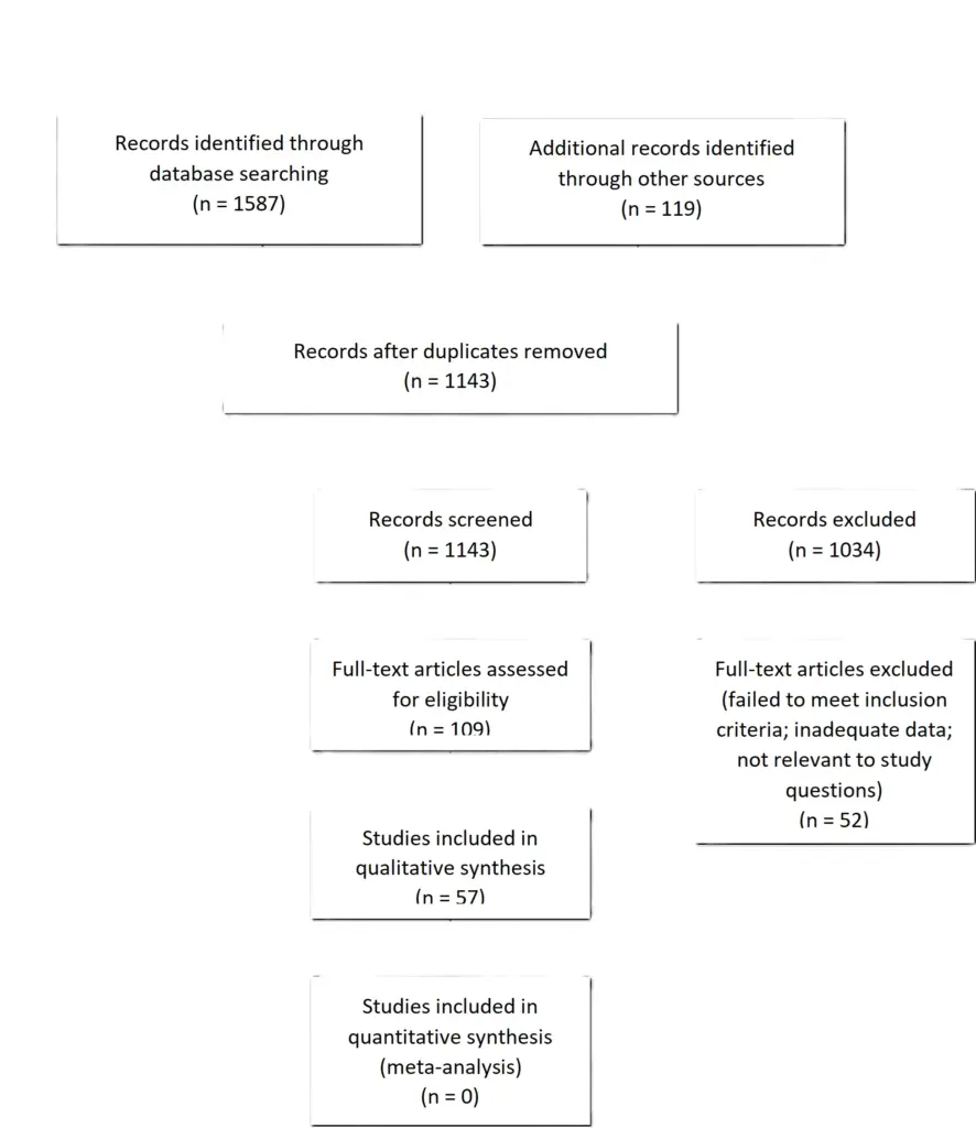
Appendix IV. Evidence Tables
Table 1 – Screening imaging in unilateral hearing loss
| Author (Year) | Study Description | Data Class | Conclusions |
| W. Yang (2020) | Pt pop: 1249 pts w/sudden sensorineural hearing loss (SSNHL) underwent MRI (1.5T or 3T) including coronal T2, axial T1, axial high-resolution T2 | III | Results: 14 (1.12%) pts were found to have VS. Screening MRI found to be more cost-effective than auditory brainstem response (ABR) test Authors Conclusions: Noncontrast, high-resolution MRI was found to be a sensitive and more cost-effective screening tool for VS Comments and Conclusions: Class III data secondary to the retrospective nature of the analysis and lack of blinding or a validation cohort |
| T. Fujita(2019) | Pt pop: 499 pts w/SSNHL underwent MRI (1.5T or 3T) including coronal T2, axial T1, axial high-resolution T2; reviewed by experienced radiologists/otologists | III | Results: 15 (3%) pts exhibited tumors on the ipsilateral side of the SNHL Authors Conclusions: Noncontrast, high-resolution three-dimensional T2WI enables accurate evaluation of CN VII/VIII within the CP angle Comments and Conclusions: Class III data secondary to the retrospective nature of the analysis and lack of blinding or a validation cohort |
Table 2 – Surveillance imaging protocols
| Author (Year) | Study Description | Data Class | Conclusions |
| S. Currie (2019) | Pt pop: 50 pts w/VS who underwent initial radiographic observation and whose MRIs included enhanced T1 and high-resolution T2 sequences were retrospectively reviewed by 2 neuroradiologists, with attention to differences in VS measurements, inter-observer reliability, and potential cost savings for non-enhanced MRI protocols | III | Results: Mean VS diameter measurements were not significantly different on enhanced T1 sequences, as compared to high-resolution T2 sequences. Inter- and intra-observer reliability concordances were excellent (0.99; >=0.98). Mean cost reduction of an unenhanced protocol was estimated at 37% Authors Conclusions: High-resolution T2 imaging provides comparable reliability to enhanced T1 sequences in follow-up assessment of observed VS, and is associated with a significant potential reduction in costs Comments and Conclusions: Class III data secondary to the retrospective nature of the analysis and lack of blinding or a validation cohort |
| D. H. Coelho (2018) | Pt pop: 50 randomly selected patients w/observed VS whose MRIs included both enhanced T1 and high-resolution T2 imaging underwent retrospective re-review by blinded neuroradiologists, with attention to VS measurement accuracy and correlation, inter-observer reliability, and differential costs and charges | II | Results: VS size measurements were highly correlated between sequences, with no significant difference in mean size estimates, and very high inter-observer reliability. Cost and charge savings of $148 and $1248 were estimated per patient per scan in an unenhanced protocol Authors Conclusions: High-resolution T2 imaging provides comparable reliability to enhanced T1 sequences in follow-up assessment of observed VS, and is associated with a significant potential reduction in costs and charges Comments and Conclusions: Class II achieved through blinded comparison of VS size measurements on enhanced T1 and high-resolution T2 images |
| M. Forgues (2018) | Pt pop: 26 VS patients with 107 MRIs were reviewed by 3 neuroradiologists, with T2 measurements compared to enhanced T1 measurements with respect to accuracy in detecting tumor growth | III | Results: T2 sequences detected growth observed on enhanced T1 sequences 88% of the time. Average measurement error between T2- and T1-weighted images was 1.27mm, or 10.4% of mean VS maximal diameter. T2 specificity was 88.2%; sensitivity was 77.8% Authors Conclusions: T2-weighted MRI is highly accurate, specific, and sensitive for detecting growth in observed VS, but is less specific and sensitive than enhanced T1 imaging, with an error of approximately 10% on routine linear measurements Comments and Conclusions: Class III data secondary to the retrospective nature of the analysis and lack of blinding or validation cohort |
| K. Buch (2018) | Pt pop: 251 observed VS underwent retrospective MRI re-review, in order to analyze differences in enhanced T1 and high-resolution T2 sequences in the measurement of VS, or the detection of clinically significant radiographic changes during patient follow-up | III | Results: No significant difference was observed in the measurements of VS made using enhanced T1 or high-resolution T2 images. Cystic and hemorrhagic components were better visualized on high-resolution T2 sequences Authors Conclusions: High-resolution T2 imaging provides comparable reliability to enhanced T1 sequences in follow-up assessment of observed VS Comments and Conclusions: Class III data secondary to the retrospective nature of the analysis and lack of blinding or a validation cohort |
Table 3 – Timing of imaging protocols for observed vestibular schwannoma
| Author (Year) | Study Description | Data Class | Conclusions |
| D. Borsetto (2022) | Pt pop: 354 pts w/sporadic VS managed w/radiographic observation and at least 10 years of total radiographic follow-up and at least 5 years of initial freedom-from-growth underwent Bayesian analysis to determine conditional probability of late growth for patients without early growth | III | Results: Late growth was observed in 12 VS (3.4%) and not significantly associated with extra-canalicular disease. The yearly conditional probabilities of late growth at years 6, 7, 8, 9, and 10 were 2.28%, 1.35%, 0.8%, 0.47%, and 0.27%, respectively Authors Conclusions: Among VSs without observed growth in the first 5 years of radiographic surveillance, the conditional probability is approximately 2% or less, and continues to decrease over time. The authors recommend MRI surveillance at 6 months, then annually for 3 years, then twice at 2-year intervals, then one final scan at 3 years later, with no further imaging indicated if no growth observed over at least 10 years of follow-up Comments and Conclusions: Class III data secondary to the retrospective nature of the analysis and lack of blinding or a validation cohort |
| J. P. Marinelli (2022) | Pt pop: 952 pts w/sporadic VS who underwent initial radiographic observation were assessed for volumetric tumor expansion | III | Results: 622 patients demonstrated ≥20% volumetric growth, with growth-free survival rates at 1, 3, and 5 years of 66%, 30%, and 20%, respectively. Among 405 patients observed after initial growth, 210 continued to grow, w/subsequent growth-free survival rates at 1, 3, and 5 years of 77%, 37%, and 24%, respectively. Large VS volume or growth rate was significantly associated w/sustained growth. Authors Conclusions: Most observed VS appear to grow during early follow-up; however, initial growth does not reliably predict sustained growth, but larger tumors with rapid early expansion are the most likely to continue to progress Comments and Conclusions: Class III data secondary to the retrospective nature of the analysis and lack of blinding or a validation cohort |
| M. A. Hentschel (2021) | Pt pop: 1217 pts w/sporadic VS undergoing radiographic observation for at least 3 years after presentation were assessed with regard to risk factors for growth using multiple imputation, regression, and decision curve analysis techniques | III | Results: 653 pts demonstrated VS growth at follow-up (54%). Increased risk of VS growth was significantly associated w/imbalance, tinnitus, higher Koos grade, shorter symptomatic interval, and larger tumor diameter. More than half of observed VS demonstrated ≥2mm of linear growth within 3 years of diagnosis. Authors Conclusions: Larger size, higher grade, and more aggressive symptomatology may predict early growth Comments and Conclusions: Class III data secondary to the retrospective nature of the analysis and lack of blinding or a validation cohort |
| J. S. Kim (2021) | Pt pop: 118 pts w/sporadic VS undergoing initial observation were assessed for risk factors predicting tumor growth using survival analysis techniques | III | Results: 5-year cumulative incidence rate for VS growth was 41.3%. Increased risk of growth was associated with larger/cisternal tumors, and early hearing loss Authors Conclusions: Early VS growth occurs in a large minority of observed VS, in particular larger tumors, or those associated with rapid-onset early hearing loss Comments and Conclusions: Class III data secondary to the retrospective nature of the analysis and lack of blinding or a validation cohort |
| H. Ruiz-Garcia (2021) | Pt pop: 243 pts w/sporadic VS initially managed with observation were assessed for predictors of radiographic tumor control and freedom-from-intervention | III | Results: Local control rates at 1, 5, and 10 years were 91%, 67%, and 58%, respectively. Larger tumors involving the CPA were significantly more likely to require treatment, in particular among younger patients Authors Conclusions: Although young patients w/larger VS are at increased risk of tumor growth requiring neurosurgical intervention, the majority of tumors did not demonstrate significant growth at 10 years Comments and Conclusions: Class III data secondary to the retrospective nature of the analysis and lack of blinding or a validation cohort |
| E. J. Patel (2021) | Pt pop: 13 sporadic, untreated VS that demonstrated spontaneous VS regression during observation, w/focused assessment of potential predictors of tumor regression | III | Results: Mean VS size reduction was 36%; 5 VS had a relative decrease of >40%, while 8 had <40% decrease, a difference that was not associated with differences in tumor size, regression rate, or audiometric parameters Authors Conclusions: Spontaneous VS regression is rare, with <4% incidence among sporadic VS, and occurs unpredictably in a relatively heterogenous patient population Comments and Conclusions: Class III data secondary to the retrospective nature of the analysis and lack of blinding or a validation cohort |
| M. Kleijwegt (2020) | Pt pop: 836 pts w/sporadic VS who underwent initial radiographic surveillance with annual MRI | III | Results: 169 pts required neurosurgical intervention during observation (20%) at a mean of 2.19 years after diagnosis. Risk factors for intervention included short duration hearing loss, imbalance, extra-canalicular tumor extension, and cystic tumor architecture Authors Conclusions: A significant minority of pts w/ observed VS may require intervention during the early follow-up period. VS that are large, cystic, or associated with imbalance or rapid-onset hearing loss may be at increased risk of progression, requiring closer follow-up Comments and Conclusions: Class III data secondary to the retrospective nature of the analysis and lack of blinding or a validation cohort |
| M. Sethi (2020) | Pt pop: 341 pts w/sporadic VS managed with initial observation and at least 5 years of follow-up, analyzed using conditional (e.g., Bayesian) probability techniques | III | Results: During the 5-year study period, 139 VS grew. Annual conditional probabilities of future growth, given no past growth, were 21%, 12%, 9%, 3%, and 2%. The conditional probability of growth was higher among extra-canalicular tumors in the first year alone Authors Conclusions: Most pts w/observed VS that fail observation will grow in the early follow-up period. VS that do not grow during the initial 4 years of observation have an estimated annual probability of <2% for future progression Comments and Conclusions: Class III data secondary to the retrospective nature of the analysis and lack of blinding or a validation cohort |
| M. Fieux (2020) | Pt pop: 336 pts w/sporadic VS (stage I or II) undergoing initial observation with annual MRI | III | Results: 125 VS progressed during the study period. Size at diagnosis (OR=2.6) and IAC filling (OR=7.7) were significantly associated with increased risk of VS growth Authors Conclusions: A large minority of observed VS demonstrate growth in early follow-up. Significant predictors of early growth included tumor size and IAC filling at time-of-diagnosis Comments and Conclusions: Class III data secondary to the retrospective nature of the analysis and lack of blinding or a validation cohort |
| J. P. Marinelli (2020) | Pt pop: 85 sporadic VS with documented growth that underwent continued radiographic observation | III | Results: 40 pts demonstrated subsequent volumetric expansion at a median 1.7y; however, annual proportions of pts in the sample w/sustained freedom from subsequent growth were 93%, 67%, 54%, 44%, and 41% Authors Conclusions: Most VS w/observed growth did not demonstrate sustained volumetric tumor expansion over the ensuing 5 years of follow-up Comments and Conclusions: Class III data secondary to the retrospective nature of the analysis and lack of blinding or a validation cohort |
| R. J. Macielak (2019) | Pt pop: 361 pts w/sporadic VS, initially managed with MRI surveillance | III | Results: 172 VS grew during the during observation, 14 of which occurred ≥5 years after initial diagnosis. Among patients with delayed growth, the latest was observed at 11.1 years, and the fastest growth rate was 1.33mm/y Authors Conclusions: Delayed growth is uncommon, but occurs in at least 8% of pts with initially observed VS. Correspondingly, lifelong surveillance is likely required for these VS Comments and Conclusions: Class III data secondary to the retrospective nature of the analysis and lack of blinding or a validation cohort |
| S. D’Haese (2019) | Pt pop: 61 pts w/sporadic VS were managed w/initial observation via annual MRI | III | Results: VS growth prompting intervention was observed in 31 pts (56%), and in 87% of them this occured within 3 years of diagnosis. Early and rapid growth were associated with larger VS size at time-of-diagnosis Authors Conclusions: Annual MRI is recommended for observed VS, in particular during the first 3 years after diagnosis. Patients with larger tumors on presentation are at increased risk of rapid or significant growth Comments and Conclusions: Class III data secondary to the retrospective nature of the analysis and lack of blinding or a validation cohort |
| D. Borsetto (2019) | Pt pop: 112 pts w/sporadic VS age 70 years and over at diagnosis, managed with initial observation at 6 months and then annually for at least 3 years | III | Results: 32 pts had tumor growth; 26 received additional treatment; 6 underwent further observation. All growth was observed within 42 months of diagnosis. Extra-canalicular tumors were significantly more likely to grow Authors Conclusions: For pts ≥70 years w/sporadic VS undergoing initial surveillance, annual MRI is recommended for 3-4 years, after which consideration may be given to discontinuing further studies Comments and Conclusions: Class III data secondary to the retrospective nature of the analysis and lack of blinding or a validation cohort |
| Z. Schnurman (2019) | Pt pop: 212 pts w/sporadic VS undergoing initial observation had 699 MRIs obtained over 2-11 years of follow-up, which were re-reviewed with volumetric analysis | III | Results: 66% of VS demonstrated growth during follow-up, 33% were stable, 1% shrank. Rapid growth was noted in 30%. Mean follow-up interval was 25 months Authors Conclusions: Most VS demonstrate volumetric growth, with up to 30% demonstrating rapid early growth, while 34% did not grow or shrank. Growth, but not growth rate, was significantly associated with larger VS size at time-of-diagnosis Comments and Conclusions: Class III data secondary to the retrospective nature of the analysis and lack of blinding or a validation cohort |
Table 4 – High-field MRI for preoperative assessment
| Author (Year) | Study Description | Data Class | Conclusions |
| N/A | N/A | N/A | N/A |
Table 5 – Tractography for preoperative assessment
| Author (Year) | Study Description | Data Class | Conclusions |
| N. Ung (2021) | Pt pop: 11 pts w/sporadic VS, mean average max tumor diameter 2.82cm | III | Results: DTI was accurate in 90.9% (10/11) of patients. Post-op 72.7% (8/11) had HB score of I or II, 18.2% (2/11) had HB III, 1 had HB IV Authors Conclusions: DTI successfully and accurately localized VII in most patients Comments and Conclusions: Class III data secondary to the retrospective nature of the analysis and lack of blinding or a validation cohort |
| M. Castellaro (2020) | Pt pop: 5 pts w/sporadic VS (mean volume 854.4 mm3) operated on via translabyrinthine approach. Multishell diffusion MRI was compared with the more standard single-shell diffusion MRI, and facial nerve tractography was performed using a probabilistic algorithm | III | Results: In the SS-MRI method, CNVII was able to be correctly visualized in 3/5 patients, in the MS-MRI technique, CNVII was able to correctly seen in 4/5 cases Authors Conclusions: Use of MS-dMRI protocol for a probabilistic tracking of FN course could be an aid for better presurgical planning of CNVII location as compared with SS-dMRI Comments and Conclusions: Class III data secondary to the retrospective nature of the analysis and lack of blinding or a validation cohort |
| T. Szmuda (2020) | Pt pop: 38 pts w/CPA tumor (32 VS, 5 meningioma, 1 epidermoid cyst) operated on via retrosigmoid approach (mean tumor diameter 29.6 mm) had CNVII course simulated before surgery using StealthViz/transferred to Medtronic S7 neuronavigation and reconstructed by DTI-fibre tracking (DTI-FT) | II | Results: CNVII was correctly predicted in 81.6% of pts. Reliability of DTI-FT for CNVII location did not depend on tumor size. Shape of CNVII (compact shape) was the dominant component influencing accuracy (p=0.03) Authors Conclusions: CNVII course can be predicted correctly in the majority of patients w/a CPA tumor, but far from the desired goal in neurosurgery Comments and Conclusions: Class II achieved through prospective analysis of reliability of CNVII localization by DTI-FT |
| L. Epprecht (2019) | Pt pop: 17 small CPA tumors (13 VS) are analyzed to localize CNVII using DTI super-resolution (SRR) vs. normal single plane DTI | III | Results: SRR increases separability of CN VII and VIII (16/17 vs. 0/17, p=0.008) Authors Conclusions: SRR improves resolution of VII and VIII in CPA compared w/single plane DTI in pts w/CPA pathologies that are small and non-operative Comments and Conclusions: Class III data secondary to the retrospective nature of the analysis and lack of blinding or a validation cohort |
| O. N. Churi (2019) | Pt pop: 40 pts w/CPA tumors (31 VS, 5 epidermoid, 2 meningioma, 2 trigeminal schwannoma) who underwent initial resection | III | Results: Accuracy of DTI as confirmed by intraoperative findings: CNVII (85%, 34/40), CNV (85%, 34/40), CNVIII (75%, 12/16) Authors Conclusions: DTI accurately localized CNVII and CNV in the majority of cases; localization of CNVIII was less successful and less accurate Comments and Conclusions: Class III data secondary to the retrospective nature of the analysis and lack of blinding or a validation cohort |
| R. Samala (2019) | Pt pop: 86 pts w/VS (>3cm) randomized to group 1 (DTI done pre-op and surgeon was informed of VII position) and group 2 (DTI not done) | II | Results: DTI group: Intraoperatively, CNVII was in the same location as seen on DTI in 39/40 patients (97.5%). CNVII was preserved anatomically and functionally in 36/40 (90%) Non-DTI group: VII was preserved anatomically/functionally in 29/46 (63%) (P= 0.002) Authors Conclusions: DTI visualization of CNVII may portend better outcomes w/regard to CNVII preservation after craniotomy for VS resection Comments and Conclusions: Class II achieved through randomization into two groups (with/without preoperative assessment of CNVII location on DTI) and prospective analysis |
| H. Li (2017) | Pt pop: 19 pts w/sporadic VS (mean tumor diameter 41mm) who underwent preoperative DTI | III | Results: Successful fiber tract localization occurred in 18/19 patients (95%). In 17/18 cases (94%) intra-operative navigation confirmed DTT localization. CNVII was located in the anterior middle part of the tumor in 7 cases Authors Conclusions: DTI was able to localize CNVII in most cases. When DTI did localize CNVII, the accuracy of that localization was validated intraoperatively Comments and Conclusions: Class III data secondary to the retrospective nature of the analysis and lack of blinding or a validation cohort |
| A. Zolal (2017) | Pt pob: Probabilistic non-tensor-based tractography was used in 21 pts w/large VS | III | Results: There was a 81% intra-operative correlation for CNVII, and 33% correlation w/CNVII Authors Conclusions: In the majority of cases, the position of CNVII, but not CNVIII, could be estimated using the probabilistic tractography Comments and Conclusions: Class III data secondary to the retrospective nature of the analysis and lack of blinding or a validation cohort |
| O. Hilly (2016) | Pt pop: 113 pts w/normal IAC/CPA anatomy were used to compare tractography using T1/T2 to determine the location of CNVII. A second group of 28 pts w/IAC/CPA tumors that were not treated surgically was used to evaluate location of the displaced CNVII. In 21 pts, preoperative localization of CNVII on MR-DTI imaging was compared with intraoperative findings | III | Results: CNVII was visualized on DTI imaging in 95% (20/21) cases. Concordance between intraoperative findings and tractography results was present in 90% of these patients (18/20). Authors Conclusions: CNVII tractography was found to be feasible and accurate in pts w/VS between 2 and 4.6 cm Comments and Conclusions: Class III data secondary to the retrospective nature of the analysis and lack of blinding or a validation cohort |
| F. Song (2016) | Pt pop: 15 pts (mean tumor size 31.9 mm) w/preoperative DTI | III | Results: In 14/15 (92.9%) of pts the DTI location was concordant with intra-operative visualization. In those consistent cases, the location of CNVII was anterior (including upper, middle, lower) in 11/14 Authors Conclusions: DTI-FT is effective in localizing CNVII in pts w/VS Comments and Conclusions: Class III data secondary to the retrospective nature of the analysis and lack of blinding or a validation cohort |
| S. A. Borkar (2016) | Pt pop: 20 pts w/sporadic VS >3cm w/preoperative DTI | III | Results: In 16/18 cases the DTI location of CNVII corresponded to intra-operative findings Authors Conclusions: DTI can be used to reliably visualize CNVII Comments and Conclusions: Class III data secondary to the retrospective nature of the analysis and lack of blinding or a validation cohort |
| J. Ma (2016) | Pt pop: 9 pts w/sporadic VS >3cm | III | Results: In 8/9 cases (89%) CNVII was able to be visualized on DTI. And in 100% of these the location was confirmed intraoperatively Authors Conclusions: DTI was able to localize CNVII in most cases; in all cases where DTI localized CNVII, the accuracy of that localization was validated intraoperatively Comments and Conclusions: Class III data secondary to the retrospective nature of the analysis and lack of blinding or a validation cohort |
| M. Yoshino (2015) | Pt pop: 22 pts w/VS in whom VII and VIII could be identified during surgery | III | Results: Used to confirm location: DTI alone: CNVII 3/22 (13.6%), CNVIII 14/22 (63.6%) CE-FEISTA alone: CNVII 13/22 (63.6%), CNVIII 1/22 (4.5%) DTT + multifused CE-FIESTA: CNVII 14/22 (63.6%), CNVIII 14/22 (63.6%) Authors Conclusions: Using DTT and multifused CE-FIESTA, authors were able to increase the number of VS pts for whom location of CNVII and CNVIII nerves were able to be predicted Comments and Conclusions: Class III data secondary to the retrospective nature of the analysis and lack of blinding or a validation cohort |
| M. Yoshino (2015) | Pt pop: 11 pts w/sporadic VS (mean tumor size 27mm) w/preoperative DTT | III | Results: In 10/11 cases (91%) a visualized fiber tract was seen on DTT exiting the brainstem and entering IAC. In 3/11 (27%) the tract was confirmed to be CNVII intra-operatively, in 6/11 (55%) this was CNVIII Authors Conclusions: DTT allows for visualization of CNVII and CNVIII preoperatively, but is unable to predict between which nerve it is, or if findings represent noise Comments and Conclusions: Class III data secondary to the retrospective nature of the analysis and lack of blinding or a validation cohort |
| P. H. Wei (2015) | Pt pop: 23 pts w/sporadic VS and preoperative DTT and CISS w/contrast | III | Results: DTT correctly identified CNVII as verified by intra-operative EMG 21/23 times (91.3%), even in cases where CNVII had become membranoid or when it was located between the capsule and tumor parenchyma Authors Conclusions: With a CNVII identification rate of 91.30%, DTT may be helpful in predicting the course of CNVII near a large VS, even in complex situations, such as a membranoid CNVII or a CNVII that traverses underneath the tumor capsule Comments and Conclusions: Class III data secondary to the retrospective nature of the analysis and lack of blinding or a validation cohort |
Table 6 – Timing of imaging protocols after vestibular schwannoma resection
| Author (Year) | Study Description | Data Class | Conclusions |
| J. M. Gonzalez-Darder (2020) | Pt pop: 30 pts w/VS treated w/microsurgical resection, all via retrosigmoid craniotomy, with postoperative radiographic extent-of-resection assessed at 6 months by contrast-enhanced MRI | III | Results: Residual tumor was noted in 11 pts (36.7%), w/all residual volumes <0.5cm3. During annual follow-up over at least 6 years, only 1 pt had progression of residual; no pt w/o nodular enhancing residuum demonstrated postoperative tumor recurrence Authors Conclusions: Postoperative MRI at 6 months appears adequate for initial extent-of-resection assessment. Annual imaging is very sensitive for capturing recurrence and progression events in follow-up Comments and Conclusions: Class III data secondary to the retrospective nature of the analysis and lack of blinding or a validation cohort |
| M. Fieux (2020) | Pt pop: 135 pts w/ VS treated w/microsurgical resection and NTR or STR achieved (i.e., not GTR), followed w/annual contrast-enhanced MRI from 1 to 5 years after surgery, using volumetric analysis | III | Results: Progression was observed in 27 patients (23.3%), with a nearly 5-fold higher odds of progression observed after STR, as compared to NTR Authors Conclusions: Postoperative MRI at 12 months appears adequate for initial extent-of-resection assessment. Annual imaging is very sensitive for capturing recurrence and progression events in follow-up. Increased residual volume is associated with higher risk of progression after STR/NTR Comments and Conclusions: Class III data secondary to the retrospective nature of the analysis and lack of blinding or a validation cohort |
| J. D. Breshears (2020) | Pt pop: 66 pts w/VS treated w/microsurgical resection and STR achieved, followed w/contrast-enhanced MRI and volumetric analysis immediately after surgery, as well as at 6-months, and then 12-month intervals. Patients receiving up-front SRS (<12 months) were excluded | III | Results: Progression was noted in 22 patients (30%) at a median of 3.1 years. Residual tumor volume and residual disease within the IAC were significantly associated with progression (OR=2.0, OR=3.7, respectively) Authors Conclusions: Although pts were assessed at 0-, 6-, and 12-months, followed by annual updates, retreatment for disease progression prior to 24 months was rare Comments and Conclusions: Class III data secondary to the retrospective nature of the analysis and lack of blinding or a validation cohort |
| M. E. Miller (2017) | Pt pop: 220 pts w/VS treated w/microsurgical resection | III | Results: 8 pts (4.1%) demonstrated radiographic progression, of whom 4 underwent retreatment. No patient was retreated at less than 1-2 years after resection; most were retreated at 5 and 10 years postoperatively Authors Conclusions: MRI surveillance after VS resection is recommended at 1, 5, and 10 years Comments and Conclusions: Class III data secondary to the retrospective nature of the analysis and lack of blinding or a validation cohort |
| Y. Tomita (2015) | Pt pop: 33 pts w/residual sporadic VS after STR | III | Results: At 3-year follow-up, growth had been observed in 10 of 33 VS after STR. Preoperative tumor volume was not related to postoperative tumor growth after STR; however, initial postoperative tumor volume was significantly associated with regrowth. No patient underwent repeat intervention during the 3-year initial postoperative follow-up period Authors Conclusions: Annual MRI surveillance is a safe and reasonable postoperative protocol for at least 3 years after surgery, w/no clinical action taken prior to 3 years Comments and Conclusions: Class III data secondary to the retrospective nature of the analysis and lack of blinding or a validation cohort |
Table 7 – Timing of imaging protocols after vestibular schwannoma SRS
| Author (Year) | Study Description | Data Class | Conclusions |
| O. Conlan (2022) | Pt pop: 159 pts w/sporadic VS treated w/initial SRS | III | Results: The mean time following SRS when regrowth began was 42 months, w/a mean growth rate of 0.48 mm/month; this growing rate was higher than the pre-SRS growth rate, but was not statistically significant (p = 0.8) Authors Conclusions: The authors did not identify any re-growth after the 6-year mark; thus, the 10-year period is reasonable, justifiable and supported by the presented data to stop w/further follow-up imaging Comments and Conclusions: Class III data secondary to the retrospective nature of the analysis and lack of blinding or a validation cohort |
| O. Fouard (2022) | Pt pop: 52 pts w/sporadic VS treated w/initial LINAC radiosurgery | III | Results: 1 tumor was stable, 26.9% had continuous shrinkage, transient tumor enlargement was observed in 63.5% (first peak at 6-12 months, late peak at 3-4 years), true progression suspected in 4 (7.7%). Only 1 patient required salvage radiotherapy Authors Conclusions: A significant tumor expansion observed on 3 sequential MRI scans after year 3 may suggest treatment failure. Long-term follow-up is therefore necessary and salvage treatment should be reserved until after year 6 Comments and Conclusions: Class III data secondary to the retrospective nature of the analysis and lack of blinding or a validation cohort |
| T. Ton (2021) | Pt pop: 42 pts w/sporadic VS (mean tumor size 16.9mm) treated w/initial SRS | III | Results: Prior to SRS the average growth rate was 0.26 mm/month, while post-SRS the growth rate was 0.05 mm/month and -0.16 mm/month at the time of the first scan (average time 11.0 months) and second scan (average time 22.3 months), respectively (p < 0.001) Authors Conclusions: Unless clinically indicated, MRI post-SRS at less than 1 year has no clinical value Comments and Conclusions: Class III data secondary to the retrospective nature of the analysis and lack of blinding or a validation cohort |
| E. Ermis (2021) | Pt pop: 53 pts w/sporadic VS treated with initial SRS (mean pre-SRS target volume 0.6 cm3) | III | Results: 1 pt had clinical/radiological progression at last f/u (55 months) and had salvage surgery. No other pt underwent salvage treatment. Two other pts had radiological progression at 24 and 36 months after SRS, but no clinical symptoms so they were not operated on Authors Conclusions: Advocate for presence of clinical deterioration as indication of salvage treatment in addition to progressive volume Comments and Conclusions: Class III data secondary to the retrospective nature of the analysis and lack of blinding or a validation cohort |
| A. Perry (2020) | Pt pop: 226 pts w/sporadic VS treated w/SRS, 91% without prior treatment (mean treatment volume 760 mm3) | III | Results: 0% with clinical action occurring by 6 months and 12 months post-SRS Authors Conclusions: Six-month post-SRS MRI has no detectable impact on patient care. Imaging should be done 12 months post-GKRS, or if clinical symptoms arise or worsen Comments and Conclusions: Class III data secondary to the retrospective nature of the analysis and lack of blinding or a validation cohort |
| T. Hasegawa (2020) | Pt pop: 615 pts w/sporadic VS treated w/SRS, 82% without prior surgery | III | Results: 42 pts (7%) required salvage resection for tumor enlargement (mean 42 months post-SRS), 17 pts had clinical directed care post-SRS (Ommaya into cyst, surgical resection, VPS) due to clinical symptoms Authors Conclusions: Clinically directed care is not common post-SRS, particularly in the first 12-24 months. If it is required, it typically presents with a clinical symptom, and is uncommonly pursued based on radiographic findings alone Comments and Conclusions: Class III data secondary to the retrospective nature of the analysis and lack of blinding or a validation cohort |
| M. H. Khattab (2020) | Pt pop: 55 pts w/sporadic VS treated w/SRS | III | Results: 1 asymptomatic patient received a repeat dose of SRS at 30 months due to increased tumor size at 24 months. Another asymptomatic patient received a repeat dose of SRS at 31 months due to tumor progression Authors Conclusions: Repeat SRS, whether in a symptomatic or asymptomatic pt, was uncommon in this cohort Comments and Conclusions: Class III data secondary to the retrospective nature of the analysis and lack of blinding or a validation cohort |
| J. D. Breshears (2019) | Pt pop: 118 pts w/sporadic VS treated w/SRS, 87% without prior surgical treatment (median treatment tumor volume of 0.73 cm3) | III | Results: 7 pts had salvage treatment after SRS (6 surgery, 1 repeat SRS) and all were related to tumor growth and/or clinical symptoms at median of 2.7 years following initial SRS Authors Conclusions: It can take as long as 6.9 yrs for 90% of tumors w/ pseudoprogression to shrink back to treatment volume. Also, 90% had peaked in size by 3.5 yrs following SRS. Moreover, 90% of tumors w/pseudoprogression had begun enlarging by 3.2 yrs following SRS. Of those enlarging after the first year, 45% were regressing by 4 yrs, and 77% by 6 yrs post-treatment Comments and Conclusions: Class III data secondary to the retrospective nature of the analysis and lack of blinding or a validation cohort |
Appendix V. Conflicts of Interest
| Task Force Member | Disclosure |
| Julie Honaker PhD, AuD | Nothing to Disclose |
| Ben Allen Strickland, MD | Nothing to Disclose |
| Eric J. Lehrer, MD | Servier Pharmaceuticals, Novocure Inc. |
| Sheryl Green, MBBCh | Nothing to Disclose |
| John P. Marinelli MD | Medtronic |
| Christopher S. Graffeo MD, MS | Nothing to Disclose |
| Isabelle M. Germano, MD, MBA | Brianlab |
| Mateo Ziu, MD | Omniscient Neurotechnology America Ltd; GT Medical Technologies, Inc |
| Walavan Sivakumar, MD | Stryker Corporation |
| Sherwin Tavakol, MD | IRRAS USA, Inc.; Globus Medical, Inc. |
| Lucas Paul Carlstrom, MD, PhD | Kuros Biosciences USA, Inc |
| Jamie J. Van Gompel, MD | Medtronic, Cadence |
| Ian Dunn, MD | Nothing to Disclose |
| Jeffrey J. Olson, MD | Verastem, Inc., Research Grant American Cancer Society, Editorial Consultant; Azurity Pharmaceuticals, Inc. |
| Ghazal S. Daher MD | Nothing to Disclose |
| Matthew L. Carlson, MD | Cochlear Americas, Advanced Bionics, Stryker Corporation; iotaMotion, Inc.; Stryker Corporation |
| Neil S. Patel, MD | Cochlear Americas, Zeiss, Viridian Therapeutics, IotaMotion, Inc. |
| Michael Sughrue, MD | Omniscient Neurotechnology America Ltd |
| Constantinos G. Hadjipanayis, MD, PhD | Stryker Corporation; Integra LifeSciences Corporation; Omniscient Neurotechnology America Ltd |
| Jeffrey Jacob, MD | Stryker Corporation; KLS; Synthes |
6. The Role of Radiosurgery and Radiation Therapy in the Management of Patients with Vestibular Schwannomas
Sponsored by: Congress of Neurological Surgeons (CNS) and the Section on Tumors
Endorsement: Reviewed for evidence-based integrity and endorsed by the American Association of Neurological Surgeons (AANS) and Congress of Neurological Surgeons (CNS)
Authors: Isabelle M. Germano, MD, MBA1; Sheryl Green, MBBCh2; Eric J. Lehrer, MD2; Mateo Ziu, MD3; Jeffrey J. Olson, MD4
1Department of Neurological Surgery, Icahn School of Medicine at Mount Sinai, New York, New York, USA
2 Department of Department of Radiation Oncology, Icahn School of Medicine at Mount Sinai, New York, New York, USA
3 Department of Neurological Surgery, Inova Health System, Fairfax, VA, USA,
4 Department of Neurosurgery, Emory University School of Medicine, Atlanta, Georgia, USA
Corresponding Author contact information:
Isabelle M. Germano, MD,MBA
Department of Neurological Surgery
Icahn School of Medicine at Mount Sina
One G. Levy Place, Box 113
New York, New York 10029
E-mail: isabelle.germano@mountsinai.org
Keywords: Radiosurgery, Fractionated radiotherapy, Gamma Knife, LINAC, radiation, vestibular schwannoma
Running title: Radiosurgery for Vestibular Schwannomas
Abbreviations:
GK: Gamma Knife
LINAC: Linear accelerator
MRI: Magnetic resonance imaging
NF2: Neurofibromatosis type
PTA: Pure tone average
SRS: Stereotactic radiosurgery
SRT: Stereotactic radiotherapy
VS: Vestibular schwannoma
ABSTRACT
Background: Radiosurgery (SRS) is an established modality for treatment of adult patients with vestibular schwannomas (VS). The aim of this work is to provide an updated literature review on this topic.
Objective: To review the literature published since the last guideline on this topic.
Methods: OVID Medline and Embase were searched for the period January 1, 2015, to
May 20, 2022, using search terms and search strategies to identify pertinent abstracts. These were then screened using published exclusion/inclusion criteria to identify full-text review articles. Evidence tables were constructed using data derived from full-text reviews and recommendations made from the evidence derived.
Results: From the total 1035 abstracts identified, 26 full-text articles met inclusion/exclusion criteria and were included in this update. Four new level III recommendations stemmed from this work. In adult patients with sporadic intracanalicular or <2cm VS, SRS should not be recommended as superior to observation alone for hearing preservation. In adult patients with sporadic VS treated with SRS, cochlear dose constraint should be considered as it provides better hearing preservation than no constraint. In the same population, single fraction SRS should be recommended rather than hypo-fractionated SRS (hfSRS: >1 and <5 fractions) as it results in decreased cranial nerve dysfunction. Finally, adult patients with sporadic VS undergoing SRS should be informed that SRS does not result in an increased number of secondary malignancies compared to the rate expected in the overall population.
Conclusion: Recent published literature provides new recommendations for the treatment of adult patients with VS with SRS.
SUMMARY OF RECOMMENDATIONS
Unchanged Questions and Recommendations From the Prior Version of These Guidelines
SRS Technology
Question
Is there a difference in outcome based on SRS equipment used: Gamma Knife (GK) versus linear accelerator (LINAC)-based SRS versus proton beam?
Target Population
This recommendation applies to all adults with VS who are candidates for SRS treatment.
Recommendation
There are no studies that compare two or all 3 modalities. Thus, recommendations on outcome based on modality cannot be made.
Radiographic Follow-Up after SRS and Retreatment
Question
What is the best time sequence for follow-up images after SRS?
Target Population
This recommendation applies to all adults with VS who underwent SRS treatment.
Recommendation
Level III: Follow-up imaging should be obtained at intervals after SRS based on clinical indications, a patient’s personal circumstances, or institutional protocols. Long-term follow-up with serial MRIs to evaluate for recurrence is recommended. No recommendations can be given regarding the interval of these studies.
Question
Is there a role for retreatment?
Target Population
This recommendation applies to all adults with VS who show radiographic progression after SRS treatment.
Recommendation
Level III: When there has been progression of tumor after SRS, SRS can be safely and effectively performed as a retreatment.
Neurofibromatosis Type 2 (NF2)
Question
What are the indications for SRS in patients with NF2?
Target Population
This recommendation applies to all adults with VS who have a diagnosis of NF2.
Recommendation
Level III: SRS is a treatment option for patients with NF2 whose VS are enlarging and/or causing hearing loss.
Questions and Recommendations Updated from the Prior Guidelines
SRS versus observation
Question: In adult patients with imaging findings consistent with sporadic intracanalicular <2cm VS without tinnitus, does SRS provide better hearing preservation than observation?
Target Population: Adult patients with imaging findings consistent with sporadic intracanalicular <2cm VS without tinnitus.
Recommendation: Level III. In adult patients with sporadic intracanalicular or <2cm VS, SRS should not be recommended as superior to observation alone for hearing preservation.
SRS and cochlear dose constraint
Question: In adult patients with imaging findings consistent with sporadic VS undergoing SRS does cochlear dose constraint provide better hearing preservation than no constraint?
Target Population: Adults with imaging finding consistent with sporadic VS undergoing SRS.
Recommendation: Level III. In adult patients with imaging findings consistent with sporadic VS undergoing SRS cochlear dose constraint should be considered as it provides better hearing preservation than no constraint. For single fraction SRS, a cochlear dose < 4Gy is associated with hearing preservation. Cochlear doses >4.2Gy are associated with higher risk of hearing loss. For fractionated SRT, cochlear dose < 35Gy is associated with hearing preservation.
SRS and Secondary Malignancies
Question: In adult patients with imaging findings consistent with VS does single fraction SRS result in higher number of secondary malignancies compared to the rate expected in the overall population?
Target Population: Adults with imaging finding consistent with VS who have undergone single fraction SRS.
Recommendation: Level III. Adult patient with sporadic VS undergoing SRS should be informed that SRS does not result in an increased number of secondary malignancies compared to the rate expected in the overall population.
New Question and Recommendation Not Included in the Prior Guidelines
SRS technique
Question: In adult patients with imaging findings consistent with VS does single fraction SRS provide better hearing preservation and/or other cranial nerve deficits that other radiation schemes, such as hypofractionation SRS (hfSRS) and/or conventional fractionated stereotactic radiotherapy (fSRT).
Target Population: Adults with imaging findings consistent with VS.
Recommendation: Level III. In adult patients with imaging findings consistent with sporadic VS single fraction SRS should be recommended rather than hypo-fractionated SRS (hfSRS: >1 and <5fractions) as it results in decreased cranial nerve dysfunction. There is insufficient data to provide a recommendation about SRS versus fSRS with regard to radiographic control, hearing preservation, and/or other cranial nerved deficits.
INTRODUCTION
Goals and Rationale
This clinical guideline has been created to improve patient care by outlining the appropriate information gathering and decision-making processes involved in the treatment of patients with VS. Neurosurgical care is provided in many different settings by many different providers. This guideline has been created as an educational tool to guide qualified physicians through a series of diagnostic and treatment decisions in an effort to improve the quality and efficiency of care.
Objectives
VS represent 8% of all primary brain neoplasms and approximately 16% of benign brain
tumors1. The aim of this study is to provide an update on the topic of SRS and radiation therapy for adults patients with VS since the previous publication on this topic2, with literature search ending on December 31, 2014. In this update, four questions were developed to provide clinically relevant information on the role of SRS used in adult patients with VS. In particular, the first question addresses the role of SRS on hearing preservation compared to observation in adult patients with intracanalicular or <2cm VS. The second question assesses the role of cochlear sparing on hearing preservation. The third question focuses on identifying ifSRS provides better hearing preservation and/or decreased other cranial nerve deficits compared to other radiation modalities. The final question focuses on the role of SRS on tumorigenesis.
Methodology
As first step, the guidelines task force reviewed the questions from the 2018 publications and modified them to PICO (patient/intervention/comparison/outcome) format. With these as guidance the guidelines task force initiated a systematic review of the literature and evidence-based guideline update relevant to the treatment of patients with VS. Through objective evaluation of the evidence and transparency in the process of making recommendations, this evidence-based clinical practice guideline was developed for the diagnosis and treatment of adult patients with VS. These guidelines are developed for educational purposes to assist practitioners in their clinical decision-making processes. Additional information about the methods utilized in this systematic review is provided below.
Literature Search
The task force members identified search terms/parameter and a medical librarian implemented the literature search, consistent with the literature search protocol, using the OVID Medline and Embase databases from 1/1/2015 to 5/20/2022 and limited to English language. Details of the strategies are noted in Appendix 1.
Inclusion/Exclusion Criteria
Articles were retrieved and included only if they met specific inclusion/exclusion criteria. These criteria were also applied to articles provided by guideline task force members who supplemented the electronic database searches with articles from their own files. To reduce bias, these criteria were specified before conducting the literature searches.
Articles that do not meet the following criteria were, for the purposes of this evidence-based clinical practice guideline, were excluded. To be included as evidence in the guideline, an article had to be a report of a study that:
- Appeared in a peer-reviewed publication or a registry report;
- Enrolled a minimum of 3 patients;
- Was of humans;
- Was published on or after January 1, 2015;
- Quantitatively presented results;
- Was not an in vitro study;
- Was not a biomechanical study;
- Was not performed on cadavers;
- Was published in English;
- Was not a systematic review, meta-analysis, or guideline developed by others
Systematic reviews or meta-analyses conducted by others, or guidelines developed by others were not included as evidence to support this review due to the differences in article inclusion/exclusion criteria specified compared to the criteria specified by the Guidelines Task Force.
The qualifying evidence derived from the searches was then collected and compared to the evidence already present in the 2018 publications, i.e., through the end of 2014, to determine if the recommendations could remain unchanged, needed updating, or if whole new recommendations were warranted.
Assessment for Risk of Bias
Our search generated a list of abstracts, which were screened, and those articles that addressed our identified questions underwent full independent review by the authors. Reviewers were critical in their assessment, specifically with regard to the study design, prospective character, size of study population, and baseline characteristics between study groups which could account for survivorship bias, selection bias, and appropriate statistical analyses of reported data. Additional bias could stem by the fact that age might have a neurodegenerative effect on hearing. Thus, its effects are difficult to separate from those of the treatment used. Finally, clinical practice could bias how patients are treated. For example, in some centers asymptomatic patients with intra-canalicular VS may be treated upfront.
The quality of evidence was rated using an evidence hierarchy for each of four different study types: therapeutic, prognostic, diagnostic, and decision modeling. Additional information regarding the hierarchy classification of evidence can be located here: https://www.cns.org/guidelines/guideline-procedures-policies/guideline-development-methodology.
Revision Plans
In accordance with the National Academy of Medicine’s standards for developing clinical practice guidelines and criteria specified by the former National Guideline Clearinghouse, the task force will monitor related publications following the release of this document and will revise the entire document and/or specific sections if new evidence shows that a recommended intervention causes previously unknown substantial harm; that a new intervention is significantly superior to a previously recommended intervention from an efficacy or harms perspective; or that a recommendation can be applied to new populations. In addition, the task force will confirm within five years from the date of publication that the content reflects current clinical practice and the available technologies for the evaluation and treatment for patients with VS.
RESULTS
Based on review of the new literature starting on January 1 2015 through May 20, 2022 it was apparent that there was new data warranting update of three questions and recommendation wording from the prior guideline related to SRS versus observation, SRS and cochlear dose constraint, and SRS and secondary malignancies. There was also additional data that warranted an additional new question addressing SRS techniques. The question for these four topics were revised to PICO format. There was no new literature that warranted an update or additional recommendations for the topics of SRS technology, radiographic follow-up and retreatment and NF2 and therefore the questions for these and their subsequent recommendations were left as they stood in the first version of the guidelines for VS.
From the total 1035 abstracts identified, 26 full-text articles met inclusion/exclusion criteria and were included in this update. Four new level III recommendations stemmed from this work. In adult patients with sporadic intracanalicular or <2cm VS, SRS should not be recommended as superior to observation alone for hearing preservation. In adult patients with sporadic VS treated with SRS, cochlear dose constraint should be considered as it provides better hearing preservation than no constraint. In the same population, single fraction SRS should be recommended rather than hypo-fractionated SRS (hfSRS: >1 and <5 fractions) as it results in decreased cranial nerve dysfunction. Finally, adult patients with sporadic VS undergoing SRS should be informed that SRS does not result in an increased number of secondary malignancies compared to the rate expected in the overall population.
Radiosurgery versus observation
Question: In adult patients with imaging findings consistent with sporadic intracanalicular or <2cm VS s without tinnitus, does radiosurgery provide better hearing preservation than observation?
Recommendation: Level III. In adult patients with sporadic intracanalicular or <2cm VS s, radiosurgery should not be recommended as superior to observation alone for hearing preservation.
Summary of previous recommendations
In the previously published guidelines on the role of radiosurgery versus observation to treat sporadic VSs in the adult population without radiographic progression, it was recommended that small tumors <2cm without tinnitus are observed, as observation does not have a negative impact on tumor growth or hearing preservation2. This was a Level III recommendation based on the available data. The previous data included retrospective reviews, all of which were Class III evidence.
Study selection and characteristics
The initial search strategy included the review of 24 full text papers. From these, three articles were included in the final review for question 1 and included in the evidence Table 1 below. Data extraction included study design, class of evidence, total number of patients, study parameters, hearing outcome, and author’s conclusions.
Ismail et al3 published a retrospective review of a cohort of 247 patients with intracanalicular or < 2 cm VSs. One group (n = 140) underwent watchful-waiting management with a mean follow up of 5.9 + 1.6 years and the other group (n = 107) underwent stereotactic radiosurgery (SRS) treatment with mean follow up of 7.1 + 1.9 years. American Association of Otolaryngology Head and Neck Surgery Pure Tone Audiograms (AAOHNS PTA) and deterioration of Speech Discrimination Score (SDS) were used as primary outcome measures. Deterioration of hearing was noticed in all patients. AAOHNS PTA decreased significantly more in patients that underwent SRS (4.62 dB/year) versus 1.90 dB/year for watchful waiting group (p < 0.001). The authors concluded that hearing outcomes are significantly better for patients undergoing watchful waiting. This is a retrospective study providing class III evidence.
Miller et al4, in a retrospective study reviewed 123 patients treated by observation (n = 89) or SRS (n = 34). They evaluated SDS, ASAOHNS PTA and Penn Acoustic Neuroma Quality of Life (PANQOL). Patients that were treated with SRS had worst hearing scores than patients in observation group. After radiation therapy their PANQOL improved. Both groups regressed to similar PANQOL total and hearing domain score at 400 weeks. It is unclear from the authors’ conclusions whether SRS is recommended to improve PANQOL since both groups’ outcome are similar at 400 weeks.
Milner et al5, aimed to assess audiological outcomes in 69 patients with growing VS (> 15% of tumor volume in 1 year). Twenty-four patients were in the watchful waiting group and 46 underwent SRS treatment. Patients in the watchful waiting group experienced a slower growth rate than the patients in SRS group (21%/year vs 178%/year respectively). AAOHNS PTA deterioration and Gardner-Robertson class deterioration showed higher percentage of deterioration after SRS. The authors concluded that audiological deterioration was worst for patients that underwent SRS. However, the later had larger tumors (1749mm3 vs 341mm3). The authors recommended that SRS should not be considered as an early treatment option if the sole aim is to attempt to preserve functional hearing.
Synthesis
Based on the three retrospective single center reviews providing Class III evidence, a level III recommendation was made that radiosurgery for adult patients with sporadic intracanalicular or <2cm VS does not provide hearing preservation benefit superior to observation alone. Of note, another retrospective single center study provided data on hearing deterioration after SRS stratifying by volume size6. Since there was no direct comparison with observation, this was not included in the evidence table. The Authors concluded that smaller tumors have a better outlook at hearing preservation after SRS since 33% of patients with volume <1.2cm3 developed hearing deterioration after SRS compared to the 49% in the larger tumor cohort.
Radiosurgery and cochlear dose constraint
Question: In adult patients with imaging findings consistent with sporadic VSs undergoing radiosurgery does cochlear dose constraint provide better hearing preservation than no constraint?
Recommendation: Level III. In adult patients with imaging findings consistent with sporadic VSs undergoing radiosurgery cochlear dose constraint should be considered as it provides better hearing preservation than no constraint. For single fraction SRS, a 4Gy cochlear dose is associated with hearing preservation. Cochlear doses >4.2Gy are associated with higher risk of hearing loss. For fractionated SRT, cochlear dose < 35Gy is associated with hearing preservation.
Summary of previous recommendations
In the previously published guidelines on the role of radiosurgery to treat sporadic VS in the adult population yielded a Level III recommendation that for single fraction SRS a dose <13Gy should be used to facilitate hearing preservation and minimize new onset or worsening of pre-existing cranial nerve deficits2. This level III recommendation was based om retrospective reviews, all of which were class III evidence. Due on new and emerging interest in determining specific doses delivered to the cochlea, in the current guidelines we examine the specific question of cochlear dose and its impact on hearing preservation
Study selection and characteristics
The initial search strategy included the review of 52 full text papers. From these 11 articles were included in the final review for question 2 and included in the evidence Table 2. Data extraction included study design, class of evidence, total number of patients, study parameters, treatment delivered, and author’s conclusions.
Maksimoski et al7 conducted a single institution retrospective study of 133 patients treated between 1998-2019 for VS with GKRS. They observed that all measures of radiation dose to the cochlea, such as maximum dose (p = 0.196), average dose (p = 0.104), and minimum dose (p=0.263) were not associated with hearing loss. This class III evidence supports a level III recommendation.
Ju et al8 conducted a single institution retrospective study of 41 patients with VS who underwent SRS to 18Gy in 3 fractions on the CyberKnife radiosurgery platform. Hearing preservation was excellent and was observed in over 90% of patients. Larger tumor volumes, higher cochlear doses, and lower cochlear volumes were associated with poorer hearing outcomes. This class III evidence supports a level III recommendation.
Frischer et al9 conducted a large single institution retrospective study of 557 patients with VS who underwent SRS alone or combination microsurgery-SRS. They observed that the median dose to the cochlea was a predictor of Gardner Robertson hearing class at follow-up (p = 0.029). Additionally, patients whose median cochlear doses exceeded 6Gy had higher rates of non-serviceable hearing at follow-up compared to those who had a median dose < 6 Gy (p = 0.027). This class III evidence supports a level III recommendation.
Patel et al10 conducted a single institution retrospective study of 100 patients with VS, where 43 underwent SRS and 57 underwent fractionated radiation treatment (50.4 Gy in 28 fractions). In patients undergoing SRS, a cochlea dose of > 5 Gy was a strong predictor of hearing loss with a sensitivity and specificity of 100% and 90%, respectively. In patients undergoing fractionated radiotherapy, a minimal cochlea dose > 35 Gy was an excellent predictor of hearing loss with a specificity and sensitivity of 91% and 50%, respectively. This class III evidence supports a level III recommendation.
Van Linge et11 al conducted a single institution retrospective comparative study with 611 patients collected from 5 institutions, where SRS (12 Gy in a single fraction) was compared to fractionated radiotherapy (54 Gy in 30 fractions) in patients who underwent primary treatment for VS. They observed that the volume of cochlea getting at least 90% of the prescription dose (V90 EQD2) was predictive of loss of functional hearing on univariate (HR: 1.01; 95% CI: 1.00-1.013; p = 0.027) and multivariate (HR: 1.01; 95% CI: 1.00-1.02; p = 0.001) analysis. This class III evidence supports a level III recommendation.
Chung et al12 conducted a single institution retrospective study of 38 patients with VS, where 14 underwent SRS and 24 underwent fractionated radiotherapy. In patients who underwent SRS, those who developed decreased hearing preservation received a significantly higher minimum dose to the cochlea (7.41 vs. 4.24 Gy; p = 0.02). Additionally, when the minimum cochlear dose exceeded 6 Gy, there was a significant risk of decreased hearing preservation (OR: 32; p = 0.02). This class III evidence supports a level III recommendation.
Hasegawa et al13conducted a single institution retrospective study of 92 patients with VS. All patients underwent single fraction SRS to a dose of 12-13 Gy. The median mean dose to the cochlea was 4 Gy. On univariate and multivariate analysis (both p < 0.001), increasing dose to the cochlea was associated with decreased hearing preservation. This class III evidence supports a level III recommendation.
Pan et al14 conducted a single institution retrospective study of 93 patients with VS who underwent GK radiosurgery. They observed that the cochlea dose was a significant predictor of hearing preservation in patients with serviceable hearing following SRS. This class III evidence supports a level III recommendation.
Lin et al15conducted a retrospective study of 100 patients with VS who underwent single fraction SRS to 12-13 Gy. They observed that the only dosimetric predictor of hearing preservation was a mean cochlear dose < 4 Gy (p = 0.02). This class III evidence supports a level III recommendation.
Watanabe et al16 conducted a retrospective study of 183 patients who underwent GKRS for VS. They observed that a mean cochlear dose > 4.2 Gy (p = 0.03) was associated with worse hearing preservation following treatment. This class III evidence supports a level III recommendation.
Mousavi et al 17conducted a single center retrospective study in 254 patients. They reported that atients in the group with no subjective hearing loss had significantly higher rates of hearing preservation compared to patients with subjective hearing loss with similar cochlear dose. This class III evidence supports a level III recommendation
Synthesis
In 10 of the 11 studies summarized above class III evidence that dose constraint to the cochlea is associated with hearing preservation is provided. This leads to a level III recommendation that in adult patients with imaging findings consistent with sporadic VSs undergoing radiosurgery cochlear dose constraint should be considered as it provides better hearing preservation than no constraint. For single fraction SRS, a cochlear dose < 4Gy is associated with hearing preservation13,15. Cochlear doses >4.2Gy are associated with higher risk of hearing loss9,10,12,16. For fractionated SRT, cochlear dose <35Gy is associated with hearing preservation. Only one of 11 studies provides contradictory class III evidence that maximum, average, and minimum doses to the cochlea are not associated with hearing loss following GKRS and is not deemed to alter the conclusions of the other eleven studies.
Radiosurgery technique
Question: In adult patients with imaging findings consistent with VSs (VS) does single fraction radiosurgery (SRS) provide better hearing preservation and/or other cranial nerve deficits that other radiation schemes, such as hypofractionation SRS (HfSRS) and/or conventional fSRT
Recommendation: Level III. In adult patients with imaging findings consistent with sporadic VSs single fraction SRS should be recommended rather than hypo-fractionated SRS (HfSRS: >1 and <5fractions) as it results in decreased cranial nerve dysfunction.There is insufficient data to provide a recommendation about SRS versus fSRS with regard to radiographic control, hearing preservation, and/or other cranial nerved deficits.
Summary of previous recommendations
In the previously published guidelines on the role of radiosurgery technology to treat sporadic VS in the adult population treated with SRS and fSRT showed no difference in radiographic control and clinical outcome2. Therefore, recommendations were not given. This lack of recommendation was based retrospective reviews, all of which were Class III evidence. Based on new emerging interest in determining the role of hypofractioned SRS, defined as >1 fraction and <5 fractions, in the current guidelines we separated this radiation scheme from conventional single fraction SRS and created a new question addressing the technique.
Study selection and characteristics
The initial search strategy included the review of 47 full text papers. From these, 10 articles were included in the final review for question 3 and included in the evidence Table 3 below. Data extraction included study design, class of evidence, total number of patients, study parameters, treatment scheme delivered, and author’s conclusions.
Kuchler et al18 reports on 149 patients treated with SRS/HfSRS (12Gy/1 fraction and 18Gy/3 fractions) compared to 87 patients treated with fractionated radiation therapy (fSRT, 57.6Gy/32 fractions) and 25 patients treated with fractionated proton therapy (FPT, 54Gy RBE/32 fractions). No statistical difference between treatment groups with regard to hearing preservation rates was observed. Facial and trigeminal nerve symptoms after RT were mild but the highest rates were observed in FPT patients. However, potential selection bias with larger tumor volumes treated by FPT could have influenced the results. This study provides class III evidence supporting level III recommendation.
Puataweepong et al19 conducted a class III trial comparing outcomes of 20 patients treated with SRS (12Gy/1 fraction) and 100 patients treated with HSRT (18Gy/3 fractions). Treatment was delivered utilizing the CyberKnife radiosurgery platform. Among 28 patients with serviceable hearing in the HSRT group, 5 and 8-year hearing preservation rates were 87% and 65% respectively. No factor was significantly associated with hearing preservation rates on univariable and multivariable analyses. Rates of non-auditory complications were 4.3% in the SRS group and 15% in the HSRT group. Koos grade III and IV tumors were associated with higher risk of non-auditory complications. This study provides class III evidence supporting level III recommendation.
Diaz et al20 conducted a class III trial evaluating 37 patients treated with SRS (12Gy/1 fraction) and 99 patients treated with hypoFSRT (39 patients receiving 18-21Gy/3 fractions and 60 patients receiving 25Gy/5 fractions). No significant difference in hearing preservation or cranial nerve injury was observed with SRS versus hypoFSRT. This study provides class III evidence supporting level III recommendation.
Khattab et al21 report a single institution retrospective study of 56 patients with VS who underwent single fraction SRS (12.5-16 Gy), 3-fraction SRS (7 Gy x 3), or 5-fraction SRS (4.5-5.5 Gy x 5). Significantly better audiologic outcomes were observed in multifraction vs. single fraction schemes (p = 0.009), albeit with short follow up. Mean or maximum cochlear doses did not predict for changes in speech awareness threshold. This study provides class III evidence supporting level III recommendation.
Singh et al 22 conducted a phase III trial from a prospectively collected international database comparing 12 patients treated with SRS (12.25Gy/1 fraction) and 52 patients treated with fractionated SRS (fSRS, 18Gy/3 fractions or 25Gy/5 fractions) using the Cyberknife robotic system. No significant difference was seen in hearing preservation rates. fSRS was associated with higher likelihood of cranial nerve dysfunction. This study provides class II evidence supporting level II recommendation.
Udawatta et al23 conducted a class III trial of 21 patients treated with SRS (12Gy/1 fraction), 33 patients treated with fSRT (50.4Gy/28 fractions) and 6 patients treated with HfSRS (25Gy/5fx). Hearing preservation rates 69.2% fSRT, 37.5% SRS and 100% HfSRT (p=0.025). HfSRT showed better hearing preservation versus SRS or fSRT but was associated with significantly more non-auditory symptoms, i.e., other cranial nerve dysfunction. This trial did not report trigeminal and facial nerve outcomes and has a small number of patients. This study provides class III evidence supporting level III recommendation.
Tang et al24 reported on 487 patients treated with single session (12.5Gy) and 74 patients treated with multisession SRS ( 6.7Gy/3 fractions over 3 consecutive days) using the GK system. Propensity score matching was used to compare the 2 groups with 29 patients from each group selected. There was a trend toward improved hearing preservation rates in the multi session group. On multivariate analysis the linear internal auditory canal length was the only significant predictor for hearing loss after multi session GKRS, odds ratio 0.3868. No significant differences were seen in trigeminal or facial nerve complications between the two groups. This study provides class III evidence supporting level III recommendation.
Lo et al25 performed a class III trial evaluating 136 patients treated with SRS (12Gy/1 fraction) and 17 patients treated with fSRT (50Gy/25 fractions). In the 49 patients with serviceable hearing treated with fSRT, hearing preservation was 55% at 3 years. On multivariable analysis, better pre-treatment ipsilateral pure-tone average was significantly associated with hearing preservation (p=0.04). 10-year actuarial rates of RT induced trigeminal nerve dysfunction 25% after SRS and 12% after fSRT (p=0.01). 10-year actuarial rates of RT induced facial nerve dysfunction 15% after both SRS and fSRT. This study provides class III evidence supporting level III recommendation.
Kessel et al26conducted a class III trial of 56 patients treated with SRS (12Gy/1 fraction) and 128 patients treated with fractionated (FSRT, 54Gy/30 fractions). The difference in hearing preservation was not significant (p=0.3). No difference in facial nerve (p=0.5) and trigeminal nerve (p=0.3) dysfunction. This study provides class III evidence supporting level III recommendation.
Combs et al27 performed a class III trial of 169 patients treated with SRS (13Gy/1 fraction) and 291 patients treated with FSRT (57.6Gy/32 fractions). No significant difference in hearing preservation, facial or trigeminal nerve dysfunction was noted between the SRS and FSRT groups.
Synthesis
One study providing class II evidence22 supported by another study providing class III evidence19 provides evidence of decreased cranial nerve dysfunction other than acoustic with SRS compared to hypo-fractionated SRS (HfSRS: >1 and <5 fractions). One study21 provides class III evidence of better audiologic outcomes after HfSRS (3 or 5 fractions compared to single fraction SRS, albeit with short follow up. These three studies combined support a level III recommendation that in adult patients with imaging findings consistent with VSs single fraction SRS might result in decreased cranial nerve dysfunction compared to hypo-fractionated SRS (HfSRS: >1 and <5 fractions). The remaining 8 studies failed to provide evidence of consistent differences in outcome comparing SRS versus fSRT. Therefore, at the present time there is insufficient data to provide a recommendation about SRS versus fSRS in regards to radiographic control, hearing preservation, and/or other cranial nerved deficits. One recent study28 reports on proton therapy indicating higher number of trigeminal and facial nerve deficits compared to SRS and/or fSRT. The possibility of selection bias with larger tumor volumes treated by proton therapy that could have influenced the results cannot be excluded.
Radiosurgery and Secondary Malignancies
Question: In adult patients with imaging findings consistent with VSs does single fraction radiosurgery result in higher number of secondary malignancies compared to the rate expected in the overall population?
Recommendation: Level III. Adult patient with sporadic VSs undergoing radiosurgery should be informed that radiosurgery does not result in an increased number of secondary malignancies compared to the rate expected in the overall population.
Summary of previous recommendations
In the previously published guidelines on the topic of tumorigenesis after radiosurgery in adult patients with VS a level III recommendation based on clinical evidence III was issued stating that patients should be informed that there is a minimal risk of tumorigenesis after SRS2. Two papers met eligibility criteria for this topic. Hasegawa et al29 reported on 440 patients treated with GK, including patients with NF2, with 1/440 developing a malignant transformation. Rowe et al30 reported 1 cases of malignant transformation and 1 of new tumor formation (glioblastoma) in 146 VS treated in 118 patients. Compared to the national incidence in the UK, the predictive incidence would have been 2.5 cases over the same follow up period.
Study selection and characteristics
The initial search strategy included the review of 16 full text papers. From these, 2 articles were included in the final review for question 4 and included in the evidence Table 4 below. Data extraction included study design, class of evidence, total number of patients, outcome and author’s conclusions.
Wolf et al31 performed a multi-center retrospective study with 1011 adult patients with VS treated with GK in 5 centers. The total number of patients studied including other pathologies equaled 4905 patients. The reported cumulative incidence of developing radiosurgery-associated malignancy, either new or a transformation of a benign tumor, was 0.045% over 10 years. The Authors concluded that the risk of developing a CNS tumor after SRS is similar to the risk in the general population. This study is the largest number of patient-years accumulated prospectively to estimate the incidence of malignant transformation and secondary malignancy in patients treated with GK radiosurgery since 1987 at five major radiosurgery centers. This class 3 evidence supports a level 3 recommendation.
Pollock et al32 reported on 358 adult patients with VS treated with GK. No radiation induced tumors were identified in 11,262 patient-years (total patients N= 1142). Of 358 VS patients, 1 (0.3%) had malignant transformation. The authors concluded that risk of a radiation-induced tumor developing after SRS was 0.0% at 5 years, 0.0% at 10 years, and 0.0% at 15 years. This class III evidence supports a level III recommendation.
Additionally, 6 case reports were identified describing progression to a malignant peripheral nerve sheet tumor (MPNST) in adult patients with sporadic VS: 2/6 patients did not receive radiation33,34, 1/6 patient received fractionated radiation35, and 3/6 received SRS36,37,38. One additional case report described a glioblastoma occurring in a patient who had previously received SRS treatment for a sporadic VS39.
Synthesis
The exposure to radiation in the range of 1- to 10Gy can result in sublethal DNA damage capable of inducing oncogenic mutation32. For this reason, the question of tumorigenesis after radiosurgery for VS is of clinical importance for both onset of new tumors and/or malignant transformation of the treated tumor. In particular, malignant peripheral nerve sheath tumors (MPNST) have an estimated incidence of 0.017 per million people per year with 1 VN-MPNST for every 1041 VS39. A recent study by Havik et al40 reported on genetic alterations associated with malignant transformation of sporadic VS in 4 tumors, two after GK radiation and two were radiation-naive. The authors report no consistent mutational signature associated with ionizing radiation and suggest the existence of a pre-malignant VS genomic signature. Class III evidence from the papers included in this review support a level III recommendation that adult patient with sporadic VS undergoing radiosurgery should be informed that radiosurgery does not result in an increased number of secondary malignancies compared to the rate expected in the overall population.
Recommendation: Level III. Adult patient with sporadic VSs undergoing radiosurgery should be informed that radiosurgery does not result in an increased number of secondary malignancies compared to the rate expected in the overall population.
CONCLUSIONS
Radiosurgery remains a heavily used therapeutic approach for treatment of adults with VS. This updates on the role of SRS for adult patients with VS yields to four new level III recommendations. These findings underscore the need to continue updating the guidelines every five years as recommended by the Academy of Medicine, previously Institute of Medicine.
KEY ISSUES FOR FUTURE INVESTIGATION
The use of national and international registries documenting patients’ selection, treatment and outcome might be helpful to further corroborate the current recommendations and perhaps add new evidence. With greater availability of proton centers, data for the VS adult population will accrue to help treating physician identifying the possible advantages and disadvantages over photon therapy. Finally, recognized barriers that might skew treatment need to be further explored.
Conflicts of Interest
All Guideline Task Force members were required to disclose all potential COIs prior to beginning work on the guideline, using the COI disclosure form of the AANS/CNS Joint Guidelines Review Committee. The CNS Guidelines Committee and Guideline Task Force Chair reviewed the disclosures and either approved or disapproved the nomination and participation on the task force. The CNS Guidelines Committee and Guideline Task Force Chair may approve nominations of task force members with possible conflicts and restrict the writing, reviewing, and/or voting privileges of that person to topics that are unrelated to the possible COIs. See Appendix V for a complete list of disclosures.
Disclosure of Funding
These evidence-based clinical practice guidelines were funded exclusively by the Congress of Neurological Surgeons, which received no funding from outside commercial sources to support the development of this document.
Disclaimer of Liability
This clinical systematic review and evidence-based guideline was developed by a physician volunteer task force as an educational tool that reflects the current state of knowledge at the time of completion. Each chapter is designed to provide an accurate review of the subject matter covered. This guideline is disseminated with the understanding that the recommendations by the authors and consultants who have collaborated in their development are not meant to replace the individualized care and treatment advice from a patient’s physician(s). If medical advice or assistance is required, the services of a competent physician should be sought. The proposals contained in these guidelines may not be suitable for use in all circumstances. The choice to implement any particular recommendation contained in these guidelines must be made by a managing physician in light of the situation in each particular patient and on the basis of existing resources.
Acknowledgments:
The guidelines task force would like to acknowledge the CNS Guidelines Committee for their contributions throughout the development of the guideline, the AANS/CNS Joint Guidelines Review Committee, as well as the contributions Trish Rehring, MPH, Director for Evidence-Based Practice Initiatives for the CNS, and Janet Waters, MLS, BSN, RN, for assistance with the literature searches. Throughout the review process, the reviewers and authors were blinded from one another. At this time the guidelines task force would like to acknowledge the following individual peer reviewers for their contributions: Patti Raksin, Tjoumakaris, Andrew Carlson, Neil Majmundar, Jeff Mullin and Koji Ebersole.
References
- Ostrom QT, Gittleman H, Farah P, et al. CBTRUS statistical report: Primary brain and central nervous system tumors diagnosed in the United States in 2006-2010. Neuro-oncology. Nov 2013;15 Suppl 2:ii1-56.
- Germano IM, Sheehan J, Parish J, Atkins T, Asher A, Hadjipanayia C, Burri S, GreenS, Loson JJ. Congress of Neurological Surgeons Systematic Review and Evidence-Based Guidelines on the Role of Radiosurgery and Radiation Therapy in the Management of Patients With Vestibular Schwannomas. Neurosurgery. 2018 Feb 1;82(2):E49-E51
- Ismail O, Sobhy O, Assal S, Sanghera P, Begg P, Irving R. Comparing Hearing Outcomes in Irradiated and Conservatively Managed Vestibular Schwannoma. Otol Neurotol. 2022;43(3):e374-e381.
- Miller LE, Brant JA, Chen J, Kaufman AC, Ruckenstein MJ. Hearing and Quality of Life Over Time in Vestibular Schwannoma Patients: Observation Compared to Stereotactic Radiosurgery. Otol Neurotol. 2019;40(8):1094-1100.
- Milner TD, Locke RR, Kontorinis G, Crowther JA. Audiological Outcomes in Growing Vestibular Schwannomas Managed Either Conservatively, or With Stereotactic Radiosurgery. Otol Neurotol. 2018;39(2):e143-e150.
- Johnson S, Kano H, Faramand A, et al. Long term results of primary radiosurgery for vestibular schwannomas. J Neurooncol. 2019;145(2):247-255.
- Maksimoski M, Bajaj A, Giri S, Sharpe LM, Kalapurakal J, Micco AG. Long-Term Hearing Outcomes From Gamma Knife Treatment for Vestibulocochlear Nerve Schwannomas in a Large, Tertiary Care, Academic Hospital. Otol Neurotol. 2021;42(10):1553-1559.
- Ju D-T, Feng S-W, Hsieh C-C, Yang Y-J, Chen Y-H, Tang C-T. Hearing preservation after cyberknife stereotactic radiosurgery for vestibular schwannomas. Journal of Medical Sciences. 2019;39(1).
- Frischer JM, Gruber E, Schoffmann V, et al. Long-term outcome after Gamma Knife radiosurgery for acoustic neuroma of all Koos grades: a single-center study. J Neurosurg. 2018:1-10.
- Patel KS, Ng E, Kaur T, et al. Increased cochlear radiation dose predicts delayed hearing loss following both stereotactic radiosurgery and fractionated stereotactic radiotherapy for vestibular schwannoma. J Neurooncol. 2019;145(2):329-337.
- van Linge A, van Os R, Hoekstra N, et al. Progression of hearing loss after LINAC-based stereotactic radiotherapy for vestibular schwannoma is associated with cochlear dose, not with pre-treatment hearing level. Radiat Oncol. 2018;13(1):253.
- Chung LK, Ung N, Sheppard JP, et al. Impact of Cochlear Dose on Hearing Preservation following Stereotactic Radiosurgery and Fractionated Stereotactic Radiotherapy for the Treatment of Vestibular Schwannoma. J Neurol Surg B Skull Base. 2018;79(4):335-342.
- Hasegawa T, Kato T, Yamamoto T, et al. Long-term hearing outcomes after gamma knife surgery in patients with vestibular schwannoma with hearing preservation: evaluation in 92 patients with serial audiograms. J Neurooncol. 2018;138(2):283-290.
- Pan SY, Liu SA, Sun MH, et al. Outcome of hearing preservation related to tumor morphologic analysis in acoustic neuromas treated by gamma knife radiosurgery. Radiat Oncol. 2017;12(1):134.
- Lin RH, Wang TC, Lin CD, et al. Predictors of hearing outcomes following low-dose stereotactic radiosurgery in patients with vestibular schwannomas: A retrospective cohort review. Clin Neurol Neurosurg. 2017;162:16-21.
- Watanabe S, Yamamoto M, Kawabe T, et al. Stereotactic radiosurgery for vestibular schwannomas: average 10-year follow-up results focusing on long-term hearing preservation. J Neurosurg. 2016;125(Suppl 1):64-72.
- Mousavi SH, Kano H, Faraji AH, et al. Hearing preservation up to 3 years after gamma knife radiosurgery for Gardner-Robertson class I patients with vestibular Schwannomas. Neurosurgery. 2015;76(5):584-590; discussion 590-581.
- Kuchler M, El Shafie RA, Adeberg S, et al. Outcome after Radiotherapy for Vestibular Schwannomas (VS)-Differences in Tumor Control, Symptoms and Quality of Life after Radiotherapy with Photon versus Proton Therapy. Cancers (Basel). 2022;14(8).
- Puataweepong P, Dhanachai M, Swangsilpa T, et al. Long-term clinical outcomes of stereotactic radiosurgery and hypofractionated stereotactic radiotherapy using the CyberKnife((R)) robotic radiosurgery system for vestibular schwannoma. Asia Pac J Clin Oncol. 2022;18(5):e247-e254.
- Soderlund Diaz L, Hallqvist A. LINAC-based stereotactic radiosurgery versus hypofractionated stereotactic radiotherapy delivered in 3 or 5 fractions for vestibular schwannomas: comparative assessment from a single institution. J Neurooncol. 2020;147(2):351-359.
- Khattab MH, Sherry AD, Whitaker R, et al. A Retrospective Cohort Study of Longitudinal Audiologic Assessment in Single and Fractionated Stereotactic Radiosurgery for Vestibular Schwannoma. Neurosurgery. 2019;85(6):E1078-E1083.
- Singh R, Ansinelli H, Jenkins J, Davis J, Sharma S, Vargo JA. Stereotactic radiosurgery and fractionated stereotactic radiosurgery for vestibular schwannomas: A comparison of clinical outcomes from the RSSearch patient registry. J Radiosurg. SBRT 2019;6(1):19-26.
- Udawatta M, Kwan I, Preet K, et al. Hearing Preservation for Vestibular Schwannomas Treated with Stereotactic Radiosurgery or Fractionated Stereotactic Radiotherapy. World Neurosurg. 2019;129:e303-e310.
- Tang X, Zheng M, Tang H, et al. Hearing outcomes between multi-session and single-session radiosurgery for vestibular schwannoma: a single center study. Translational Cancer Research. 2018;7(4):1092-1102.
- Lo A, Ayre G, Ma R, Hsu F, Akagami R, Mckenzie M, Valev B, Gete E, Vallieres I, Nichols A. Population-Based Study of Stereotactic Radiosurgery or Fractionated Stereotactic Radiation Therapy for Vestibular Schwannoma: Long-Term Outcomes and Toxicities. Int J Radiat Oncol Biol Phys. 2018 Feb 1;100(2):443-451.
- Kessel KA, Fischer H, Vogel MM, et al. Erratum to: Fractionated vs. single-fraction stereotactic radiotherapy in patients with vestibular schwannoma : Hearing preservation and patients’ self-reported outcome based on an established questionnaire. Strahlenther Onkol. 2017;193(2):171.
- Combs SE, Engelhard C, Kopp C, et al. Long-term outcome after highly advanced single-dose or fractionated radiotherapy in patients with vestibular schwannomas – pooled results from 3 large German centers. Radiother Oncol. 2015;114(3):378-383.
- Kuchler M, El Shafie RA, Adeberg S, et al. Outcome after Radiotherapy for Vestibular Schwannomas (VS)-Differences in Tumor Control, Symptoms and Quality of Life after Radiotherapy with Photon versus Proton Therapy. Cancers (Basel). 2022;14(8).
- Hasegawa T, Kida Y, Kato T, Iizuka H, Yamamoto T. Factors associated with hearing preservation after Gamma Knife surgery for vestibular schwannomas in patients who retain serviceable hearing. Journal of neurosurgery. Dec 2011;115(6):1078-1086.
- Rowe J, Grainger A, Walton L, Radatz M, Kemeny A. Safety of radiosurgery applied to conditions with abnormal tumor suppressor genes. Neurosurgery. May 2007;60(5):860-864; discussion 860-864.
- Wolf A, Naylor K, Tam M, et al. Risk of radiation-associated intracranial malignancy after stereotactic radiosurgery: a retrospective, multicentre, cohort study. Lancet Oncol. 2019;20(1):159-164.
- Pollock BE, Link MJ, Stafford SL, Parney IF, Garces YI, Foote RL. The Risk of Radiation-Induced Tumors or Malignant Transformation After Single-Fraction Intracranial Radiosurgery: Results Based on a 25-Year Experience. Int J Radiat Oncol Biol Phys. 2017;97(5):919-923.
- Bashir A, Poulsgaard L, Broholm H, Fugleholm K. Late malignant transformation of vestibular schwannoma in the absence of irradiation: case report. J Neurosurg. 2016;125(2):372-377.
- Belyaev A, Usachev D, Shimansky V, et al. Spontaneous Transformation of Vestibular Schwannoma into Malignant Peripheral Nerve Sheath Tumor. Asian J Neurosurg. 2018;13(3):810-813.
- Simmermacher S, Vordermark D, Kegel T, Strauss C. Malignization of a vestibular schwannoma 13 years after radiation therapy. HNO. 2017;65(Suppl 2):153-157.
- Wolf A, Naylor K, Tam M, et al. Risk of radiation-associated intracranial malignancy after stereotactic radiosurgery: a retrospective, multicentre, cohort study. Lancet Oncol. 2019;20(1):159-164.
- Tish S, Ross L, Habboub G, Roser F, Recinos PF. Malignant triton tumor diagnosed twelve years after radiosurgically treated vestibular schwannoma. Clin Neurol Neurosurg. 2019;183:105367.
- De Jesus O, Sanchez Jimenez JG, Santiago Quinones G, Velez R. Malignant peripheral nerve sheath tumour transformation of histological benign vestibular schwannoma after stereotactic radiosurgery in patients without neurofibromatosis. BMJ Case Rep. 2021;14(11).
- Kapurch JR, Jacob JT, Carlson ML, Atkinson JL, Raghunathan A, Link MJ. Temporal Lobe Gliosarcoma After Gamma Knife Radiosurgery for Vestibular Schwannoma. Otol Neurotol. 2016;37(8):1143-1147.
- Carlson ML, Glasgow AE, Jacob JT, Habermann EB, Link MJ. The Short-Term and Intermediate-Term Risk of Second Neoplasms After Diagnosis and Treatment of Unilateral Vestibular Schwannoma: Analysis of 9460 Cases. Int J Radiat Oncol Biol Phys. 2016;95(4):1149-1157.
- Havik AL, Bruland O, Miletic H, et al. Genetic alterations associated with malignant transformation of sporadic vestibular schwannoma. Acta Neurochir (Wien). 2022;164(2):343-352.
Appendix I: Literature Searches
Search Strategies
Ovid MEDLINE(R)
1 ((Stereotactic or stereotaxic) adj5 (radiotherap* or radiation)).mp. [mp=title, abstract, original title, name of substance word, subject heading word, floating sub-heading word, keyword heading word, organism supplementary concept word, protocol supplementary concept word, rare disease supplementary concept word, unique identifier, synonyms] 12210
2 Radiosurgery/ or RADIOSURG*.ti,ab,kw. or RADIO-SURG*.ti,ab,kw. 23970
3 (CYBERKNIFE* or cyber-knife* or cyber knife*).mp. 1773
4 (gamma knife* or linac or linear accelerator*).ti,ab,kw. 14362
5 Radiotherapy/ and (stereotactic* or stereotaxic*).mp. [mp=title, abstract, original title, name of substance word, subject heading word, floating sub-heading word, keyword heading word, organism supplementary concept word, protocol supplementary concept word, rare disease supplementary concept word, unique identifier, synonyms] 701
6 Radiotherapy, Intensity-Modulated/ 12455
7 (helical tomotherap* or intensity modulated arc therap* or intensity-modulated arc therap* or intensity-modulated radiotherapy* or volumetric modulated arc therap* or volumetric-modulated arc therap* or IMRT).ti,ab,kw. 15297
8 Radiation Dose Hypofractionation/ 1046
9 (HYPOFRACTIONAT* or HYPO-FRACTIONAT*).mp. 4903
10 or/1-9 54280
11 exp Neuroma, Acoustic/ 8763
12 ((vestib* or acoustic) adj3 (neuroma* or neurilemmoma* or neurilemoma* or neurinoma* or tumor* or tumour* or schwannoma*)).mp. [mp=title, abstract, original title, name of substance word, subject heading word, floating sub-heading word, keyword heading word, organism supplementary concept word, protocol supplementary concept word, rare disease supplementary concept word, unique identifier, synonyms] 11046
13 (acoustic nerve cancer* or acoustic neurofibroma* or acusticus neurinoma* or auditory nerve neurinoma* or ear schwannoma* or angle tumor* or angle tumour* or cerebellopontine angle tumor* or neurinoma of the acoustic nerve or neurosensory deafness* or sensoryneural deafness* or sensory neural deafness*).ti,ab,kw. 1211
14 11 or 12 or 13 12489
15 limit 14 to english language 10469
16 Animals/ not Humans/ 4974929
17 15 not 16 10374
18 comment/ or editorial/ or letter/ or review/ or systematic review/ 5102112
19 17 not 18 8685
20 exp adolescent/ or exp child/ or exp infant/ 3849849
21 exp Adult/ 7797507
22 20 not 21 2052582
23 19 not 22 8366
24 limit 23 to dt=20150101-20220522 2297
25 in vitro techniques/ 387712
26 Culture Techniques/ 47809
27 Drug Evaluation, Preclinical/ 54481
28 Disease Models, Animal/ 383220
29 Xenograft Model Antitumor Assays/ 44247
30 24 not (25 or 26 or 27 or 28 or 29) 2275
31 10 and 30 494
EMBASE
(‘radiosurgery’/exp OR radiosurg*:ti,ab,kw,de OR ‘radio-surgery’:ti,ab,kw,de OR ((stereotactic OR stereotaxic) NEAR/5 (radiotherap* OR radiation*)) OR (‘radiotherapy’/exp AND (stereotactic* OR stereotaxic*)) OR ‘cyberknife’/exp OR ‘cyberknife’:ti,ab,kw OR ‘cyber knife’:ti,ab,kw OR ‘gamma knife’/exp OR ‘gamma knife’:ti,ab,kw OR gammaknife:ti,ab,kw OR perfexion:ti,ab,kw OR ‘linear accelerator’/exp OR ‘linear accelerator’:ti,ab,kw OR linac:ti,ab,kw OR ‘intensity modulated radiation therapy’/exp OR ‘intensity modulated radiation therapy’:ti,ab,kw OR imrt:ti,ab,kw OR ‘intensity modulated arc therapy’:ti,ab,kw OR ‘intensity modulated radiotherapy’:ti,ab,kw OR ‘intensity modulated photon radiotherapy’:ti,ab,kw OR ‘intensity modulated therapy’:ti,ab,kw OR ‘intensity-modulated radiation therapy’:ti,ab,kw OR ‘intensity-modulated radiotherapy’:ti,ab,kw OR ‘tomotherapy’/exp OR tomotherap*:ti,ab,kw OR ‘hypofractionation’/exp OR hypofractionat*:ti,ab,kw,de OR ‘hypo-fractionation’:ti,ab,kw OR ‘hypofractionated radiotherapy’/exp) AND (‘acoustic nerve cancer’:ti,ab,kw OR ‘acoustic nerve neurinoma’:ti,ab,kw OR ‘acoustic nerve tumor’:ti,ab,kw OR ‘acoustic nerve tumour’:ti,ab,kw OR ‘acoustic neurofibroma’:ti,ab,kw OR ‘acusticus neurinoma’:ti,ab,kw OR ‘auditory nerve neurinoma’:ti,ab,kw OR ‘ear schwannoma’:ti,ab,kw OR ‘angle tumor’:ti,ab,kw OR ‘angle tumour’:ti,ab,kw OR ‘neurinoma of the acoustic nerve’:ti,ab,kw OR ‘neurosensory deafness’:ti,ab,kw OR ‘sensoryneural deafness’:ti,ab,kw OR ‘sensory neural deafness’:ti,ab,kw OR ((vestib* OR acoustic) NEAR/3 (neuroma* OR neurilemmoma* OR neurilemoma* OR neurinoma* OR tumor* OR tumour* OR schwannoma*))) AND [english]/lim NOT (‘animal’/exp NOT ‘human’/exp) NOT (‘juvenile’/exp NOT ‘adult’/exp) NOT (‘letter’/exp OR ‘editorial’/exp OR ‘conference paper’/exp OR ‘review’/exp) NOT (‘case report’/exp NOT ‘case control study’/exp) NOT ((‘acoustic nerve cancer’:ti,ab,kw OR ‘acoustic nerve neurinoma’:ti,ab,kw OR ‘acoustic nerve tumor’:ti,ab,kw OR ‘acoustic nerve tumour’:ti,ab,kw OR ‘acoustic neurofibroma’:ti,ab,kw OR ‘acusticus neurinoma’:ti,ab,kw OR ‘auditory nerve neurinoma’:ti,ab,kw OR ‘ear schwannoma’:ti,ab,kw OR ‘angle tumor’:ti,ab,kw OR ‘angle tumour’:ti,ab,kw OR ‘neurinoma of the acoustic nerve’:ti,ab,kw OR ‘neurosensory deafness’:ti,ab,kw OR ‘sensoryneural deafness’:ti,ab,kw OR ‘sensory neural deafness’:ti,ab,kw OR ((vestib* OR acoustic) NEAR/3 (neuroma* OR neurilemmoma* OR neurilemoma* OR neurinoma* OR tumor* OR tumour* OR schwannoma*))) AND [english]/lim NOT (‘animal’/exp NOT ‘human’/exp) NOT (‘juvenile’/exp NOT ‘adult’/exp) NOT (‘letter’/exp OR ‘editorial’/exp OR ‘conference paper’/exp OR ‘review’/exp) NOT (‘case report’/exp NOT ‘case control study’/exp) AND ‘conference abstract’/it) AND [01-01-2015]/sd NOT (‘preclinical study’/exp OR ‘animal experiment’/de OR ‘in vitro study’/exp)
Appendix II: Rating Evidence Quality
Classification of Evidence on Therapeutic Effectiveness and Levels of Recommendation
| Class I Evidence Level I (or A) Recommendation | Evidence from one or more well-designed, randomized controlled clinical trial, including overviews of such trials. |
| Class II Evidence Level II (or B) Recommendation | Evidence from one or more well-designed comparative clinical studies, such as non-randomized cohort studies, case-control studies, and other comparable studies, including less well-designed randomized controlled trials. |
| Class III Evidence Level III (or C) Recommendation | Evidence from case series, comparative studies with historical controls, case reports, and expert opinion, as well as significantly flawed randomized controlled trials. |
Appendix III: PRISMA Flowchart
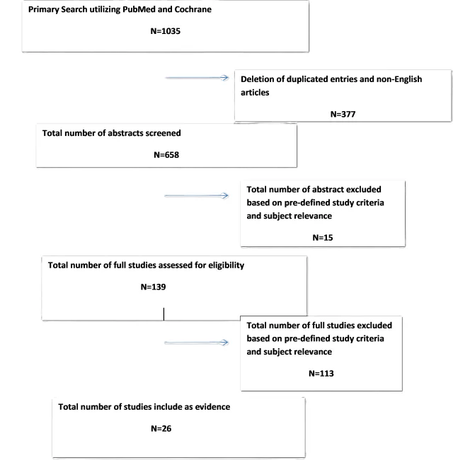
Appendix IV. Evidence Tables
Table 1: Radiosurgery versus observation in adult patients with intracanalicular or <2cm VSs without tinnitus
| Author/year | Study Description | Data Class | Conclusion |
| Ismail et al3, 2022 | Retrospective comparison review of two patients cohorts with intracanalicular or < 2 cm VS Single institution 247 patients: 140 – watchful waiting, mean follow up 5.9 +/- 1.6 years 107 – SRS, mean follow up 7.1 +/- 1.9 years SDS and AAOHNS PTA hearing threshold deterioration used as primary outcomes | III | Data Summary: Mean tumor size increase: – watchful waiting: from 9.59 +/- 4.89 mm to 9.80 +/- 5.44 mm (p = 0.926) – SRS: 11.30 +/- 5.02 mm to 13.68 +/- 4.96 mm (p < 0.001) Deterioration of hearing overtime: – watchful wait: Mean PTA decrease of 1.90 dB/yr – SRS: Mean PTA increase of 4.62 dB/yr (p <0.001) SDS available for 46 watchful wait patients and 47 for SRS patients SDS decline for SRS patients was 4.54%/year for watchful wait was 1.56%/yr Serviceable hearing preservation: – Watchful waiting: 16 (34.8%) at time of presentation and 11 (23.9%) after 5.7 years (p = 0.025) – SRS: 13 (27.7%) at presentation and 2 (4.3%) at the end of 6.9 years (p = 0.001) Author’s Conclusions: Hearing outcomes of watchful waiting strategy are significantly better than those managed by SRS. Patients with < 2cm VS will have better hearing outcomes if managed conservatively Comments Retrospective single institution study. |
| Miller et al2, 2019 | Retrospective comparison of two patient cohorts with VS Single institution 123 patients with VS included – SRS: 34 patients – Observation: 89 Main outcome measures were PTA, SDS, PANQOL Score | III | Data Summary: Mean Tumor Size: Observation patients vs SRS was 16.2 mm vs. 7.7 mm respectively (p < 0.001) Mean PTA at diagnosis was worse for SRS patients (50 +/- 20 dB) than observation group (36 +/- 20 dB) SDS at diagnosis was worse for SRS (42 +/- 32%) vs observation group (72 +/- 29%) PANQOL score was higher at baseline in observation group (532 +/- 115) than in the SRS group (455 +/- 120) (p = 0.006) SRS group had higher probability to progress to class D hearing compared to observation (HR 7.1, p = 0.005) Rate of change of PANQOL scores in SRS group improved in total at a rate of 23.92/yr (p = 0.005) and hearing 2.39/yr (p = 0.04) domain score when compared with observation Both groups regress to similar PANQOL total and hearing domain scores at 400 weeks Author’s Conclusions: PANQOL scores were higher at baseline for observation group. Overtime they increased for SRS group and decreased for Observation group, resulting in equivalent scores for both groups at 400 weeks. Comments Retrospective review. Not clear what conclusions to deduct from this study. |
| Milner et al3, 2018 | Retrospective comparison review patients with growing VS (>15% tumor volume in 1 year) treated either with SRS or watchful waiting Single institution 69 patients with growing VS: 24 – Watchful waiting 46 – SRS (GK 12 – 13 Gy marginal dose) Mean follow up: 69.6 months for SRS and 71.7 months for Watchful waiting cohort AAOHNS PTA hearing threshold deterioration and Gardner-Robertson class deterioration overtime were used as primary outcome measurements | III | Data Summary: SRS group: Tumor growth rate before treatment 178%/yr watchful wait group growth rate 21%/year Mean tumor size: SRS: 1749.4 mm3 [range 12 – 9504 mm3] Watchful wait: 341.5 mm3 [range 6 – 1785 mm3] AAOHNS PTA deterioration for SRS (mean = 32.5 dB) and watchful waiting (mean 26.8 dB) (t = 0.33, SE = 7.44, p=0.745). Gardner-Robertson class deterioration for SRS demonstrated in 13/43 patients (65.1%), for watchful wait 13/23 (56.5%) (Adj OR = 2.63, 95% CI 0.21 – 32.5, p = 0452) Functional hearing was maintained in 8 (53.3%) of watchful wait patients and 7 (25.9%) SRS patients (X2 = 3.15, df = 1, p = 0.075) Mean rate of progression to loss of functional hearing was 23.1 months in SRS patients and 26.7 months in watchful waiting patients (Adj. HR 0.37, 95% CI 0.11 – 1.26, p = 0.11) Author’s Conclusions: Audiological deterioration is greater amongst patients treated with SRS that had preserved hearing at diagnosis. Patients with lower AAOHNS PTA and functional hearing threshold at diagnosis are more likely to develop a deterioration in hearing threshold and functional hearing. SRS should not be advocated as an early treatment modality in growing VS if sole aim is to attempt to preserve functional hearing Comments Retrospective study with inherent biases such as SRS cohort was larger and had faster growing tumors. |
Abbreviations: RT = Radiation Therapy; VS = Vestibular Schwannoma; AAOHNS PTA = American Association of Otolaryngology – Head and Neck Surgery Pure Tone Audiograms; SDS = Speech discrimination score; Mo = months; SRS = Stereotactic RadioSurgery; PTA = Pure Tone Average; PANQOL = Penn Acoustic Neuroma Quality of Life;
**volume 1.2cm3=diameter 1.32cm
Table 2: Radiosurgery and cochlear dose constraint in adult patients with imaging findings consistent with sporadic VSs undergoing radiosurgery.
| Author/year | Study Description | Data Class | Conclusion |
| Maksimoski et al7, 2021 | Study Description: Retrospective study of patients with VS Patient Population: N = 133 patients with VS Treatment: All patients underwent GKRS | III | Results: All measures of radiation dose, such as maximum cochlear dose (p = 0.196), average cochlear dose (p = 0.104) and minimum cochlear dose (p = 0.263) were not associated with hearing loss. Author Conclusion: Maximum, average, and minimum doses to the cochlea are not associated with hearing loss following GKRS. Comments and Conclusions: Class III due to retrospective single center study. Maximum, average, and minimum doses to the cochlea are not associated with hearing loss following GKRS. |
| Ju et al8, 2019 | Study Description: Retrospective study in patients with VS Single center Patient Population: N = 41 patients with VS Treatment: CyberKnife SRS to 18 Gy in 3 fractions | III | Results: In 37/41 (90%) patients hearing was either preserved or improved following treatment. Higher cochlear radiation doses was associated with worse hearing outcomes. Author Conclusion: 18 Gy delivered in 3 fractions in patients with VS results in excellent hearing preservation. Comments and Conclusions: Class III due to retrospective single institution study with relatively small numbers of patients. fSRS is associated with excellent hearing preservation in patients with VS. Hearing outcomes are likely further improved by minimizing dose to the cochlea. |
| Frischer et al9, 2019 | Study Description: Retrospective study of patients with VS Patient Population: N = 557 patients with VS Treatment: N = 452 underwent SRS alone and N = 105 underwent microsurgery-SRS | III | Results: On multivariate analysis, median dose to the cochlea was a predictor of GR class at follow-up (p = 0.029). Patients whose median cochlear dose exceeded 6 Gy had a higher rate of nonserviceable hearing at last follow up compared to those who had a median cochlear dose < 6 Gy (p = 0.027). Author Conclusion: Minimizing cochlear doses appears to be beneficial for hearing preservation; however, intracanalicular tumors should not be undertreated in favor of lowering the cochlea dose. Comments and Conclusions: Large Retrospective study with mixed patient population SRS alone and SRS as after surgery. Cochlear median doses < 6 Gy appear to be associated with improved hearing outcomes. |
| Patel et al10, 2019 | Study Description: Retrospective study of patients with VS Patient Population: N = 100 patients Treatment: N = 43 underwent SRS and N = 57 underwent fractionated radiotherapy (50.4 Gy in 28 fractions) | III | Results: Patients who developed a decrease in their GR score and those who lost serviceable hearing following SRS/fractionated radiotherapy were noted to have significantly higher minimal doses to the cochlea. In patients undergoing SRS, a cochlea dose > 5 Gy was an excellent predictor of hearing loss with a sensitivity and specificity of 100% and 90%, respectively. In patients undergoing fractionated radiotherapy, a minimal cochlea dose >35 Gy was an excellent predictor of hearing loss with a specificity and sensitivity of 91% and 50%, respectively. Authors Conclusion: Minimal dose to the cochlea with thresholds < 5 Gy and 35 Gy for SRS and fractionated radiotherapy, respectively may improve hearing preservation following radiation treatment for VS. Comments and Conclusions: Class III due to single institution retrospective design with small sample size. Cochlear dose thresholds of 5 Gy and 35 Gy for SRS and fractionated radiotherapy, respectively may be associated with improved hearing preservation following radiation treatment for VS. |
| Van Linge et al11, 2018 | Study Description: Retrospective comparative study in patients with VS Single center Patient Population: N = 94 adult patients with VS Treatment” SRS in N = 67 patients to a dose of 12 Gy in a single fraction Fractionated Radiotherapy (fSRT) in N = 27 patients to 54 Gy in 30 fractions | III | Results: On both univariable and multivariable analyses, the volume of cochlea getting at least 90% of the prescription dose (V90 EQD2) was significantly associated with loss of functional hearing, increase in baseline GR class, and deterioration in PTA. No differences were observed by fractionation scheme. Authors Conclusion: Limiting the V90 to the cochlea may reduce progression of hearing loss Comments and Conclusions: Retrospective study comparing SRS to fractionated radiotherapy. Class III evidence due to retrospective nature, limited sample size, and patients all treated at a single institution. Limiting cochlea V90 may reduce hearing loss. |
| Chung et al12, 2018 | Study Description: Retrospective case control study of patients with VS Single center Patient Population: N = 38 patients with VS Treatment: N = 14 patients underwent SRS (12 Gy x 1) or fractionated radiotherapy (N = 24, fSRT 50.4 Gy in 28 fractions) | III | Results: In patients who underwent SRS, those who developed decreased hearing received a significantly higher minimum cochlear dose (7.41 vs. 4.24 Gy; p = 0.02). When the minimum cochlear dose exceeded 6 Gy, there was a significant risk of decreased hearing preservation (OR: 32; p = 0.02) In patients who underwent fractionated radiotherapy, no differences in cochlear dose were observed for patients with decreased hearing. Author Conclusion: Higher minimum cochlea dose was predictive of decreased hearing preservation in patients undergoing SRS for VS. Comments and Conclusions: Class III due to small sample size and single institution retrospective nature. Higher minimum doses to the cochlea in patients undergoing SRS is predictive of worsened hearing preservation. |
| Hasegawa et al13, 2018 | Study Description: Retrospective study of patients with VS Patient Population: N = 92 patients with VS Treatment: All underwent GKRS to a median margin dose of 12 Gy | III | Results: The median mean cochlear dose for all patients was 4 Gy. On univariable (p < 0.001) and multivariable (p < 0.001) analysis, mean cochlear dose was associated with hearing preservation. Author Conclusion: Higher mean cochlear doses are associated with decreased hearing preservation. Comments and Conclusions: Class III due to single institution retrospective study. Increasing mean cochlear dose is associated with worse hearing preservation in patients with VS undergoing SRS. |
| Pan et al14, 2017 | Study Description: Retrospective study of patients with VS Patient Population: N = 93 patients with VS Treatment: Upfront GKRS in all patients to a margin dose of 12 Gy | III | Results: The dose to the cochlea was a statistically significant predictor of hearing preservation in patients with serviceable hearing following SRS. Authors Conclusion: Cochlea dose is a significant predictor of hearing preservation in patients with serviceable hearing following SRS. Comments and Conclusions: Class III due to small sample size and single institution retrospective design. Cochlea dose is a significant predictor of hearing preservation. |
| Lin et al15, 2017 | Study Description: Retrospective Study of Patients with VS Patient Population: N = 100 patients with VS Treatment: All patients underwent single fraction SRS to 12-13 Gy | III | Results: Mean cochlear dose < 4 Gy was the only significant dosimetric predictor of hearing preservation (p = 0.02) Authors Conclusion: The mean dose to the cochlea is the most crucial factor for hearing preservation following SRS for VS. Comments and Conclusions: Class III due to single institution retrospective design. Maintaining a mean cochlear dose of < 4 Gy is associated with improved hearing outcomes following SRS for VS. |
| Watanabe et al16, 2016 | Study Description: Retrospective study of patients with VS Patient Population: N = 183 patients with VS Treatment: GKRS in all patients | III | Results: A mean cochlear dose of > 4.2 Gy (p = 0.03) was associated with worse hearing preservation. Authors Conclusion: Mean cochlear dose > 4.2 Gy is a significant predictor of hearing preservation. Comments and Conclusions: Class III due to single institution retrospective design. A mean cochlear dose of > 4.2 Gy is associated with worse hearing preservation following GKRS for VS. |
| Mousavi et al17, 2015 | Study Description: Retrospective study in patients with VS Single center Patient Population: N = 68 patients with GR Class I hearing; N = 25 had no subjective hearing loss and N = 43 has subjective hearing loss prior to GKRS Treatment: SRS to 12.5 Gy x 1 | III | Results: No statistically significant difference between the two groups was observed for mean cochlear dose or median tumor margin dose. Patients in the group with no subjective hearing loss had significantly higher rates of hearing preservation. Median cochlear dose, median tumor marginal dose, and median maximum dose were not associated with worse hearing outcomes. Author Conclusion: At 2- to 3-years following GKRS, patients without subjective hearing loss or a PTA < 15 dB had higher rates of hearing preservation. Comments and Conclusions: Class III due to single institution retrospective nature. Patients without subjective hearing loss or a PTA < 15 dB had higher rates of grade I or II hearing preservation 2- to 3-years post GKRS. |
Abbreviations: ASHL: acute sensorineural hearing loss; EQD2: equivalent dose in 2 Gy fractions; GKRS: Gamma Knife Radiosurgery; GR: Gardner Robertson hearing scale; Gy: gray; HR: hazard ratio; PTA: pure tone average; OR: odds ratio; SAT: speech awareness threshold; SRS: stereotactic radiosurgery; VS: vestibular schwannoma
Table 3. Hearing preservation and /or other cranial nerve deficits after radiosurgery delivered as single fraction radiosurgery (SRS) and/or hypofractionation SRS (HfSRS) and/or conventional fSRT in adult patients with imaging findings consistent with VSs .
| Author/Year | Study Description | Data Class | Conclusion |
| Kuchler et al18, 2022 | Retrospective review Single institution Objective: Evaluate differences in tumor control, symptoms and quality of life after SRS/HfSRT (149 patients) vs. fractionated radiation therapy (fSRT) (87 patients) versus fractionated proton therapy (FPT) (25 patients) Median doses: SRS/fSRS 12Gy/1fx and 18Gy/3fx fSRT 57.6Gy/32 fx FPT 54Gy (RBE)/32 fx Median follow-up 38 months QOL assessment via questionnaire Median tumor volume: SRS/HFSRT 0.65ccm fSRT 1.59ccm FPT 3.93ccm | III | Results: Hearing preservation rate 97% at 12 months and 87% at 60 months with no statistical difference between treatment groups (p=0.31). Facial and trigeminal nerve symptoms after SRS/fSRT were mild but with highest rates in FPT patients. Facial n: 6.9% after SRS/fSRT vs.27.8% after FPT. Trigeminal n: 10.8% after SRS/fSRT vs.20% after FPT. The majority of FPT patients who developed nerve related symptoms had large VS with brainstem contact (20%) or compression (60- 80%). In patients with SRS/HFSRT lower risk for facial and trigeminal nerve impairment was seen with fSRTversus SRS, but the difference was not statistically significant (p=0.66 and p =0.4 respectively) Authors conclusions: SRS/fSRS, fSRT and FPT show similar overall clinical and functional outcomes. Cranial nerve impairment rates vary, potentially due to selection bias with large VS in the fSRT and FPT group. Comments and conclusions: Class III evidence due to retrospective study from single institution and due to cross-sectional study in a population treated over 10 years. Relatively small number of patients treated with proton therapy. The available data on FPT are biased as these patients had significantly larger lesions and were more symptomatic at time of treatment. No significant difference in outcomes was observed but higher rates of cranial nerve complications were observed in patients with large VS treated with fractionated therapy. |
| Puataweepong et al19, 2022 | Retrospective review Single institution Treatment delivered with CyberKnife Objective: Compare long-term clinical outcomes with SRS (23 patients) versus HfSRS (100 patients). SRS was used for patients with non-serviceable hearing and Koos grade I-III tumors. HfSRS was used for patients with serviceable hearing and Koos grade III-IV tumors. Median dose: SRS 12Gy/1fx HfSRS 18Gy/3fx Median tumor volume: SRS 0.92 cm3 and HSRT 2.9 cm3 Median follow-up 72 months | III | Results: Among 28 patients with serviceable hearing in the HSRT group, the 5 and 8-year hearing preservation rates were 87% and 65% respectively. The median time to hearing deterioration was 71 months. No factor was significantly associated with hearing preservation rates on univariable and multivariable analyses. The rate of non-auditory complications was 4.3% in the SRS group and 15% in the HfSRS group. Koos grade III and IV was associated with non- auditory complications. Authors conclusions: fSRS delivered via CyberKnife may result in acceptable hearing preservation rates albeit with a higher incidence of non-auditory complications in Koos III/IV patients. Comments and conclusions: Class III evidence due to retrospective study from single institution. Patient numbers with serviceable hearing at time of treatment were small. There is a higher incidence of non-auditory complications in patients with Koos III/IV in the reported population. |
| Soderlund Diaz et al20, 2020 | Retrospective review Single institution Objective: To assess outcomes after LINAC based radiosurgery (SRS) (37 patients) and fractionated radiosurgery (fSRS) (99 patients) and identify possible differences in outcomes SRS median dose 12Gy/1fx fSRS 18-21Gy/3fx (39patients) or 25Gy/5fx (60 patients) Median PTV volume 2.5cm3 (range 0.6-10.8). PTV was significantly larger in the hypofSRS 5 fraction group with median volume of 3.9cm3 Median follow-up was 57 months (but was significantly shorter in the SRS group at 41 months p=<0.001) | III | Results: Overall hearing preservation 35% (mean audiometric follow-up of 33 months). No significant difference in hearing preservation after SRS vs. fSRS (p=0.15) No difference in trigeminal nerve toxicity incidence was observed between SRS and fSRs (p=0.61). No difference was found between the treatment groups for facial nerve toxicity (p=0.35) Authors conclusions: Both SRS and fSRS are effective treatment options with no differences in hearing preservation or neurologic toxicity. SRS recommended for small to medium sized VS. fSRS is an alternative option including those with larger tumors. Dose fractionation for fSRS regimen is yet to be refined as patients treated with both 6Gy and 7Gy per fraction but analyzed together. Comments and conclusions: Class III evidence due to being a retrospective study from single institution. Excellent long-term outcomes seen with 3-5 fraction fSRS. Further studies needed to refine optimal patient selection criteria for SRS versus fSRS and dose fractionation for fSRS requires further validation. |
| Khattab et al21, 2019 | Study Description: Retrospective study in patients with VS Patient Population: N = 56 patients with VS Treatment: Single fraction SRS in N = 12 patients (12.5-16 Gy) 3-fraction SRS in N = 12 patients (7 Gy x 3) 5-fraction SRS in N = 31 patients (4.5-5.5 Gy x 5) | III | Results: Multifraction SRS was associated with better audiologic outcomes compared to single fraction (p = 0.009). Both cochlear mean dose (p = 0.85) and cochlear max dose (p=0.82) were not associated with a change in speech awareness threshold (SAT). Authors Conclusion: Multifraction SRS may provide better hearing preservation than single fraction SRS in patients with VS Comments and Conclusions: Class III due to small number of patients and a retrospective, single institution cohort. Multifraction SRS may provide better hearing preservation in patients with VS compared to single fraction SRS. |
| Singh et al22, 2019 | Multi institutional analysis with data obtained from a prospectively collected international database of VS patients treated with SRS and fSRS Objective: Compare clinical outcomes following SRS (12 patients) and fractionated stereotactic radiosurgery (fSRS) (52 patients). All patients were treated with CyberKnife robotic radiosurgery system Median # of fractions 3 (range of 1-5) Median prescription dose: 1 fx 12.25Gy 3 fx 18Gy 5 fx 25Gy GTV median 1.09 cc (range 0.008–34.8) Median follow-up 30.4 months (range 7.5–107) Potential factors predictive of toxicity were estimated using the Kaplan-Meier method, Cox proportional hazard model and binary logistics regression with propensity score weighting | II | Results: In regards to hearing loss, no statistically significant difference identified between SRS (0%) and fSRS (13.5%) (p=0.14) despite significantly longer median follow-up in the SRS group. fSRS was associated with a higher likelihood of toxicities (42.3% versus 8.3% for SRS; p=0.054), including trigeminal and facial nerve dysfunction. Author conclusions: Equivalent local control achieved but higher cranial nerve toxicity with fSRS. Comments and conclusions: Class II evidence from multi-center prospective data. Specifics regarding treatment planning unknown and may have varied amongst institutions. Follow up time was limited. |
| Udawatta et al23, 2019 | Retrospective chart review Single institution Objective: Primary – To determine differences in preservation of serviceable hearing for patients treated with SRS (21 patients), fSRT (33 patients) or HfSRT (6 patients) SRS median dose 12Gy/1 fraction HfSRT median dose 25Gy/5fractions FSRT median dose 50.4Gy/28 fractions Median follow-up 31 months, but significantly shorter in the HfSRT group (median 9 months) The SRS cohort had a higher baseline incidence of non-serviceable hearing compared with other cohorts before RT (p=0.001) | III | Results: Significant differences in hearing preservation rates as an effect of treatment type using Fisher exact test (p=0.025). Hearing retention rates 69.2% for FSRT, 37.5% for SRS and 100% for HfSRT patients. SRS cohort showed shorter time to hearing deterioration compared with FSRT and HfSRT cohorts. Authors conclusions: fSRT showed better hearing preservation versus SRS but was also associated with significantly more non auditory symptoms that can exert a negative impact on patient’s quality of life. No radiation related symptoms seen in the HfSRT group however patient numbers were small and follow-up time-limited. Future studies needed to determine whether HfSRT can be used as a valid alternative to fSRT. Comments and conclusions: Class III evidence due to retrospective nature, and single institution study. Benefit of HfSRT limited by small patient numbers and limited follow-up. fSRT demonstrated better hearing preservation than SRS but with greater negative impact on quality of life. |
| Tang et al24, 2018 | Retrospective review Single institution Objective: To determine differences in hearing outcomes between single session (487 patients) and multisession (74 patients) GK radiosurgery (GKRS) Treatment Single session: Mean marginal dose 12.5Gy prescribed to the 45-60% isodose line Multi session: 3 sessions over 3 consecutive days with mean marginal dose per session of 6.7Gy prescribed to the 49- 70% isodose line Single session: Average tumor volume 3.03cm3 (range: 0.24–9.4) All test session: Average tumor volume 2.81 cm3 (range: 0.16–9.77 Follow-up: The mean follow-up for audiometry was 16 months in a single session group and 21 months in the multi session group. Propensity score matching was used to compare the 2 groups -29 patients from each group were selected for study. Patient’s age, tumor volume, pre-GKRS pure-tone average and radiographic follow-up were not significantly different between the 2 groups (p>0.05) | III | Results: Hearing preservation rates were analyzed using both “<20dB change in PTA” and GR class I or II criteria. No significant difference in hearing preservation rates were identified between the 2 groups (PTA: 69% versus 65.5%, p=0.08; GR class: 51.9% versus 45%, p= 0.1015). There was a trend toward improved hearing preservation rates in the multi session group. The audiometric follow-up time in multi session groups was significantly longer than in single session group (21 versus 16 months, p= 0.0385). On multivariate analysis the linear internal auditory canal (IAC) length was the only significant predictor for hearing loss after multi session GKRS; odds ratio 0.3868. No significant difference seen in trigeminal or facial nerve complications between the 2 groups. Authors conclusions: Multi session GKRS is a safe and effective treatment option but hearing preservation rates were not superior to single session GKRS. Patients with longer IAC length may benefit from multi session treatment in terms of hearing preservation. Comments and conclusions: Class III evidence due to retrospective study and single institution. Follow-up period limited. No significant difference in hearing outcomes between the two groups. |
| Lo et al25, 2018 | Retrospective review Population-based study Objective: Evaluate long-term outcomes and toxicities of SRS (136 patients) and fSRT (17 patients) SRS dose 12Gy/1fx FSRT dose 50Gy/25fx FSRT used for tumors >3cm and for patients with serviceable hearing (Gardner–Robertson classes I and II) Mean tumor volume: SRS 2.cm3 and FSRT 3.6cm3 Median follow-up 7.7years | III | Results: In the 49 patients with serviceable hearing treated with FSRT, hearing preservation was 55% at 3 years and 29% at 7 years. In multivariable analysis, better pretreatment ipsilateral pure-tone average was significantly associated with hearing preservation (p=0.04). 10 year actuarial rates of RT induced trigeminal nerve dysfunction were 25% after SRS, compared with 12% after FSRT (p=0.01). 10-year actuarial rates of RT induced facial nerve dysfunction were 15% after both SRS and FSRT. Authors conclusions: Both SRS and SRT provide excellent long-term local control however SRS was associated with higher rates of trigeminal nerve dysfunction. Even with FSRT, hearing preservation declined steadily with long-term audiometric follow-up. Comments and conclusions: Class III evidence due to retrospective review of population based study and the small number of patients treated with FSRT. Long-term hearing preservation rates decline despite the use of FSRT. |
| Kessel et al26, 2016 | Retrospective review Single institution Surveys assessing symptoms and quality of life sent to patient with a return rate of 76%. Objective: To assess hearing preservation and QOL outcomes for VS patients treated with SRS (56 patients) and fSRT (128 patients) Treatment: SRSmedian dose 12Gy/1 fraction Fractionated fSRT median dose 54Gy/30fractions Median PTV volume 1.96 ml (range at 0.09-41.1 ml) Median follow-up: 7.5 years (range 0-14.4years). Of patients who returned the survey, median follow-up was 9.6 years. | III | Results: In SRS group the median hearing preservation was 36.3 months (range 2.3-13.7 years). Hearing worsened in 30%. In fSRT group median hearing preservation was 48.7 months (range 0-13.8 years). The difference in hearing preservation was not significant between SRS and FSRT (p=0.3). No difference in facial and trigeminal nerve dysfunction was noted between the SRS and FSRT groups (facial nerve p=0.5 and trigeminal nerve p=0.1). The survey response results correlated well with information documented in patient charts for facial and trigeminal nerve toxicity but significant differences noted in reporting of hearing impairment. Authors conclusions: Patient self-reported outcome during follow-up is of high value. fSRT can be offered independent of tumor size whilst SRS should be reserved for treatment of smaller lesions. Comments and conclusions: Class III evidence due to this being a retrospective study from a single institution. Patient self-reported outcomes were valuable but the questionnaire requires further validation. No significant difference in hearing preservation was noted between the two groups. |
| Combs et al27, 2015 | Retrospective review Pooled results from 3 large centers Objective: To evaluate local control, hearing preservation and cranial nerve toxicity for patients treated with SRS (169 patients) or FSRT (291 patients) SRS median dose 13Gy/1fx fSRT median dose 57.6Gy/32fx Median tumor diameter 15 mm Median follow-up 67 months | III | Results: Long-rank test used to evaluate intergroup differences in outcome. Loss of useful hearing was 14% for fSRT and 16% for SRS group. For patients treated with SRS ≤13Gy hearing deterioration was 13%. No difference in trigeminal and facial nerve toxicity between SRS and fSRT groups. Authors conclusions: When chosen diligently based on tumor volume, pre-treatment characteristics and volume- dependent dose-prescription), both treatments may be considered equally effective. Comments and conclusions: Class III evidence due to retrospective study with pooled results from 3 centers. No significant difference in outcomes between the 2 groups. |
Abbreviations: SRS= stereotactic radiosurgery; fx= fraction, fSRS= fractionated stereotactic radiosurgery (<5 fx); fSRT=fractionated stereotactic radiotherapy (>10 fx); HfSRT= hypo-fractionated stereotactic radiotherapy (>5fx and <10fx)
Table 4: Radiosurgery and secondary malignancies in adult patients with imaging findings consistent with VSs.
| Author/year | Study Description | Data Class | Conclusion |
| Wolf et al35, 2018 | Study Description: Retrospective multi-center study of patients with VS Patient Population: N = 1011 patients with VS Total N= 4905 Treatment: All patients underwent GKRS Participating Centers: N=5 | III | Results: The cumulative incidence of developing radiosurgery-associated malignancy, either new or a transformation of a benign tumor, is 0,045% over 10 years. Author Conclusion: The risk of developing a CNS tumor after SRS is similar to the risk of the general population. Comments and Conclusions: Class III due to retrospective study. |
| Pollock et al31, 2017 | Study Description: Retrospective single center study of patients with VS Patient Population: N = 358 patients with VS Treatment: All patients underwent GKRS | III | Results: No radiation induced tumors were identified in 11,262 patient-years (total patients N= 1142). Of 358 VS patients, 1 (0.3%) had malignant transformation. Author Conclusion: The risk of a radiation-induced tumor developing after SRS was 0.0% at 5 years, 0.0% at 10 years, and 0.0% at 15 years. Comments and Conclusions: Class III due to retrospective single center study. |
Abbreviations: GKRS: Gamma Knife Radiosurgery
Appendix V. Conflicts of Interest
| Task Force Member | Disclosure |
| Julie Honaker PhD, AuD | Nothing to Disclose |
| Ben Allen Strickland, MD | Nothing to Disclose |
| Eric J. Lehrer, MD | Servier Pharmaceuticals, Novocure Inc. |
| Sheryl Green, MBBCh | Nothing to Disclose |
| John P. Marinelli MD | Medtronic |
| Christopher S. Graffeo MD, MS | Nothing to Disclose |
| Isabelle M. Germano, MD, MBA | Brianlab |
| Mateo Ziu, MD | Omniscient Neurotechnology America Ltd; GT Medical Technologies, Inc |
| Walavan Sivakumar, MD | Stryker Corporation |
| Sherwin Tavakol, MD | IRRAS USA, Inc.; Globus Medical, Inc. |
| Lucas Paul Carlstrom, MD, PhD | Kuros Biosciences USA, Inc |
| Jamie J. Van Gompel, MD | Medtronic, Cadence |
| Ian Dunn, MD | Nothing to Disclose |
| Jeffrey J. Olson, MD | Verastem, Inc., Research Grant American Cancer Society, Editorial Consultant; Azurity Pharmaceuticals, Inc. |
| Ghazal S. Daher MD | Nothing to Disclose |
| Matthew L. Carlson, MD | Cochlear Americas, Advanced Bionics, Stryker Corporation; iotaMotion, Inc.; Stryker Corporation |
| Neil S. Patel, MD | Cochlear Americas, Zeiss, Viridian Therapeutics, IotaMotion, Inc. |
| Michael Sughrue, MD | Omniscient Neurotechnology America Ltd |
| Constantinos G. Hadjipanayis, MD, PhD | Stryker Corporation; Integra LifeSciences Corporation; Omniscient Neurotechnology America Ltd |
| Jeffrey Jacob, MD | Stryker Corporation; KLS; Synthes |
7. Surgical Resection for the Treatment of Patients with Vestibular Schwannomas: Update
Sponsored by: Congress of Neurological Surgeons (CNS) and the Section on Tumors
Endorsement: Reviewed for evidence-based integrity and endorsed by the American Association of Neurological Surgeons (AANS) and Congress of Neurological Surgeons (CNS)
Authors:
Jamie J. Van Gompel, MD1,2 Lucas P. Carlstrom MD, PhD1,Constantinos G. Hadjipanayis, MD, PhD3, Christopher Graffeo MD4, Neil Patel MD5, Matthew L. Carlson, MD1,2, Jeffrey Jacob MD6, Jeffrey J. Olson, MD7
Affiliations:
- Department of Otorhinolaryngology, Mayo Clinic School of Medicine, Rochester, Minnesota, USA
- Department of Neurologic Surgery, Mayo Clinic, Rochester, Minnesota, USA
- Department of Neurosurgery, University of Pittsburgh Medical Center, Pittsburgh, USA
- Department of Neurosurgery, University of Oklahoma, Oklahoma City, Oklahoma, USA
- Department of Otorhinolaryngology, University of Utah Hospital, Salt Lake City, UT, USA
- Department of Neurologic Surgery, Michigan Head & Spine Institute, Southfield, MI, USA
- Department of Neurosurgery, Emory University School of Medicine, Atlanta, Georgia, USA
Corresponding Author:
Jamie J. Van Gompel, MD
Department of Otorhinolaryngology
Mayo Clinic School of Medicine
200 1st St SW
Rochester, MN 55905
Keywords: Acoustic neuroma, microsurgery, stereotactic radiosurgery, surgical resection, vestibular schwannoma
Running Title: Updated CNS guidelines: Surgical therapy for sporadic vestibular schwannomas
No part of this manuscript has been published or submitted for publication elsewhere.
Abbreviations
AAO-HNS American Academy of Otolaryngology-Head and Neck Surgery
AN Acoustic Neuroma
CPA Cerebellopontine Angle
DHI Dizziness handicap inventory
FN Facial nerve
GKRS Gamma Knife radiosurgery
GTR Gross total resection
HB House–Brackmann
HP Hearing preservation
IAC Internal auditory canal
IC Intracanalicular
IOM Intraoperative monitoring
MF Middle fossa
NTR Near total resection
QOI Quality of life
RS Retrosigmoid
SDS Speech discrimination score
SRS Stereotactic radiosurgery
STR Subtotal resection
TL Translabyrinthine
VS Vestibular schwannoma
ABSTRACT
Background: Surgical intervention remains an important option in the management of a vestibular schwannoma (VS)s. Development of a systematic approach to choose the most appropriate route for this intervention, based upon existing published evidence, is an important goal.
Objective: To review the literature published since the 2018 CNS Guideline on surgical intervention for patients with sporadic VS s and utilize this information to update that set of recommendations.
Methods: Literature in the PubMed and Medline databases from January 2015 through February 2022 was searched for manuscripts pertaining to surgical intervention for VS s. Those manuscripts meeting inclusion criteria were then analyzed for creation of recommendations in response to a set of updated questions.
Results: The resultant findings included a considerable amount of data that did not alter the recommendations form the 2018 publication on this topic. Thus, recommendations stating hearing preservation (HP) surgery via the middle fossa (MF) or retrosigmoid (RS) approach may be considered in individuals with good preoperative hearing as an alternative to simple observation remain. Additionally, if microsurgical resection is necessary after SRS, it is recommended that patients be counseled that there is an increased likelihood of a subtotal resection (STR) and decreased FN function. In some questions, insufficient data was present to create an answer and that is stated.
Conclusion: This guideline demonstrates surgical intervention for VS s represents a range of options and the choice of the intervention depends on the specific aspects of the lesion and the individual that harbors them. Objective refinement of those choices will require thoughtful research design by investigations that wish to address those items for which we still have insufficient information.
UPDATED QUESTIONS WITH UNCHANGED RECOMMENDATIONS FROM THE PRIOR VERSION OF THIS GUIDELINE
Question 1 In patients with a sporadic VS and serviceable hearing, is RS craniotomy as effective as MF craniotomy for facial nerve (FN) preservation with gross total resection (GTR)?
Target Population
These recommendations apply to adults with sporadic VS who are candidates for microsurgical resection via the RSRS or MF MF approach.
Recommendation
There is insufficient evidence to support superiority of either the MF or RS approach for complete VS resection and FN preservation when serviceable hearing is present.
Question 2 In patients with a sporadic VS without serviceable hearing is RS craniotomy as effective as translabyrinthine (TL) approach for facial nerve (FN) preservation and GTR?
Target Population
This recommendation applies to adults with sporadic VSs who are candidates for microsurgical resection via the RS or TL approach.
Recommendation
There is insufficient evidence to support superiority of either the RS or TL approach for complete VS resection and FN preservation when serviceable hearing is not present.
Question 3 In patients with a sporadic VS is there an optimal surgical approach (TL, RS, or MF) for tumors > 2cm in greatest diameter, compared to tumors < 2cm in greatest diameter for maintenance of FN function and GTR?
Target Population
This recommendation applies to adults with sporadic VSs who are candidates for microsurgical resection via the TL, RS, or MF approach.
Recommendation
There is insufficient data to support one approach over another for microsurgical tumor resection in terms of superiority of FN outcomes or gross total tumor resections.
Question 4 In patients with a sporadic VS who are eligible for microsurgical resection (typically less than 1.5 cm), should microsurgery be considered compared to observation for long term tumor control?
Target Population
This recommendation applies to adults with sporadic VSs who are eligible to undergo microsurgical resection or observation.
Recommendation
There are insufficient data to support surgery be the primary treatment for this subclass of VS.
Question 5 In patients with sporadic VS s who are eligible for microsurgical resection with serviceable hearing should microsurgery be considered compared to observation for long term HP?
Target Population
This recommendation applies to adults with a sporadic VSs undergoing microsurgical resection via the MF or RS approach.
Recommendation
Level III: HP surgery via the MF or the RS approach may be attempted in patients with good preoperative hearing and remains an option compared to initial observation.
Question 6 In patients with sporadic VS who are eligible for microsurgical resection; should STR followed by potential stereotactic radiosurgery (SRS) be considered compared to GTR for improved FN outcomes by House-Brachmann (HB)scale?
Target Population
This recommendation applies to adults with a sporadic VS who are eligible for microsurgical resection.
Recommendation
There is insufficient evidence to support STR followed by SRS provides comparable or favorable hearing and FN preservation compared to patients who undergo a complete surgical resection.
Question 7 In patients with sporadic VS schwannomas who are eligible for microsurgical resection with preoperative balance issues; should microsurgery be considered compared to stereotactic radiation to improve balance problems?
Target Population
This recommendation applies to adults with a sporadic VS who are candidates for microsurgical resection or SRS treatment.
Recommendation
There is insufficient evidence to support either surgical resection or SRS for treatment of preoperative balance problems.
Question 8 In patients with sporadic VS who are eligible for microsurgical resection with preoperative trigeminal neuralgia; should microsurgery be considered compared to stereotactic radiation to improve trigeminal neuropathy?
Target Population
This recommendation applies to adults with a sporadic VS who are candidates for microsurgical resection or SRS treatment.
Recommendation
Level III: Surgical resection of VSs may be used to better relieve symptoms of trigeminal neuralgia compared to SRS in patients with sporadic VSs.
Question 9 In patients with a sporadic VS who are eligible for microsurgical resection; does microsurgery after stereotactic radiation compared to microsurgery without stereotactic radiation lead to worse FN outcomes?
Target Population
This recommendation applies to adults with a sporadic VS who are candidates for microsurgical resection after SRS treatment.
Recommendation
Level III: If microsurgical resection is necessary after SRS, it is recommended that patients be counseled that there is an increased likelihood of a STR and decreased FN function.
Introduction
Sporadic VSs present many surgical challenges and the treatment is very nuanced as described in the prior CNS guidelines addressing surgery and VS.1 Much has been written through the years regarding their surgical management which was summarized in our first CNS guidelines surgical article.1 We now update the guidelines to include papers from 2015 to 2022 to further clarify the questions and update the evidence supporting the importance of surgery in the management of VS.
Rationale
Complete tumor removal and cranial nerve preservation are the goals of any VS surgical resection. The success of surgical resection of VSs may be impacted by the surgical approach and serviceable hearing status of the patient, tumor size and location, NF2 status, multidisciplinary team management, combination treatment with SRS, previous SRS treatment, and other preoperative symptoms. Sporadic VSpresent many surgical challenges and the treatment is very nuanced as previously described in the prior CNS guidelines addressing surgery and VS.1 Much has been written through the years regarding their surgical management which was summarized in our first CNS guidelines surgical article.1 The guidelines have been updated to include papers from 2015 to 2022 to further clarify the questions of importance relative to surgery of VS.
Objectives
The objectives of this guideline are to assess both comparative and noncomparative studies of surgical management of VSs based on the following questions which were modified from the original guidelines which reported literature up to 2015 and made into PICO format which was not used in the first guidelines1: Two of the eleven questions in the 2015 version, one dealing with management of neurofibromatosis type 2 schwannomas and another dealing with multidisciplinary tumor management, were not included in this update. Neither was amenable to PICO format and each had disparate preliminary search results and were therefore deemed unlikely to yield useful recommendations suitable for this update.
Methodology
The evidence-based clinical practice guideline taskforce members and the Joint Tumor Section of the American Association of Neurological Surgeons (AANS) and the Congress of Neurological Surgeons (CNS) have prioritized writing the updated guidelines for management of VSs. A series of authors for the development of guidelines related to surgical management of VSs were identified and screened for conflict of interest. This group in turn agreed on a set of questions addressing the topic at hand and conducted a systematic review of the literature relevant to the surgical management of VSs. Additional details of the systematic review are provided below and within the introduction and methodology chapter of the guideline (add link).
Literature Search
The task force collaborated with a medical librarian to search for articles published from January 2015 through February 2022. Two electronic databases, Ovid Medline and EMBASE (see below), were searched. Strategies for searching electronic databases were constructed by the Evidence-based clinical practice guideline taskforce members and the medical librarian using standard strategies to identify relevant studies.6–13
The authors supplemented the searches of electronic databases with manual screening of the bibliographies of all retrieved publications. The authors also searched the bibliographies of recent systematic reviews and other review articles for potentially relevant citations. All articles identified were subject to the study selection criteria listed below. As noted above, the guideline committee also examined lists of included and excluded studies for errors and omissions. The authors went to great lengths to obtain a complete set of relevant articles. Having a complete set ensured that this guideline is not based on a biased subset of articles.
Inclusion/Exclusion Criteria
A total of 810 citations were manually reviewed by the team with specific inclusion and exclusion criteria as outlined below. Two independent reviewers evaluated and abstracted full text data for each article, and the 2 sets of data were compared for agreement by a third party. Inconsistencies were re-reviewed, and disagreements were resolved by consensus. Citations that considered adult patients focusing on surgical treatment of VSs were considered. To be included in this guideline, an article must be a report of a study that:
- Investigated patients suspected of having spontaneous VSs
- Patients ≥18 years of age
- Was of humans
- Published between January 1, 2015, and December 31, 2021
- Quantitatively presented results
- Was not an in vitro study (for novel molecular markers, in vitro studies were included on patient samples)
- Was not a biomechanical study
- Was not performed on cadavers
- Was published in English
- Was not a meeting abstract, editorial, letter, or commentary
- Examined mixed pathologies or if so, the data pertaining to VSs was abstractable from the rest of the tumor types in the paper
- Had five or more patients or patient samples
- Primary studies that did not include national abstracted big databases like SEER or NSQIP
- Did not involve primary study of novel technologies or new applications of technologies (e.g., flexible, and rigid endoscope, laser fibers, monitoring devices or techniques, etc.)
- Did not mainly focus on variant approach techniques, patient positioning, or major tumor variability (including implanted devices)
- Did not involve primary categorization of data by metrics not relevant to the study questions (e.g., patient age, unique radiographic features, quality of life (QOI) questionnaires, etc.)
- Primarily NF2 population focused papers were excluded to ensure this guideline focuses on sporadic VS
The authors did not include systematic reviews, guidelines, or meta-analyses conducted by others. These documents are developed using different inclusion criteria than those specified in this guideline. The summary of the search and screening process is provided in the PRISMA diagram in Appendix III.
Assessment for Risk of Bias
All the literature reviewed was class III evidence (i.e., case series or retrospective comparative studies with flawed historical controls, prospective case control series with flawed control groups, prospective comparative studies with incomplete data or study design flaws). Because the data analyzed were all class III, bias could be present because of selective case choice for study and selective results reporting, lack or loss of information over time, the biases of the interpreting investigator in regard to the study, publication bias regarding positive studies or positive cases, misclassification, survivorship bias, publication bias, recognition that data collected in this retrospective or prospective manner does not imply causation, selection bias, attrition bias, change in methods over time, ascertainment bias, hidden agenda bias, and variability caused by random error related to problems with unintentional data entry oversight and neglect.
The quality of evidence was rated using an evidence hierarchy for each of four different study types; therapeutic, prognostic, diagnostic, and decision modeling. These hierarchies are shown in Appendix II: Rating Evidence Quality. Additional information regarding the hierarchy classification of evidence can be located here: https://www.cns.org/guidelines/guideline-procedures-policies/guideline-development-methodology.
Revision Plans
In accordance with the Institute of Medicine’s standards for developing clinical practice guidelines and criteria specified by the National Guideline Clearinghouse, the task force will monitor related publications following the release of this document and will revise the entire document and/or specific sections “if new evidence shows that a recommended intervention causes previously unknown substantial harm; that a new intervention is significantly superior to a previously recommended intervention from an efficacy or harms perspective; or that a recommendation can be applied to new populations.”2 In addition, the task force will confirm within five years from the date of publication that the content reflects current clinical practice and the available technologies for the evaluation and treatment for patients with perioperative spinal disease.
RESULTS
Microsurgical Approach and Presence or Absence of Serviceable Hearing
Question 1: In patients with sporadic VSs and serviceable hearing, is RS craniotomy as effective as MF craniotomy for FN preservation with GTR?
Target Population
This recommendation applies to adults with sporadic VSs who candidates for microsurgical resection via the RS or MF approach.
Recommendation
There is insufficient evidence to support superiority of either the MF or RS approach for complete VS resection and FN preservation when serviceable hearing is present.
Study Selection and Characteristics
The initial search strategy included 810 candidate citations. A total of 600 citations were removed because they did not meet the inclusion/exclusion criteria on title and abstract review. After title and abstract review, 210 articles remained for full-text review. From these 210 articles, eight articles remained after the inclusion/exclusion criteria and were applicable to question 1 and are summarized in Table 1 below. Data extraction included study design, class of evidence, total number of patients, study selection parameters, mean or median tumor size, mean or median follow-up, other study results and exclusion of NF2.
Results of Individual Studies, Discussion of Study Limitations, and Risk of Bias
Two main microsurgical approaches were analyzed for FN function preservation in VS patients when serviceable hearing was present at the time of surgery. Both the RS and MF approaches afford the opportunity to preserve hearing during VS resection. All the studies analyzed were retrospective and had class 3 evidence. The HB scale was used to classify FN function results. At least 12 months of clinical follow-up of patients was also required to be included in the final analysis.
Successful HP and FN function were found in patients undergoing an MF microsurgical approach for resection of their VS in general, however the average tumor sizes were smaller in those cases managed with MF than with RS. The MF approach is selected mainly for patients with intrameatal VS tumors. Functional HP rates of 34% to 100% were reported with the MF approach.3-9 FN function preservation rates (HB I) were between 78% and 95%.3-9
The RS approach also provided excellent HP and FN function after VS resection.3,6,7HP rates between 14% and 70% were found in patients undergoing an RS approach.3,6,7,9,10 FN function preservation rates ranged between 81% and 92%.3,6,9,10
There were 4 studies analyzing both the MF and RS approaches for VS resection that included data on HP and FN function.3,6,7,9In those studies, HP was higher with the MF approach, while FN function preservation was greater with the RS approach. All selected publications were either retrospective or nonrandomized prospective studies, there is a high probability of treatment selection bias in these groups. While there is some overlap of tumors eligible for RS and MF, there are clearly situations in which some surgeons believe one is better than the other and further there are more attempts at treating larger tumors via the RS to see if hearing can be maintained. In these studies, while mean tumor size can be assessed individual tumor volumes and locations of course cannot be abstracted therefore it is impossible to compare MF and RS directly.
Synthesis
Both the MF and RS surgical approaches can permit preservation of hearing and FN function. Small, lateral-based VS tumors in the IAC may permit greater HP by an MF approach in which vestibular nerves are not resected with the tumor.
The evidence for this guideline was drawn from studies with class III evidence; currently, no class I or II evidence exists to guide recommendations for this subject. These data should be used when counseling patients regarding the probability of long-term maintenance of serviceable hearing and FN preservation after microsurgery for sporadic VSs. Thus, there is insufficient evidence to support superiority of either the MF or RS approach for complete VS resection and FN preservation when serviceable hearing is present.
Question 2: In patients with sporadic VSs without serviceable hearing is RS craniotomy effective as TL approach for FN preservation and GTR?
Target Population
This recommendation applies to adults with sporadic VSs who are candidates for microsurgical resection via the RS or TL approach.
Recommendation
There is insufficient evidence to support superiority of either the RS or TL approach for complete VS resection and FN preservation when serviceable hearing is not present.
Study Selection and Characteristics
The initial search strategy included 810 candidate articles. A total of 600 articles were removed because they were outside the date range specified by the inclusion/exclusion criteria. After title and abstract review, 210 articles remained for full-text review. From these, 210 articles were included in the final review for question 2. Eight articles remained after the inclusion/exclusion criteria were applied and are included in Table 2 below. Data extraction included study design, class of evidence, total number of patients, study selection parameters, mean or median tumor size, mean or median follow-up.
Results of Individual Studies, Discussion of Study Limitations, and Risk of Bias
Two microsurgical approaches (RS and TL) were analyzed to determine the best approach for VS resection and FN function preservation in patients with nonserviceable hearing who had ≥12 months of clinical follow-up after their surgery. Most of the studies in this analysis classified normal to good FN function as HB grade I/II.
A total of 3 studies described the RS approach and provided detailed FN functional preservation rates in patients who are candidates for VS resection.11-13 Two studies described the TL approach for VS resection and FN functional preservation.14,15Three studies compared the TL approach with the RS approach for VS patients undergoing surgery with nonserviceable hearing.3,12,16
Among patients undergoing an RS approach and complete VS resection, normal FN function (HB I) ranged from 84% to 92%, while good FN function (HB I/II) ranged from 77 to 96%.10,11,13 The size of the tumor was a confounding variable as the larger sized tumors had lower FN function preservation. Among patients undergoing a TL approach, FN function preservation rates (HB I) ranged from 29% to 95%.14,15 No studies demonstrated within a single study statistically significant FN outcomes by approach.3
Because all the selected publications were either retrospective or nonrandomized prospective studies, there is a substantial risk of treatment selection bias. Tumor selection by approach also comes into play when comparing RS or TL approaches. Surgeon preference may be biased toward an RS approach because the TL approach usually requires the assistance of a neurotologist. In addition, larger VS tumors (>3 cm) have been typically resected by an RS approach instead of a TL approach because of the smaller bony opening with a TL approach. However, some groups prefer the TL approach for large VS tumors and contend that tumor size is not an obstacle when using extended or modified TL approaches.
Synthesis
Both the TL and RS approaches permit FN function preservation in patients with no serviceable hearing undergoing complete removal of VSs. The evidence for this guideline was drawn from studies with class III evidence; currently, no class I or II evidence exists to guide recommendations on this subject. These data should be used when counseling patients regarding the probability of FN preservation after microsurgery for sporadic VSs when nonserviceable hearing is present.
Question 3: In patients with sporadic VSs is there an optimal surgical approach (TL, RS, or MF) for tumors > 2cm in greatest diameter, compared to tumors < 2cm in greatest diameter for maintenance of FN function and GTR?
Target population
This recommendation applies to adults with sporadic VSs who are candidates for microsurgical resection via the TL, RS, or MF approach.
Recommendation
There is insufficient data to support one approach over another for microsurgical tumor resection in terms of superiority of FN outcomes or gross total tumor resections.
Study Selection and Characteristics
The initial search strategy included 810 candidate articles. A total of 600 articles were removed because they were outside the date range specified by the inclusion/exclusion criteria. After title and abstract review, 210 articles remained for full-text review. From these, 210 articles were included in the final review for question resulting in 23 articles incorporated into table 3.
Results of Individual Studies, Discussion of Study Limitations, and Risk of Bias
The key results of individual studies are outlined in Table 3 below and are summarized within the guideline recommendations. In total, there were 23 retrospective studies with proper clinical follow-up of ≥12 months.
RS reported good FN outcomes between 72% and 98%, and TL reported good FN outcomes between 75% and 98% .3,6,7,9-12,17-32 Kiyofugi et al. looked specifically at very large VS over 4 cm and small VS under 1 cm, noting good FN outcomes in large tumors were 50% and in those that were less than 1 cm near 99%.29 Data from the 23 retrospective studies largely corroborated these results.3,6,7,9-12,17-32 Collectively, these data demonstrate that tumor size is among the most reliable prognostic factors for HP and FN function after microsurgery of VSs.
Surgeons’ choice based on tumor size and location comes into play when comparing RS or TL craniotomy. Some surgeons will manage any tumor size through a TL, while others believe giant VS should be approached through a RS. Therefore, when comparing outcomes, it is critical that the same size class is compared between approaches because size is one of the primary predictors FN outcome. Further, the authors report substantial variation in the GTR rates which would substantially impact FN outcome rates. Finally, reporting bias must be considered. Specifically, series with better patient outcomes are more likely to be reported compared to series with mediocre or suboptimal surgical results.
Synthesis
Class III evidence supports the conclusion that tumor size is a strong predictor of FN preservation after microsurgery resection, however one does not recommend a specific approach over another.
The evidence for this guideline was drawn from studies with class III evidence. Currently, no class I or II evidence exists to guide recommendations on this subject. These data should be used when counseling patients regarding the probability of long-term maintenance of FN preservation after microsurgery for sporadic VSs.
Small Intracanalicular(IC) Vs. Tumor and Surgical Resection
Question 4: In patients with sporadic VSs who are eligible for microsurgical resection (typically less than 1.5 cm), should microsurgery be considered compared to observation for long term tumor control?
Target Population
This recommendation applies to adults with sporadic VSs who are candidates for microsurgical resection.
Recommendation
There are insufficient data to support surgery be the primary treatment for this subclass of VS.
Study Selection and Characteristics
The initial search strategy included 810 candidate articles. A total of 600 articles were removed because they were outside the date range specified by the inclusion/exclusion criteria. After title and abstract review, 210 articles remained for full-text review. From these, 210 articles were included in the final review with 6 studies ultimately addressing question 4.
Results of Individual Studies, Discussion of Study Limitations, and Risk of Bias
While little controversy accompanies the management of large VSs that abut or compress the brainstem, the appropriateness of surgery for IC VSs continues to inspire debate. The natural history of this subset of tumors, when studied independently, appears to be that growth and some degree of hearing loss is expected over reported follow-up intervals.
Interestingly, hearing loss was similar across patients with stable, growing, and shrinking tumors.
Schwartz et al. reported on 100 patients undergoing TL resection with 97% achieving GTR, 96% had HB 1 or 2 at last follow up with no recurrences seen.14 Anaizi et al. reported on 80 patients with small VS, GTR was achieved in 89% of RS, 88% of TL, and 100% of MF with 95% HB 1 or 2 FN outcome at last follow up, 2 recurrences occurred that underwent SRS treatment.7 Chiluwal et al had similar results with a relatively limited 30 patient series as did Huo et all in a series of 138 patients.9,25 Hunter et al. reported a series of 564 observed VS with a median follow up of 22.9 months.33 33% ultimately went on to some form of treatment, 22% underwent surgery and 11% SRS.33 Patro et al had both observed cases (120) and surgically managed cases (100), and they noted observation did not seem to impact outcome of ultimate surgery.34
Synthesis
Excellent rates of resection, FN preservation function results, and HP have been reported after surgery for IAC VSs. However, there are insufficient data to support a firm recommendation that surgery be the primary treatment for this subclass of VS, especially considering close observation does not appear to impact long term FN outcomes. However, observation may impact HP success. A comparison study between surgery, observation, and SRS for IAC VSs may provide better evidence to support one treatment over the other.
Routine HP and VS Surgical Resection
Question 5: In patients with sporadic VSs who are eligible for microsurgical resection with serviceable hearing should microsurgery be considered compared to observation for long term HP?
Target Population
This recommendation applies to adults with both sporadic VSs undergoing microsurgical resection via the MF or RS approach.
Recommendation
Level 3: HP surgery via the MF or the RS approach may be attempted in patients with good preoperative hearing and remains an option compared to initial observation.
Study Selection and Characteristics
The initial search strategy included 810 candidate articles. A total of 600 articles were removed because they were outside the date range specified by the inclusion/exclusion criteria. After title and abstract review, 210 articles remained for full-text review. From these 210 articles 25 scientific articles applied to question 5.
Results of Individual Studies, Discussion of Study Limitations, and Risk of Bias
The 25 retrospective studies, representing class III data were summarized and included in Table 5. Notably, tumor size appears to be the best predictor of postoperative HP with smaller tumors more commonly associated with preserved hearing regardless of approach.35
MF HP rates were reported between 25% to 87%.7-9,35-44 RS HP rates were reported between 29% and 100% (commonly larger tumor sizes reported).7,9,25,35,37,40,41,45-51 Observation was associated with a 24% chance of decline in the observation period (mean observation 34.8 months) to nonserviceable hearing.52,53
Synthesis
Class III evidence suggests HP surgery using the MF or the RS approach for removal of small to medium VSs can result in good preoperative hearing function. The definition of hearing success after VS resection remains controversial. Many audiologic classification schemes have been developed to determine “HP,” and the fact that there are multiple schemes indicates that none is universally accepted.
VS STR Followed by SRS
Question 6: In patients with sporadic VSs who are eligible for microsurgical resection; should STR followed by potential SRS be considered compared to GTR for improved FN outcomes by HB scale?
Target Population
This recommendation applies to adults with sporadic VSs who are candidates for microsurgical resection.
Recommendation
There is insufficient evidence to support STR followed by SRS provides comparable or favorable hearing and FN preservation compared to patients who undergo a complete surgical resection.
Study Selection and Characteristics
The initial search strategy included 810 candidate articles. A total of 600 articles were removed because they were outside the date range specified by the inclusion/exclusion criteria. After title and abstract review, 210 articles remained for full-text review. From these 210 articles 7 full-text articles were applied to this question. (Table 6).
Results of Individual Studies, Discussion of Study Limitations and Risk of Bias
Of the 7 included articles reviewed, all were retrospective reviews of patients who underwent radiosurgery after receiving subtotal VS resection and provided class III information. All of the papers discussed tumor control rate, and each discussed variably FN function or HP. None of the articles offer direct comparison to a GTR group but cite historical outcomes from other papers in their discussion.
Landry et al. reported a series of 5 patients that underwent planned STR with follow up SRS of 205 VS patients.54 All patients had HB1 function postoperatively.54 Mackenzie et al. reported on 63 patients with planned STR then SRS in which 72% were HB1 or 2 and serviceable hearing was maintained in 29%.19 Iwai et al reported on 40 patients with large VS, with planned RS STR then SRS.55 10 year tumor control with this management technique as 86%, with 4 patients requiring salvage surgery.55 HB 1 or 2 was achieved in 95% of patients.55 Additionally studies supported NTR when possible with FN preservation with reasonable rates of local control.12,56-58
All of these studies were retrospective and are therefore subject to the inherent bias associated with any retrospective analysis. None of the included studies had their own internal control of patients undergoing GTR, but instead included some comparison to the results of other studies or largely generalized averages of HP and local tumor control. Without randomization, there was certainly inherent differences in surgical decision making and anatomy that would play a role in whether a patient received primary GTR or STR. It is unknown what effect these pretreatment variables would have on outcomes regardless of treatment approach. The number of included studies is small studying a small number of patients providing low level of evidence.
Synthesis
When a VS is treated with STR followed by radiosurgery either primarily or because of tumor remnant growth, tumor control rates are reasonable however FN preservation rates can be unpredictable. At this time due to low numbers, the effectiveness of this strategy is difficult to ascertain. However, if the goal of treatment is FN preservation in the short term this strategy appears to achieve that compared to historical GTR at all costs.
Additional Analysis/Future Research
Future studies directly comparing GTR to STR plus radiosurgery with regard to outcomes for similar patients with similar tumors on a prospective basis in regard to cranial nerve function as well as long-term tumor control would provide the strongest data to address the stated question.
VS Resection and Preoperative Balance Difficulties
Question 7:In patients with sporadic VSs who are eligible for microsurgical resection with preoperative balance issues; should microsurgery be considered compared to stereotactic radiation to improve balance problems?
Target Population
This recommendation applies to adults with sporadic VSs who are candidates to undergo microsurgical resection or SRS treatment.
Recommendation
There is insufficient evidence to support either surgical resection or SRS for treatment of preoperative balance problems.
Study Selection and Characteristics
The initial search strategy included 810 candidate articles. A total of 600 articles were removed because they were outside the date range specified by the inclusion/exclusion criteria. After title and abstract review, 210 articles remained for full-text review. From these, 210 articles were included in the final review and 4 were pertinent to question 7 (reported in table 7).
Results of Individual Studies, Discussion of Study Limitations, and Risk of Bias
It was observed that 45 to 89% of patients had resolution of vestibular symptoms after surgery with these 4 studies.7,19,31,59 Overall, there was a trend for improvement in a core group of patients after surgery however the overall rate of vestibular symptom worsening after surgery was not reported therefore it is difficult to draw any conclusion from these studies.
Synthesis
Vestibular symptoms seem to worsen in a minority of patients treated with both methods of therapy as previously reported in the prior guidelines with a broader depth of studies available in those older studies to support this contention. However, in this guideline with the available data we are able to support a statement that there will be improvement in vestibular symptoms in 45 to 89% of surgical patients. A single study to determine the factors associated with improved balance after treatment is worthy of further exploration. Presently, there are limited data to support using SRS or microsurgery with the goal of improving balance, and what data exist are fraught with the expected selection biases, especially related to tumor size. In general, smaller tumors are treated with SRS and larger tumors are surgically resected. Tumor size, as a result, can be perceived as a significant confounding variable. In addition, the existing literature suggests that vestibular dysfunction is likely to be related to tumor size and patient age, among other factors. This makes the exact relationship between treatment modality and balance problems difficult to infer.
VS Resection and Trigeminal Neuralgia
Question 8: In patients with sporadic VSs who are eligible for microsurgical resection with preoperative trigeminal neuralgia; should microsurgery be considered compared to stereotactic radiation to improve trigeminal neuropathy?
Target Population
This recommendation applies to adults with sporadic VSs who candidates for are microsurgical resection or SRS treatment.
Recommendation
Level III: Surgical resection of VSs may be used to better relieve symptoms of trigeminal neuralgia compared to SRS in patients with sporadic VSs.
Study Selection and Characteristics
The initial search strategy included 810 candidate articles. A total of 600 articles were removed because they were outside the date range specified by the inclusion/exclusion criteria. After title and abstract review, 210 articles remained for full-text review. From these, 210 articles were included in the final review and 4 met criteria for question 8.
Results of Individual Studies, Discussion of Study Limitations, and Risk of Bias
There were few studies available to address this question, in the first guidelines 3 reports met criteria and in this update there were 4. Landry et al. reported 67% of patients improved trigmenial neuralgia symptoms with surgery at last follow up.54 MacKenzie et al. and Won et al. reported similar improvement in trigeminal neuropathy of 33% of patients with surgery.19,26 Huang et al. reported a 69% improvement of patients with preoperative symptoms.20,31 Notably no included studies had information regarding the outcome of trigeminal neuralgia after SRS treatment in this update.
Synthesis of Results/Discussion
In the prior guidelines three studies of VSs treated with surgical resection had excellent results with >87.5% of patients reporting, at minimum, partial relief of trigeminal pain. These additional four studies report less optimistic at improvements, ranging from 69% down to 33%. However, these studies did not primarily address trigeminal neuropathy so therefore the outcome of surgery may not have been fully reported in these studies. Furthermore, no additional papers in this grouping reported outcomes of patients with preexisting trigeminal neuralgia and its outcome after SRS.
VS Surgical Resection after initial SRS Treatment
Question 9: In patients with sporadic VSs who are eligible for microsurgical resection does microsurgery after stereotactic radiation compared to microsurgery without stereotactic radiation lead to worse FN outcomes?
Target Population
This recommendation applies to adults with sporadic VS who are candidates for microsurgical resection after SRS treatment.
Recommendation
Level III: If microsurgical resection is necessary after SRS, it is recommended that patients be counseled that there is an increased likelihood of a STR and decreased FN function.
Study Selection and Characteristics
The initial search strategy included 810 candidate articles. A total of 600 articles were removed because they were outside the date range specified by the inclusion/exclusion criteria. After title and abstract review, 210 articles remained for full-text review. From these, 210 articles were included in the final review for question 9 and 7 met criteria as applied to the question.
Results of Individual Studies, Discussion of Study Limitations, and Risk of Bias
Overall, perhaps due to devascularization or potentially due to wanting to avoid retreatment, FN outcomes are poorer after resection after SRS and STR appear to be common.60 Aboukais et al. reported on 11 patients in which 8 they achieved GTR after SRS, final facial function was good (HB1-2) in 64%.60 Kay-Rivest et al. reported on an additional 7 patients in which GTR was achieved in 43%, with 71% maintaining good facial function.61 Lee et al. reported on 6 patients failing SRS, 66% had poor facial function that was new after surgery.62 Nonaka et al. reported on 39 patients failing SRS, after salvage surgery 69% received GTR, 20% of patients had worse postoperative facial function at last follow up.63 Breshears et al reported on 10 patients whom failed SRS, 80% had good postoperative FN outcome, GTR was performed in 70%.64 Wise et al. reported on 37 patients that failed SRS and underwent MS.65 Only 49% of patients received a GTR, however no cases of tumor regrowth was seen in the follow up period which was a median follow up of 36 months.65 73% of patients had good postoperative facial function.65
All studies that were included in this analysis were retrospective in nature and therefore have biases inherent in that study method and provide class III data. In particular, many studies included anecdotal or relative evaluations of the extent of tumor adherence and difficulty of surgery. Thus, this study question is inherently subject to treatment and selection bias.
Synthesis
Class III evidence supports STR in patients with previous radiation to preserve FN function. The evidence for this guideline was drawn from studies with class III evidence. Currently there are no class I or II evidence to guide recommendations on this topic. There were multiple studies with anecdotal reports on the experience of surgical resection after radiation, although there was no consensus that surgery was more difficult after radiation. The class III evidence that was available suggests that STR should be considered to preserve FN function if surgery is considered necessary after previous radiation therapy.
DISUCSSION
These guidelines reflect contemporary surgical management approaches for VSs (VS) in patients without NF2. The treatment strategy for VS involves careful clinical assessment, considering factors such as age, overall health, and preoperative hearing status, which significantly influence treatment recommendations. Dividing tumors into small (less than or equal to 1.5 cm), medium (greater than 1.5 to 2.5 cm), and large (greater than 2.5 cm) VS allows a framework to apply categories in which observation, surgery or/and SRS, and surgery can be applied respectively. Treatment also must weigh heavily preoperative hearing status as a major determinant of final recommendations.
Most of the current controversy in VS management lies in the small VS, or tumors less than or equal to 1.5 cm, treatment category where current practice allows for observation, surgery or treatment with SRS. Question 1 updates an ongoing debate, where MF and RS operations are at this time excellent options weighing patient specific factors for VS complete tumor resection, serviceable hearing, and FN preservation. Question 3 further explores the individual value of TL, RS, and MF approaches for VS and what is clear is that practice differences and preferences make it difficult to establish if any approach as a standalone is truly superior to another. Question 4 looks at the complicated question as to whether observation aimed at the best rate of short term HP impacts long term tumor control which it does not appear to do, Question 5 extends this analysis looking at the value of hearing over time, therefore observation as an initial strategy continues to be a validated option for patients presenting with small VS. Ultimately, for small VS these guidelines support current practice.
In medium sized tumors, greater than 1.5 cm to 2.5 cm, treatment is currently recommended with either SRS or surgical removal as prior studies supported observation in this cohort resulted in worse long term FN outcomes. This finding is not absolute as pointed out by the excellent analysis by Dr. Macielak showing the asymptotic outcome is probably around 1.7 cm.66 Further, for medium sized tumors surgical management begins to weigh the advantages of STR and combined therapy of surgery and SRS as outlined in questions 3 and 6. Notably there is an ongoing trial to assess if STR followed by SRS is a good treatment strategy, this has been slow to enroll and results are not available yet.
For larger tumors (greater than 2.5 cm) surgery remains the mainstay with TL and RS seemingly having similar results in available retrospective series as addressed in question 2 and question 3. Further, preoperative balance issues do not appear to be impacted by surgical approach or strategy and is addressed in question 7. Overall, these guidelines support current standard of care surgical practice.
FUTURE RESEARCH
To advance and clarify surgical questions regarding the management of these incredibly complex tumors, either detailed multicenter surgical registry data or well-designed randomized trials will need to be performed. Given the variance in size, presenting hearing, location of tumors ultimately controlling for statistical variability would be difficult to answer a generalized surgical question. However the goal of a national or international registry may be one of the best ways to understand best practices going forward and also still allow patient autonomy in decision making. Moreover, in review of this updated literature SRS papers routinely report growth control, pseudoprogression, and results of salvage surgery with 7th nerve outcomes, however outcomes relative to trigeminal neuropathy, vestibulopathy, balance, and QOI are lacking.
CONCLUSIONS
Surgical intervention for VSs will remain a mainstay in the management of this disease for the foreseeable future. The indications for surgery, techniques to accomplish it, and supportive measures available are likely to change over time. Clinical and basic research in imaging, optics, surgical tools, and monitoring can be expected to deliver changes will improve safety and disease control. For instance, these developments may eventually sufficient information for some of the questions in this guideline that are currently not easily answered. This data may eventually show the superiority in the safety and ability to remove more tumor via either TL, MF or RS approaches allowing one of them to emerge as a superior approach over the others. Better planned studies of combinations of approaches such as observation to certain point, followed by surgery, or partial surgical resection followed by radiation therapy may provide longer periods of functional preservation. The planned updates on this set of guidelines will capture these changes to improve the likelihood of their dissemination to the medical community.
Conflicts of Interest
All Guideline Task Force members were required to disclose all potential COIs prior to beginning work on the guideline, using the COI disclosure form of the AANS/CNS Joint Guidelines Review Committee. The CNS Guidelines Committee and Guideline Task Force Chair reviewed the disclosures and either approved or disapproved the nomination and participation on the task force. The CNS Guidelines Committee and Guideline Task Force Chair may approve nominations of task force members with possible conflicts and restrict the writing, reviewing, and/or voting privileges of that person to topics that are unrelated to the possible COIs. See Appendix V for a complete list of disclosures.
Disclosure of Funding
These evidence-based clinical practice guidelines were funded exclusively by the Congress of Neurological Surgeons, which received no funding from outside commercial sources to support the development of this document.
Disclaimer of Liability
This clinical systematic review and evidence-based guideline was developed by a physician volunteer task force as an educational tool that reflects the current state of knowledge at the time of completion. Each chapter is designed to provide an accurate review of the subject matter covered. This guideline is disseminated with the understanding that the recommendations by the authors and consultants who have collaborated in their development are not meant to replace the individualized care and treatment advice from a patient’s physician(s). If medical advice or assistance is required, the services of a competent physician should be sought. The proposals contained in these guidelines may not be suitable for use in all circumstances. The choice to implement any particular recommendation contained in these guidelines must be made by a managing physician in light of the situation in each particular patient and on the basis of existing resources.
Acknowledgments:
The guidelines task force would like to acknowledge the CNS Guidelines Committee for their contributions throughout the development of the guideline, the AANS/CNS Joint Guidelines Review Committee, as well as the contributions Trish Rehring, MPH, Director for Evidence-Based Practice Initiatives for the CNS, and Janet Waters, MLS, BSN, RN, for assistance with the literature searches. Throughout the review process, the reviewers and authors were blinded from one another. At this time the guidelines task force would like to acknowledge the following individual peer reviewers for their contributions: Patti Raksin, Tjoumakaris, Andrew Carlson, Neil Majmundar, Jeff Mullin and Koji Ebersole.
REFERENCES
- Hadjipanayis CG, Carlson ML, Link MJ, et al. Congress of Neurological Surgeons Systematic Review and Evidence-Based Guidelines on Surgical Resection for the Treatment of Patients With Vestibular Schwannomas. Neurosurgery. 2018;82(2):E40-e43.
- Ransohoff DF, M. Pignone, and H.C. Sox, . How to decide whether a clinical practice guideline is trustworthy. . JAMA. 2013;309(2):139-140.
- Lee S, Seol HJ, Park K, et al. Functional Outcome of the Facial Nerve After Surgery for Vestibular Schwannoma: Prediction of Acceptable Long-Term Facial Nerve Function Based on Immediate Postoperative Facial Palsy. World neurosurgery. 2016;89:215-222.
- Raheja A, Bowers CA, MacDonald JD, et al. Middle Fossa Approach for Vestibular Schwannoma: Good Hearing and Facial Nerve Outcomes with Low Morbidity. World neurosurgery. 2016;92:37-46.
- Xian-Hao J, Zhen G, Ya-Sheng Y, Wei-Dong Z. Resection of Vestibular Schwannoma Through Middle Cranial Fossa Approach with Endoscopic Assistance. World neurosurgery. 2021.
- Zhang Z, Nguyen Y, De Seta D, et al. Surgical treatment of sporadic vestibular schwannoma in a series of 1006 patients. Trattamento chirurgico degli schwannomi vestibolari: risultati su una serie di 1006 pazienti. 2016;36(5):408-414.
- Anaizi AN, DiNapoli VV, Pensak M, Theodosopoulos PV. Small Vestibular Schwannomas: Does Surgery Remain a Viable Treatment Option? Journal of neurological surgery Part B, Skull base. 2016;77(3):212-218.
- Kosty JA, Stevens SM, Gozal YM, et al. Middle Fossa Approach for Resection of Vestibular Schwannomas: A Decade of Experience. Operative neurosurgery (Hagerstown, Md). 2019;16(2):147-158.
- Huo Z, Chen J, Wang Z, Zhang Z, Wu H. Prognostic Factors of Long-Term Hearing Preservation in Small and Medium-Sized Vestibular Schwannomas After Microsurgery. Otology & neurotology : official publication of the American Otological Society, American Neurotology Society [and] European Academy of Otology and Neurotology. 2019;40(7):957-964.
- Mastronardi L, Cacciotti G, Roperto R, Di Scipio E, Tonelli MP, Carpineta E. Position and Course of Facial Nerve and Postoperative Facial Nerve Results in Vestibular Schwannoma Microsurgery. World neurosurgery. 2016;94:174-180.
- Hoshide R, Faulkner H, Teo M, Teo C. Keyhole retrosigmoid approach for large vestibular schwannomas: strategies to improve outcomes. Neurosurgical focus. 2018;44(3):E2.
- Troude L, Boucekine M, Montava M, Lavieille J-P, Regis J-M, Roche P-H. Predictive Factors of Early Postoperative and Long-Term Facial Nerve Function After Large Vestibular Schwannoma Surgery. World neurosurgery. 2019;127:e599-e608.
- Breun M, Nickl R, Perez J, et al. Vestibular Schwannoma Resection in a Consecutive Series of 502 Cases via the Retrosigmoid Approach: Technical Aspects, Complications, and Functional Outcome. World neurosurgery. 2019;129:e114-e127.
- Schwartz MS, Lekovic GP, Miller ME, Slattery WH, Wilkinson EP. Translabyrinthine microsurgical resection of small vestibular schwannomas. Journal of neurosurgery. 2018;129(1):128-136.
- de Boer NP, Koot RW, Jansen JC, et al. Prognostic Factors for the Outcome of Translabyrinthine Surgery for Vestibular Schwannomas. Otology & neurotology : official publication of the American Otological Society, American Neurotology Society [and] European Academy of Otology and Neurotology. 2021;42(3):475-482.
- Tawfik KO, Alexander TH, Saliba J, Mastrodimos B, Cueva RA. Predicting Long-Term Facial Nerve Outcomes After Resection of Vestibular Schwannoma. Otology & neurotology : official publication of the American Otological Society, American Neurotology Society [and] European Academy of Otology and Neurotology. 2020;41(10):e1328-e1332.
- Sepehrnia A, Borghei-Razavi H. Vestibular schwannoma between 1 and 3 cm: importance of the tumor size in surgical and functional outcome. Clinical neurology and neurosurgery. 2015;129:21-26.
- Liu S-w, Jiang W, Zhang H-q, et al. Intraoperative neuromonitoring for removal of large vestibular schwannoma: Facial nerve outcome and predictive factors. Clinical neurology and neurosurgery. 2015;133:83-89.
- MacKenzie R, Sporns P, Zoubi T, et al. The difficulty of predicting clinical outcome after intended submaximal resection of large vestibular Schwannomas. Journal of clinical neuroscience : official journal of the Neurosurgical Society of Australasia. 2018;50:62-68.
- Huang X, Xu M, Xu J, et al. Complications and Management of Large Intracranial Vestibular Schwannomas Via the Retrosigmoid Approach. World neurosurgery. 2017;99:326-335.
- Boublata L, Belahreche M, Ouchtati R, et al. Facial Nerve Function and Quality of Resection in Large and Giant Vestibular Schwannomas Surgery Operated By Retrosigmoid Transmeatal Approach in Semi-sitting Position with Intraoperative Facial Nerve Monitoring. World neurosurgery. 2017;103:231-240.
- Grahnke K, Garst JR, Martin B, Leonetti JP, Anderson DE. Prognostic Indices for Predicting Facial Nerve Outcome following the Resection of Large Acoustic Neuromas. Journal of neurological surgery Part B, Skull base. 2017;78(6):454-460.
- Stastna D, Mannion R, Axon P, et al. Facial Nerve Function Outcome and Risk Factors in Resection of Large Cystic Vestibular Schwannomas. Journal of Neurological Surgery, Part B: Skull Base. 2021.
- Rujimethapass S, Ananthanandorn A, Karnchanapandh K, Wongsirisuwan M, Gunnarat I, Segkhaphant N. Surgical Outcomes After Total or Subtotal Resection of Large Vestibular Schwannoma: A Single-Institution Experience. Brain tumor research and treatment. 2022;10(2):108-112.
- Chiluwal AK, Rothman A, Svrakic M, Dehdashti AR. Surgical outcome in smaller symptomatic vestibular schwannomas. Is there a role for surgery? Acta neurochirurgica. 2018;160(11):2263-2275.
- Won S-Y, Kilian A, Dubinski D, et al. Microsurgical Treatment and Follow-Up of KOOS Grade IV Vestibular Schwannoma: Therapeutic Concept and Future Perspective. Frontiers in oncology. 2020;10:605137.
- Zumofen DW, Guffi T, Epple C, et al. Intended Near-Total Removal of Koos Grade IV Vestibular Schwannomas: Reconsidering the Treatment Paradigm. Neurosurgery. 2018;82(2):202-210.
- Kim KH, Cho Y-S, Seol HJ, et al. Comparison between retrosigmoid and translabyrinthine approaches for large vestibular schwannoma: focus on cerebellar injury and morbidities. Neurosurgical review. 2021;44(1):351-361.
- Kiyofuji S, Neff BA, Carlson ML, Driscoll CLW, Link MJ. Large and small vestibular schwannomas: same, yet different tumors. Acta neurochirurgica. 2021;163(8):2199-2207.
- Refaat MI, Abdallah OY. Surgical Outcome of Giant Vestibular Schwannomas: A Retrospective Analysis. Indian Journal of Neurosurgery. 2021;10(3):190-193.
- Huang X, Xu J, Xu M, et al. Functional outcome and complications after the microsurgical removal of giant vestibular schwannomas via the retrosigmoid approach: a retrospective review of 16-year experience in a single hospital. BMC neurology. 2017;17(1):18.
- Killeen DE, Barnett SL, Mickey BE, Hunter JB, Isaacson B, Kutz JW, Jr. The Association of Vestibular Schwannoma Volume With Facial Nerve Outcomes After Surgical Resection. The Laryngoscope. 2021;131(4):E1328-E1334.
- Hunter JB, Francis DO, O’Connell BP, et al. Single Institutional Experience With Observing 564 Vestibular Schwannomas: Factors Associated With Tumor Growth. Otology & neurotology : official publication of the American Otological Society, American Neurotology Society [and] European Academy of Otology and Neurotology. 2016;37(10):1630-1636.
- Patro A, Totten DJ, Sherry AD, et al. Outcomes of Initial Observation Versus Upfront Microsurgical Resection for Small to Medium-sized Vestibular Schwannomas. Otology & neurotology : official publication of the American Otological Society, American Neurotology Society [and] European Academy of Otology and Neurotology. 2021;42(9):1408-1413.
- Wallerius KP, Macielak RJ, Lawlor SK, et al. Hearing Preservation Microsurgery in Vestibular Schwannomas: Worth Attempting in “Larger” Tumors? The Laryngoscope. 2021.
- Aihara N, Murakami S. Enlargement of the Internal Auditory Canal and Hearing Preservation in the Middle Fossa Approach for Intracanalicular Vestibular Schwannomas. World neurosurgery. 2015;84(6):1950-1955.
- Wilkinson EP, Roberts DS, Cassis A, Schwartz MS. Hearing Outcomes after Middle Fossa or Retrosigmoid Craniotomy for Vestibular Schwannoma Tumors. Journal of neurological surgery Part B, Skull base. 2016;77(4):333-340.
- Quist TS, Givens DJ, Gurgel RK, Chamoun R, Shelton C. Hearing preservation after middle fossa vestibular schwannoma removal: are the results durable? Otolaryngology–head and neck surgery : official journal of American Academy of Otolaryngology-Head and Neck Surgery. 2015;152(4):706-711.
- La Monte OA, Tawfik KO, Khan U, Schwartz M, Friedman R. Analysis of Hearing Preservation in Middle Cranial Fossa Resection of Vestibular Schwannoma. Otology & neurotology : official publication of the American Otological Society, American Neurotology Society [and] European Academy of Otology and Neurotology. 2022;43(3):395-399.
- Zanoletti E, Cazzador D, Faccioli C, et al. Multi-option therapy vs observation for small acoustic neuroma: hearing-focused management. Acta otorhinolaryngologica Italica : organo ufficiale della Societa italiana di otorinolaringologia e chirurgia cervico-facciale. 2018;38(4):384-392.
- Dowling EM, Patel NS, Lohse CM, et al. Durability of Hearing Preservation Following Microsurgical Resection of Vestibular Schwannoma. Otology & neurotology : official publication of the American Otological Society, American Neurotology Society [and] European Academy of Otology and Neurotology. 2019;40(10):1363-1372.
- Ichimasu N, Kohno M, Nakajima N, et al. Long-term prognosis of preserved useful hearing after surgery in patients with vestibular schwannoma: a study of 91 cases. Acta Neurochirurgica. 2020;162(11):2619-2628.
- Ahmed S, Arts HA, El-Kashlan H, Basura GJ, Thompson BG, Telian SA. Immediate and Long-term Hearing Outcomes With the Middle Cranial Fossa Approach for Vestibular Schwannoma Resection. Otology & neurotology : official publication of the American Otological Society, American Neurotology Society [and] European Academy of Otology and Neurotology. 2018;39(1):92-98.
- Roche JP, Woodson EA, Hansen MR, Gantz BJ. Ultra Long-Term Audiometric Outcomes in the Treatment of Vestibular Schwannoma With the Middle Cranial Fossa Approach. Otology & neurotology : official publication of the American Otological Society, American Neurotology Society [and] European Academy of Otology and Neurotology. 2018;39(2):e151-e157.
- Mendelsohn D, Westerberg BD, Dong C, Akagami R. Clinical and Radiographic Factors Predicting Hearing Preservation Rates in Large Vestibular Schwannomas. Journal of neurological surgery Part B, Skull base. 2016;77(3):193-198.
- Bozhkov Y, Shawarba J, Feulner J, et al. Prediction of Hearing Preservation in Vestibular Schwannoma Surgery According to Tumor Size and Anatomic Extension. Otolaryngology–head and neck surgery : official journal of American Academy of Otolaryngology-Head and Neck Surgery. 2022;166(3):530-536.
- Abboud T, Regelsberger J, Matschke J, Jowett N, Westphal M, Dalchow C. Long-term vestibulocochlear functional outcome following retro-sigmoid approach to resection of vestibular schwannoma. European Archives of Oto-Rhino-Laryngology. 2016;273(3):719-725.
- Zanoletti E, Mazzoni A, Frigo AC, Borsetto D, Cazzador D. Hearing Preservation Outcomes and Prognostic Factors in Acoustic Neuroma Surgery: Predicting Cutoffs. Otology & neurotology : official publication of the American Otological Society, American Neurotology Society [and] European Academy of Otology and Neurotology. 2020;41(5):686-693.
- Zhu W, Chen H, Jia H, et al. Long-Term Hearing Preservation Outcomes for Small Vestibular Schwannomas: Retrosigmoid Removal Versus Observation. Otology & neurotology : official publication of the American Otological Society, American Neurotology Society [and] European Academy of Otology and Neurotology. 2018;39(2):e158-e165.
- Tawfik KO, Alexander TH, Saliba J, Mastrodimos B, Cueva RA. The Effect of Tumor Size on Likelihood of Hearing Preservation After Retrosigmoid Vestibular Schwannoma Resection. Otology & neurotology : official publication of the American Otological Society, American Neurotology Society [and] European Academy of Otology and Neurotology. 2020;41(10):e1333-e1339.
- Han M-S, Jung S, Lim S-H, et al. What Clinicians Should Consider to Determine a More Beneficial Treatment Strategy for Small to Medium Sized Vestibular Schwannoma With Serviceable Hearing: A Single Surgeon’s Long-term Outcome of Microsurgery and Gamma Knife Radiosurgery. Otology & neurotology : official publication of the American Otological Society, American Neurotology Society [and] European Academy of Otology and Neurotology. 2020;41(8):1122-1130.
- Jethanamest D, Rivera AM, Ji H, Chokkalingam V, Telischi FF, Angeli SI. Conservative management of vestibular schwannoma: Predictors of growth and hearing. The Laryngoscope. 2015;125(9):2163-2168.
- Jia H, Sterkers O, Pavillon-Maisonnier C, et al. Management and Outcomes of Sporadic Vestibular Schwannoma: A Longitudinal Study Over 12 Years. The Laryngoscope. 2021;131(3):E970-E976.
- Landry AP, Yang K, Wang JZ, Gao AF, Zadeh G. Outcomes in vestibular schwannoma treated with primary microsurgery: Clinical landscape. Journal of clinical neuroscience : official journal of the Neurosurgical Society of Australasia. 2022;96:138-146.
- Iwai Y, Ishibashi K, Watanabe Y, Uemura G, Yamanaka K. Functional Preservation After Planned Partial Resection Followed by Gamma Knife Radiosurgery for Large Vestibular Schwannomas. World neurosurgery. 2015;84(2):292-300.
- Strickland BA, Ravina K, Rennert RC, et al. Intentional Subtotal Resection of Vestibular Schwannoma: A Reexamination. Journal of Neurological Surgery, Part B: Skull Base. 2020;81(2):136-141.
- Lee WJ, Lee JI, Choi JW, et al. Optimal Volume of the Residual Tumor to Predict Long-term Tumor Control Using Stereotactic Radiosurgery after Facial Nerve-preserving Surgery for Vestibular Schwannomas. Journal of Korean medical science. 2021;36(16):e102.
- Jeltema HR, Bakker NA, Bijl HP, Wagemakers M, Metzemaekers JDM, van Dijk JMC. Near total extirpation of vestibular schwannoma with salvage radiosurgery. The Laryngoscope. 2015;125(7):1703-1707.
- Samii M, Metwali H, Gerganov V. Efficacy of microsurgical tumor removal for treatment of patients with intracanalicular vestibular schwannoma presenting with disabling vestibular symptoms. Journal of neurosurgery. 2017;126(5):1514-1519.
- Aboukais R, Bonne N-X, Touzet G, Vincent C, Reyns N, Lejeune J-P. Progression of vestibular schawnnoma after GammaKnife radiosurgery: A challenge for microsurgical resection. Clinical neurology and neurosurgery. 2018;168:77-82.
- Kay-Rivest E, Golfinos JG, McMenomey SO, et al. Outcomes of Salvage Resection and Radiosurgery Following Failed Primary Treatment of Vestibular Schwannomas. Otolaryngology–head and neck surgery : official journal of American Academy of Otolaryngology-Head and Neck Surgery. 2022;166(5):957-963.
- Lee H-J, Kim MJ, Koh SH, Chang WS, Moon IS. Comparing Outcomes Following Salvage Microsurgery in Vestibular Schwannoma Patients Failing Gamma-knife Radiosurgery or Microsurgery. Otology & neurotology : official publication of the American Otological Society, American Neurotology Society [and] European Academy of Otology and Neurotology. 2017;38(9):1339-1344.
- Nonaka Y, Fukushima T, Watanabe K, Friedman AH, Cunningham CD, 3rd, Zomorodi AR. Surgical management of vestibular schwannomas after failed radiation treatment. Neurosurgical review. 2016;39(2):303-312.
- Breshears JD, Osorio JA, Cheung SW, Barani IJ, Theodosopoulos PV. Surgery After Primary Radiation Treatment for Sporadic Vestibular Schwannomas: Case Series. Operative neurosurgery (Hagerstown, Md). 2017;13(4):441-447.
- Wise SC, Carlson ML, Tveiten OV, et al. Surgical salvage of recurrent vestibular schwannoma following prior stereotactic radiosurgery. The Laryngoscope. 2016;126(11):2580-2586.
- Macielak RJ, Wallerius KP, Lawlor SK, et al. Defining clinically significant tumor size in vestibular schwannoma to inform timing of microsurgery during wait-and-scan management: moving beyond minimum detectable growth. Journal of neurosurgery. 2021:1-9.
- Tawfik KO, Alexander TH, Saliba J, Ren Y, Mastrodimos B, Cueva RA. Preoperative Sudden Hearing Loss May Predict Hearing Preservation After Retrosigmoid Resection of Vestibular Schwannoma. Otology & neurotology : official publication of the American Otological Society, American Neurotology Society [and] European Academy of Otology and Neurotology. 2021;42(6):923-930.
- Mastronardi L, Campione A, Boccacci F, et al. Koos grade IV vestibular schwannomas: considerations on a consecutive series of 60 cases—searching for the balance between preservation of function and maximal tumor removal. Neurosurgical Review. 2021.
- Troude L, Boucekine M, Balossier A, et al. Is salvage surgery for large vestibular schwannomas after failed gamma knife radiosurgery more challenging? Neurosurgical review. 2022;45(1):751-761.
Appendix I: Literature Searches
Search Strategies
Ovid Medline
1 exp Craniotomy/ 17095
2 retrosigmoid*.mp. 1649
3 (Craniectom* or craniotom*).ti,ab,kw. 20595
4 Cranial Fossa, Middle/ 899
5 (middle adj3 fossa).mp. [mp=title, abstract, original title, name of substance word, subject heading word, floating sub-heading word, keyword heading word, organism supplementary concept word, protocol supplementary concept word, rare disease supplementary concept word, unique identifier, synonyms] 3709
6 TRANSLABRYNTHINE.mp. 3
7 TRANSLABYRINTHIN*.mp. 1149
8 (trans-labyrinthine or TL approach* or translabyrinth or translabyrinthal or translabyrinthian or translabyrinthic).ti,ab,kw. 99
9 (MICROSURG* or MICRO-SURG*).mp. 44091
10 (microscale surg* or microscopic surg*).ti,ab,kw. 353
11 or/1-10 76196
12 exp Neuroma, Acoustic/ 8763
13 ((vestib* or acoustic) adj3 (neuroma* or neurilemmoma* or neurilemoma* or neurinoma* or tumor* or tumour* or schwannoma*)).mp. [mp=title, abstract, original title, name of substance word, subject heading word, floating sub-heading word, keyword heading word, organism supplementary concept word, protocol supplementary concept word, rare disease supplementary concept word, unique identifier, synonyms] 11046
14 (acoustic nerve cancer* or acoustic neurofibroma* or acusticus neurinoma* or auditory nerve neurinoma* or ear schwannoma* or angle tumor* or angle tumour* or cerebellopontine angle tumor* or neurinoma of the acoustic nerve or neurosensory deafness* or sensoryneural deafness* or sensory neural deafness*).ti,ab,kw. 1211
15 12 or 13 or 14 12489
16 limit 15 to english language 10469
17 Animals/ not Humans/ 4974929
18 16 not 17 10374
19 comment/ or editorial/ or letter/ or review/ or systematic review/ 5102112
20 18 not 19 8685
21 exp adolescent/ or exp child/ or exp infant/ 3849849
22 exp Adult/ 7797507
23 21 not 22 2052582
24 20 not 23 8366
25 limit 24 to dt=20150101-20220522 2297
26 in vitro techniques/ 387712
27 Culture Techniques/ 47809
28 Drug Evaluation, Preclinical/ 54481
29 Disease Models, Animal/ 383220
30 Xenograft Model Antitumor Assays/ 44247
31 25 not (26 or 27 or 28 or 29 or 30) 2275
32 11 and 31 658
Embase.com
(‘craniotomy’/exp OR craniotom*:ti,ab,kw,de OR ‘craniectomy’/exp OR craniectom*:ti,ab,kw OR ‘retrosigmoid approach’/exp OR retrosigmoid:ti,ab,kw,de OR ‘middle cranial fossa’/exp OR ‘middle cranial fossa’:ti,ab,kw OR ‘fossa cranialis media’:ti,ab,kw OR (middle NEAR/2 fossa) OR ‘translabyrinthine approach’/exp OR translabyrinth*:ti,ab,kw OR ‘tl approach’:ti,ab,kw OR translabrynthine*:ti,ab,kw OR ‘microsurgery’/exp OR microsurg*:ti,ab,kw OR ‘micro-surgery’:ti,ab,kw OR ‘microscale surgery’:ti,ab,kw OR ‘microscopic surgery’:ti,ab,kw) AND (‘acoustic nerve cancer’:ti,ab,kw OR ‘acoustic nerve neurinoma’:ti,ab,kw OR ‘acoustic nerve tumor’:ti,ab,kw OR ‘acoustic nerve tumour’:ti,ab,kw OR ‘acoustic neurofibroma’:ti,ab,kw OR ‘acusticus neurinoma’:ti,ab,kw OR ‘auditory nerve neurinoma’:ti,ab,kw OR ‘ear schwannoma’:ti,ab,kw OR ‘angle tumor’:ti,ab,kw OR ‘angle tumour’:ti,ab,kw OR ‘neurinoma of the acoustic nerve’:ti,ab,kw OR ‘neurosensory deafness’:ti,ab,kw OR ‘sensoryneural deafness’:ti,ab,kw OR ‘sensory neural deafness’:ti,ab,kw OR ((vestib* OR acoustic) NEAR/3 (neuroma* OR neurilemmoma* OR neurilemoma* OR neurinoma* OR tumor* OR tumour* OR schwannoma*))) AND [english]/lim NOT (‘animal’/exp NOT ‘human’/exp) NOT (‘juvenile’/exp NOT ‘adult’/exp) NOT (‘letter’/exp OR ‘editorial’/exp OR ‘conference paper’/exp OR ‘review’/exp) NOT (‘case report’/exp NOT ‘case control study’/exp) NOT ((‘acoustic nerve cancer’:ti,ab,kw OR ‘acoustic nerve neurinoma’:ti,ab,kw OR ‘acoustic nerve tumor’:ti,ab,kw OR ‘acoustic nerve tumour’:ti,ab,kw OR ‘acoustic neurofibroma’:ti,ab,kw OR ‘acusticus neurinoma’:ti,ab,kw OR ‘auditory nerve neurinoma’:ti,ab,kw OR ‘ear schwannoma’:ti,ab,kw OR ‘angle tumor’:ti,ab,kw OR ‘angle tumour’:ti,ab,kw OR ‘neurinoma of the acoustic nerve’:ti,ab,kw OR ‘neurosensory deafness’:ti,ab,kw OR ‘sensoryneural deafness’:ti,ab,kw OR ‘sensory neural deafness’:ti,ab,kw OR ((vestib* OR acoustic) NEAR/3 (neuroma* OR neurilemmoma* OR neurilemoma* OR neurinoma* OR tumor* OR tumour* OR schwannoma*))) AND [english]/lim NOT (‘animal’/exp NOT ‘human’/exp) NOT (‘juvenile’/exp NOT ‘adult’/exp) NOT (‘letter’/exp OR ‘editorial’/exp OR ‘conference paper’/exp OR ‘review’/exp) NOT (‘case report’/exp NOT ‘case control study’/exp) AND ‘conference abstract’/it) AND [01-01-2015]/sd NOT (‘preclinical study’/exp OR ‘animal experiment’/de OR ‘in vitro study’/exp)
Appendix II: Rating Evidence Quality
Classification of Evidence on Therapeutic Effectiveness and Levels of Recommendation
| Class I Evidence Level I (or A) Recommendation | Evidence from one or more well-designed, randomized controlled clinical trial, including overviews of such trials. |
| Class II Evidence Level II (or B) Recommendation | Evidence from one or more well-designed comparative clinical studies, such as non-randomized cohort studies, case-control studies, and other comparable studies, including less well-designed randomized controlled trials. |
| Class III Evidence Level III (or C) Recommendation | Evidence from case series, comparative studies with historical controls, case reports, and expert opinion, as well as significantly flawed randomized controlled trials. |
Classification of Evidence on Prognosis and Levels of Recommendation
| Class I Evidence Level I (or A) Recommendation | All 5 technical criteria above are satisfied. |
| Class II Evidence Level II (or B) Recommendation | Four of five technical criteria are satisfied. |
| Class III Evidence Level III (or C) Recommendation | Everything else. |
Classification of Evidence on Diagnosis and Levels of Recommendation
| Class I Evidence Level I (or A) Recommendation | Evidence provided by one or more well-designed clinical studies of a diverse population using a “gold standard” reference test in a blinded evaluation appropriate for the diagnostic applications and enabling the assessment of sensitivity, specificity, positive and negative predictive values, and, where applicable, likelihood ratios. |
| Class II Evidence Level II (or B) Recommendation | Evidence provided by one or more well-designed clinical studies of a restricted population using a “gold standard” reference test in a blinded evaluation appropriate for the diagnostic applications and enabling the assessment of sensitivity, specificity, positive and negative predictive values, and, where applicable, likelihood ratios. |
| Class III Evidence Level III (or C) Recommendation | Evidence provided by expert opinion or studies that do not meet the criteria for the delineation of sensitivity, specificity, positive and negative predictive values, and, where applicable, likelihood ratios. |
Classification of Evidence on Clinical Assessment and Levels of Recommendation
| Class I Evidence Level I (or A) Recommendation | Evidence provided by one or more well-designed clinical studies in which interobserver and/or intraobserver reliability is represented by a Kappa statistic > 0.60. |
| Class II Evidence Level II (or B) Recommendation | Evidence provided by one or more well-designed clinical studies in which interobserver and/or intraobserver reliability is represented by a Kappa statistic > 0.40. |
| Class III Evidence Level III (or C) Recommendation | Evidence provided by one or more well-designed clinical studies in which interobserver and/or intraobserver reliability is represented by a Kappa statistic < 0.40. |
Appendix III: PRISMA Flowchart
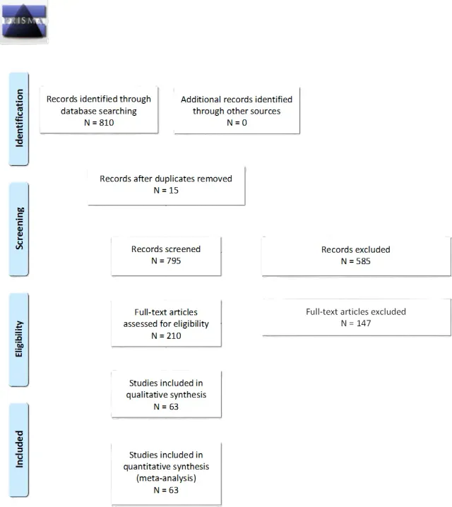
Appendix IV. Evidence Tables
Table 1. RS versus MF Approach for FN Preservation
| Author, Year | Results | Data Class | Conclusion |
| Xian-hao5 et al 2022 | Single institution, retrospective case series of 19 patients who underwent MF approach (with or without endoscopic assistance) for VS. M/F 42/58%. All patients had AAO-HNS class C or better hearing (68% A-B). All patients had HB1 preop. Follow up mean 52 months (range 48-60). | III | GTR obtained in all MF with endoscope, and 75% standard MF patients. MF standard: 75% had immediate postop HB1, 25% HB4. At last f/u 83% HB1, 17% HB4. MF w/ endoscope: 100% had HB1-2 immediately postop (86% HB1). At last f/u, 86% had HB1, 14% had HB3. Endoscope found residual in 43% cases. Residual of MRI found on 25% standard MF. AAO-HNS A-B hearing preserved in 66% (2/3) of MF with endoscope, 50% (6/12) standard MF. Authors conclusions: Using an endoscope in VS resection through the MCF approach could facilitate complete removal of the lesion while minimizing the risk of hearing loss and facial paralysis. The endoscope assisted MCF approach is especially suitable for removing an IC VS with lateral extension involving the space below the transverse crest Comments and Conclusions: This is class III data based on the retrospective nature of data collection and the associated inherent biases. |
| Huo9 et al 2019 | Single institution, retrospective case series of 138 patients that underwent MS for small/medium VS via (71) RS or (67) MF for HP. Minimum follow up was 2 years. 79% of patients had at least AAO-HNS A-B hearing. | III | 97.8% of tumors underwent GTR, no difference between approaches. Postoperative hearing levels were preserved (AAO-HNS A-B) in 41.2% of those with preop hearing. Hearing outcomes were significantly better in patients with normal intraoperative I wave on ABR. Hearing loss within 6 months had a positive effect on postoperative hearing. Better preoperative hearing and tumors from SVN were correlated with better postoperative hearing outcomes. The different surgical approaches (RS or MF) resulted in no significant differences in postoperative hearing. Good FN (HB1-2) was obtained in 92% RS and 90% MF; no differences in early or last HB scores. Authors conclusions: Better preoperative hearing, shorter hearing loss period, tumors from SVN, and normal intraoperative I wave are prognostic factors for serviceable hearing. RSA and MFA are effective and safe for tumor removal and HP. Comments and Conclusions: The retrospective nature of the data yields class III data. |
| Kosty8 et al 2019 | Single institution, retrospective case series of 63 patients that underwent MF approach for VS. The mean postoperative follow up was 21 ± 21 months (range 5-78). The mean tumor size was 10 ± 4 mm. 76% of patients had preop AAO-HNS hearing class A-B. All patients preop had HB1-2. | III | GTR was achieved in 97% of patients. In 2 patients, a capsular rind was left on the C. Tumor recurred in 1 of these patients. Three additional patients had recurrent tumor (6.3% recurrence rate). Of the 4 recurrences, 3 are being observed with serial imaging. One patient underwent hypofractionated radiation and achieved tumor quiescence. The tumor control rate was 98.5%. 51 patients (81%) achieved an HB 1 outcome and 11/63 (17%) achieved HB 2 FN outcome at last follow-up. The remaining patient suffered a complete FN transection requiring a cable nerve graft (great auricular nerve), ultimately achieving an HB V. The serviceable and usable HP rates were 54% and 50%, respectively. Some residual hearing was preserved in 71% of patients. Authors conclusion: In our series, the MCF approach for VS provided excellent rates of tumor and FN function, with durable serviceable HP Comments and Conclusions: The study provides class III data based on the retrospective nature of the data. |
| Lee et al 20163 | Single institution, retrospective case series of 353 patients. Male/Female 44/56%. Tumor size mean 3 cm (range 5-60). Preop symptoms: 56% hearing loss, 25% vertigo/balance issues, 17% trigeminal neuropathy, 5% facial weakness. 76% underwent RS, 23% TL, 1% MF. Follow up was at least 2 years after surgery. | III | Extent of resection: 52% GTR, 18% NTR, 30% STR. Disease recurrence/progression: 8% GTR, 16% NTR, 25% STR; 17% RS, 7% TL, 20% MF approaches. HB score: immediate (HB1 n= 100, HB2 n= 69, HB3 n= 92, HB4-6 n= 92), 1 year (HB1 n= 139, HB2 n= 91, HB3 n= 44, HB4-6 n= 37), >2 years (HB1 n= 113, HB2 n= 74, HB3 n= 34, HB4-6 n= 30). HB1-2 75% after 2 years. Approach and HB grade 1-3: immediate post op (RS 70%, TL 82%, MF 60%), 2 years (RS 84%, TL 92%, MF 100%); no statistical differences. HP achieved at 1 year in 12 of 88 patients (14%) Author Conclusion: H-B grade of immediate postoperative facial palsy can predict facial palsy at long-term follow-up. H-B grade 3 immediate postoperative facial palsy is the lowest tolerable grade that guarantees functional improvement on long-term follow-up. Planned FN preservation surgery followed by radiosurgery is thought to be optimal treatment in patients with vestibular schwannoma for both tumor control and FN function Comments and Conclusions: This is class III data based on the retrospective nature of data collection and the associated inherent biases. |
| Raheja et al 20164 | Single institution, retrospective case series of 78 patients that underwent MF approach for VS. M/F 47/53%. Mean tumor size 7.5 mm (1-17.2). Preop: AAO-HNS A-B 78%, C 18%, D 4%. HB1 95%, HB2 5% Mean follow up was 15 months (0.25-132). | III | GTR obtained in 100% patients, no disease recurrence noted. 76% retained AAO-HNS hearing A-B. HB1 76% HB2 14% HB3 7% HB4-6 3% Author conclusion: Preliminary results from this single-center retrospective study of patients undergoing MFA for resection of VS showed that good HP and FN outcomes could be achieved with few complications. These results suggest that resection via the MFA is a rational alternative to watchful waiting or SRS Comments and Conclusions: The retrospective nature of the data yields class III data. |
| Mastronardi et al 201610 | Single institution, retrospective case series of 100 that underwent RS approach for VS. M/F 53/47%. Mean tumor size 2.4 cm. Follow up minimum 6 months. | III | GTR/NTR 68%, STR 25%, <STR 7%. HP (AAO-HNS A-B) in 30 of 43 patients with good preop hearing (70%). HB grade immediate: – HB1 58%, HB2 19%, HB3-6 22% >6 months: – HB1 92%, HB2 4%, HB3-6 4% Author conclusion: The anterior position and course and adhesion of the FN to the tumor capsule were the 2 factors most strongly associated with worse postoperative FN results. Comments and Conclusions: The study provides class III data based on the retrospective nature of the data. |
| Zhang6 et al 2016 | Single institution, retrospective case series of 1006 VS patients. 63% TL, 20% % RS, 9% MF, 8% transotic. Study evaluated practiced changes over time (1990-2006).42% had serviceable preop hearing. | III | GTR 99.4%. 1.2% had disease recurrence; mean interval 2.8 years (range 1-6). Postop HB at 1 year: HB1-2 – TL 88%, RS81%, MF 78%, TO 82% HB3-4 – T: 11%, RS19%, MF 22%, TO 18% HB5-6: TL 1%, zero for RS/MF/TO. HP rate was 62%, 34% had serviceable hearing (no delineation of approach). Authors conclusions: Surgical outcomes of sporadic vestibular schwannoma have improved concerning FN function outcomes, HP and cerebrospinal fluid (CSF) leaks, mainly due to the neuro-otological team’s experience. Functional results after complete microsurgical removal of large VS depend on experience gained on small VS removal Comments and Conclusions: This is class III data based on the retrospective nature of data collection and the associated inherent biases. |
| Anaizi7 et al 2016 | Retrospective case series, single institution, 80 patients with small (Koos grade 1-2) that underwent RS (52%), TL (40%), or MF (8%) for VS. M/F 44/56%. 92% of patients had some hearing loss at presentation, 49% had serviceable hearing. 43% were observed for 1 year. 36% patients presented with vertigo/balance issues. Follow up mean 34 months. | III | 89% GTR, 11% NTR (GTR: RS 89%, TL 88%, MF 100%). 2 of NTR demonstrated growth on next interval scan and received SRS. 95% patients had HB1-2 at last follow up. At last follow up, 36% retained serviceable hearing that had it preop (37% RS, 25% MF). 93% reported resolution in vertigo/imbalance. Patients with postop complications (5% RS, 18% TL, 33% MF) Authors conclusions: As one of the largest contemporary surgical series of small vestibular schwannomas, we discuss some nuances to help refine treatment algorithms. Although observation and radiosurgery have established roles, our results reinforce microsurgery as a viable, safe option for a subgroup of patients Comments and Conclusions: The retrospective nature of the data yields class III data. |
Abbreviations: cm = centimeter; Preop = preoperative; RS retrosigmoid; TL = Translabyrinthine; MF = middle fossa; GTR = gross total resection; NTR = near total resection; STR = subtotal resection; HB = House Brachmann; n = number; AAO-HNS, American Academy of Otolaryngology-Head and Neck Surgery; CNAP = cochlear nerve action potential; FN = facial nerve; GR= Gardner– Robertson; HP = hearing preservation; IAC = internal auditory canal; PTA = pure tone average; VS = vestibular schwannoma; WRS = word recognition score; M = male; F = female; obs = observation; m = month
Table 2. RS versus TL Approach for FN Preservation
| Author, Year | Results | Data Class | Conclusions |
| de Boer15 et al 2020 | Single institution, retrospective case series of 596 patients that underwent TL for VS. Mean follow-up after surgery was 50 m (median, 36 m; range, 3–209 m) | III | The extent of tumor removal was GTR in 32%, NTR in 58%, and STR in 10%. In 5.5% (33/596) of patients the tumor recurred. STR, young age, and tumor progression preoperatively significantly increased the risk of recurrence, whereas tumor size or histologic composition did not. Mean follow-up until the diagnosis of recurrence was 46 m (median, 39; range, 6–131 m). Salvage treatment for recurrences consisted of second surgery in eight patients and radiotherapy in 25 patients. A good postoperative FN function (HB1–2) was achieved in 85% at 1 year. The risk of postoperative FN paresis or paralysis increased with tumor size (HB1-2 90% IC, 79% small, 76% medium, 69% mod large, 54% large, 47% giant), but was not associated with the extent of tumor removal, histologic composition, or patient demographics. Authors conclusions: TL surgery is an effective treatment for VS, with a good local control rate and FN outcome. The extent of tumor removal is a clinically relevant predictor for tumor recurrence, as are young patient age and preoperative tumor progression. A large preoperative tumor size is associated with a higher risk of postoperative FN paresis or paralysis Comments and Conclusions: The study provides class III data based on the retrospective nature of the data. |
| Tawfik67 et al 2020 | Single institution, retrospective case series of 290 VS patients undergoing MS, 158 (54%) TL, 131 (45%) RS. Mean 37.7 months of follow up. | III | GTR was achieved in 98% of patients, no difference between RS and TL. Four of seven patients who underwent NTR had tumors more than or equal to 40 mm in maximal diameter (all TL). Long term facial outcome at last follow up: HB1-2: 84% TL, 98% RS HB3: 13% TL, 2% RS HB4-6: 4% TL, <1% RS. When accounting for tumor size, the TL and retrosigmoid approaches yield equivalent FN and extent of resection results. Authors conclusions: In patients with VS and retained serviceable hearing, SHL is an independent predictor of HP after RS microsurgical resection when the cochlear nerve is preserved Comments and Conclusions: This is class III data based on the retrospective nature of data collection and the associated inherent biases. |
| Breun13 et al 2019 | Single institution, retrospective case series of 502 VS operated via RS approach. F 53%. 36% small, 64% large (T3B or T4). 73% had preop useful hearing. 12% had preop facial weakness. Follow up not detailed. | III | GTR 70%, NTR 26.9%, PTR 4%. HP in 43% with small tumors, 23% large tumors. 86% HB1-3 in small tumors 77% large tumors. HB1 GTR 26%, NTR 8%, PTR 2% HB2 GTR 21%, NTR 6%, PTR <1% HB3 GTR 10%, NTR 5%, PTR <1% HB4-6 GTR 12%, NTR 8%, PTR 1% Authors conclusions: In a standardized setting, the semi-sitting position allowed a safe approach. This setting offers the advantage of bimanual tumor nerve handling by the surgeon and an optimal visualization of important functional structures Comments and Conclusions: The retrospective nature of the data yields class III data. |
| Troude12 et al 2019 | Single institution, retrospective case series of 169 large VS (>3 cm) operated via RS approach. M/F 40/60%. 10% had preop facial weakness, 13% had nervous intermediate symptoms, 25% had preop GR 1-2 class hearing.36% had imbalance issues, 4% had trigeminal neuralgia. 36% operated through TL, 64% through RS approach. Follow up mean 62 months (54-71). | III | 11% GTR, 59% NTR, 21% STR, 9% PTR. No tumor recurrence for GTR. Mean delay till recurrence/progression 37 months for NTR/STR/PTR. Of 143 patients with non-GTR, 66 followed obs, 77 underwent adjuvant SRS. Tumor control 82% in obs, 81% in SRS; 7-year PFS in obs 76%, SRS78%. Immediate postop: HB1 45% HB2 22% HB3 11% HB4-6 22%. 3 months: HB1 57% HB2 17% HB3 11% HB4-6 15% Last f/u: HB1 68% HB2 16% HB3 15% HB4-6 1%. Surgical approach (RS vs TL) was not predictive of postop HB grade. Authors conclusions: As long as the extent of resection or additional Gamma Knife surgery have not been identified as predictive risk factors of postoperative FN palsy, we suggest that optimal resection is the main option for patients harboring large VS Comments and Conclusions: The study provides class III data based on the retrospective nature of the data. |
| Hoshide11 et al 2018 | Retrospective case series, single institution, 45 patients underwent keyhole RS approach for VS >3 cm. M/F 47/53. Mean tumor size 4.4 cm (3-7.5). Pre-op: 78% had hearing loss, 98% had HB1. Follow up mean 49m (14-145). | III | NTR/GTR 100%. No patients required reoperation for tumor recurrence. 40% of patients experienced transient facial weakness. At last follow up: HB1-2 84% HB3 9% HB4-6 7% Author conclusion: It is the experience of the senior author that complete or near-complete resection of large VSs can be successfully achieved via a keyhole approach. In this series of 45 large VSs, a greater extent of resection was achieved while demonstrating high rates of FN preservation and low approach-related and postoperative complications compared with the literature Comments and Conclusions: This is class III data based on the retrospective nature of data collection and the associated inherent biases. |
| Schwartz14 et al 2018 | Single institution, retrospective case series of 100 patients that underwent TL resection of VS. M/F 45/55%. 74% had severe hearing loss. Tumor size 1.3 cm (0.4-2.0). | III | 97 patients (97%) underwent GTR. 70 patients had at least 1 year follow up, without any disease recurrence. 99% had initial HB1-2 after surgery. 1 patient had HB4 post-op, with 7 developing delayed FN palsy; 2 patients had HB3 and 2 had HB5 at last follow up. Authors conclusions: It is the experience of the senior author that complete or near-complete resection of large VSs can be successfully achieved via a keyhole approach. In this series of 45 large VSs, a greater extent of resection was achieved while demonstrating high rates of FN preservation and low approach-related and postoperative complications compared with the literature Comments and Conclusions: The retrospective nature of the data yields class III data. |
| Lee3 et al 2016 | Single institution, retrospective case series of 353 patients. M/F 44/56%. Tumor size mean 3 cm (range 5-60). Preop symptoms: 56% hearing loss, 25% vertigo/balance issues, 17% trigeminal neuropathy, 5% facial weakness. 76% underwent RS, 23% TL, 1% MF. Follow up was at least 2 years after surgery. | III | Extent of resection: 52% GTR, 18% NTR, 30% STR. Disease recurrence/progression: 8% GTR, 16% NTR, 25% STR; 17% RS, 7% TL, 20% MF approaches. HB score: immediate (HB1 100, HB2 69, HB3 92, HB4-6 92), 1 year (HB1 139, HB2 91, HB3 44, HB4-6 37), >2 years (HB1 113, HB2 74, HB3 34, HB4-6 30). HB1-2 75% after 2 years. Approach and HB grade 1-3: immediate post op (RS 70%, TL 82%, MF 60%), 2 years (RS 84%, TL 92%, MF 100%); no statistical differences. Authors conclusions: H-B grade of immediate postoperative facial palsy can predict facial palsy at long-term follow-up. H-B grade 3 immediate postoperative facial palsy is the lowest tolerable grade that guarantees functional improvement on long-term follow-up. Planned FN preservation surgery followed by radiosurgery is thought to be optimal treatment in patients with VS for both tumor control and FN function Comments and Conclusions: The study provides class III data based on the retrospective nature of the data. |
| Mastronardi10 et al 2016 | Single institution, retrospective case series of 100 that underwent RS approach for VS. 57% patients had no serviceable preop hearing. Follow up minimum 6 months. | III | GTR/NTR 68%, STR 25%, <STR 7%. HB grade immediate HB1 58% HB2 19% HB3-6 22%) >6 months HB1 92% HB2 4% HB3-6 4%) Authors Conclusions: The AS pattern was most common for smaller VSs. The A position and course and adhesion of the FN to the tumor capsule were the 2 factors most strongly associated with worse postoperative FN result. Comments and Conclusions: This is class III data based on the retrospective nature of data collection and the associated inherent biases. |
Abbreviations: cm = centimeter; Preop = preoperative; RS retrosigmoid; TL = Translabyrinthine; MF = middle fossa; GTR = gross total resection; NTR = near total resection; STR = subtotal resection; HB = House Brachmann; n = number; AAO-HNS, American Academy of Otolaryngology-Head and Neck Surgery; CNAP = cochlear nerve action potential; FN = facial nerve; GR= Gardner– Robertson; HP = hearing preservation; IAC = internal auditory canal; PTA = pure tone average; VS = vestibular schwannoma; WRS = word recognition score; M = male; F = female; obs = observation; m = month
Table 3. Comparison of Surgical Approach, Tumor Size and FN Functional Preservation
| Author, Year | Results | Data Class | Conclusions |
| Rujimethapass24 et al 2022 | Single institution, retrospective case series of 48 patients that underwent resection of large (>3cm, mean size 3.8 cm) or small (<3cm, mean size 1.5 cm) VS. In large lesions, 97% patients had hearing loss, 67% gait imbalance, 50% trigeminal neuropathy. In small lesions, 100% hearing loss, 5.6% gait imbalance, 11% trigeminal neuropathy. | III | At 1 month, 30% of large and 83.3% small had HB1-3. At 1 year follow up, 40% large and 94.4% small VS patients had HB1-3. 2). Comparing FN outcome at 1 year between GTR/NTR and STR showed HB1-3 in 7 patients (46.7%) in GTR/NTR group and 5 patients (33.3%) in STR group, HB4-6 was found in 8 (53.3%) and 10 (66.7%) in GTR/NTR group and STR group, respectively. Large-sized VS (≥3 cm) microsurgical resection had significantly poorer FN outcomes than those who had VS <3 cm. Authors conclusions: In patients with large size VS, microsurgical resection had poor FN outcomes compared with those of their counterparts with small to medium size VS. Planned STR with postoperative radiosurgery might attain superior FN outcomes and result in better QOI in subjects with large VS Comments and Conclusions: This is class III data based on the retrospective nature of data collection and the associated inherent biases. |
| Stastna23 et al 2022 | Single institution, retrospective case series of 125 patients with large cystic VS largely operated through TL (mean volume 9.5 cm3, range 1.8-52.7). M/F 52/48%. 98% had TL, 2% had RS. | III | GTR obtained in 62.4%, NTR in 34.4%, STR in 3.2%. Trend towards more NTR/STR in RS than TL. 3 months postop, 52% had HB1, 22.8% had HB2, 15.5% HB3-4, 9.6% HB5-6. 1 year postop, 76% had HB1-2, 16% HB3-4, 8% HB5-6. Poor FN outcome associated with larger size (>25 cm3). Authors Conclusions: Our study confirmed that microsurgery of cystic VS has worse outcomes of FN preservation and extent of resection compared with solid VS. Greater attention should be paid to the above-mentioned risk factors. Comments and Conclusions: The retrospective nature of the data yields class III data. |
| Kiyofuji29 et al 2021 | Single institution, retrospective case series of 48 large (>4 cm) and 38 small (<1 cm) VS. Small tumors presented more commonly with tinnitus and sudden hearing loss. RS was selected in 43 patients (89.6%), while TL approach (TL) was used in 5 patients (10.4%) in large VS. In small VS, RS was selected in 17 patients (44.7%), while TL was used in 19 patients (50%) and MF approach in 2 (5.3%). | III | Patients with large VS underwent more STR than small VS (50.0% vs. 2.6%). In small VS, GTR/NTR achieved in 97.4%, while GTR/NTR obtained in 50%. The rate of recurrence/progression needing treatment was not different between the groups (12.5% in large vs. 7.9% in small). In large VS, recurrence/progression was noted in 6 patients (12.5%) at a median of 35 months (range 13–84 months): four cases were progressions following STR, and two cases were recurrences following GTR and NTR. In small VS, recurrence was noted in 3 patients (7.9%, p = 0.49) at a median of 53 months (range 44–95 months). Two recurrences followed GTR, while one patient experienced recurrence after NTR. All recurrences were treated with SRS. These three patients were followed at a median of 36 months (range 0–55) after SRS, and they did not encounter further recurrence/progression. In large VS, 24 patients (50%) had good (HB I-II) facial function at last follow-up, while in small VS, only one patient demonstrated unsatisfactory postoperative facial function. In large VS, patients who underwent STR had better FN function at last follow-up than those who underwent GTR/NTR (66.7% vs. 33.3%) Large tumors have more unsatisfactory outcomes in FN function and postoperative hearing despite maximal efforts undertaken toward function-preservation strategy; however, similar tumor control was achieved. Authors conclusions: Large and small VS present differently. LTG showed more unsatisfactory outcomes in FN function and postoperative hearing despite maximal efforts undertaken toward function-preservation strategy; however, similar tumor control was achieved Comments and Conclusions: The study provides class III data based on the retrospective nature of the data. |
| Kim28 et al 2021 | Single institution, retrospective case series of 86 patients comparing RS vs TL approach for large (>3 cm) VS. 53 underwent RS, 33 underwent TL. Median follow up 34.5 months. 96% (RS) and 97% (TL) had preop HB1-2. | III | GTR: 8% RS, 18% TL. NTR: 55% RS, 55% TL. STR: 37% RS, 27% TL. Tumor recurrence/progression: 4% RS, 3% TL. 82% RS, 88% TL preservation of FN function. 26% HP RS. Surgical outcomes, including the extent of resection, tumor recurrence, and FN preservation, showed no significant differences between the two groups. Patients who underwent the RS approach showed a marginal trend for postoperative lower cranial nerve (LCN) dysfunction. Both surgical approaches show equivalent surgical and clinical outcomes. Authors conclusions: Both surgical approaches show equivalent surgical outcomes. Notably, the TL approach for large VS has advantages in that it reduces cerebellar injury and related morbidities. Comments and Conclusions: This is class III data based on the retrospective nature of data collection and the associated inherent biases. |
| Mastronardi68 et al 2021 | Single institution, retrospective case series of 60 patients with Koos grade IV VS (>3 cm) that underwent RS approach. In 11 cases, a preoperative HB 2-4 FN deficit was present. | III | GTR/NTR was accomplished in 46 cases (76.7%), 65.8% in cases with, and 95.4% without tight adhesion of capsule to nervous structures. At a mean follow-up of 59.3 months, a recurrence/regrowth of residue was observed in 8 (13.3%) cases operated on with STR/PTR: in 2 of them, a second surgery was necessary for large cystic transformation, 2 and 4 years after the first operation, respectively. In the other 6 cases, the initial growth of tumor was stable on serial scans. At last follow-up, 34 (56.7%) patients had a normal postoperative FN outcome (HB1), 9 (15%) were HB2, 8 (13%) HB3, and 9 (15,0%) HB4-6. The GTR/NTR resection of solid and low-bleeding VS, without tight capsule adhesion, was associated with better FN outcome. Long-term FN results seem to be worse in patients with cystic Koos grade IV VS, in cases with tight capsule adherences to nervous structures and in high-bleeding tumors. Authors conclusions: Microsurgery of Koos grade IV VS seems to be associated with more than acceptable functional results, with high rate of T and NT removal of tumor. Long-term FN results seem to be worse in patients with cystic Koos grade IV VS, in cases with tight capsule adherences to nervous structures and in high-bleeding. Comments and Conclusions: The retrospective nature of the data yields class III data. |
| Refaat30 et al 2021 | Single institution, retrospective case series of 15 giant (>4.5 cm) VS operated via RS. 4 patients had preop facial weakness, and 1 of those had complete palsy. Median follow up was 12 months (range 7-14). | III | GTR in 73.3%, STR 26.7%. There was no tumor recurrence/progression (although short follow up). Twelve cases (80%) had postoperative facial palsy, HB<3 in 5 cases (33.3%), and >3 in 7 cases (46.7%). Authors conclusions: Large and giant VSs are still commonly met in neurosurgical practice in developing countries; they have different behaviors and presentations from those of smaller tumors. Both patient and surgeon expectations from surgery should be toward no mortality and mild or no morbidities. |
| Killeen32 et al 2020 | Single institution, retrospective case series of 167 VS patients that underwent TL (76.7%), MF (14.4%), RS (7.2%) or other (1.8%) approaches. The median tumor diameter and volume were 25.3 mm(range: 4.1–47.1 mm) and 3.17 cm3 (range: 0.01–30.6 cm3), respectively. The median follow-up was 24.2 months (range: 12–114.2 months). | III | GTR was performed in 79% of cases, with residual tumor identified on MRI in 17% of cases. For patients with tumors <3 cm3, 92.7% had grade 1 or 2 facial function after at least 1 year follow-up, compared to 81.2% for those with tumors >3 cm3 (OR = 2.9). Logistic regression OR for postop facial weakness: TL 0.18, MF 1.14, RS 0.09 (not significant). Tumor volume >3 cm3 was predictive of facial weakness on multivariate regression analysis (OR = 7.4) when controlling for surgical approach, internal auditory canal extension, anterior extension, age, gender, and extent of resection. Tumor conclusions: Tumor volume >3 cm3 is associated with worse FN outcomes 12 months following surgical resection. Comments and Conclusions: The study provides class III data based on the retrospective nature of the data. |
| Won26 et al 2020 | Single institution, retrospective case series of 58 patients with Koos grade IV tumors (mean volume 17.1 +/- 9.2 cm3) operated via RS approach. At admission, 49 patients (96.1%) had good FN function (HB 1–3) and 2 patients (3.9%) had poor FN function (HB 4–6). Follow up after surgery median time of 28 months (range 4.3– 53.8). | III | 12 (21%) patients underwent GTR. There was no tumor recurrence in these patients at last follow up. In those 46 patients with residual tumors, stable disease was documented in 21 patients (45.7%), tumor regression in 12 patients (26.1%) and residual tumor progression in 11 patients (23.9%). After surgery, good FN function was observed in 66.7%. At follow-up, the number of patients with good FN function had increased, now present in 82.4%; however, pre-existing FN palsy (HB 5 or 6) had neither improved after surgery nor at follow-up examination. Authors conclusions: Subtotal tumor resection is a good therapeutic concept in patients with KOOS IV VS resulting in a high rate of good hearing and FN function and a very low rate of subsequent tumor progression. The goal of surgery should be to achieve more than 87% of tumor resection to keep residual tumor progression low. Comments and Conclusions: This is class III data based on the retrospective nature of data collection and the associated inherent biases. |
| Huo9 et al 2019 | Single institution, retrospective case series of 138 patients that underwent MS for small/medium VS via (71) RS or (67) MF for HP. Minimum follow up was 2 years. 79% of patients had at least AAO-HNS A-B hearing. | III | 97.8% of tumors underwent GTR, no difference between approaches. Postoperative hearing levels were preserved (AAO-HNS A-B) in 41.2% of those with preop hearing. Hearing outcomes were significantly better in patients with normal intraoperative I wave on ABR. Hearing loss within 6 months had a positive effect on postoperative hearing. Better preoperative hearing and tumors from SVN were correlated with better postoperative hearing outcomes. The different surgical approaches (RS or MF) resulted in no significant differences in postoperative hearing. Good FN (HB1-2) as obtained in 92% RS and 90% MF; no differences in early or last HB scores. Authors conclusions: Better preoperative hearing, shorter hearing loss period, tumors from SVN, and normal intraoperative I wave are prognostic factors for serviceable hearing. RSA and MFA are effective and safe for tumor removal and HP. Comments and Conclusions: The retrospective nature of the data yields class III data. |
| Troude12 et al 2019 | Single institution, retrospective case series of 169 large VS (>3 cm) operated via RS approach. M/F 40/60%. 10% had preop facial weakness, 13% had nervous intermediate symptoms, 25% had preop GR 1-2 class hearing.36% had imbalance issues, 4% had trigeminal neuralgia. 36% operated through TL, 64% through RS approach. Follow up mean 62 months (54-71). | III | 11% GTR, 59% NTR, 21% STR, 9% PTR. No tumor recurrence/growth at last f/u in 83%. Mean delay till recurrence/progression 37 months. Of 143 patients with non-GTR, 66 followed obs, 77 underwent adjuvant SRS. Tumor control 82% in obs, 81% in SRS. 7 year PFS in obs 76%, SRS78%. Complementary adjuvant SRS not predicted of remnant growth. Immediate postop: HB1 45%, HB2 22%, HB3 11%, HB4-6 22%. 3 months: HB1 57%, HB2 17%, HB3 11%, HB4-6 15%. Last f/u: HB1 68%, HB2 16%, HB3 15%, HB4-6 1%. Surgical approach (RS vs TL) was not predictive of postop HB grade. Authors conclusions: As long as the extent of resection or additional Gamma Knife surgery have not been identified as predictive risk factors of postoperative FN palsy, we suggest that optimal resection is the main option for patients harboring large VS Comments and Conclusions: The study provides class III data based on the retrospective nature of the data. |
| Chiluwal25 et al 2018 | Single institution, retrospective case series of 30 patients with small VS (Hannover grade T1-3b, mean 1.7 cm) operated through RS. 57% had AAO-HNS class 1-2. | III | 90% underwent GTR. All patients had HB1 immediately postop. At 1 year, 97% had HB1-2, 3% had HB3. 59% of those with preop hearing preserved it postop ta 3 months (hearing preserved in smaller lesions, all T1-2 patients). 7 patients with hearing had f/y >2 years (range 24-70), all maintained class A-B hearing, one did go from A to B. Authors conclusions: Although both observation and radiosurgery are valid options in the management of smaller size VSs, surgical treatment seems to offer a high rate of FN preservation, a reasonable rate of hearing sparing, and a high total resection rate. Clinicians should consider surgical treatment as a valid option in the initial management of symptomatic small VSs in younger patients. Comments and Conclusions: This is class III data based on the retrospective nature of data collection and the associated inherent biases. |
| Hoshide11 et al 2018 | Retrospective case series, single institution, 45 patients underwent keyhole RS approach for VS >3 cm. M/F 47/53. Mean tumor size 4.4 cm (3-7.5cm). Pre-op: 78% had hearing loss, 98% had HB1. Follow up mean 49 m (14-145 m). | III | 100% NTR/GTR (not distinguished). 40% of patients experienced transient facial weakness. At last F/U, 84% had HB1-2, 9% HB3, 7% HB 4-6. Authors conclusions: It is the experience of the senior author that complete or near-complete resection of large VSs can be successfully achieved via a keyhole approach. In this series of 45 large VSs, a greater extent of resection was achieved while demonstrating high rates of FN preservation and low approach-related and postoperative complications compared with the literature Comments and Conclusions: The retrospective nature of the data yields class III data. |
| MacKenzie19 et al 2018 | Single institution retrospective case of 63 patients with planned preop STR through RS approach. Preop: 96% HB1-2, 58% serviceable hearing, 46% vertigo, 46% cerebellar balance issues, 27% trigeminal symptoms. Mean tumor volume 7.7 cm (0.74-41.44). Median follow-up 3 months. | III | Extent of resection: STR 81%, NTR 19%. Discussion of adjuvant SRS, but no clear numbers provided. Immediate post-op: 71% HB1-2, 13% HB3, 16% HB4-6. At last follow up: 72% HB1-2, 10% HB3, 18% HB4-6. Authors conclusions: Intended submaximal resection provides satisfactory neurological outcome for patients with large VS. Risk factors for postoperative neurological deterioration remain unclear. Comments and Conclusions: The study provides class III data based on the retrospective nature of the data. |
| Zumofen27 et al 2018 | Single institution, retrospective case series of 44 patients who underwent NTR of Koos grade IV VS via RS (mean tumor volume 10.9 cm3). At baseline HB1 (75%), HB2 (23%), HB4 (2%). Follow up mean 22 months (range 0.5-72). | III | The mean extent of resection was 89%. At the last radiological follow-up, the residual tumor had become smaller or remained the same size in 84% of patients. Mean time to volumetric progression 31 months (range 19-50); 18-24 months (7%), 24-36 (5%), 36-72 (16%). Volumetric progression was negatively correlated with the original extent of resection and positively correlated with postoperative residual tumor volume. At first clinical follow up (mean 26 days, range 7-126), 50% had HB1, 30% HB2, 5% HB3, 15% HB4-6. At the last clinical follow-up, FN function was good (HB) I-II] in 89%, fair (HB III) in 9%, and poor (HB IV-VI) in 2% of the patients. Authors conclusions: Intended near-total removal results in excellent preservation of FN function and has a low recurrence rate. Any progressive residual tumor may be treated by radiosurgery Comments and Conclusions: This is class III data based on the retrospective nature of data collection and the associated inherent biases. |
| Boublata21 et al 2017 | Single institution, retrospective case series of 151 large/giant VS (>3 cm) operated via RS approach. M/F 28/72%. 42% had preop hearing loss. 49% had balance issues. 3% had trigeminal neuralgia. Mean follow up 28 months (range 3-54). | III | GTR 83%, STR 14%, PTR 3%. FN anatomically intact in 99%. Immediate postop, 76% HB1-2, 18% HB3-4, 6% HB5-6. After two years: 82% HB1-2, 14% HB3-4, 4% HB5-6. Authors conclusions: The development of anesthesia techniques and microsurgery and the systematic use of IOM of the FN have allowed us to move from a life preservation era to another era of preservation of function Comments and Conclusions: The retrospective nature of the data yields class III data. |
| Huang20 et al 2017 | Single institution, retrospective case series of 1167 patients with large (> 3 x 2 cm) approached through a RS craniotomy. M/F 46/54%. 60% had preop hearing of AAO-HNS A or B. Follow up mean 57 months (range 6-187). | III | 86% GTR, 14% NTR, 0.2% STR. 4.9% had recurrent tumor at last follow up. Anatomical FN preservation was 93%. Immediate postop: 13% HB1, 23% HB2, 46% HB3, 36% HB4-6. For GTR: HB1-2 35%, Hb3 47%, 36% HB4-6. NTR: HB1-2 44%, HB3 37%, 19% HB4-6. At last follow up: 32% HB1, 56% HB2, 6% HB3, 6% HB4-6. 81% lost complete hearing after surgery. Serviceable HP was 12%, and at last follow up 6% had class A-B hearing. Authors conclusions: The key factors for reducing surgical complications include careful assessment of the functions of acoustic and FNs as well as a thorough understanding of anatomy via the RS approach before operation, skillful microsurgical technique, and monitoring of multiple cranial nerves during resection. Comments and Conclusions: The study provides class III data based on the retrospective nature of the data. |
| Grahnke22 et al 2017 | Single institution, retrospective case series of 105 large (>2.5 cm) VS who underwent TL or RS approach. M/F 57/43%. Mean tumor diameter 3.4 cm. All patients presented with hearing loss. Mean follow up 36.5 months. | III | Resection: 33% STR, 67% GTR. 68% GTR had HB1-2, 60% STR had HB1-2. HB1-2 by approach: 75% RS, 60% TL, 60% combined RS-TL post-op; at 1 year: 92, 88, 86% (RS, TL, combined). Immediate postop, 65% had HB1-2. At 1 month, HB1-2 72% and at 1 year or last follow up 89% HB1-2. 11 patients (11%) experience HB6 postop, of those, 4 improved to HB3, 1 improved to HB4, 2 to HB5, 1 showed no improvement. No meaningful difference in FN outcomes between the RS, TL, and combined approaches for FN outcomes or tumor control long term. Authors conclusions: Our prognostic index may be useful to assess the risk of FN injury preoperatively for large acoustic neuromas, while also providing information about the tumor-nerve relationship. Comments and Conclusions: This is class III data based on the retrospective nature of data collection and the associated inherent biases. |
| Huang31 et al 2017 | Single institution, retrospective case series of 657 giant (>4 cm) VS treated via RS approach. The other most frequent clinical symptoms were facial paresthesia/trigeminal neuropathy (453 cases, 68.9%), balance issues/disequilibrium (293 cases, 44.6%), facial paralysis (HB III + IV 204 cases, 31.1%). Mean follow up 60 months (range 6-191). | III | GTR in 556 patients (84.6%); NTR was achieved in 99 patients (15.1%). The mortality rate is 0.6%. The FN was preserved anatomically in 589 cases (89.7%). Good FN functional outcome (HB1-2) postoperatively was achieved in 216 patients (32.9%). Other 308 cases (46.9%) were HB3, and 133 patients (20.2%) were HB4-6. Authors conclusions: Trends in the data lead the authors to suggest that the microsurgical technique, intraoperative nerve monitoring, and multidisciplinary cooperation, were the keys to improving prognostic outcomes in giant intracranial VS patients. Comments and Conclusions: The retrospective nature of the data yields class III data. |
| Anaizi7 et al 2016 | Retrospective case series, single institution, 80 patients with small (Koos 1-2) that underwent RS (52%), TL (40%), or MF (8%) for VS. M/F 44/56%. 92% of patients had some hearing loss at presentation, 49% had serviceable hearing. 43% were observed for 1 year. Follow up mean 34 months. | III | 89% GTR, 11% NTR (GTR: RS 89%, TL 88%, MF 100%). 2 of NTR demonstrated growth on next interval scan and received SRS. 95% patients had HB1-2 at last follow up. At last follow up, 36% retained serviceable hearing that had it preop (37% RS, 25% MF). Patients with postop complications (5% RS, 18% TL, 33% MF) Authors conclusions: As one of the largest contemporary surgical series of small VSs, we discuss some nuances to help refine treatment algorithms. Although observation and radiosurgery have established roles, our results reinforce microsurgery as a viable, safe option for a subgroup of patients. Comments and Conclusions: The study provides class III data based on the retrospective nature of the data. |
| Lee3 et al 2016 | Single institution, retrospective case series of 353 patients. M/F 44/56%. Tumor size mean 3 cm (range 5-60). 5% patients had preop facial weakness. 76% underwent RS, 23% TL, 1% MF. Follow up was at least 2 years after surgery. | III | Extent of resection: 52% GTR, 18% NTR, 30% STR. Disease recurrence/progression: 8% GTR, 16% NTR, 25% STR; 17% RS, 7% TL, 20% MF approaches. HB score: Immediate (HB1 100, HB2 69, HB3 92, HB4-6 92), 1 year (HB1 139, HB2 91, HB3 44, HB4-6 37) >2 years (HB1 113, HB2 74, HB3 34, HB4-6 30). HB1-2 75% after 2 years. Approach and HB grade 1-3: immediate post op (RS 70%, TL 82%, MF 60%), 2 years (RS 84%, TL 92%, MF 100%); no statistical differences in approach. Authors conclusions: H-B grade of immediate postoperative facial palsy can predict facial palsy at long-term follow-up. H-B grade 3 immediate postoperative facial palsy is the lowest tolerable grade that guarantees functional improvement on long-term follow-up. Planned FN preservation surgery followed by radiosurgery is thought to be optimal treatment in patients with VS for both tumor control and FN function Comments and Conclusions: This is class III data based on the retrospective nature of data collection and the associated inherent biases. |
| Zhang6 et al 2016 | Single institution, retrospective case series of 218 VS that were >4cm in diameter and underwent RS craniotomy. M/F 50/50%. 95% had preop hearing loss. Follow up mean 40 months (12-72). | III | 29% GTR, 51% NTR, 64% STR. During F/U, 20 patients had disease progression (treated with SRS). Progression/recurrence rate: 3% GTR, 8% NTR, 24% STR. HB grade: Immediate (1: 6%, 2 41%, 3 27%, 4-6 27%). At 3 months HB1-2 40% GTR, 62% NTR, 64% STR At last follow up HB1-2 obtained in 59% of GTR, 80% NTR, 83% STR. Authors conclusions: Surgical outcomes of sporadic VS have improved concerning FN function outcomes, HP and cerebrospinal fluid (CSF) leaks, mainly due to the neuro-otological team’s experience. Functional results after complete microsurgical removal of large VS depend on experience gained on small VS removal. Comments and Conclusions: The retrospective nature of the data yields class III data. |
| Sepehrnia17 et al 2015 | Multi-institution, retrospective case series of 446 patients with VS <2 cm (n=292; M/F 54/46%; mean follow up 63 months) or >2 (n=154, M/F 57/43%, mean follow up 67 months). All patients operated through RS craniotomy. | III | All patients had GTR in both cohorts. VS <2 cm: 94% HB1, 6% HB2. 51% HP (GR1-3). 34% serviceable HP. VS >2cm: 78% HB1, 20% HB2, 2% HB3. 34% HP (GR1-3). 27% preservation of serviceable hearing. Authors conclusions: Even a small increase in tumor size correlated with a significant reduction in good hearing and facial preservation postoperatively, which implies that tumor removal should be performed at the earliest stage possible. Furthermore, these results contradict recommending the wait-and-see approach for intra/extrameatal tumors. Comments and Conclusions: The study provides class III data based on the retrospective nature of the data. |
| Liu18 et al 2015 | Single institution, retrospective case series of 106 large (>3 cm) VS operated through a RS approach (tumor size range 3.0-5.7 cm). 2-year follow up. | III | 82% GTR, 14% STR. No recurrence at 2 years for all GTR. All STR showed tumor growth by 2 years. 98% patients had anatomic preservation of CN7; 3 patients had mild laceration of CN7 due to tumor adherence. 3 patients had HB6 post-op, 2 improved to HB3 at 2 years, and one to HB4. Even within large VS, larger tumor size increased risk of worse HB score. No difference between cystic/solid lesions. Authors conclusions: Indicative factors of both immediate and long-term postoperative FN function in large VSs include tumor size, intraoperative train time, start to final FMEP ratios and proximal to distal MRA ratios. Comments and Conclusions: This is class III data based on the retrospective nature of data collection and the associated inherent biases. |
Abbreviations: cm = centimeter; Preop = preoperative; RS retrosigmoid; TL = Translabyrinthine; MF = middle fossa; GTR = gross total resection; NTR = near total resection; STR = subtotal resection; HB = House Brachmann; n = number; AAO-HNS, American Academy of Otolaryngology-Head and Neck Surgery; CNAP = cochlear nerve action potential; FN = facial nerve; GR= Gardner– Robertson; HP = hearing preservation; IAC = internal auditory canal; PTA = pure tone average; VS = vestibular schwannoma; WRS = word recognition score; M = male; F = female; obs = observation; m = month
Table 4. Microsurgery Compared to Observation for Tumor Control in Small Tumors
| Author, Year | Results | Data Class | Conclusions |
| Patro34 et al 2021 | Single institution, retrospective case series of 220 VS patients, 120 initially observed, 100 underwent microsurgery | III | Median time from consultation to microsurgery was 7.3 months (IQR 2.2–18.2), with 174 (79%) achieving GTR, 33 (15%) achieving NTR, and 13 (6%) achieving STR. There was no significant difference by logistic regression for STR, FN function at 2-3 weeks or 12 months, FN interventions, major complications, minor complications, tumor recurrence rate, or salvage therapy. Time from initial consultation to surgery did not significantly impact the probability of postoperative outcomes: STR, FN function at 2-3 weeks or 12 months, FN interventions, major complications, minor complications, tumor recurrence rate, or time to salvage therapy. Patients most frequently proceeded with surgery due to tumor growth alone (67%), followed by tumor growth and worsening symptoms (23%), worsening symptoms alone (8%), and patient preference without any tumor growth or worsening symptoms (2%). Median growth prior to microsurgery was 3.6 mm (IQR 1.4–5.4). Patients with worse hearing, larger tumor volume, and brainstem compression were more likely to pursue upfront microsurgery. A watchful waiting period does not appear to worsen outcomes and can be considered for patients with better hearing and smaller tumors without brainstem compression. Authors conclusions: Patients with worse hearing, larger tumor volume, and brainstem compression were more likely to pursue upfront microsurgery. A watchful waiting period does not appear to worsen outcomes and can be considered for patients with better hearing and smaller tumors without brainstem compression Comments and Conclusions: This is class III data based on the retrospective nature of data collection and the associated inherent biases. |
| Huo9 et al 2019 | Single institution, retrospective case series of 138 patients that underwent MS for small/medium VS via (71) RS or (67) MF for HP. Minimum follow up was 2 years. 79% of patients had at least AAO-HNS A-B hearing. | III | 97.8% of tumors underwent GTR, no difference between approaches. Postoperative hearing levels were preserved (AAO-HNS A-B) in 41.2% of those with preop hearing. Hearing outcomes were significantly better in patients with normal intraoperative I wave on ABR. Hearing loss within 6 months had a positive effect on postoperative hearing. Better preoperative hearing and tumors from SVN were correlated with better postoperative hearing outcomes. The different surgical approaches (RS or MF) resulted in no significant differences in postoperative hearing. Good FN (HB1-2) was obtained in 92% RS and 90% MF; no differences in early or last HB scores. Authors conclusions: Better preoperative hearing, shorter hearing loss period, tumors from SVN, and normal intraoperative I wave are prognostic factors for serviceable hearing. RSA and MFA are effective and safe for tumor removal and HP. Comments and Conclusions: The retrospective nature of the data yields class III data. |
| Chiluwal25 et al 2018 | Single institution, retrospective case series of 30 patients with small VS (Hannover grade T1-3b, mean 1.7 cm) operated through RS. 57% had AAO-HNS class 1-2. | III | 90% underwent GTR, 2 patients required salvage treatment (SRS) due to growth. Twenty-three patients also had MRI beyond 2 years postoperatively, with no new changes compared to 1-year follow-up. All patients had HB1 immediately postop. At 1 year, 97% had HB1-2, 3% had HB3. 59% of those with preop hearing preserved it postop ta 3 months (hearing preserved in smaller lesions, all T1-2 patients). 7 patients with hearing had follow up >2 years (range 24-70), all maintained class A-B hearing, one did go from A to B. Authors conclusions: Although both observation and radiosurgery are valid options in the management of smaller size VSs, surgical treatment seems to offer a high rate of FN preservation, a reasonable rate of hearing sparing, and a high total resection rate. Clinicians should consider surgical treatment as a valid option in the initial management of symptomatic small VSs in younger patients. Comments and Conclusions: The study provides class III data based on the retrospective nature of the data. |
| Schwartz14 et al 2018 | Single institution, retrospective case series of 100 patients that underwent TL resection of VS. M/F 45/55%. 26% had preop serviceable hearing. Tumor size 1.3 cm (0.4-2.0). | III | 97 patients (97%) underwent GTR. 70 patients had at least 1 year follow up, without any disease recurrence. 1 patient had HB4 post-op, with 7 developing delayed FN palsy; 2 patients had HB3 and 2 had HB 5 at last follow up. Authors conclusions: TL resection of small VSs provides excellent results in terms of complication avoidance, tumor control, and FN outcomes. This is a hearing-destructive operation that is advocated for selected patients. Comments and Conclusions: This is class III data based on the retrospective nature of data collection and the associated inherent biases. |
| Anaizi7 et al 2016 | Retrospective case series, single institution, 80 patients with small (Koos 1-2) that underwent RS (52%), TL (40%), or MF (8%) for VS. M/F 44/56%. 92% of patients had some hearing loss at presentation, 49% had serviceable hearing. 43% were observed for 1 year. 36% patients presented with vertigo/balance issues. Follow up mean 34 months. | III | 89% GTR, 11% NTR (GTR: RS 89%, TL 88%, MF 100%) . 2 of NTR demonstrated growth on next interval scan and received SRS. 95% patients had HB1-2 at last follow up. At last follow up, 36% retained serviceable hearing that had it preop (37% RS, 25% MF). At last follow up, 93% reported resolution in vertigo/inbalance. Patients with postop complications (5% RS, 18% TL, 33% MF) Authors conclusions: As one of the largest contemporary surgical series of small VSs, we discuss some nuances to help refine treatment algorithms. Although observation and radiosurgery have established roles, our results reinforce microsurgery as a viable, safe option for a subgroup of patients. Comments and Conclusions: The retrospective nature of the data yields class III data. |
| Hunter33 et al 2016 | Single institution, retrospective case series of 564 observed VS. Median age 59.2 years; 53.5% female. Median f/u 22.9 months (11.7-42.7). Median tumor diameter 1 cm. | III | In all, 40.8% of tumors demonstrated growth and 32.1% underwent intervention (21.5% microsurgery, 10.5% radiation) during the surveillance period. By 22 months from baseline, 50% of the tumors had experienced growth or had undergone intervention. VS growth was associated with older patients (60.2 years vs. 58.2 years, p=0.02), those presenting with symptoms of asymmetric hearing loss (82.6% vs. 73.7%). Authors conclusions: To date, this is the largest series of observed VS reported in the literature. Risk of VS growth is significantly increased among patients who present with larger tumors and who have concomitant disequilibrium. Comments and Conclusions: The study provides class III data based on the retrospective nature of the data. |
Abbreviations: cm = centimeter; Preop = preoperative; RS retrosigmoid; TL = Translabyrinthine; MF = middle fossa; GTR = gross total resection; NTR = near total resection; STR = subtotal resection; HB = House Brachmann; n = number; AAO-HNS, American Academy of Otolaryngology-Head and Neck Surgery; CNAP = cochlear nerve action potential; FN = facial nerve; GR= Gardner– Robertson; HP = hearing preservation; IAC = internal auditory canal; PTA = pure tone average; VS = vestibular schwannoma; WRS = word recognition score; M = male; F = female; obs = observation; m = month
Table 5. Microsurgical Resection Compared to Observation for HP
| Author, Year | Results | Data Class | Conclusions |
| La Monte39 et al 2022 | Single institution, retrospective case series of 63 patients that underwent MF for HP of VS. All patients had preop WRS >50%. | III | Hearing preserved in 58.7% of patients. Better preop hearing predicted higher rate of HP, however no mention of long-term hearing rates. Comments and Conclusions: This is class III data based on the retrospective nature of data collection and the associated inherent biases. Comments and Conclusions: The study provides class III data based on the retrospective nature of the data, |
| Bozhkov46 et al 2021 | Single institution, retrospective case series of 138 VS operated via RS. Mean tumor size 2.0 cm. Preop hearing in 70.3%. Follow up at 3 months. | III | 76% GTR, 17%NTR, 7% STR. 22.5% serviceable hearing in those with it preop. 55% AAO-HNS A-B preop to 15% postop at 3 months. Small tumors (intrameatal, Koos grade 1) versus tumors <12 mm (Erlangen grade 1) had functional HP rates of 100% versus 83.3%. Good HP rates in small tumors, however short-postop follow up. Authors conclusions: Surgery on small VSs can achieve excellent HP. Different grading has a significant influence on and correlates with postoperative HP. Tumor size seems more important than anatomic relationship. Comments and Conclusions: The retrospective nature of the data yields class III data. |
| Jia53 et al 2021 | Single institution, retrospective case series of 201 VS patients that underwent observation (120, 67.5%), MS (72, 37%), or SRS. 64% had serviceable hearing at first clinical interaction. | III | After 6 years, among the 31 VS still under observation, serviceable hearing declined from 58% at diagnosis to 48% at the last visit. Among 27 VS with initial class A, 20 VS (74%) remained class A (8 still obs), and seven other presented hearing loss (5 still under obs). For 24 VS with initial class B, 17 VS (71%) remained class B (7 still under observation), and the remaining six VS lost hearing to class C (3 still under obs, and 3 operated on because VS was growing) and one to class D (later SRS). Serviceable hearing at diagnosis was found in 60% of patients, with a subsequent low rate of deterioration in small and mid-sized observed tumors that did not implicate a change of policy. Delayed microsurgery on growing VS achieved similar FN function outcomes as immediate surgery and does not preclude attempting to preserve residual hearing. Authors conclusions: This longitudinal study of a large number of VS, which were diagnosed over a short period of time and followed for 12 years, provides new information on both the natural history of these benign tumors and individual patient concerns. This study recommends use of the WaS policy for small and mid-sized VS before active therapeutic decision making Comments and Conclusions: The study provides class III data based on the retrospective nature of the data. |
| Macielak35 et al 2021 | Single institution, retrospective case series, 603 patients underwent microsurgery, M/F 50/50%, tumor size 1.8 (1.2-2.5), 40% pre-op AAO-HNS A-B. Follow up not detailed. | III | 18% had serviceable hearing at last F/U. Smaller tumor size, 1.2 (0.8-1.5) vs 1.8 (1.2-2.4) cm lead to increased HP. Authors conclusions: The probability of incurring less optimal microsurgical outcomes begins to significantly increase at 14-20 mm of CPA extension. Although many factors ultimately influence decision-making, when considering timing of microsurgical resection, using a size threshold range as depicted in this study offers an evidence-based approach that moves beyond reflexively recommending treatment for all tumors after detecting >= 2 mm of tumor growth on serial MRI studies. Comments and Conclusions: The study provides class III data based on the retrospective nature of the data.. |
| Wallerius35 et al 2021 | Single institution, retrospective case series of 243 patients with serviceable preop hearing who underwent MS. Fifty (21%) tumors were confined to the IAC, and the median tumor size was 16.2 mm (IQR 11.3–23.2) for tumors with CPA extension. 92% underwent RS, 8% MF. Median time from MS to audiogram f/u 4.1 months (IGR 3.2-29.8). | III | Serviceable hearing was maintained in 64% of patients with tumors confined to the IAC, 28% with CPA extension <15 mm, and 9% with CPA extension ≥15 mm. On multivariable analysis, the odds ratios of acquiring nonserviceable hearing postop for tumors extending <15 mm and ≥15 mm into the cerebellopontine angle were 5.75 (95% confidence interval [CI] 2.13–15.53) and 22.11 (95% CI 7.04–69.42), respectively, compared with IC tumors. The strongest predictor of HP with microsurgery after multivariable adjustment is tumor size. Approximately 10% of patients with tumors ≥15 mm of CPA extension will retain serviceable hearing after microsurgery. When categorized by tumor size, there was a strong inverse relationship between tumor size and likelihood of successful HP. Authors conclusions: The strongest predictor of HP with microsurgery after multivariable adjustment is tumor size. Approximately 10% of patients with tumors >=15 mm of cerebellopontine angle extension will retain serviceable hearing after microsurgery. Furthermore, HP techniques offer cochlear nerve preservation and cochlear patency allowing for possible future cochlear implantation. An attempt at HP, including avoiding surgical approaches that necessarily sacrifice hearing, is worthwhile even in larger tumors if serviceable hearing is present preoperatively Comments and Conclusions: This is class III data based on the retrospective nature of data collection and the associated inherent biases. |
| Han51 et al 2020 | Single institution, retrospective case series of 267 VS patients managed with MS or SRS. 21% were small to medium sized tumors (<25 mm). 51 patients had serviceable hearing for inclusion here, 21 undergoing RS, and 30 SRS. | III | In the MS group, the HP rate was 71.4% and the tumor control rate was 100% at a median interval of 41.5 months. MS/RS had a HP rate at 5 years of 71.4% (higher than SRS at 53.3%). Authors conclusions: MS was more suitable for patients who are younger, have good physical status, good preoperative hearing status including AAO-HNS class B, and medial type VS. GKS was more suitable for patients who are elderly, have poor physical status, preoperative AAO-HNS class A hearing. Comments and Conclusions: The retrospective nature of the data yields class III data. |
| Ichimasu42 et al 2020 | Single institution, retrospective case series of 91 VS operated on for HP. Mean age, 39.5 years; mean tumor size, 18.9 mm) | III | At last follow-up (mean 63.0 months), useful hearing was maintained in 79 patients (87%), and the hearing class remained unchanged during the follow-up period in 40 patients (44%). Significant predictors of useful hearing maintenance were AAO-HNS class A immediately after surgery, improvement of ABR, and the absence of postoperative DPOAE deterioration. Postoperative DPOAE deterioration correlated with hearing class deterioration. Despite hearing being preserved in VS patients immediately after surgery, Thirteen percent lost their useful hearing during the long follow-up period, and hearing class worsened in 55% of the patients. Authors conclusions: Despite hearing being preserved in VS patients immediately after surgery, Thirteen percent lost their useful hearing during the long follow-up period, and hearing class worsened in 55% of the patients. This study, which analyzed one of the largest series of VS patients, demonstrated that retrocochlear condition is a key factor for useful hearing maintenance. In patients with VS who have preserved hearing function, regular postoperative monitoring of hearing function is as important as regular MRI. Comments and Conclusions: The study provides class III data based on the retrospective nature of the data. |
| Tawfik50 et al 2020 | Single institution, retrospective case series of 153 patients that underwent HP RS surgery for VS. Mean follow up 41.6 months. | III | Hearing was preserved and lost in 64 (41.8%) and 89 (58.2%) patients, respectively. HP rates were higher for intrameatal tumors than for tumors with extrameatal extension (57.6% versus 29.4%). Tumor size (per mm increase) was a negative predictor of HP. Preop AAO-HNS class was also predictive of HP. Class A hearing (compared with class B hearing) was the strongest positive risk factor for HP. Authors conclusions: In patients with VS and retained serviceable hearing, SHL is an independent predictor of HP after RS microsurgical resection when the cochlear nerve is preserved Comments and Conclusions: This is class III data based on the retrospective nature of data collection and the associated inherent biases. |
| Zanoletti48 et al 2020 | Single institution, retrospective case series of 100 patients operated through RS for HP. | III | Preoperative hearing class was preserved after surgery in 31% (AAO-HNS class A-B). According to the AAO-HNS classification, the tumor size in CPA, PTA, and speech discrimination score cutoffs for predicting good postoperative hearing function were 7 mm, 21 dB, and 90%, respectively. On multivariable analysis, tumor size and PTA were independent prognostic factors for preserving postoperative hearing. Authors conclusions: The estimated cutoffs for tumor size and PTA were independently associated with HPS. These factors should be prospectively investigated before they are adopted as selection criteria for HPS. Comments and Conclusions: The retrospective nature of the data yields class III data. |
| Dowling41 et al 2019 | Single institution, retrospective case series of 43 patients who had serviceable hearing (AAO-HNS class A-B) after MS of VS (RS or MF). | III | The median immediate postoperative pure-tone average (PTA) and word recognition score (WRS) were 31 dB and 95%, respectively. At last follow-up, the median PTA was 38 dB with a median change of 5 dB from initial postoperative audiogram, and the median WRS was 90% with a median change of 0% from initial postop audiogram. Eight patients developed non-serviceable hearing at a median of 4.1 years following microsurgical resection (IQ range, 2.9–7.0). The median duration of hearing follow-up for the 35 patients who maintained serviceable hearing was 3.1 years (IQ range, 2.2–7.5). Tumor control was achieved in 41 (95%) patients. The rate of maintaining serviceable hearing at 5 years was 81%. Authors conclusions: Microsurgical resection provides excellent tumor control and durable long-term hearing in those with AAO-HNS class A or B hearing postoperatively. The paradigm of proactive microsurgical resection-when the tumor is small and hearing is good-hinges on the surgeon’s ability to preserve residual hearing in a very high percentage of cases at or near preoperative hearing levels to maintain an advantage over conservative observation with regard to long-term HP. Comments and Conclusions: The study provides class III data based on the retrospective nature of the data. |
| Huo9 et al 2019 | Single institution, retrospective case series of 138 patients that underwent MS for small/medium VS via (71) RS or (67) MF for HP. Minimum follow up was 2 years. 79% of patients had at least AAO-HNS A-B hearing. | III | 97.8% of tumors underwent GTR, no difference between approaches. Postoperative hearing levels were preserved (AAO-HNS A-B) in 41.2% of those with preop hearing. Hearing outcomes were significantly better in patients with normal intraoperative I wave on ABR. Hearing loss within 6 months had a positive effect on postoperative hearing. Better preoperative hearing and tumors from SVN were correlated with better postoperative hearing outcomes. The different surgical approaches (RS or MF) resulted in no significant differences in postop hearing. Good FN (HB1-2) were obtained in 92% RS and 90% MF; no differences in early or last HB scores. Authors conclusions: Better preoperative hearing, shorter hearing loss period, tumors from SVN, and normal intraoperative I wave are prognostic factors for serviceable hearing. RSA and MFA are effective and safe for tumor removal and HP. Comments and Conclusions: This is class III data based on the retrospective nature of data collection and the associated inherent biases. |
| Kosty8 et al 2019 | Single institution, retrospective case series of 63 patients that underwent MF approach for VS. The mean postoperative follow up was 21 ± 21 mo (range 5-78). The mean tumor size was 10 ± 4 mm. 76% of patients had preop AAO-HNS hearing class A-B. All patients had HB1-2. | III | The serviceable and usable HP rates were 54% and 50%, respectively. Some residual hearing was preserved in 71% of patients. GTR was achieved in 97% of patients. Three patients had recurrent tumor (6.3% recurrence rate). Of the 4 recurrences, 3 are being observed with serial imaging. One patient underwent hypofractionated radiation and achieved tumor quiescence. The long-term tumor control rate was 98.5%. Authors conclusions: In our series, the MCF approach for VS provided excellent rates of tumor and FN function, with durable serviceable HP. Comments and Conclusions: The retrospective nature of the data yields class III data. |
| Ahmed43 et al 2018 | Single institution, retrospective case series of 155 VS that underwent MF for HP. Seventy-one patients with measurable hearing met criteria for long-term data analysis. | III | Class A or B hearing was preserved in 70% of the entire cohort after recovery. AAO-HNS class A-B HP was 82% at 3 to 5 years and declined thereafter. The rate of word recognition score class I or II HP was 98% at 3 to 5 years and declined less rapidly thereafter. Patients with preoperative Class A hearing had significantly higher rates of successful HP at all postop intervals. Delayed hearing loss occurs in a progressively increasing fashion but speech understanding remains durable for a majority of patients whose hearing is initially preserved with the MF approach. Authors conclusions: delayed hearing loss occurs in a progressively increasing fashion but speech understanding remains durable for a majority of patients whose hearing is initially preserved with the MCF approach. Comments and Conclusions: The study provides class III data based on the retrospective nature of the data. |
| Chiluwal25 et al 2018 | Single institution, retrospective case series of 30 patients with small VS (Hannover grade T1-3b, mean 1.7 cm) operated through RS. 57% had AAO-HNS class 1-2. | III | 90% underwent GTR. All patients had HB1 immediately postop. At 1 year, 97% had HB1-2, 3% had HB3. 59% of those with preop hearing preserved it postop at 3 months (hearing preserved in smaller lesions, all T1-2 patients). 7 patients with hearing had f/y >2 years (range 24-70), all maintained class A-B hearing, one did go from A to B. Authors conclusions: Although both observation and radiosurgery are valid options in the management of smaller size VSs, surgical treatment seems to offer a high rate of FN preservation, a reasonable rate of hearing sparing, and a high total resection rate. Clinicians should consider surgical treatment as a valid option in the initial management of symptomatic small VSs in younger patients. Comments and Conclusions: This is class III data based on the retrospective nature of data collection and the associated inherent biases. |
| Zhu49 et al 2018 | Single institution, retrospective case series of 110 patients with small VS (<15 mm) that underwent hearing-preservation surgery with RS approach for VS, and 160 patients that underwent serial observation, were reviewed | III | Preoperative hearing levels of the RS surgery patients were Class A in 49 patients, Class B in 43 patients, and Class C in 18 patients. In all RS surgery patients, 97.3% maintained the same level during postoperative follow-up (mean follow-up time was 49 +/- 28 months) and 78.2% had complete radiologic and audiometric data at least 4 years follow-up for review. In the 4 years follow-up surgery group, postoperative hearing levels were Class A, B, C, and D for 22, 11, 18, and 35 patients, and postoperative rates of preservation of serviceable and useful hearing were 59.3% and 47.1%, respectively. In serial observation group, mean follow-up time was 35 +/- 33 months. Overall mean tumor growth rate was 1.08 +/- 2.3 mm/yr; serviceable HP rate of 98 patients was 54.1% (53/98) at the 5-year end point and 48.7% (37/76) at the 7-year end point. Better preoperative hearing predicted a higher rate of postoperative HP; patients without fundal extension were more likely to achieve HP than those with fundal extension. Authors conclusions: Tumor removal should be the first treatment option for patients with small VSs and preserved hearing, especially for young patients with good hearing; RS approach is an effective and safe approach for small VSs removal with excellent functional outcomes; better preoperative hearing predicted a higher rate of postoperative HP; patients without fundal extension were more likely to achieve HP than those with fundal extension, but no difference had been detected when RS removal assisted with endoscope was performed; patients with small tumors originating from SVN were more likely to achieve HP compared with those with IVN-originating tumors. Comments and Conclusions: The retrospective nature of the data yields class III data. |
| Zanoletti40 et al 2018 | Single institution, retrospective case series of 91 patients with VS. 71% underwent planned observation, 22% underwent HP RS/MF, 7% underwent TL. Median follow up 25 months. | III | OBS: 25% abandoned observation for active intervention after tumor growth (20% TL surgery, 5% SRS). 28% hearing deteriorated, only coinciding with tumor growth in 35% of cases. Cumulative hazard of tumour growth after diagnosis was 3.4% for intrameatal tumors and 15.6% for extrameatal tumors in the first year, rising to 12.3% and 26.2%, respectively, in the first two years. Hearing-preserving RS/MF: 68% maintained AAO-HNS class A-B. HPS should be considered when preoperative hearing and tumour size are within the ranges of PTA ≤ 30 dB, SDS ≥ 70% and ≤10 mm in the CPA. Otherwise, observation seems to be the choice, as shown by the poor results of pre-treatment unfavorable cases. Under a wait-and-see policy, hearing remains adequately stable in the short term, but seems to become unsatisfactory over time. Authors conclusions: In this cohort of patients undergoing MCF resection of VS, rates of HP were higher for patients with excellent preoperative hearing. Postoperatively, +HP patients reported improved hearing-related PANQOL scores compared to -HP patients Comments and Conclusions: The study provides class III data based on the retrospective nature of the data. |
| Abboud47 et al 2016 | Single institution, retrospective case series of 64 patients that underwent RS approach for VS. Mean tumor size 2.2 cm (range 0.5-5.0). Follow up mean 77 months (range 15-159). 66% had preop serviceable hearing. | III | 43% of those with preop serviceable hearing maintained it postop. At last follow up, 5% had GRHS class 1, 24% class 2. Logistic regression identified younger patient age and smaller tumour size as independent factors associated with improved rates of postoperative serviceable hearing, with the risk of developing non-serviceable hearing postoperatively increasing by a factor of 1.7 for every 10 patient years and a factor of 2.2 for every 10 mm in tumor size. Authors conclusions: Resection of VS via the retro-sigmoid approach is associated with improvement in postoperative vertiginous symptoms. Absence of central compensation leads to increased postoperative balance disturbances. Preservation of serviceable postoperative hearing is associated with good preoperative hearing status, younger age, and smaller tumors. Comments and Conclusions: This is class III data based on the retrospective nature of data collection and the associated inherent biases. |
| Mendelsohn45 et al 2016 | Single institution, retrospective case series of 85 patients with large (>3cm VS) who underwent RS approach with planned HP attempt. M/F 51/49%. 51% had preop serviceable hearing. Older age, DM, HTN risk factors for worse preop hearing function. | III | Postop HP was 42%. Large IAC tumor volume associated with reduced HP. CSF cleft in IAC improving hearing outcomes. 77% had HB1-2 immediately postop, at last follow up 86% had HB1-2. Authors conclusions: Systemic comorbidities may influence hearing loss preoperatively in patients with large VSs. The absence of tinnitus may reflect hearing reserve and propensity for HP. Preoperative radiographic features did not predict HP despite some associations with postoperative facial weakness. Comments and Conclusions: The retrospective nature of the data yields class III data. |
| Anaizi7 et al 2016 | Retrospective case series, single institution, 80 patients with small (Koos 1-2) that underwent RS (52%), TL (40%), or MF (8%) for VS. M/F 44/56%. 92% of patients had some hearing loss at presentation, 49% had serviceable hearing. 43% were observed for 1 year. 36% patients presented with vertigo/balance issues. Follow up mean 34 months. | III | 89% GTR, 11% NTR (GTR: RS 89%, TL 88%, MF 100%); 2 of NTR demonstrated growth on next interval scan and received SRS. 95% patients had HB1-2 at last follow up. At last follow up, 36% retained serviceable hearing that had it preop (37% RS, 25% MF). Authors conclusions: As one of the largest contemporary surgical series of small VSs, we discuss some nuances to help refine treatment algorithms. Although observation and radiosurgery have established roles, our results reinforce microsurgery as a viable, safe option for a subgroup of patients. Comments and Conclusions: The study provides class III data based on the retrospective nature of the data. |
| Wilkinson37 et al 2016 | Single institution, retrospective review of 377 patients that underwent MF (305) vs RS (75) approach for VS with the goal of understanding hearing outcomes. No differences in pre-op hearing, larger tumors in RS (1.78 cm) than MF group (0.97 cm). M/F 52/48% (MF), 40/60% (RS). Mean times to last audiometric follow-up were MF 1.0 year and RS 0.7 years. | III | Mean decline in hearing from preoperative to last follow-up was greater in the RS group (55.5 dB in PTA and 45.6% in discrimination) than the MF group (38.9 dB and 31.7%). The effect of surgical approach on hearing outcome remained after controlling for tumor size. FN outcomes and cerebrospinal fluid leak rates were not significantly different. GTR in 97% MF, 93% RS. Authors conclusions: Loss of hearing was greater with the RS approach than the MF approach, even when accounting for differences in tumor size. Postoperative FN function and other complications did not differ between approaches. Comments and Conclusions: The study provides class III data based on the retrospective nature of the data. |
| Aihara36 et al. 2015 | Single institution, retrospective case series of 48 patients undergoing MF approach for IC VS. M/F 52/48%. Median tumor volume 1.4 cm. Follow up hearing test 2 weeks after surgery. | III | HP in 34 of 48 (70.8%) patients (AAO-HNS class A-B) at 2 weeks post-op. Authors conclusions: Enlargement of the IAC on coronal reconstruction computed tomography scan before surgery can predict HP using the MF approach. Patients without IE may represent good surgical candidates for the MF approach Comments and Conclusions: The retrospective nature of the data yields class III data. |
| Quist38 et al 2015 | Single institution, retrospective review of 57 VS that underwent MF approach for HP. Preop serviceable hearing present in 86%. | III | Immediate postoperative serviceable hearing was maintained in 27 (55%) patients, with an average PTA and WRS of 31 dB (5-50 dB) and 96% (70%-100%), respectively. Five-year follow-up was available for 16 of the 27 patients. Twelve (75%) of the 16 patients maintained serviceable hearing with an average PTA and WRS of 35 dB (4-49 dB) and 95% (84%-100%), respectively. Of the 16 subjects who did maintain class A or B hearing, the mean change in PTA and WRS was 5 dB and 0.4%, respectively. Of the 4 patients who did not maintain class A/B hearing, average change in PTA and WRS was 16 dB (4.5-23 dB) and 16% (0%-40%), respectively. For patients who undergo MF resection of VS in whom serviceable hearing is preserved after surgery, there is a high rate of long-term HP. Authors conclusions: For patients with VS in whom serviceable hearing is preserved following the MCF approach, the long-term hearing outcome remains durable in most patients Comments and Conclusions: The study provides class III data based on the retrospective nature of the data. |
| Jethanamest52 et al 2015 | Single institution, retrospective case series of 94 patients that underwent serial observation for VS. Mean follow up 34.8 months. Mean tumor growth rate 1.14 mm/year. | III | While undergoing observation, 22.3% of patients underwent a change in management strategy to microsurgical excision or stereotactic radiotherapy. For patients with initial serviceable hearing, 24.3% observed a decline to a nonserviceable level. No significant clinical factors were identified to predict changes in hearing. Survival analysis revealed that, for patients with serviceable hearing at onset of observation, the median time to hearing loss worsening to a nonserviceable level was 76 months, and the median time to detecting tumor growth (1 mm/year) was 67 months Authors conclusions: Serial observation of VS is a viable treatment strategy for selected patients, with two-thirds of patients electing to continue this management option after 5 years. Disequilibrium as a presenting symptom may be associated with subsequent tumor growth Comments and Conclusions: This is class III data based on the retrospective nature of data collection and the associated inherent biases. |
Abbreviations: cm = centimeter; Preop = preoperative; RS retrosigmoid; TL = Translabyrinthine; MF = middle fossa; GTR = gross total resection; NTR = near total resection; STR = subtotal resection; HB = House Brachmann; n = number; AAO-HNS, American Academy of Otolaryngology-Head and Neck Surgery; CNAP = cochlear nerve action potential; FN = facial nerve; GR= Gardner– Robertson; HP = hearing preservation; IAC = internal auditory canal; PTA = pure tone average; VS = vestibular schwannoma; WRS = word recognition score; M = male; F = female; obs = observation; m = month
Table 6. STR and Radiation Compared to Total Resection for FN Function Preservation
| Author, Year | Results | Data Class | Conclusions |
| Landry54 et al 2022 | Single institution retrospective case of 205 VS patients, 5 underwent planned STR and adjuvant radiation. No hearing information. 4 patients had preoperative trigeminal symptoms. | III | All patients had HB1-2 preop. 2 patients went from HB1 to 2 post-op, and 1 patient with preop HB2 recovered to HB1. 1 patient had recovery of trigeminal symptoms postoperatively. Authors conclusions: We comprehensively explore the clinical landscape of surgically treated VS and highlight important outcome predictors and disease subgroups. This may have important implications in risk stratifying these challenging cases Comments and Conclusions: This is class III data based on the retrospective nature of data collection and the associated inherent biases. |
| Lee57 et al 2021 | Single institution, retrospective case series of 68 patients that underwent STR VS resection followed by planned adjuvant SRS with a minimum follow up of 24 months. The median residual TV was 2.5 cm3 (range: 0.3–27.4). The median follow-up period after the first adjuvant GKRS was 64 months (range: 25.7–152.4). Sixty-seven (99%) patients had a preoperative good FN function. | III | Eight (12%) patients showed tumor progression. In multivariate analyses, residual tumor volume was associated with tumor progression. A good FN function was observed in 50 (74%) patients in the immediate postoperative period and 54 (81%) patients at the time of the first GKRS. Two patients experienced worsening of the FN function after GKRS. One patient showed worsened FN function (H-B grade 3 → 4) due to the tumor bleeding after the first GKRS. Another patient showed worsened FN function (H-B grade 3 → 5) after the second GKRS after the additional surgery. At the last follow-up, 57 (84%) patients maintained a good FN function. Residual tumor volume was not associated with good FN function during the immediate postoperative period or at the last follow-up. Preservation of FN function was not correlated with the extent of resection. Authors conclusions: In this study, residual TV was associated with tumor progression in VS after adjuvant GKRS following STR. As preservation of FN function is not correlated with the extent of resection, optimal volume reduction is imperative to achieve long-term tumor control. Our findings will help surgeons predict the prognosis of residual VS after FN-preserving surgery. Comments and Conclusions: The retrospective nature of the data yields class III data. |
| Troude12 et al 2019 | Single institution, retrospective case series of 169 large VS (>3 cm) operated via RS approach. M/F 40/60%. 10% had preop facial weakness, 13% had nervous intermediate symptoms, 25% had preop GR 1-2 class hearing.36% had imbalance issues, 4% had trigeminal neuralgia. 36% operated through TL, 64% through RS approach. Follow up mean 62 months (54-71). | III | 11% GTR, 59% NTR, 21% STR, 9% PTR. No tumor recurrence at last f/u in 83% in GTR. Mean delay till recurrence/progression 37 months in NTR/STR/PTR. Of 143 patients with non-GTR, 66 followed obs, 77 underwent adjuvant SRS. Tumor control 82% in obs, 81% in SRS. 7-year PFS in obs 76%, SRS78%. Complementary adjuvant SRS not predicted of remnant growth. Immediate postop: HB1 45%, HB2 22%, HB3 11%, HB4-6 22%. 3 months: HB1 57%, HB2 17%, HB3 11%, HB4-6 15%. Last f/u: HB1 68%, HB2 16%, HB3 15%, HB4-6 1%. Surgical approach (RS vs TL) was not predictive of postop HB grade. No differences in HB grade between obs and adjuvant SRS treatment. Authors conclusions: As long as the extent of resection or additional Gamma Knife surgery have not been identified as predictive risk factors of postoperative FN palsy, we suggest that optimal resection is the main option for patients harboring large VS Comments and Conclusions: The study provides class III data based on the retrospective nature of the data. |
| MacKenzie19 et al 2018 | Single institution retrospective case of 63 patients with planned preop STR through RS approach. Preop: 96% HB1-2, 58% serviceable hearing, 46% vertigo, 46% cerebellar balance issues, 27% trigeminal symptoms. Mean tumor volume 7.7 cm (0.74-41.44). Median follow-up 3 months. | III | Immediate post-op: 71% HB1-2, 13% HB3, 16% HB4-6. Serviceable hearing 29%. 27% vertigo, 22% cerebellar balance issues, 100% trigeminal symptoms. At last follow up: 72% HB1-2, 10% HB3, 18% HB4-6. Serviceable hearing 14%. 94% vertigo, 29% cerebellar balance issues, 12% trigeminal symptoms. Authors conclusions: Intended submaximal resection provides satisfactory neurological outcome for patients with large VS. Risk factors for postoperative neurological deterioration remain unclear Comments and Conclusions: This is class III data based on the retrospective nature of data collection and the associated inherent biases. |
| Iwai55 et al 2015 | Single institution retrospective case of 40 patients with large VS (>2.5 cm) treated with planned RS STR followed by SRS 1-12 months after surgery (median 3 months; median dose 12 Gy). 93% had hearing loss, 33% had facial weakness (13 patients, 11 had HB2, 2 had HB3). | III | Follow up median after SRS was 65 months (18-156 months). HB1 92.5%, HB2: 2.5%, 5% HB4-6. Tumor control 3 year (92%), 5 year (86%), and 10 year 86%), 4 patients (10%) required salvage surgery. Authors conclusions: Planned partial removal of large VS followed by GKS achieved a high rate of FN and HP. To achieve long-term tumor growth control, the tumor volume at GKS after planned partial surgical resection should be smaller than 6 cm3. Our results revealed that patients with HP postoperatively have a chance of maintaining hearing function, even though the possibility exists of deterioration by long-term follow-up after surgical intervention and GKS. Furthermore, some patients with severe hearing loss before treatment have the chance of hearing improvement, even those with large VS Comments and Conclusions: The retrospective nature of the data yields class III data. |
| Jeltema-Rinck58 et al 2015 | Single institution, retrospective case series of 55 patients that underwent RS. 81.8% underwent NTR/STR, and 7 patients were deemed necessary at follow up for SRS (follow up mean 35.4 months) | III | Normal FN function (HB I) was preserved in 30 patients (57.7%), 17 patients (32.7%) experienced a permanent mild FN deficit (HB II, III), and five patients (9.6%) experienced a severe FN deficit (HB grade IV–VI). Seventeen patients (32.7%) experienced a permanent mild FN deficit (HB II or III), whereas five patients (9.6%), including three patients needing adjuvant SRS, suffered a severe permanent FN deficit (HB IV, V, or VI) at the last follow-up. The seven patients with growth who underwent salvage SRS had no complications of the treatment, including any or further deterioration of FN function. Authors conclusions: Initial observation after near total surgical removal of VS is a feasible strategy, with only a minority requiring salvage radiosurgery during follow-up Comments and Conclusions: The study provides class III data based on the retrospective nature of the data. |
Abbreviations: cm = centimeter; Preop = preoperative; RS retrosigmoid; TL = Translabyrinthine; MF = middle fossa; GTR = gross total resection; NTR = near total resection; STR = subtotal resection; HB = House Brachmann; n = number; AAO-HNS, American Academy of Otolaryngology-Head and Neck Surgery; CNAP = cochlear nerve action potential; FN = facial nerve; GR= Gardner– Robertson; HP = hearing preservation; IAC = internal auditory canal; PTA = pure tone average; VS = vestibular schwannoma; WRS = word recognition score; M = male; F = female; obs = observation; m = month
Table 7. Microsurgical Resection Compared to SRS to Improve Balance
| Author, Year | Results | Data Class | Conclusions |
| MacKenzie19 et al 2018 | Single institution retrospective case of 63 patients with planned preop STR through RS approach. Preop: 96% HB1-2, 58% serviceable hearing, 46% vertigo, 46% cerebellar balance issues, 27% trigeminal symptoms. Mean tumor volume 7.7 cm (0.74-41.44). Median follow-up 3 months. | III | Immediate post-op: 71% HB1-2, 13% HB3, 16% HB4-6. Serviceable hearing 29%. 27% vertigo, 22% cerebellar balance issues, 100% trigeminal symptoms. At last follow up: 72% HB1-2, 10% HB3, 18% HB4-6. Serviceable hearing 14%. 94% vertigo, 29% cerebellar balance issues, 12% trigeminal symptoms. Cerebellar/balance symptoms resolved in 18 of 29 patients (62%) after surgical decompression. Authors conclusions: Intended submaximal resection provides satisfactory neurological outcome for patients with large VS. Risk factors for postoperative neurological deterioration remain unclear Comments and Conclusions: This is class III data based on the retrospective nature of data collection and the associated inherent biases. |
| Samii59 et al 2017 | Retrospective, single institution case series of 19 patients with primarily vestibular symptoms at presentation evaluating surgery via RS craniotomy for largely intracannalicular VS. M/F 53/47%. Duration of pre-op symptoms 15 months (range 2-48). All patients had disabling vertigo and dizziness (Dizziness Handicap Inventory/DHI >53; mean 66, range 54-94). F/u 1 year. | III | 3 months: 12 patients (63%) had complete resolution of vestibular symptoms at 3 months post-op (DHI 9.8, range 2-44). 1 year: 17 patients 89% had resolution of vertigo (4.3, range 0-32). At 1 year, all patients had negative Rhomberg and Unterberger’s tests. Authors conclusions: Disabling vestibular dysfunction that affects QOI should be considered an indication for surgery, even in otherwise asymptomatic patients with IC VS. Surgical removal of the tumor is safe and very effective in regard to symptom relief. All patients had excellent FN function within 1 year after surgery, with a very good chance of HP. Comments and Conclusions: The retrospective nature of the data yields class III data. |
| Anaizi7 et al 2016 | Retrospective case series, single institution, 80 patients with small (Koos 1-2) tumors that underwent RS (52%), TL (40%), or MF (8%) for VS. M/F 44/56%. 92% of patients had some hearing loss at presentation, 49% had serviceable hearing. 43% were observed for 1 year. 36% patients presented with vertigo/balance issues. Follow up mean 34 months. | III | 89% GTR, 11% NTR (GTR: RS 89%, TL 88%, MF 100%). 2 of NTR demonstrated growth on next interval scan and received SRS. 95% patients had HB1-2 at last follow up. At last, follow up, 36% retained serviceable hearing that had it preop (37% RS, 25% MF). Patients with postop complications (5% RS, 18% TL, 33% MF) 93% reported resolution in vertigo/imbalance. Authors conclusions: As one of the largest contemporary surgical series of small VSs, we discuss some nuances to help refine treatment algorithms. Although observation and radiosurgery have established roles, our results reinforce microsurgery as a viable, safe option for a subgroup of patients. Comments and Conclusions: The study provides class III data based on the retrospective nature of the data. |
| Huang31 et al 2017 | Single institution, retrospective case series of 657 giant (>4 cm) VS treated via RS approach. The other most frequent clinical symptoms were facial paresthesia/trigeminal neuropathy (453 cases, 68.9%), balance issues/disequilibrium (293 cases, 44.6%), facial paralysis (HB III + IV 204 cases, 31.1%). Mean follow up 60 months (range 6-191). | III | GTR in 556 patients (84.6%); NTR was achieved in 99 patients (15.1%). The mortality rate is 0.6%. The main short-term complication included ‘new’ deafness (47.6%). The FN was preserved anatomically in 589 cases (89.7%). Good FN functional outcome (HB1-2) postop was achieved in 216 patients (32.9%). Other 308 cases (46.9%) were HB3, and 133 patients (20.2%) were HB4-6. Facial numbness/trigeminal neuropathy improved from 68.9% to 15.7%. Balance issues/disequilibrium improved from 44.6% to 6.71% at last follow up. |
Abbreviations: cm = centimeter; Preop = preoperative; RS retrosigmoid; TL = Translabyrinthine; MF = middle fossa; GTR = gross total resection; NTR = near total resection; STR = subtotal resection; HB = House Brachmann; n = number; AAO-HNS, American Academy of Otolaryngology-Head and Neck Surgery; CNAP = cochlear nerve action potential; FN = facial nerve; GR= Gardner– Robertson; HP = hearing preservation; IAC = internal auditory canal; PTA = pure tone average; VS = vestibular schwannoma; WRS = word recognition score; M = male; F = female; obs = observation; m = month
Table 8. Microsurgery Compared to SRS for Trigeminal Neuropathy
| Author, Year | Results | Data Class | Conclusions |
| Landry54 et al 2021 | Single institution retrospective case of 205 VS patients, 41 having undergone previous surgery; 164 included for this review. M/F 45/55%. Mean tumor size 2.9. Mean preop HB 1.1. No hearing information. 51 patients had trigeminal neuropathy/CN5 symptoms pre-op. 94 patients underwent RS, 70 underwent TL approaches. Mean follow up 60 months (0-179). | III | 75 patients underwent STR, and 71 underwent GTR. Mean HB 2.3 (SD 1.4) at final follow up. No difference in HB between TL and RS. Large tumor size (>3cm), higher Koos grade, preop edema, and GTR resulted in worse FN outcomes. TL had higher rate of GTR than RS, but thought to be patient selection. 17 patients (33.3%) had trigeminal neuropathy at last follow up; with 34 improving after surgery (66.7%). Authors conclusions: We comprehensively explore the clinical landscape of surgically treated VS and highlight important outcome predictors and disease subgroups. This may have important implications in risk stratifying these challenging cases Comments and Conclusions: This is class III data based on the retrospective nature of data collection and the associated inherent biases. |
| Won26 et al 2020 | Single institution, retrospective case series of 58 patients with KOOS grade IV tumors (mean volume 17.1 +/- 9.2 cm3) operated via RS approach. Most common clinical symptoms at admission were hearing loss in 54 patients (93.4%) followed by vertigo in 26 patients (44.8%), facial dysesthesia in 26 patients (44.8%), ataxia in 24 patients (41.4%), imbalance in 16 patients and headache in 10 patients (17.2%). Only 1 patient (1.7%) was asymptomatic at presentation. At admission, 49 patients (96.1%) had good FN function (HB 1–3) and 2 patients (3.9%) had poor FN function (HB 4–6). Among 51 patients, 22 patients (43.1%) displayed trigeminal neuropathy prior to surgery. Follow up median time of 28 months (range 4.3– 53.8) and a mean time of 33.7 months (SD 35.6). | III | 12 (21%) patients underwent GTR. There was no tumor recurrence in these patients at last follow up. In those 46 patients with residual tumors, stable disease was documented in 21 patients (45.7%), tumor regression in 12 patients (26.1%) and residual tumor progression in 11 patients (23.9%). After surgery, good FN function was observed in 34 patients (96.1 vs 66.7%). At follow-up, the number of patients with good FN function had increased, now present in 42 patients (96.1 vs 82.4%, p=0.01); however, pre-existing FN palsy (HB 5 or 6) had neither improved after surgery nor at follow-up examination. After surgery, the number of patients with trigeminal dysfunction was significantly reduced (43.1 vs 23.5%) and at last follow-up, a further reduction was noted (43.1 vs 17.6%). Authors Conclusions: Subtotal tumor resection is a good therapeutic concept in patients with KOOS IV VS resulting in a high rate of good hearing and FN function and a very low rate of subsequent tumor progression. The goal of surgery should be to achieve more than 87% of tumor resection to keep residual tumor progression low Comments and Conclusions: The retrospective nature of the data yields class III data. |
| MacKenzie19 et al 2018 | Single institution retrospective case of 63 patients with planned preop STR through RS approach. Preop: 96% HB1-2, 58% serviceable hearing, 46% vertigo, 46% cerebellar balance issues, 27% trigeminal symptoms. Mean tumor volume 7.7 cm (0.74-41.44). Median follow-up 3 months. | III | Immediate post-op: 71% HB1-2, 13% HB3, 16% HB4-6. Serviceable hearing 29%. 27% vertigo, 22% cerebellar balance issues, 100% trigeminal symptoms. At last follow up: 72% HB1-2, 10% HB3, 18% HB4-6. Serviceable hearing 14%. 94% vertigo, 29% cerebellar balance issues, 12% trigeminal symptoms. Frequency of trigeminal nerve function clearly improved after surgery in about one third of the patients, whereas CN III, IV or VI deficits rarely improved. No correlations between worsening of trigeminal symptoms, signs of cerebellar compression or CN III, IV or VI deficits and any of the analyzed variables were found Authors conclusions: Intended submaximal resection provides satisfactory neurological outcome for patients with large VS. Risk factors for postoperative neurological deterioration remain unclear Comments and Conclusions: The study provides class III data based on the retrospective nature of the data. |
| Huang31 et al 2017 | Single institution, retrospective case series of 657 giant (>4 cm) VS treated via RS approach. The other most frequent clinical symptoms were facial paresthesia/trigeminal neuropathy (453 cases, 68.9%), balance issues/disequilibrium (293 cases, 44.6%), facial paralysis (HB III + IV 204 cases, 31.1%). Mean follow up 60 months (range 6-191). | III | GTR in 556 patients (84.6%); NTR was achieved in 99 patients (15.1%). The mortality rate is 0.6%. The main short-term complication included ‘new’ deafness (47.6%). The FN was preserved anatomically in 589 cases (89.7%). Good FN functional outcome (HB1-2) postoperatively was achieved in 216 patients (32.9%). Other 308 cases (46.9%) were Hb3, and 133 patients (20.2%) were HB4-6. Facial numbness/trigeminal neuropathy improved from 68.9% to 15.7% at last follow up. Authors conclusions: Trends in the data lead the authors to suggest that the microsurgical technique, intraoperative nerve monitoring, and multidisciplinary cooperation, were the keys to improving prognostic outcomes in giant intracranial VS patients. Comments and Conclusions: This is class III data based on the retrospective nature of data collection and the associated inherent biases. |
Abbreviations: cm = centimeter; Preop = preoperative; RS retrosigmoid; TL = Translabyrinthine; MF = middle fossa; GTR = gross total resection; NTR = near total resection; STR = subtotal resection; HB = House Brachmann; n = number; AAO-HNS, American Academy of Otolaryngology-Head and Neck Surgery; CNAP = cochlear nerve action potential; FN = facial nerve; GR= Gardner– Robertson; HP = HP; IAC = internal auditory canal; PTA = pure tone average; VS = vestibular schwannoma; WRS = word recognition score; M = male; F = female; obs = observation; m = month
Table 9. Microsurgery After Stereotactic Radiation and FN Preservation
| Author, Year | Results | Data Class | Conclusions |
| Kay-Rivest61 et al 2022 | Single institution retrospective case series of 7 patients that underwent MS after SRS. Mean age 62 years, 2 male/7 female. Median interval between SRS and MS was 42 months. 1 underwent RS, 6 TL. Median follow up after MS 15 months (3-52). | III | GTR 3 patients (43%). 2 patients had worsened HB, one went from HB2 to HB4, the other went from went HB1 to HB6. In the operative reports reviewed, salvage MS was described as more difficult than usual, with lack of the normal tissue planes or ‘‘sticky’’ planes. Authors conclusions: For MS recurrences/residuals, SRS is the mainstay of treatment and does not preclude facial function recovery. If salvage microsurgery is required, an alternate approach should be considered. For SRS failures, when MS is required, less-than GTR may be preferable, and reirradiation is a potential safe alternative. Comments and Conclusions: The retrospective nature of the data yields class III data. |
| Troude69 et al 2022 | Single institution, retrospective case series of 23 patients that underwent failed GKS followed by MS (and a control cohort of 170 that underwent MS allocated to obs or adjuvant GKS). The mean delay between GKS and salvage surgery was 66 months (median 47 months). Nine (39%) patients underwent surgery via TL and 14 (61%) via RS approach. The overall mean follow-up was 74 months (range 12–175). | III | Two patients (9%) underwent GTR, 9 underwent NTR (39%), 9 (39%) STR, and 3 (13%) PTR. Tumor control was achieved in 91% and 83% of cases with a mean follow-up of 74 and 63 months in the GKS failure and the genuine VS populations, respectively. The 1-, 5-, and 7-year progression-free survival were 100%, 95%, and 85% respectively in the GKS failure group and 97%, 80%, and 81% in the genuine VS group. Immediately after surgery, among the 21 patients with normal preoperative FN function, 19 (90%) retained a good FN function (HB1-2), 1 (5%) displayed an intermediate FN function (HB3), and 1 (5%) a poor FN function (HB4-6). No patient exhibited a delayed FN palsy. At last follow-up examination, good HB grade I and II FN function was observed in 20 patients (95%). Only one patient (5%) presented moderate deficit (HB3). Despite significant modifications of the microsurgical environment associated to salvage surgery after GKS failure, a functional nerve-sparing resection is an effective strategy to optimize the results on FN function, with similar long-term tumor control to those observed in the genuine VS population. Authors conclusions: Despite significant modifications of the microsurgical environment associated to salvage surgery after GKS failure, a functional nerve-sparing resection is an effective strategy to optimize the results on FN function, with similar long-term tumor control to those observed in the genuine VS population. Comments and Conclusions: The study provides class III data based on the retrospective nature of the data. |
| Aboukais60 et al 2018 | Single institution retrospective case series of 11 patients that underwent TL microsurgery after VS progression after primary SRS. Mean SRS dose 11.8 Gy (11-12), mean f/u 9.4 years (4-13), mean duration between SRS and progression was 32 months (18-72). | III | 8 of the 11 patients obtained GTR, 3 patients had tumor residual left on the FN due to adherence. Formal FN anatomic preservation was noted in 9 of 11 patients (82%). No tumor progression was noted at 26 month follow up. At one year, 7 patients had HB1-2, 1 had HB3, and 3 had HB4. Authors conclusions: Salvage surgery of recurrent VS after failed initial GKS remains a good treatment. However, FN preservation is more challenging in this case and small tumor remnant could be sometimes deliberately left. Comments and Conclusions: This is class III data based on the retrospective nature of data collection and the associated inherent biases. |
| Breshears64 et al 2017 | Single institution, retrospective case series of 10 patients who underwent previous radiation followed by MS for VS. 4 underwent RS, 6 TL. Median follow up 15 months (range 0-85). All patients had preop HB1. Median interval from radiation to MS 3 years (range 2-6) | III | Eight of 10 patients had a postoperative HB1 at a median follow-up of 14 months, while 2 patients had HB4. GTR was achieved in 7 of 10 cases, NTR was achieved in 2 cases, and STR was achieved in 1 case. Salvage surgery is a safe and effective option after failure of primary radiation. Authors conclusions: Salvage surgery is a safe and effective option after failure of primary radiation and may offer benefits over repeat radiosurgery. Comments and Conclusions: The retrospective nature of the data yields class III data. |
| Lee62 et al 2017 | Single institution, retrospective case series of 6 patients who underwent salvage MS following VS progression. All patients were operated via the TL/TO. Pre-salvage median tumor size 2.8 (range 1.8-3.7), median SRS dose 13 (range 12.5-14), median duration from SRS to MS 38 months (range 19-168). Preop HB 1 in 3 patients, HB2 in 2 patients, and HB3 in 1 patient. | III | The decision to perform salvage surgery was based on the progressive worsening of neurologic symptoms (three patients), continuous tumor growth after 2 years of follow-up (two patients), and cystic degeneration (one patient). 3 patients underwent NTR, 3 STR. There was no evidence of disease progression at 1 year. Post-op HB: HB1 in 1 patient, HB2 in 1 patient, HB3 in 3 patients. 66% of patients demonstrated worsening of HB grade. Authors conclusions: H-B grade of immediate postoperative facial palsy can predict facial palsy at long-term follow-up. H-B grade 3 immediate postoperative facial palsy is the lowest tolerable grade that guarantees functional improvement on long-term follow-up. Planned FN preservation surgery followed by radiosurgery is thought to be optimal treatment in patients with VS for both tumor control and FN function Comments and Conclusions: The study provides class III data based on the retrospective nature of the data. |
| Nonaka63 et al et al 2016 | Single institution, retrospective case series of 39 patients that failed SRT that underwent MS. 36 patients (92.3 %) demonstrated steady tumor growth after SRT. Two (5.1 %) patients with slight increase of the mass underwent surgical resection because of development of unbearable facial pain. Preexisting FN palsy or weakness was seen in 3 out of 39 patients (7.7 %) at the time of surgery. | III | Severe adhesions between the tumor capsule and cranial nerves, vessels, and the brainstem were observed in 69.2 %. GTR obtained in 33.3%, NTR in 35.9%, and STR in 30.8%. Patients with preop HB weakness had no worsening of facial weakness following surgery. Seven out of 36 patients (19.4 %) developed a new FN weakness after surgery. HB 3 was seen in two patients and grade 4 in four patients, and one patient had grade 5 FN palsy. Authors conclusions: Findings suggest that patients with VS who fail SRT with either tumor progression or worsening of clinical symptoms will have an increased rate of adhesions to the neurovascular structures and may have radiation-influenced neuromalacia. Salvage surgery of radiation-failed tumors is more difficult and will have a higher risk of postoperative complications. Radical total resection may not be feasible, and conservative modality of subtotal resection needs to be considered to avoid new neurologic deficits Comments and Conclusions: This is class III data based on the retrospective nature of data collection and the associated inherent biases. |
| Wise65 et al 2016 | Multi-institution, retrospective case series of 37 patients that underwent salvage MS after failed radiation for VS. Median time from radiation to surgical salvage was 36 months (range 9.6–153 months). | III | Following tumor progression after SRS, 18 (49%) patients underwent GTR, 10 (27%) underwent NTR, and nine (24%) underwent STR. There were no cases of tumor recurrence or regrowth after a median length of 26 months following microsurgical salvage (range 3–114 months). Twenty-seven (73%) patients had good postoperative FN outcome (HB1-2) at long-term follow-up. The rate of satisfactory postoperative FN function was not different between study and control subjects (73% vs. 76%); however, less-than-complete resection was utilized more frequently among previously radiated patients. Less-than-complete resection is required in a greater percentage of patients to preserve FN integrity and prevent neurological complications. Authors conclusions: Microsurgical salvage of VS following primary radiation therapy is challenging. Less-than-complete resection is required in a greater percentage of patients to preserve FN integrity and prevent neurological complications. Long-term follow-up is needed to determine the risk of delayed progression following incomplete tumor removal Comments and Conclusions: The retrospective nature of the data yields class III data. |
Abbreviations: cm = centimeter; Preop = preoperative; RS retrosigmoid; TL = Translabyrinthine; MF = middle fossa; GTR = gross total resection; NTR = near total resection; STR = subtotal resection; HB = House Brachmann; n = number; AAO-HNS, American Academy of Otolaryngology-Head and Neck Surgery; CNAP = cochlear nerve action potential; FN = facial nerve; GR= Gardner– Robertson; HP = hearing preservation; IAC = internal auditory canal; PTA = pure tone average; VS = vestibular schwannoma; WRS = word recognition score; M = male; F = female; obs = observation; m = month
Appendix V. Conflicts of Interest
| Task Force Member | Disclosure |
| Julie Honaker PhD, AuD | Nothing to Disclose |
| Ben Allen Strickland, MD | Nothing to Disclose |
| Eric J. Lehrer, MD | Servier Pharmaceuticals, Novocure Inc. |
| Sheryl Green, MBBCh | Nothing to Disclose |
| John P. Marinelli MD | Medtronic |
| Christopher S. Graffeo MD, MS | Nothing to Disclose |
| Isabelle M. Germano, MD, MBA | Brianlab |
| Mateo Ziu, MD | Omniscient Neurotechnology America Ltd; GT Medical Technologies, Inc |
| Walavan Sivakumar, MD | Stryker Corporation |
| Sherwin Tavakol, MD | IRRAS USA, Inc.; Globus Medical, Inc. |
| Lucas Paul Carlstrom, MD, PhD | Kuros Biosciences USA, Inc |
| Jamie J. Van Gompel, MD | Medtronic, Cadence |
| Ian Dunn, MD | Nothing to Disclose |
| Jeffrey J. Olson, MD | Verastem, Inc., Research Grant American Cancer Society, Editorial Consultant; Azurity Pharmaceuticals, Inc. |
| Ghazal S. Daher MD | Nothing to Disclose |
| Matthew L. Carlson, MD | Cochlear Americas, Advanced Bionics, Stryker Corporation; iotaMotion, Inc.; Stryker Corporation |
| Neil S. Patel, MD | Cochlear Americas, Zeiss, Viridian Therapeutics, IotaMotion, Inc. |
| Michael Sughrue, MD | Omniscient Neurotechnology America Ltd |
| Constantinos G. Hadjipanayis, MD, PhD | Stryker Corporation; Integra LifeSciences Corporation; Omniscient Neurotechnology America Ltd |
| Jeffrey Jacob, MD | Stryker Corporation; KLS; Synthes |