





1. Introduction and Methods
Sponsored by: The Congress of Neurological Surgeons and the Section on Tumors
Affirmation of Educational Benefit by: The Congress of Neurological Surgeons and the American Association of Neurological Surgeons
Jeffrey J. Olson, MD,1 Steven N. Kalkanis, MD,2 and Timothy C. Ryken, MD, MS3
- Department of Neurosurgery, Emory University School of Medicine, Atlanta, Georgia
- Department of Neurosurgery, Henry Ford Health System, Detroit, Michigan
- Section of Neurosurgery, Dartmouth-Hitchcock Medical Center, Lebanon, New Hampshire
Correspondence:
Jeffrey J. Olson, MD
Department of Neurosurgery
Emory University School of Medicine
1365B Clifton Road, NE,
Atlanta, GA 30322
E-mail: jolson@emory.edu
Disclaimer of Liability
This clinical systematic review and evidence-based guideline was developed by a multidisciplinary physician volunteer task force and serves as an educational tool designed to provide an accurate review of the subject matter covered. These guidelines are disseminated with the understanding that the recommendations by the authors and consultants who have collaborated in their development are not meant to replace the individualized care and treatment advice from a patient’s physician(s). If medical advice or assistance is required, the services of a competent physician should be sought. The proposals contained in these guidelines may not be suitable for use in all circumstances. The choice to implement any particular recommendation contained in these guidelines must be made by a managing physician in light of the situation in each particular patient and on the basis of existing resources.
ABSTRACT
Background: The Congress of Neurological Surgeons systematic review and evidence-based clinical practice parameter guidelines for the treatment of adults with metastatic brain tumors was first published in 2010. Based upon the time elapsed since that publication, an update of this set of guidelines based upon literature published since is now indicated.
Objectives: The objective of these guidelines was to establish the best evidence-based management of metastatic brain tumors over all commonly used diagnostic and treatment modalities in regularly encountered clinical situations.
Methods: Literature searches regarding the management of metastatic brain tumors with whole brain radiation therapy, surgery, stereotactic radiosurgery, chemotherapy, prophylactic anticonvulsants, steroids, instances of multiple brain metastases, and emerging and investigational therapies were carried out to answer questions designed by consensus of a multidisciplinary writing group.
Results: Recommendations were created and their strength linked to the quality of the literature data available thus creating an evidence-based guideline. Importantly, shortcomings and biases to the literature data are addressed to provide guidance for future investigation and improvements in the management of metastatic brain tumors.
Conclusions: This series of guidelines was constructed to assess the most current and clinically relevant evidence for management of metastatic brain tumors. They set a benchmark regarding the current evidence base for this management while also highlighting important key areas for future basic and clinical research, particularly on those topics for which no recommendations could be formulated.
INTRODUCTION
Background and Rationale
Guidelines on the management of metastatic brain tumors were published in 2010 and endorsed by the American Association of Neurological Surgeons (AANS) and Congress of Neurological Surgeons (CNS).1-8 A component of that set of guidelines was recognition that updates would eventually be necessary so as to allow the recommendations to be modified to stay abreast of advances in the care and management of metastatic brain tumors. This updated set of guidelines has been created in response to that recognition.9-16
Although there are data to show that it is declining in incidence, cancer remains an important health problem as it is estimated there were more than 1.6 million new cancer cases in the United States in 2015.17 Although there a number of ways of measuring it, the estimated prevalence of new brain metastases in the United States is between 7and 14 persons per 100,000 based on population studies. On the basis of an official census of nearly 310 million people in the United States, the expected incidence of newly diagnosed patients with brain metastases is estimated to be between 21,651 and 43,301 annually.18, 19 Metastases from lung, breast, and melanoma primary tumors make up the bulk of the lesions identified.20 The reasons for this increase in incidence cannot be discerned exactly but is probably due to a combination of improved imaging, an increase in the prevalence of cancers prone to metastasize to the brain, and improved survival of patients with cancer.21 Between 1983 and 2009, Nieder et al. reported a decline in the incidence of lung cancer brain metastases, and an increase in the incidence of melanoma, colorectal, and kidney brain metastases, as well as the relative stability in the incidence of breast cancer brain metastases cases.22
These guidelines include sections similar to those previously published, including topics such as surgery, radiation and chemotherapy. Additionally, the task force concluded that there would be value in adding sections on the management of multiple metastases and radiation necrosis.
The methods and style used here are adapted from and similar to other guidelines projects endorsed by the AANS and CNS. This coherence and repetitive nature is intentionally used for the purposes of reproducibility and streamlining the administration of their creation. Each section was developed with recognition of the Preferred Reporting Items for Systematic Reviews and Meta-Analyses (PRISMA) checklist items.23 The manner in which these points are addressed varies by section depending on the nature of the information available.
By way of definition, this systematic review and subsequent set of guidelines defines brain metastases as solid metastases to the brain from systemic cancer. The definition excludes leptomeningeal metastatic disease.
Objectives and Guideline Panel Development
Recognizing the important health impact of metastatic brain tumors along with the lack of consensus among various treatment options, the Joint Tumor Section recommended that evidence-based guidelines be developed as a top priority, for the diagnosis, management, and treatment of patients with metastatic brain tumors. The objectives of these guidelines are to establish the best evidence-based management of metastatic brain tumors in terms of whole brain radiation therapy, surgery, stereotactic radiosurgery, chemotherapy, prophylactic anticonvulsants, steroids, and instances of multiple brain metastases. Because the management of these tumors remains imperfect, it was also recommended that information on promising emerging therapies be assessed in the same manner to determine the possible application of these findings.
Having identified the topical objectives, the Guidelines Committee of the Joint Tumor Section then recruited experts in the field from each of the parent organizations as lead authors of each section (Table 1). These authors, in turn, recruited experts in non-neurosurgical specialties relevant to the field of management and therapy chosen. The authors were provided with training on the method of guideline development as used in this guideline set, using stepwise written instructions and then providing direct guidance as needed for each writer. The senior authors and CNS Guidelines Manager then worked with them on a step-by-step basis to confirm that the methods were followed as the literature was collected and assessed, and the documents were developed. When the authors were approached and preliminarily agreed to participate, they were asked to complete a formal conflict of interest (COI) questionnaire confirming the appropriateness of their participation. The authors also agreed to report any new conflicts of interest that might develop during the writing process. In this manner, a multidisciplinary panel of authors referred to as the Metastatic Brain Tumor Guidelines Task Force was assembled (with significant administrative, logistical, and analytical support from the CNS Guidelines Committee). The method of this evidence-based clinical practice parameter guideline has been written in a manner to be as transparent as possible using published assessment criteria.
METHODS
Topic Range of this Systematic Review and Evidence-Based Clinical Practice Guideline
Having identified authors for each topic, the members assessed the questions from the previously published guidelines.2-8 They either kept them as they were or in some cases modified and updated them, and also added additional questions to allow for assessment of the literature in a manner that would provide guidance for the management of metastatic brain tumors. These questions are presented at the beginning of each of the eight guideline chapters spanning the topics of whole brain radiation therapy, surgical resection, stereotactic radiosurgery, chemotherapy, prophylactic anticonvulsants, steroid use, management of multiple brain metastases, and emerging and investigational therapies. The questions developed for each section are summarized in Table 2.
Literature Examination Approach
A wide-ranging literature search strategy was undertaken to identify all citations relevant to the management of metastatic brain tumors. The MEDLINE (utilizing the PubMed or Ovid interface) and Embase® electronic databases were searched with additional data being gleaned from the Cochrane Database of Systematic Reviews and Cochrane Central Register of Controlled Trials. The date range was from October 2008 through December 2015 for questions that were unchanged from the guidelines published in 2010. For new questions or questions modified significantly from the 2010 publication, the date range for the searches was chosen as January 1990 through December 2015. Additionally, important articles from before this interval were reviewed and included if deemed to be critical evidence by the task force. The search strategies used a combination of controlled vocabulary terms and text words. The specifics of the searches for a given topic are outlined in each respective guideline section. Reference lists of the publications chosen for full-text review were also screened for potentially relevant studies.
Study Selection, Quality Assessment and Statistical Methods
The search of the bibliographic databases identified possibly relevant citations for a given topic and often these were large in number. The eligibility (inclusion/exclusion) criteria to screen the citations for each of the questions were determined ahead of time for each section by the respective writing group. These are documented in the individual clinical practice guideline sections in this series to assist the reader in understanding the development process. At least two authors evaluated the titles and abstracts using the inclusion and exclusion criteria with broad interpretation of the criteria being used initially so as to maximize the likelihood of capturing pertinent information. Cases of disagreement about pertinence were resolved by a third author when needed. The full-text articles of the selected abstracts were then collected and the same process of applying the eligibility criteria was carried out again with the more detailed information available in the manuscripts. Articles that met the eligibility criteria were grouped according to the questions they addressed and used to create the evidence tables and results sections. Reasons for exclusion for papers were also documented to be able to discuss pertinent problem citations in the results sections as needed.
Studies that met the eligibility criteria were subject to more detailed scrutiny. Their data were extracted by one reviewer and the extracted information was checked by one or more other reviewers. Evidence tables, reporting the extracted study information and evidence classification, were generated for all of the included studies. Evidence tables were created with the most recent data first and subsequent listings in retrograde chronological order. The table headings consisted of first author name and year, followed by a brief study description, chosen data class, and conclusion. The authors were directed to craft the data in the tables in a succinct and fact-filled manner to allow for rapid understanding of the literature entry by the readership. The literature in the evidence tables was expanded upon in the results section of each section to emphasize important points supporting its classification and contribution to recommendations. Additional information about the methods used in this systematic review can be found here.
Internal drafts of the tables and manuscripts were developed by sharing them between authors electronically, by telephone, and in person meetings. Summary and conclusion statements were included for each section, with comments on key issues for future investigation being added where pertinent. When adequate data were presented in the manuscripts, the authors made an effort to measure the agreement between observations or observers beyond chance using the kappa statistic.
AANS/CNS Evidence Classes and Levels of Recommendations
The evidence classifications were then used to create recommendations, the strength of which were graded according to the Joint Guidelines Review Committee (JGRC) Guideline Development Methodology (Tables 3-6). The class of evidence assigned to each study was based on study design (ie, Class I, II, or III). The strength of the recommendations made (ie, Level 1, 2, or 3) was directly linked to the evidence classification and took into account aspects of study quality and whether or not the plan was accomplished, not just study design. To restate, Class I evidence could be extrapolated to Level 1 recommendations or lower, Class II evidence could be extrapolated to Level 2 evidence or lower, and Class III evidence could only yield Level 3 recommendations. Specifically, the level of a recommendation made could be decreased, based on consensus input by the writing group, if there were methodological concerns regarding the studies that provided evidence for that particular recommendation. Additional information about the methods used in this systematic review can be found here.
Guideline Panel Consensus and Approval Process
As previously mentioned, a multidisciplinary task force was created for each section based on author expertise to address each of the disciplines and particular areas of therapy selected for these clinical guidelines. Each group was involved with literature selection, creation and editing of the evidence tables and results for their specific section and discipline. Using this information, the task force then drafted the recommendations in response to the questions formulated at the beginning of the process, culminating in the clinical practice guideline for their respective discipline. The draft guidelines were then circulated to the entire task force to allow for multidisciplinary feedback, discussion, and ultimately approval.
Two topics originally identified for consideration in this set of guidelines documents were eventually removed from consideration and further development. In the previous set of guidelines published in 2010, there was a section on retreatment,6 that included 2 questions. This resulted in 1 Class III recommendation for the first question, which stated that treatment should be individualized using whatever modality is deemed appropriate by the treating clinician. No recommendation could be formulated for the second question. A literature search to address 3 updated questions for retreatment of metastatic brain tumors was mounted for this update. A total of 1739 citations were generated, of which 44 were deemed worthy of full-text review. It was concluded that no meaningful new guidance could be provided for retreatment of metastatic brain tumors. Because the previous publication on retreatment provided the lowest level or no recommendations on that topic, the task force chose to not include a section on this topic in this iteration of the metastatic brain tumor management guidelines. In the other sections of this set of guidelines, some data were found to support comments on treatment of recurrent metastatic brain tumors. The readers are referred to them for elaboration.
Additionally, there was a planned section on management of radiation necrosis. A literature search to address 5 questions was mounted. This resulted in 1253 unique citations. Review of these resulted in the realization that there is not a broadly accepted definition of radiation necrosis in this disease setting, and there was no properly designed clinical research available beyond simple case series to make concrete and declarative recommendations. Based on these findings, this section was abandoned.
The completed evidence-based clinical practice guidelines for the management of metastatic brain tumors were presented to the JGRC of the AANS/CNS for peer review. The reviewers for the JGRC were vetted by Neurosurgery for suitability and expertise to serve as reviewers for the purposes of publication in that journal. The final product was then approved and endorsed by the executive committees of both the AANS and CNS prior to publication in Neurosurgery.
Figure 1 provides an outline of the key steps in the process of developing these clinical practice guidelines.
DISCUSSION
This series of guidelines was constructed to assess the most current and clinically relevant evidence for the management of metastatic brain tumors in order to set a benchmark for standard of care while also highlighting important key areas for future research. Only by designing future investigations in a high-quality manner that recognizes and overcomes prior weaknesses noted in these guidelines will advancement toward a remedy of this disease be achieved. Secondarily, the suggestions provided are set forth for conscientious use by the practicing physician who must take into account all of the unique individual conditions in the therapy of a given person during his or her illness. The application of published guidelines information is an activity that results in strong and often polarizing opinions. The guidelines presented in this current project are not meant to resolve these issues, and it is unlikely that any could accomplish such a goal. Fortunately, new research is constantly underway, and these guidelines are meant to be improved as this new evidence matures and is published. One will note that the PRISMA checklist serves as a forerunner to the 2011 Institute of Medicine Clinical Practice Guideline Development Process. An important part of that document, called Standard 8, suggests timely updating the data and recommendations.24 To that point, the data analyzed for this set of guidelines has been collected through 2015. It is estimated that the updated iteration of this guideline overall will be written in approximately 5 years with modification of this timeline dependent on emergence of important scientific and therapeutic advances.
Potential Conflicts of Interest
The Brain Metastases Guideline Update Task Force members were required to report all possible conflicts of interest (COIs) prior to beginning work on the guideline, using the COI disclosure form of the AANS/CNS Joint Guidelines Review Committee, including potential COIs that are unrelated to the topic of the guideline. The CNS Guidelines Committee and Guideline Task Force Chair reviewed the disclosures and either approved or disapproved the nomination. The CNS Guidelines Committee and Guideline Task Force Chair are given latitude to approve nominations of task force members with possible conflicts and address this by restricting the writing and reviewing privileges of that person to topics unrelated to the possible COIs. The conflict of interest findings are provided in detail in Table 7.
ACKNOWLEDGEMENTS
The authors acknowledge the CNS Guidelines Committee for its contributions throughout the development of the guideline and the AANS/CNS Joint Guidelines Review Committee for its review, comments, and suggestions throughout peer review, as well as Trish Rehring, MPH, CHES, CNS Guidelines Senior Manager, and Mary Bodach, MLIS, Senior Guidelines Specialist, for their assistance. Throughout the review process, the reviewers and authors were blinded from one another. At this time, the guidelines task force would like to acknowledge the following individual peer reviewers for their contributions: Manish Aghi, MD, PhD, Manmeet Ahuwalia, MD, Sepideh Amin-Hanjani, MD, Edward Avila, MD, Maya Babu, MD, MBA, Kimon Bekelis, MD, Priscilla Brastianos, MD, Paul Brown, MD, Andrew Carlson, MD, MS, Justin Jordan, MD, Terrence Julien, MD, Cathy Mazzola, MD, Adair Prall, MD, Shayna Rich, MD, PhD, Arjun Sahgal, MD, Erik Sulman, MD, May Tsao, MD, Michael Voglebaum, MD, Stephanie Weiss, MD, and Mateo Ziu, MD.
Disclosures
These evidence-based clinical practice guidelines were funded exclusively by the Congress of Neurological Surgeons and the Tumor Section of the Congress of Neurological Surgeons and the American Association of Neurological Surgeons, which received no funding from outside commercial sources to support the development of this document.
Table 1. Metastatic Brain Tumor Guidelines Authors
| Guideline Author | Affiliations |
|---|---|
| Christopher Alvarez-Breckenridge, MD, PhD | Department of Neurosurgery, Massachusetts General Hospital, Boston, Massachusetts |
| Mario Ammirati, MD, MBA | Department of Neurosurgery, St. Rita Medical Center, Lima, Ohio |
| David W. Andrews, MD | Department of Neurological Surgery, Thomas Jefferson University, Philadelphia, Pennsylvania |
| Priscilla K. Brastianos, MD | Department of Neurology, Massachusetts General Hospital, Boston, Massachusetts |
| Clark C. Chen, MD, PhD | Department of Neurosurgery, University of Minnesota Medical School, Minneapolis, Minnesota |
| Charles S. Cobbs, MD | Department of Neurosurgery, Ben and Catherine Ivy Center for Advanced Brain Tumor Treatment, Swedish Neuroscience Institute, Seattle, Washington |
| J. Bradley Elder, MD | Department of Neurological Surgery, The Ohio State University Wexner Medical Center, Columbus, Ohio |
| Laurie E. Gaspar, MD, MBA | Department of Radiation Oncology, University of Colorado School of Medicine, Aurora, Colorado |
| Jerome J. Graber, MD, MPH | Department of Neurology, Ben and Catherine Ivy Center for Advanced Brain Tumor Treatment, Swedish Neuroscience Institute, Seattle, Washington |
| Alia Hdeib, MD | Department of Neurosurgery, Case Western Reserve University |
| Steven N. Kalkanis, MD | Department of Neurosurgery, Henry Ford Health System, Detroit, Michigan |
| John S. Kuo, MD, PhD | Department of Neurosurgery and Mulva Clinic for the Neurosciences, Dell Medical School, University of Texas at Austin, Austin, Texas |
| George F. Lasker, MD, PhD | Department of Neurological Surgery, University of California, San Francisco, San Francisco, California |
| Mark E. Linskey, MD | Department of Surgery, University of California, Irvine, Orange, California |
| Yiping Li, MD | Department of Neurosurgery, University of Wisconsin School of Medicine and Public Health, Madison, Wisconsin |
| Simon S. Lo | Department of Radiation Oncology, University of Washington School of Medicine, Seattle, Washington |
| D. Jay McCracken, MD | Department of Neurosurgery, Emory University School of Medicine, Atlanta, Georgia |
| Michael W. McDermott, MD | Department of Neurological Surgery, University of California, San Francisco, San Francisco, California |
| Brian Nahed, MD, MSc | Department of Neurosurgery, Massachusetts General Hospital, Boston, Massachusetts |
| Jeffrey J. Olson, MD | Department of Neurosurgery, Emory University School of Medicine, Atlanta, Georgia |
| Roshan S. Prabhu, MD, MS | Southeast Radiation Oncology Group, Levine Cancer Institute, Carolinas Healthcare System, Charlotte, North Carolina |
| Robert C. Rennert, MD | Department of Neurosurgery, University of California, San Diego, San Diego, California |
| Timothy C. Ryken, MD, MS | Department of Neurosurgery, University of Kansas Medical Center, Kansas City, Kansas |
| Jonathan H. Sherman, MD | Department of Neurosurgery, The George Washington University, School of Medicine and Health Sciences, Washington, DC |
| Helen Shih, MD, MS, MPH | Department of Radiation Oncology, Massachusetts General Hospital, Boston, Massachusetts |
| Andrew E. Sloan, MD | Department of Neurological Surgery, University Hospital Cleveland Medical Center, Cleveland, Ohio |
Table 2. Questions Addressed in this Guideline
| Guideline Topic | Questions |
|---|---|
| Whole Brain Radiation Therapy | |
| If WBRT is used, is there an optimal dose/fractionation schedule? | |
| What impact does tumor histopathology or molecular status have on the decision to use WBRT, the dose fractionation scheme to be utilized, and its outcomes? | |
| Separate from survival outcomes, what are the neurocognitive consequences of WBRT, and what steps can be taken to minimize them? | |
| Does the addition of WBRT after surgical resection or radiosurgery improve progression-free or overall survival outcomes when compared to surgical resection or radiosurgery alone? | |
| Surgical Resection | |
| Should patients with newly diagnosed metastatic brain tumors undergo surgery, SRS, or WBRT? | |
| Should patients with newly diagnosed metastatic brain tumors undergo surgical resection followed by WBRT, SRS, or another combination of these modalities? | |
| Should patients with recurrent metastatic brain tumors undergo surgical resection? | |
| Does the surgical technique (en bloc resection or piecemeal resection) affect recurrence? | |
| Does the extent of surgical resection (gross total resection or subtotal resection) affect recurrence? | |
| Stereotactic Radiosurgery | |
| Should patients with newly diagnosed metastatic brain tumors undergo SRS compared with other treatment modalities? | |
| What is the role of SRS after open surgical resection of brain metastasis? | |
| What is the role of SRS alone in the management of patients with 1 to 4 brain metastases? | |
| What is the role of SRS alone in the management of patients with more than 4 brain metastases? | |
| Chemotherapy | |
| Should patients with brain metastases receive chemotherapy in addition to WBRT for the treatment of their brain metastases? | |
| Should patients with brain metastases receive chemotherapy in addition to SRS for the treatment of their brain metastases? | |
| Should patients with brain metastases receive chemotherapy alone? | |
| Prophylactic Anticonvulsants | |
| Do prophylactic AEDs decrease the risk of seizures in non-surgical patients with brain metastases who are otherwise seizure free? | |
| Do prophylactic AEDs decrease the risk of seizures in patients with brain metastases and no prior history of seizures in the postoperative setting? | |
| Steroids | |
| Do steroids improve neurologic symptoms and/or quality of life in patients with metastatic brain tumors compared to supportive care only or other treatment options? | |
| If steroids are given, what dose should be used? | |
| Emerging Therapy | |
| What evidence is available regarding emerging and investigational treatment options for metastatic brain tumors? | |
| High Intensity Focused Ultrasound | |
| Laser Interstitial Thermal Therapy | |
| Radiation sensitizers | |
| Interstitial modalities | |
| Immune modulators | |
| Molecular targeted agents | |
| Multiple Metastases | |
| In what circumstances should WBRT be recommended to improve tumor control and survival in patients with multiple brain metastases? | |
| In what circumstances should SRS be recommended to improve tumor control and survival in patients with multiple brain metastases? | |
| In what circumstances should surgery be recommended to improve tumor control and survival in patients with multiple brain metastases? |
AED, Antiepileptic drug; SRS, stereotactic radiosurgery; WBRT, whole brain radiation therapy.
Table 3. AANS/CNS Classification of Evidence on Therapeutic Effectiveness and Levels of Recommendation
| Evidence Classification | |
| Class I | Evidence provided by one or more well-designed randomized controlled clinical trials, including overview (meta-analyses) of such trials |
| Class II | Evidence provided by well-designed observational studies with concurrent controls (eg case-control and cohort studies) |
| Class III | Evidence provided by expert opinion, case series, case reports and studies with historical controls |
| Levels of Recommendation | |
| Level 1 | Generally accepted principles for patient management, which reflect a high degree of clinical certainty (usually this requires Class I evidence which directly addresses the clinical questions or overwhelming Class II evidence when circumstances preclude randomized clinical trials) |
| Level 2 | Recommendations for patient management which reflect clinical certainty (usually this requires Class II evidence or a strong consensus of class III evidence) |
| Level 3 | Other strategies for patient management for which the clinical utility is uncertain (inconclusive or conflicting evidence or opinion) |
Table 4. AANS/CNS Classification of Evidence on Diagnosis and Levels of Recommendation
| Class I EvidenceLevel 1 Recommendation | Evidence provided by one or more well-designed clinical studies of a diverse population using a “gold standard” reference test in a blinded evaluation appropriate for the diagnostic applications and enabling the assessment of sensitivity, specificity, positive and negative predictive values, and, where applicable, likelihood ratios. |
| Class II EvidenceLevel 2 Recommendation | Evidence provided by one or more well-designed clinical studies of a restricted population using a “gold standard” reference test in a blinded evaluation appropriate for the diagnostic applications and enabling the assessment of sensitivity, specificity, positive and negative predictive values, and, where applicable, likelihood ratios. |
| Class III Evidence Level 3 Recommendation | Evidence provided by expert opinion or studies that do not meet the criteria for the delineation of sensitivity, specificity, positive and negative predictive values, and, where applicable, likelihood ratios. |
Table 5. AANS/CNS Classification of Evidence on Clinical Assessment and Levels of Recommendation
| Class I EvidenceLevel 1 Recommendation | Evidence provided by one or more well-designed clinical studies in which interobserver and/or intraobserver reliability is represented by a Kappa statistic >0.60. |
| Class II EvidenceLevel 2 Recommendation | Evidence provided by one or more well-designed clinical studies in which interobserver and/or intraobserver reliability is represented by a Kappa statistic >0.40. |
| Class III EvidenceLevel 3 Recommendation | Evidence provided by one or more well-designed clinical studies in which interobserver and/or intraobserver reliability is represented by a Kappa statistic <0.40. |
Table 6. AANS/CNS Classification of Evidence on Prognosis and Levels of Recommendation
In order to evaluate papers addressing prognosis, five technical criteria are applied:
- Was a well-defined representative sample of patients assembled at a common (usually early) point in the course of their disease?
- Was patient follow-up sufficiently long and complete?
- Were objective outcome criteria applied in a “blinded” fashion?
- If subgroups with different prognoses were identified, was there adjustment for important prognostic factors?
- If specific prognostic factors were identified, was there validation in an independent “test set” group of patients?
| Class I EvidenceLevel 1 Recommendation | All 5 technical criteria above are satisfied. |
| Class II EvidenceLevel 2 Recommendation | Four of 5 technical criteria are satisfied. |
| Class III EvidenceLevel 3 Recommendation | Everything else. |
Table 7. COI Disclosures
| Guideline Authors | Potential COI |
|---|---|
| David W. Andrews, MD | Brainlab: Consultant feeIMVAX: Stock shareholderIMVAX: Board/Trustee/Officer position (CEO) |
| Priscilla K. Brastianos, MD | Genentech: Consultant feeRoche: Consultant feeAngiochem: Consultant feeMerck: Honorarium |
| Clark C. Chen, MD, PhD | Medtronic: Grants/research supportTocagen : Consultant feeMRI Interventions: Consultant feeMonteris: Consultant FeeVarian: Honorarium |
| Jerome Graber, MD, MPH | Scientific Advisory Board, Novocure, Inc.: Consultant feeData Safety Monitoring Board, Stemedica, Inc.: Other |
| Simon S. Lo, MD | Elekta AB: Grants/research supportAccuray: HonorariumAccuray: Gifts over value of $100 |
| Brian V. Nahed, MD, MSc | Medtronic: Honorarium |
| Jeffrey J. Olson, MD | American Cancer Society: Consultant feeTakeda: Research grant Arbor Pharmaceuticals: Research grant |
| Timothy C. Ryken, MD, MS | Medtronic, Inc.: Consultant feeEBM Care, Inc.: Consultant feeArbor Pharmaceuticals, LLC: Consultant feeK2M Spine, Inc.: Consultant fee |
Figure 1. An outline of the key steps in the process of developing these clinical practice guidelines

AANS: American Association of Neurological Surgeons; CNS: Congress of Neurological Surgeons; JGRC: Joint Guidelines Review Committee
REFERENCES
1. Robinson PD, Kalkanis SN, Linskey ME, Santaguida PL. Methodology used to develop the AANS/CNS management of brain metastases evidence-based clinical practice parameter guidelines. J. Neurooncol. Jan 2010;96(1):11-16.
2. Gaspar LE, Mehta MP, Patchell RA, et al. The role of whole brain radiation therapy in the management of newly diagnosed brain metastases: a systematic review and evidence-based clinical practice guideline. J. Neurooncol. Jan 2010;96(1):17-32.
3. Kalkanis SN, Kondziolka D, Gaspar LE, et al. The role of surgical resection in the management of newly diagnosed brain metastases: a systematic review and evidence-based clinical practice guideline. J. Neurooncol. Jan 2010;96(1):33-43.
4. Ryken TC, McDermott M, Robinson PD, et al. The role of steroids in the management of brain metastases: a systematic review and evidence-based clinical practice guideline. J. Neurooncol. Jan 2010;96(1):103-114.
5. Mehta MP, Paleologos NA, Mikkelsen T, et al. The role of chemotherapy in the management of newly diagnosed brain metastases: a systematic review and evidence-based clinical practice guideline. J. Neurooncol. Jan 2010;96(1):71-83.
6. Ammirati M, Cobbs CS, Linskey ME, et al. The role of retreatment in the management of recurrent/progressive brain metastases: a systematic review and evidence-based clinical practice guideline. J. Neurooncol. Jan 2010;96(1):85-96.
7. Mikkelsen T, Paleologos NA, Robinson PD, et al. The role of prophylactic anticonvulsants in the management of brain metastases: a systematic review and evidence-based clinical practice guideline. J. Neurooncol. Jan 2010;96(1):97-102.
8. Olson JJ, Paleologos NA, Gaspar LE, et al. The role of emerging and investigational therapies for metastatic brain tumors: a systematic review and evidence-based clinical practice guideline of selected topics. J. Neurooncol. Jan 2010;96(1):115-142.
9. Nahed BV, Alvarez-Breckenridge C, Brastianos PK, et al. Congress of Neurological Surgeons Systematic Review and Evidence-Based Guidelines on the Role of Surgery in the Management of Adults With Metastatic Brain Tumors. Neurosurgery. Mar 1 2019;84(3):E152-e155.
10. Gaspar LE, Prabhu RS, Hdeib A, et al. Congress of Neurological Surgeons Systematic Review and Evidence-Based Guidelines on the Role of Whole Brain Radiation Therapy in Adults With Newly Diagnosed Metastatic Brain Tumors. Neurosurgery. Mar 1 2019;84(3):E159-e162.
11. Graber JJ, Cobbs CS, Olson JJ. Congress of Neurological Surgeons Systematic Review and Evidence-Based Guidelines on the Use of Stereotactic Radiosurgery in the Treatment of Adults With Metastatic Brain Tumors. Neurosurgery. Mar 1 2019;84(3):E168-e170.
12. Sherman JH, Lo SS, Harrod T, et al. Congress of Neurological Surgeons Systematic Review and Evidence-Based Guidelines on the Role of Chemotherapy in the Management of Adults With Newly Diagnosed Metastatic Brain Tumors. Neurosurgery. Mar 1 2019;84(3):E175-e177.
13. Ammirati M, Nahed BV, Andrews D, Chen CC, Olson JJ. Congress of Neurological Surgeons Systematic Review and Evidence-Based Guidelines on Treatment Options for Adults With Multiple Metastatic Brain Tumors. Neurosurgery. Mar 1 2019;84(3):E180-e182.
14. Ryken TC, Kuo JS, Prabhu RS, Sherman JH, Kalkanis SN, Olson JJ. Congress of Neurological Surgeons Systematic Review and Evidence-Based Guidelines on the Role of Steroids in the Treatment of Adults With Metastatic Brain Tumors. Neurosurgery. Mar 1 2019;84(3):E189-e191.
15. Chen CC, Rennert RC, Olson JJ. Congress of Neurological Surgeons Systematic Review and Evidence-Based Guidelines on the Role of Prophylactic Anticonvulsants in the Treatment of Adults with Metastatic Brain Tumors. Neurosurgery. Mar 1 2019;84(3):E195-e197.
16. Elder JB, Nahed BV, Linskey ME, Olson JJ. Congress of Neurological Surgeons Systematic Review and Evidence-Based Guidelines on the Role of Emerging and Investigational Therapties for the Treatment of Adults With Metastatic Brain Tumors. Neurosurgery. Mar 1 2019;84(3):E201-e203.
17. Siegel RL, Miller KD, Jemal A. Cancer statistics, 2015. CA Cancer J. Clin. Jan-Feb 2015;65(1):5-29.
18. U.S. Census Bureau. Population Distribution and Change: 2000 to 2010. 2011; http://www.census.gov/prod/cen2010/briefs/c2010br-01.pdf. Accessed May 19, 2017.
19. Fox BD, Cheung VJ, Patel AJ, Suki D, Rao G. Epidemiology of metastatic brain tumors. Neurosurg. Clin. N. Am. Jan 2011;22(1):1-6, v.
20. Taillibert S, Le Rhun E. Epidemiology of brain metastases. Cancer Radiother. Feb 2015;19(1):3-9.
21. Owonikoko TK, Arbiser J, Zelnak A, et al. Current approaches to the treatment of metastatic brain tumours. Nat. Rev. Clin. Oncol. Apr 2014;11(4):203-222.
22. Nieder C, Spanne O, Mehta MP, Grosu AL, Geinitz H. Presentation, patterns of care, and survival in patients with brain metastases: what has changed in the last 20 years? Cancer. Jun 01 2011;117(11):2505-2512.
23. Moher D, Liberati A, Tetzlaff J, Altman DG. Preferred reporting items for systematic reviews and meta-analyses: the PRISMA statement. J. Clin. Epidemiol. Oct 2009;62(10):1006-1012.
24. Institute of Medicine Committee on Standards for Developing Trustworthy Clinical Practice Guidelines. In: Graham R, Mancher M, Miller Wolman D, Greenfield S, Steinberg E, eds. Clinical Practice Guidelines We Can Trust. Washington (DC): National Academies Press (US); 2011.
Source: Neurosurgery, January 9, 2019
2. The Role of Surgical Resection
Sponsored by: The Congress of Neurological Surgeons and the Section on Tumors
Affirmation of Educational Benefit by: The Congress of Neurological Surgeons and the American Association of Neurological Surgeons
Brian V. Nahed, MD, MSc,1* Christopher Alvarez-Breckenridge, MD, PhD,1* Priscilla K. Brastianos, MD,2 Helen Shih, MD, MS, MPH,3 Andrew Sloan, MD,4 Mario Ammirati MD, MBA,5 John S. Kuo, MD, PhD,6 Timothy C. Ryken, MD,7 Steven N. Kalkanis, MD,8 and Jeffrey J. Olson, MD9
- Department of Neurosurgery, Massachusetts General Hospital, Boston, Massachusetts, USA
- Department of Neurology, Massachusetts General Hospital, Boston, Massachusetts, USA
- Department of Radiation Oncology, Massachusetts General Hospital, Boston, Massachusetts, USA
- Department of Neurosurgery, Case Western Reserve University, Cleveland, Ohio, USA
- Department of Neurosurgery, St. Rita Medical Center, Lima, Ohio, USA
- Department of Neurosurgery and Mulva Clinic for the Neurosciences, Dell Medical School, University of Texas at Austin, Austin, Texas, USA
- Section of Neurosurgery, Dartmouth-Hitchcock Medical Center, Lebanon, New Hampshire, USA
- Department of Neurosurgery, Henry Ford Health System, Detroit, Michigan, USA
- Department of Neurosurgery, Emory University School of Medicine, Atlanta, Georgia, USA
Correspondence:
Brian V. Nahed, MD, MSc
Massachusetts General Hospital
Department of Neurosurgery
15 Parkman Street
Wang 745
Boston, Massachusetts 02114
bnahed@partners.org
Disclaimer of Liability
This clinical systematic review and evidence-based guideline was developed by a multidisciplinary physician volunteer task force and serves as an educational tool designed to provide an accurate review of the subject matter covered. These guidelines are disseminated with the understanding that the recommendations by the authors and consultants who have collaborated in their development are not meant to replace the individualized care and treatment advice from a patient’s physician(s). If medical advice or assistance is required, the services of a competent physician should be sought. The proposals contained in these guidelines may not be suitable for use in all circumstances. The choice to implement any particular recommendation contained in these guidelines must be made by a managing physician in light of the situation in each particular patient and on the basis of existing resources.
Keywords:
Brain metastases, cerebral metastases, chemotherapy, intracranial metastatic disease, observation, radiation, recurrent metastatic brain tumors, surgery
Abbreviations
ECOG: Eastern Cooperative Oncology Group
GTR: Gross total resection
KPS: Karnofsky performance status
LMD: Leptomeningeal disease
MTR: Microscopic total resection
RPA: Recursive partitioning analysis
SRS: Stereotactic radiosurgery
STR: Subtotal resection
WBRT: Whole brain radiation therapy
No part of this manuscript has been published or submitted for publication elsewhere.
ABSTRACT
Target population: These recommendations apply to adult patients with newly diagnosed metastatic brain tumors, excluding radiosensitive tumor histologies.
Surgery for metastatic brain tumors at new diagnosis
Question: Should patients with newly diagnosed metastatic brain tumors undergo surgery, stereotactic radiosurgery (SRS), or whole brain radiation therapy (WBRT)?
Recommendations:
Level 1: Surgery + WBRT is recommended as first-line treatment in patients with single brain metastases with favorable performance status and limited extracranial disease to extend overall survival, median survival, and local control.
Level 3: Surgery + SRS is recommended to provide survival benefit in patients with metastatic brain tumors
Level 3: Multimodal treatments including either surgery + WBRT + SRS boost or surgery + WBRT are recommended as alternatives to WBRT + SRS in terms of providing overall survival and local control benefits.
Surgery and radiation for metastatic brain tumors
Question: Should patients with newly diagnosed metastatic brain tumors undergo surgical resection followed by WBRT, SRS, or another combination of these modalities?
Recommendations:
Level 1: Surgery + WBRT is recommended as superior treatment to WBRT alone in patients with single brain metastases.
Level 3: Surgery + SRS is recommended as an alternative to treatment with SRS alone to benefit overall survival.
Level 3: It is recommended that SRS alone be considered equivalent to surgery + WBRT.
Target population: These recommendations apply to adult patients diagnosed with recurrent, non-radiosensitive metastatic brain tumors.
Surgery for recurrent metastatic brain tumors
Question: Should patients with recurrent metastatic brain tumors undergo surgical resection?
Recommendation:
Level 3: Craniotomy is recommended as a treatment for intracranial recurrence after initial surgery or SRS.
Surgical technique and recurrence
Question A: Does the surgical technique (en bloc resection or piecemeal resection) affect recurrence?
Recommendation:
Level 3: En bloc tumor resection, as opposed to piecemeal resection, is recommended to decrease the risk of postoperative leptomeningeal disease when resecting single brain metastases.
Question B: Does the extent of surgical resection (gross total resection or subtotal resection) affect recurrence?
Recommendation:
Level 3: Gross total resection is recommended over subtotal resection in recursive partitioning analysis Class I patients to improve overall survival and prolong time to recurrence.
INTRODUCTION
Rationale
Surgery is recommended for brain metastases that are large, have significant perilesional edema, result in neurological deficits, and present with uncertain pathology. In addition, surgery provides tissue diagnosis, when needed. Smaller targeted craniotomies and an emphasis on minimizing postoperative deficits have led to faster operations and discharge a few days after a craniotomy. Given the limitations of radiation therapy and other targeted therapies, surgery plays a critical role for patients, the timing of which is discussed in this guideline.
METHODS
Writing Group and Question Establishment
The writers represent a multi-disciplinary panel of clinical experts encompassing neurosurgery, neuro-oncology, and radiation oncology. Together, they were recruited to develop these evidence-based practice guidelines for surgery for metastatic brain tumors. Questions were developed following salient clinical questions from the collective clinical panel. Questions were framed to build upon prior surgical guidelines for brain metastases and incorporate new developments in the field.
Literature Review
The following electronic databases were searched from January 1, 2008 to December 31, 2015: PubMed and Ovid Medline, using relevant MeSH and non-MeSH terms, including: “Metastasis”, “Metastases”, “Metastatic”, “Metastasize”, “Surgery”, “Surgical”, “Operative”, “Resect”, “Brain”, and “Brain Neoplasm.” See Appendix A for the complete search strategies.
Article Inclusion and Exclusion Criteria
Eligibility Criteria
- Peer-reviewed publications.
- Patients with newly diagnosed and recurrent brain metastases who have had surgery.
- Each study had >5 or more subjects.
- Patients
- Publications in English.
- Excluded radiosensitive tumor histologies (small cell lung cancer, lymphoma, and multiple myeloma).
Study selection and quality assessment
The search criteria were developed and abstract review was performed by two independent reviewers. Citations were independently reviewed and included if they met the a priori criteria for relevance. No discrepancies in study eligibility were noted. Corresponding full-text PDFs were obtained for all citations meeting the criteria, and were reviewed. Data were extracted by the first reviewer and verified by another, all of which were compiled into evidence tables. The tables and data were reviewed by all of the authors. Articles that did not meet the selection criteria were removed.
Evidence Classification and Recommendation Levels
Each reviewer independently determined the strength of the evidence, classified it according to the criteria described above, and a consensus level of recommendation was achieved. Additional information on the method of data classification and translation to recommendation level can be found here.
Guideline Development Process
Assessment for Risk of Bias
The literature search generated a list of abstracts, which were screened, and those articles that addressed the identified questions underwent full manuscript independent review by the authors. Reviewers were critical in their assessment of trial design, including whether the study was retrospective, a single surgeon cohort, study size, randomization of treatment, baseline characteristics between study groups that could account for survivorship bias, blindness, selection bias, and appropriate statistical analyses of reported data. Studies were also evaluated as single surgeon experiences, single institution, or multi-institution studies. Given the diversity in primary sites of metastatic brain tumors, articles were screened for their conclusions as they related to a single type of brain metastasis (eg, melanoma) or brain metastases in general (eg, lung, breast, and melanoma combined into one group). Studies were rated on the quality of the published evidence and the factors mentioned above. Level I was reserved for well-designed randomized controlled studies with clear mechanisms to limit bias. Level II recommendations described studies that were randomized control studies with design flaws leading to bias that limits the paper’s conclusions, non-randomized cohort studies, and case-control studies. Level III recommendations were reserved for single surgeon, single institutional case series, comparative studies with historical control, and randomized studies with significant flaws related to under-powered studies and statistical analysis. Additional information on study classification and recommendation development can be found here.
RESULTS
Study Selection and Characteristics
The search criteria yielded 1060 publications, which were reviewed by two authors independently. Of these, 121 studies met the eligibility criteria and were screened for inclusion. Of these, 32 studies met the criteria and specifically focused on surgery for metastatic brain tumors either at initial diagnosis or at recurrence. Figure 1 depicts the number of studies in each part of the selection and review process.
Summary of Prior Recommendations
In the previously published guidelines on surgery for the management of newly diagnosed brain metastases, two questions were answered by Level 1 recommendations. First, the question of surgical resection plus WBRT versus surgical resection alone, Kalkanis et al.1 concluded that surgery followed by WBRT represented a superior treatment modality in terms of improving tumor control at the original site of metastasis and in the brain when compared to surgical resection alone. Second, for the question of surgical resection plus WBRT versus WBRT alone, Kalkanis et al.1 concluded surgery plus WBRT is superior in patients with good performance status and limited extracranial disease.
Should patients with newly diagnosed metastatic brain tumors undergo surgery, stereotactic radiosurgery, or whole brain radiation therapy?
Results of Individual Studies, Discussion of Study Limitations, and Risk of Bias
Multiple Class III retrospective studies investigated the question of surgery versus radiation therapy as a first-line treatment for newly diagnosed brain metastases. Among these studies across various metastatic histologies, surgery resulted in significant2-10 or nearly significant11, 12 improvement in overall survival compared to either whole brain radiation therpay (WBRT) or stereotactic radiosurgery (SRS). These results were distributed among studies investigating single 3, 4, 9 and multiple brain metastases.5-8, 10-12 In these studies, patients were treated with either surgery alone8, 9, 12 or surgery plus radiation therapy. Combinations of surgery and radiation therapy included WBRT,3, 4, 6, 11 SRS,7, 12 or a combination of approaches.2, 5, 10, 13 Lindvall et al4 compared surgery plus WBRT to hypofractionated stereotactic irradiation. Surgery plus WBRT for small tumors (volumes <10 cc) may provide a survival advantage, particularly in areas of non-eloquent brain.
Several retrospective Class III studies have identified factors to consider prior to proceeding with surgery. Low Karnofsky Performance Status (KPS) was associated with poor surgical outcome in multiple studies.3, 14-16 Two Class III studies demonstrated that surgery as part of a multimodal treatment was non-inferior to WBRT plus SRS. Rades et al13 performed a matched pair analysis of 92 patients across various histologic subtypes to demonstrate equivalent 1-year local control, 1-year intracerebral control, and 1-year survival between surgery plus WBRT plus radiation boost and WBRT plus SRS. Additionally, the retrospective analysis by d’Agostino et al17 evaluated surgery plus WBRT compared to WBRT plus SRS and yielded similar rates of local control or overall survival at 1 or 5 years, suggesting equivalence of both approaches. However, the authors failed to account for tumor size or control of extracranial disease between groups, making the interpretation of these results challenging. Examples of additional limitations from these studies include treatment group imbalances,2, 6, 12 retrospective analyses,2-5, 7 non-randomization into surgical versus radiation treatment groups, variations in adjuvant therapies,9 small study size,2, 7, 8 combination of multiple tumor histologies into a single brain metastases group,3, 4 lack of control for tumor location,2, 3 lack of consideration of tumor size in enrollment criteria,3 and incomplete statistical analyses.5
Synthesis of Results
Consistent with previously published guidelines by Kalkanis et al.,1 surgery plus WBRT has been re-demonstrated as a superior treatment modality to WBRT alone.2, 3, 6. Surgery plus SRS was superior to SRS alone in multiple studies. The data for surgery versus SRS alone were conflicting8, 9, 12 and was explained in part by treatment selection bias inherent in retrospective analyses. Similar uncertainty was seen in the comparison between surgery plus WBRT and SRS alone.11. Additionally, Baykara et al6demonstrated improved overall survival in the surgery plus WBRT group compared with WBRT plus SRS, although additional studies are warranted to validate the superiority of either treatment approach. Also the strength of the conclusions about the value of combinations of these modalities is limited by the lack of randomized controlled trials addressing these questions.
Should patients with newly diagnosed metastatic brain tumors undergo surgical resection followed by WBRT, SRS, or other combination of these modalities?
Results of Individual Studies, Discussion of Study Limitations, and Risk of Bias
Two Class III studies indicate that surgery followed by WBRT results in improvement in median survival6, 18 and local failure relapse-free survival6 for surgery combined with WBRT compared to WBRT alone. However, both studies were limited in their imbalance between treatment groups 6 or lack of baseline characteristics between treatment groups.18 There are 2 Class II and 5 Class III studies to support a benefit for surgery followed by WBRT,6, 11, 17-19 SRS,20, 21 or WBRT plus SRS.20, 21 In contrast, the data for surgery followed by WBRT compared to SRS alone are less clear. The studies of Muacevic et al19 and Marko et al11 failed to demonstrate a difference between these 2 groups in terms of overall survival. However, the study by Marko et al11 demonstrated a trend towards improved mean survival in patients treated with surgery plus WBRT compared with SRS alone (20.1 months vs 12.3 months, p = .07). Surgery combined with WBRT compared with WBRT plus SRS was equivalent between groups.17 The retrospective study by d’Agostino et al17 failed to demonstrate a difference in local control or overall survival at 1 or 5 years but also failed to demonstrate an association between traditional prognostic factors and overall survival.
In a matched pair analysis for patients with 1 to 2 brain metastases, patients undergoing surgery with WBRT and an SRS boost had similar median survival, 1-year survival, and 1-year local control compared to patients undergoing WBRT and SRS.21 Similarly, Wang et al20 demonstrated in a retrospective analysis of 528 patients that surgery combined with SRS and WBRT resulted in improved overall survival compared to SRS alone on multivariate analysis but was equivalent to SRS plus WBRT or surgery plus SRS.
Synthesis of Results
Consistent with previously published guidelines by Kalkanis et al.,1 surgery plus WBRT has been re-demonstrated as a superior treatment modality to WBRT alone.2, 3, 6 Although Class III published reports suggest the benefit of surgery plus WBRT compared with WBRT alone,6, 18 findings of surgery plus WBRT compared to multimodal radiation approaches was conflicting and underpowered in class II and III studies.6, 13, 17, 19 Similarly, surgery plus SRS was shown to be superior to SRS alone7, 10, 20 but superiority among surgery plus SRS, SRS plus WBRT, or surgery plus SRS plus WBRT was not demonstrated. These findings suggest a lack of overarching evidence to support surgery plus SRS or surgery plus WBRT compared to multi-modal radiation approaches and requires interpretation of clinical features such as performance status, number of brain metastases, intracranial tumor location, and control of extracranial disease.
Should patients with recurrent metastatic brain tumors undergo surgical resection?
Results of Individual Studies, Discussion of Study Limitations, and Risk of Bias
Two Class III studies found a benefit for the role of reoperation for recurrence after an initial craniotomy for metastatic disease.22, 23 Three Class III studies have suggested a role for surgery following failed stereotactic radiotherapy.24-26 Although a time interval between SRS and resection of ≥3 months was associated with improved overall survival,24 these findings raise the concern that these patients with delayed recurrence are biased to have improved overall survival compared to short-term SRS failure. Additionally, patients with viable tumor on resection had a decreased mean survival in contrast to those patients with radiation necrosis,25 suggesting that surgery can be useful in distinguishing tumor recurrence from pseudo-progression and its associated impact on overall survival, but did not provide a comparison between surgery for recurrence compared to other treatment modalities.
Synthesis of Results
Although craniotomy for recurrence was associated with improved survival, attention should be given to preoperative functional status, age, extracranial disease, and the interval between SRS and resection.22, 24. In particular, the role of surgery for recurrence in patients >65 years of age or with an interval between SRS and surgery of <3 months is uncertain. Additionally, Obermueller et al26 suggest that surgery for recurrence after radiation in either eloquent or non-eloquent cortex leads to a higher risk of postoperative deficits. These results suggest that additional studies are warranted to investigate how resection following radiation therapy affects patients in terms of quality of life and distinguishes radiation necrosis from tumor recurrence by providing diagnostic information to guide future therapy. Moreover, these findings demonstrate the need to systemically investigate novel treatments, such as laser interstitial thermal therapy for recurrent disease that is refractory to SRS and that is located in surgically inaccessible areas.
Does surgical technique (en bloc resection or piecemeal resection) affect recurrence? Does the extent of surgical resection (gross total resection or subtotal resection) affect recurrence?
Results of Individual Studies, Discussion of Study Limitations, and Risk of Bias
En bloc resection or piecemeal resection
Three Class III studies demonstrate en bloc resection to be superior to piecemeal resection and a decreased risk of leptomeningeal disease (LMD) in single melanoma brain metastases located in the lateral ventricle,27 improved overall survival,28 a lower complication rate,29 and local recurrence, particularly in tumors < 9.71cm3.30 However, Patel et al30 demonstrated that the median volume of tumors resected by a piecemeal approach was 15.87 cm3 compared with 7.59 cm3 for en bloc resection, suggesting that these non-standardized treatment groups and associated technical limitations may have biased these results. Additional limitations from Patel et al29 were reflected in the retrospective design. For instance, there were significant differences between treatment groups requiring statistical correction, and the authors were unable to assess 30-day postoperative KPS due to incomplete clinical documentation, and there were limitations in accounting for surgical limitations that could prevent en bloc resection in eloquent cortex.
Gross total resection or subtotal resection
Consistent with the advantages of en bloc resection, gross total resection (GTR) was shown to be generally superior to subtotal resection (STR) in terms of median survival7, 26, 31 and time to local recurrence.7, 32 Of note, the improved overall survival demonstrated by Lee et al31 was found in recursive partitioning analysis (RPA) Class I patients with KPS ³70 and age <65 years with controlled primary and no extracranial metastases. There was a significant improvement in median survival for GTR plus SRS (14.1 months) compared with either STR plus SRS (7.1 months) or SRS alone (6.9 months) (p = .032).7 LMD was not associated with en bloc nor subtotal resection on univariate analysis.12 A potential limitation of studies looking at GTR and en bloc resection is the role of infiltrating tumor cells beyond the border of a brain metastasis. To address this, a Class III study found that microscopic total resection (MTR) was associated with improved local control and decreased local recurrence, but was not associated with improved overall survival compared to GTR.33
Synthesis of Results
Several studies have directly examined the role of en bloc resection and GTR in terms of improved overall survival, fewer postoperative complications, reduction of LMD, and time to local recurrence. The literature supports resection of brain metastases with the goal of GTR ideally through an en bloc approach. Future studies are warranted to investigate the role of surgical approach and LMD. In particular, identification of surgical patients who are at highest risk of developing LMD is needed. This may include tumor location, histology, and tumor features (solid, cystic, or encapsulated) and the development of techniques to reduce the risk of LMD in high-risk groups. Clinical judgment is critical to application of these considerations when the tumor resides in eloquent cortex. Additionally, prospective studies are needed to evaluate the benefit of GTR through en bloc resection for multiple brain metastases, to differentiate across multiple RPA classes, and to investigate MTR to target infiltrating tumor cells.
SUMMARY AND DISCUSSION
Multiple retrospective studies demonstrated the benefit of initial surgery compared with radiation therapy alone, particularly in patients with KPS > 70,2 younger age,7 favorable RPA class,5 and lower Eastern Cooperative Oncology Group (ECOG) score,7 control of primary tumor,8 brain metastases diameter < 4 cm,9 and complete surgical resection.7 However, conclusions regarding these findings were limited due to the lack of high-quality randomized controlled trials.
The findings of Rades13 (Class II) and D’Agostino17 (Class III) raise further questions about the role of surgery followed by adjuvant SRS and WBRT compared to WBRT plus SRS. Although a multimodal surgical approach was non-inferior to WBRT plus SRS, further studies are warranted to understand the appropriate use of surgery in terms of the number of brain metastases, tumor location, and optimal timing between surgery and adjuvant radiotherapies. Lastly, Lindvall et al raised a point regarding optimal tumor size for radiation therapy versus surgery. Although smaller tumors are typically targeted with radiotherapy rather than surgery, these authors demonstrated that surgery plus WBRT was superior to hypofractionated stereotactic irradiation for tumors <10 cc. These findings suggest that surgery plus WBRT should be considered for smaller lesions in non-eloquent cortex. The validity of these findings in a randomized controlled study is warranted, particularly given the risk of neurotoxicities associated with WBRT and the increasing use of SRS among neuro-oncologists and radiation oncologists. In particular, attention should be given towards surgery alone compared with surgery plus adjuvant SRS or surgery plus multimodal SRS + WBRT radiotherapy, as well as a determination of a lower tumor volume threshold for surgical resection.
The role of surgery for recurrence warrants further investigation with delineation between surgery and SRS as the initial treatment modality. In particular, there is a propensity towards treating patients with SRS in the setting of tumor in eloquent cortex, smaller tumor size, and an increased number of brain metastases. A current NRG study is attempting to control for these factors in a randomized fashion in order to determine if the role of surgery is most beneficial after initial surgical resection22, 23 rather than initial SRS.26 As future developments in radiographic imaging help clarify pseudo-progression following SRS, it will guide in surgical decision making with respect to concern for tumor recurrence.
Surgical technique, particularly piecemeal versus en bloc resection and GTR versus STR, was addressed in several studies. Collectively, these analyses found that en bloc resection and GTR were superior surgical approaches and that piecemeal resection was associated with an increased risk of LMD. A limitation of these studies, however, was the difference in initial tumor size between piecemeal and en bloc resection. Given limitations based on tumor size and location, an en bloc resection may not be feasible and may predispose a patient to an increased risk of postoperative complications. In addition to controlling for these factors, future studies are needed to study the role of adjuvant radiation therapy (SRS, WBRT, or both) in the setting of en bloc and piecemeal resection.
CONCLUSIONS AND KEY ISSUES FOR FUTURE INVESTIGATION
Looking towards the future, the authors found that there were several topics that were not adequately addressed in the literature. In particular, studies typically included patients with 1 to 4 brain metastases who had surgery for the largest or symptomatic lesion. Although initial publications are encouraging, additional studies are necessary to establish the settings in which there is value in the routine use of surgical resection of two or more metastases. Several studies investigated the role of surgery for recurrence after SRS or initial surgery. However, there is a lack of studies examining the role of synchronous surgical resection for multiple intracranial metastases, as well as a lack of studies examining the appropriate adjuvant radiation regimen for patients undergoing resection of these lesions.
An additional area of interest is the role of surgery in patients undergoing immunotherapy for brain metastases. Lonser et al. presented an initial retrospective analysis of patients with metastatic melanoma treated with surgery and immunotherapy (interleukin-2 [IL-2], IL-12, immunotoxin, vaccine, adoptive cell therapy, and monoclonal antibody).34 Among the cohort, adjuvant WBRT in 36% of the patients was not associated with improved survival, local, or distant brain recurrence rates. However, these findings warrant further attention as novel immunotherapeutic approaches are being applied to brain metastases. Additionally, the role of SRS, WBRT, and the combination of both adjuvant agents have not been investigated in the setting CTLA-4 and PD-1 blockade.
Advances in the management of metastatic brain tumors have led to better outcomes and longer survival. Surgery plays a large role at initial diagnosis and recurrence. Future investigation into the timing of when and how often to perform surgery while taking into account newer chemotherapeutic/immunological regimens, and radiation therapy, especially at recurrence, is critical to clearly define the role of surgery with respect to progression-free and overall survival. Lastly, emerging surgical techniques including laser interstitial therapy and minimally invasive tubular approaches are emerging surgical techniques that warrant investigation for single versus multiple brain metastases, time to adjuvant therapy, need for post-operative immunosuppressants, optimal tumor locations, and quality of life metrics as compared with conventional craniotomy.
Conflict of Interest (COI)
The Update Brain Metastases Guidelines Task Force members were required to report all possible COIs prior to beginning work on the guideline, using the COI disclosure form of the AANS/CNS Joint Guidelines Review Committee, including potential COIs that are unrelated to the topic of the guideline. The CNS Guidelines Committee and Guideline Task Force Chair reviewed the disclosures and either approved or disapproved the nomination. The CNS Guidelines Committee and Guideline Task Force Chair are given latitude to approve nominations of Task Force Members with possible conflicts and address this by restricting the writing and reviewing privileges of that person to topics unrelated to the possible COIs. The conflict of interest findings are provided in detail in the companion introduction and methods manuscript.
Disclosures
These evidence-based clinical practice guidelines were funded exclusively by the Congress of Neurological Surgeons and the Tumor Section of the Congress of Neurological Surgeons and the American Association of Neurological Surgeons, which received no funding from outside commercial sources to support the development of this document.
ACKNOWLEDGEMENTS
The authors acknowledge the Congress of Neurological Surgeons Guidelines Committee for its contributions throughout the development of the guideline and the American Association of Neurological Surgeons/Congress of Neurological Surgeons Joint Guidelines Review Committee for its review, comments, and suggestions throughout peer review. The authors would also like to acknowledge the significant contributions of Mary Bodach and Trish Rehring, as well as Martha Stone and Lisa Philpotts, medical research librarians. Throughout the review process, the reviewers and authors were blinded from one another. At this time, the guidelines task force would like to acknowledge the following individual peer reviewers for their contributions: Manish Aghi, MD, PhD, Manmeet Ahuwalia, MD, Sepideh Amin-Hanjani, MD, Edward Avila, MD, Maya Babu, MD, MBA, Kimon Bekelis, MD, Paul Brown, MD, Andrew Carlson, MD, MS, Justin Jordan, MD, Terrence Julien, MD, Cathy Mazzola, MD, Adair Prall, MD, Shayna Rich, MD, PhD, Arjun Sahgal, MD, Erik Sulman, MD, May Tsao, MD, Michael Voglebaum, MD, Stephanie Weiss, MD, and Mateo Ziu, MD.
Figure 1: PRISMA flowchart
Table 1: Evidence table
| Author, Year | Study Description | Data Class | Conclusion |
|---|---|---|---|
| Bougie et al,9 2015 | Retrospective single institution study of 115 patients with a single brain metastasis from non–small cell lung cancer who were treated initially with either surgery (43 patients) or SRS (72 patients) | III | The SRS cohort on average had smaller tumors (4.4 mL) compared with the surgery cohort (25.3 mL). Local control was the same between groups. Median survival for surgical group was 13.3 months compared with 7.8 months for SRS (p = .047). In multivariate analysis of the surgical group, brain metastasis diameter 20 Gy to the margin was associated with improved local control (p = .007). Of note, patients in both groups received variable adjuvant therapies for local and distant recurrences. |
| Patel et al,29 2015 | Single institution retrospective analysis of 1033 patients undergoing resection of a previously untreated single brain metastasis. Patients underwent either en bloc resection (62%) or piecemeal resection (38%) | III | There were significant differences between the two groups, including preoperative tumor volume, KPS, tumor functional grade, preoperative tumor volume, hemorrhagic tumor, cystic tumor, and symptoms. The 1-month mortality between groups was similar between groups. The complication rate for en bloc resection was 13%, compared to 19% for piecemeal resection (p = .007), and for major complication rates were 7% vs 10% between the two groups (p = .04). These differences were significant on multivariate analysis. The 30-day neurologic complication rate for piecemeal resection was 13% compared to 8% for en bloc resection (p = .03); however, the incidence of major neurologic complications was similar between groups. The incidence of overall complications, neurologic complications, and select neurologic complications was significantly higher for piecemeal resection in eloquent brain compared to en bloc resection; however, there was not a difference in 1-year mortality or major neurologic complications. |
| Quigley et al,7 2015 | Retrospective analysis of 162 consecutive patients with oligometastatic disease who underwent surgery + SRS boost (49 patients) or SRS alone (113 patients). Patients who received prior WBRT were excluded. | III | RPA class was statistically different between groups. The surgery + SRS group had larger maximal tumor dimension, larger treatment volume, lower average radiation dose to tumor margin, and initial tumor volume. Median survival for complete resection + SRS vs incomplete resection + SRS vs SRS alone was 14.1 months, 7.1 months, and 6.9 months respectively (p = .032). Overall survival was associated with complete surgical resection (HR = 0.55, p = .01), age (HR = 1.21/decade, p = . 37), and ECOG score (HR = 1.9, p =. 01). Time to local recurrence was associated with radiation-sensitive pathology (HR = 0.34, p = .001), treatment volume (HR = 1.078/mL, p = .002), and complete tumor resection (HR = 0.37, p = .015). Incomplete tumor resection and SRS alone had equivalent time to local recurrence and median survival. Using propensity score matching ad Cox regression demonstrated that complete resection was a significant factor in survival (HR = 0.52, p = .03) |
| Wang et al,20 2015 | Retrospective analysis of 528 patients undergoing treatment for one or multiple brain metastases among various histologies. Treatment included SRS alone (206 patients), SRS + WBRT (111 patients), surgery + SRS (109 patients), surgery + SRS + WBRT (102 patients). | III | On univariate analysis, patients treated with surgery + SRS (HR = 0.468, p < .001), SRS + WBRT (HR = 0.636, p = .001), or surgery + SRS+WBRT (HR = 0.481, p < .001) all had improved overall survival compared with SRS alone. Multivariate analysis confirmed that surgery + SRS + WBRT had the longest survival (HR = 0.467, p < .001) compared with SRS alone but was equivalent to the other bimodality approaches. Surgery + SRS without WBRT did not adversely affect survival. Predictors of survival on multivariate analysis included uncontrolled primary extra-CNS disease, age, and KPS. |
| Johnson et al,12 2016 | Single institution retrospective analysis of 330 patients treated with radiosurgery for intact (218 patients) or resected metastases (112 patients). | III | Differences between groups were notable for age, RPA class, and total tumor volume. The 1-year cumulative incidence of LMD was 5.2% for SRS alone, compared with 16.9% for surgery + SRS (p < .01). Univariate analysis of the surgical patients did not reveal predictors of LMD, including en bloc resection or subtotal resection. On multivariate analysis, prior surgery and breast cancer were significant predictors of LMD (p < .01 and p = .03, respectively). There was a trend toward increased median overall survival for surgery vs SRS alone (12.9 vs 10.6 months, p = .06) |
| Arita et al,14 2014 | Retrospective analysis of 264 surgical cases for various brain metastases to evaluate clinical characteristics that were predictive of early death after surgery (within 6 months). | III | A total of 23% of patients died within 6 months of surgery. On multivariate analysis, factors associated with early death include a decrease in postoperative KPS (<70) (p = .041), lack of postoperative systemic therapy (p < .0001), and uncontrolled extracranial disease (p = .0022). Preoperative KPS <70, pre- and postoperative RPA class were only associated with early death in univariate analysis. |
| Baykara et al,6 2014 | Single institution retrospective study of 138 patients undergoing treatment for metastatic non–small cell lung cancer. Treatment groups consisted of 44.2% receiving SRS, 24.6% SRS + WBRT, 10.8% surgery + WBRT, 12.3% WBRT. Patients had 1-4 intracranial metastases. | III | Local failure relapse-free survival for surgery + WBRT was significantly higher than WBRT alone (p < .0001). By univariate analysis, overall survival was significantly longer for surgery + WBRT compared to other treatment groups (p = .037). Median survival was significantly longer for surgery + WBRT compared with either WBRT alone (29.6 vs 16.7 months, p = .006) or SRS + WBRT (9.3 months, p = .007). |
| Obermueller et al,26 2014 | Retrospective analysis of 206 brain metastases that underwent surgery. A total of 56 patients had tumor involvement in eloquent motor areas while 150 were in noneloquent areas. | III | Cases with gross total resection had overall survival of 9.1 months compared with 7.5 months with subtotal resection (p = .08). There was no association between postoperative impairment in motor function and tumor histology. For surgery in eloquent motor cortex, there was a trend toward postoperative paresis (p = .101). Among patients with surgery in eloquent cortex, high RPA class was associated with postoperative paresis (p < .05). A similar finding was observed for surgery in noneloquent cortex (p < .001) as well. Prior treatment with radiation in the motor eloquent group led to a new postoperative deficit in 55% of patients, compared with 13% who did not have preoperative radiation (p = .01). In nonmotor eloquent group, prior treatment with radiation led to a new deficit in 28.1% of cases, compared with 14% in patients who did not have preoperative radiation (p < .05). In both groups, preoperative chemotherapy was not associated with postoperative deficits. |
| Ojerholm et al,32 2014 | Retrospective analysis of 91 patients without prior WBRT who received SRS to 96 resection cavities across multiple tumor histologies. | III | On multivariate analysis, preoperative metastases diameter >3 cm and residual or recurrent tumor at the time of SRS was associated with local failure (p = .04 and .008, respectively). Leptomeningeal carcinomatosis was associated with breast histology and infratentorial cavities (p = .024 and .012, respectively). |
| Kim et al,8 2013 | Retrospective analysis of 27 patients undergoing SRS and 11 patients treated surgically for colorectal brain metastases. | III | The surgical group had a significant improvement in local control compared with SRS (90% vs 71%, p = .006), symptom relief at 3 months (72% vs 18%, p = .005), and median overall survival (16.2 vs 5.6 months, p = .0035). In multivariate analysis, controlled primary tumor and solitary metastases were associated with prolonged overall survival (p = .038 and p = .024, respectively). Surgery was associated with longer local control (p = .034). Of note, the surgical population was significantly younger than the SRS population (56 vs 66, p = .014), treated tumors >3 cm (81% vs 7.4%, p < .001), and treated solitary tumors (100% vs 37%, p < .001). |
| Lee et al,31 2013 | Retrospective 17-year longitudinal study of 157 patients undergoing surgery for various histologic brain metastases. A total of 69.4% of patients underwent adjuvant WBRT while 10.8% of patients underwent SRS. | III | The median survival after gross total resection was 20.4 months compared with 15.1 months after subtotal resection (p = .016). Patients with stable primary extracranial disease and RPA class I had longer overall survival (p = .032, p = .022). Among patients in the RPA class I, gross total resection led to a significant increase in overall survival compared to subtotal resection (p = .022). Adjuvant treatment did not lead to an improvement in survival or clinical outcome. |
| Miller et al,23 2013 | Single institutional retrospective analysis of 34 patients with metastatic melanoma brain metastases. Among the patients, 22 had a single metastasis while 12 patients had two or more lesions. | III | Patients with single brain metastasis had a median survival of 13 months compared with 5.0 months for patients with two or more metastases (p = .014). Patients who did not receive adjuvant therapy after surgery lived significantly shorter than patients receiving postoperative radiation, chemotherapy, or immunotherapy (2 months vs 6 months, p = .014). Patients with isolated intracerebral relapse survived significantly longer than patients with systemic progression (6 months vs 3 months, p = .003). Patients receiving local therapy consisting of surgery or SRS for recurrence had improved survival compared to recurrence treated with WBRT, chemotherapy, or supportive therapy (6 months vs 3 months, p = .011). Patients with high performance status had prolonged median survival (7 months vs 1 month, p = .001). The only postoperative adjuvant treatment associated with improved overall survival was immunotherapy with interferon therapy (50 months vs 7 months, p = .039); however, only 3 patients were included in the immunotherapy cohort, and the authors caution that these patients may represent a selection bias towards patients with better prognosis. |
| Rades et al,13 2012 | Matched pair analysis comparing WBRT + radiosurgery (46 patients) compared to surgery + WBRT + boost (46 patients) for single brain metastasis. | II | No significant difference was observed for 1-year local control, 1-year intracerebral control, and 1-year survival. On univariate analysis, improved survival was associated with KPS >70 (p = .032), absence of extracerebral metastases (p = .003), RPA class I (p = .014), and GPA 3.0-4.0 (p = .01). |
| Rades et al,15 2012 | Retrospective analysis of 41 patients treated with WBRT + radiosurgery compared to 111 patients treated with surgery + WBRT for a single brain metastasis. | III | A significant difference in 1-year local control was observed between WBRT + radiosurgery (87%) compared to surgery + WBRT (56%) (p = .01). Using a Cox proportional hazards model, treated regimen remained significant (2.46, p = .005). Difference in treatment did not result in a significant difference in overall survival. On multivariate analysis, independent factors associated with overall survival included KPS, extracerebral metastases, RPA class, and GPA. |
| d’Agostino et al,17 2011 | Retrospective analysis of patients with brain metastases undergoing surgery + WBRT (50 patients) compared to WBRT + SRS (47 patients). | III | No statistically significant difference was observed in local control or overall survival at 1 or 5 years. Groups were matched for WBRT schedule, age, gender, performance status, tumor type, number of metastases (<3) but did not appear matched for tumor size. Notably, survival was not associated with RPA class, primary tumor, or number of brain lesions. |
| Elaimy et al,10 2011 | Retrospective single institution study of 275 patients treated WBRT (117 patients), SRS (65 patients), WBRT + SRS (48 patients), surgery + SRS (15 patients), surgery + WBRT (11 patients), surgery + WBRT + SRS (19 patients). | III | On multivariate analysis, improved survival was associated with SRS compared to WBRT alone (p < .001), surgery + SRS compared to SRS alone (p = .02), non–small cell lung cancer compared to melanoma or renal cell carcinoma (p < .001), and patients with breast cancer when compared to non–small cell lung cancer (p < .001). There was no association with survival and number of brain metastases or tumor volume. |
| Jung et al,5 2011 | Retrospective analysis of 126 patients with varying number of colorectal cancer brain metastases treated at a single institution. Treatment included steroids alone (20 patients), WBRT (45 patients), SRS (41 patients) and surgery + radiation (20 patients). | III | Among the four treatment modalities, surgical patients had the longest median survival (11.5 months, p < .001). However, the authors did not state whether median survival for steroids (1.5 months), WBRT (4 months), or SRS (9.5 months) were significant. Multivariate analysis demonstrated that RPA class and amount of chemotherapy prior to brain metastases was associated with survival. |
| Marko et al,11 2011 | Retrospective single institution study examining 26 patients with incidentally found non-small cell lung cancer brain metastases treated with upfront SRS alone compared to patients treated with WBRT (121 patients), WBRT + surgery (45 patients), or WBRT + SRS (15 patients). Inclusion criteria included KPS > 90, minimal neurologic symptoms, and SRS treatment within 60 days of diagnosis of the metastasis. | III | Survival among patients treated with SRS was not statistically different from comparable patients treated with WBRT or WBRT + SRS. Although not statistically significant, there was a trend towards improved mean survival in patients treated with WBRT + surgery compared to SRS alone (20.1 months vs 12.3 months, p = .07). Of note, a comparison between SRS alone and surgery + SRS was lacking. |
| Stark et al,22 2011 | Retrospective analysis of 309 patients who underwent surgery for newly diagnosed brain metastases | III | Factors associated with survival on univariate analysis included age, extracranial metastases, preoperative KPS >70, complete resection based on postoperative imaging, postoperative KPS >70, radiotherapy, and re-craniotomy for recurrence. Multivariate analysis demonstrated age (above or below 65), postoperative KPS (above or below 70), extracranial metastases, radiotherapy, and re-craniotomy for recurrence as independent factors associated with prolonged survival. Further analysis was performed using an age threshold of 65 years to stratify patient prognosis. Among patients <65, extracranial metastases, preoperative KPS (above or below 70), complete resection, radiotherapy, and recraniotomy for recurrence were identified as independent prognostic factors. |
| Hassaneen et al,28 2010 | Retrospective analysis of 29 patients undergoing craniotomy for lateral ventricle metastases. | III | Factors associated with improved survival on univariate analysis include KPS <80, single intracranial metastasis, renal cell carcinoma, and resection method (en bloc rather than piecemeal). Associations with survival time on multivariate analysis included KPS >80, primary RCC, and en bloc resection. |
| Jagannathan et al,25 2010 | Retrospective analysis of 912 patients who failed gamma knife radiation for intracranial metastases. A total of 15 patients underwent surgical resection following gamma knife. | III | Mean survival for patients in whom viable tumor was identified was significantly lower than for patients in whom only necrosis was seen (9.4 vs 15.1 months, p < .05). |
| Kalani et al,16 2010 | Retrospective analysis of 150 patients who underwent resection of solitary brain metastasis and SRS. | III | Patients with a pretreatment KPS of ≥90 had median survival of 23.2 months compared to patients with a pretreatment KPS |
| Patel et al,30 2010 | Retrospective analysis to examine factors influencing local recurrence in 570 cases who underwent surgery of a previously untreated single brain metastasis. | III | Histology of primary cancer was not predictive of local recurrence. Univariate analysis demonstrated an association for local recurrence with piecemeal resection vs en bloc resection (of 1.7, p = .03) and tumors >9.7cm3 (HR 1.7, p = .02). On multivariate analysis, en bloc resection was associated with decreased rate of local recurrence for tumors < 9.71cm3. Of note, the median volume of tumors resected by piecemeal was 15.87 cm3 compared with 7.59 cm3 for en bloc. |
| Aprile et al,18 2009 | Retrospective analysis of 30 patients with colorectal cancer brain metastases undergoing surgery (14 patients) vs surgery + WBRT (16 patients). | III | Patients with surgery + WBRT had median survival of 7.6 months vs 4.7 months for surgery alone (p = .014). On multivariate analysis, WBRT was associated with improved overall survival. Of note, statistical analysis of baseline patient population is lacking. Authors conclude that aggressive treatment is warranted in patients with adequate functional status and controlled systemic disease. |
| Kano et al,24 2009 | Retrospective analysis of 58 patients undergoing SRS followed by surgery for brain metastases. | III | On univariate analysis, factors associated with patient survival included preoperative RPA classification, KPS >70, systemic disease status, and the interval between SRS and resection (8.8 months for surgery ≥3 months after SRS vs 5.8 months for surgery 3 months) were best candidates for surgery while RPA class and systemic disease status should also be considered. |
| Lindvall et al,4 2009 | Retrospective study of the treatment of solitary brain metastases with surgery + WBRT (59 patients) vs hypofractionated stereotactic irradiation (HCSRT) (47 patients). | III | The overall median survival for surgery + WBRT was 7.9 months vs 5.0 months for HCSRT (p = .014). For patients with tumor volume |
| Suki et al,27 2009 | Retrospective analysis of leptomeningeal disease (LMD) in patients with supratentorial brain metastases undergoing SRS (285 patients), piecemeal (191 patients) or en bloc (351 patients) resection. | III | Risk of LMD was significantly higher with piecemeal resection compared to SRS (HR = 5.8, p = .002) and en bloc resection (HR = 2.7, p = .009). Melanoma was most susceptible to LMD comparing piecemeal vs en bloc (HR = 8.4, p = .007). There was no difference in LMD between en bloc resection and SRS. Additional multivariate predictors of LMD included tumor functional grade III and pre-procedure tumor volume >9.6 cc. |
| Yoo et al,33 2009 | Retrospective analysis of patients undergoing microscopic total resections (tumor resection with additional removal of ~5 mm of normal-appearing brain tissue; MTR) in noneloquent areas (43 patients) compared with patients undergoing gross total resections (GTR) in eloquent locations (51 patients). | III | MTR led to improved local control compared to GTR (local recurrence of 23.3% vs 43.1%, p = .04). Multivariate analysis demonstrated an association of decreased local recurrence with MTR and postoperative radiotherapy. Extent of surgery was not associated with overall survival on univariate or multivariate analysis. Of note, 37% of GTR patients had KPS |
| Rades et al,21 2009 | Matched-pair analysis of patients with 1 or 2 brain metastases undergoing WBRT + SRS (47 patients) compared to surgery + WBRT + boost to the operative (47 patients) | II | Median survival for surgery + WBRT + boost was 25 months compared to 15 months for WBRT + SRS. However, these results were not statistically significant (p = .19). In addition to lack of a statistically significant difference in 1-year survival, there was no different in 1-year intracerebral control rate or 1-year local control rate. On multivariate analysis, improved survival was associated with performance status, lack of extracerebral metastases, RPA class I, and interval from tumor diagnosis to WBRT. |
| Muacevic et al,19 2009 | Phase III multicenter trial comparing treatment with gamma knife (31 patients) to surgery + WBRT (33 patients). Patients ranged from 18-80 years of age, had a single brain metastasis ≤3 cm in size, KPS ≥ 0, and stable systemic disease. Primary endpoint was overall survival. Secondary endpoints were recurrence of tumor in the brain, health-related quality of life, and treatment-related toxicity. | II | Radiosurgery was associated with higher rates of distant recurrence, but difference was lost after adjusting for effects of salvage radiosurgery. No difference in overall survival, neurologic death rate, or local recurrence. Radiosurgery was associated with a shorter hospital stay, faster steroid taper, and lower rate of grade 1 or 2 toxicities. Quality of life was improved at 6 weeks’ postradiosurgery but lost after 6 months. Radiosurgery compared with surgery + WBRT yielded similar results, except for distant tumor control but could potentially be addressed by salvage radiation. |
| Ogawa et al,2 2008 | Retrospective analysis of 65 patients with breast cancer brain metastases. 11 patients underwent surgery followed by radiotherapy while 54 patients were treated by radiotherapy alone. | III | Univariate and multivariate analysis demonstrated an improvement in 1-year overall survival and brain metastases progression/recurrence-free survival for patients with KPS ≥70, surgery + radiotherapy (73% vs 19% 1-year overall survival), and chemotherapy following radiotherapy. |
| Rades et al,3 2008 | Retrospective analysis of 195 patients with single brain metastases treated with surgery followed by WBRT (99 patients) compared to WBRT alone (96 patients). | III | Median survival for surgery + WBRT was 11.5 months compared with 8 months for WBRT alone (p < .001). On multivariate analysis, surgery was associated with improved overall survival, local control, and control within the entire brain but not with improved distant intracranial control. |
CNS, central nervous system; ECOG, Eastern Cooperative Oncology Group; GTR, gross total resection; HCSRT, hypofractionated stereotactic irradiation; HR, hazard ratio; KPS, Karnofsky Performance Status; LMD, leptomeningeal disease; MTR, microscopic total resection; RCC, renal cell carcinoma; RPA, recursive partitioning analysis; SRS, stereotactic radiosurgery; WBRT, whole brain radiation therapy.
Appendix A: Primary Search Strategies
OVID MEDLINE, searched on Aug 9, 2016:
1. brain neoplasms/
2. brain neoplasms/su
3. (brain neoplasm$ or brain tumor$ or brain tumour$ or brain cancer or brain lesion$).ti,ab.
4. (surgery or surgical or operative or resect$).ti,ab.
5. Neoplasm Metastasis/
6. (Metastasis or Metastases or metastatic or metastasize$ or metastasise$).ti,ab.
7. 1 and 4 and (5 or 6)
8. 2 and (5 or 6)
9. 3 and 4 and (5 or 6)
10. 7 or 8 or 9
11. age-18-and-under/
12. (pediatr$ or paediatr$ or child$ or infan$ or adolesc$).ti,ab,hw,jn,jw,de.
13. 11 or 12
14. 10 not 13
15. (brain or surgery or surgical or operative or resect$ or metas$).ti.
16. 14 and 15
17. (“more than 1” or “1 or more” or multiple).ti,ab.
18. (case report$ or comment or editorial or letter or news or patient education handout or portraits).pt,ti.
19. 16 not 18
20. limit 19 to (english language and yr=”2008 – 2015″)
21. 17 and 20
22. 20 or 21
PUBMED (NLM), searched on August 17, 2016:
(((Metastasis[Title] OR Metastases[Title] OR metastatic[Title] OR metastasize*[Title] OR metastasise*[Title])) AND (surgery[Title] OR surgical[Title] OR operative[Title] OR resect*[Title])) AND brain[Title]
OR
((((Metastasis[Title] OR Metastases[Title] OR metastatic[Title] OR metastasize*[Title] OR metastasise*[Title])) AND (surgery[Title] OR surgical[Title] OR operative[Title] OR resect*[Title]))) AND Brain Neoplasms [Majr]
NOT: ((case report*[Publication Type] OR comment[Publication Type] OR editorial[Publication Type] OR letter[Publication Type] OR news[Publication Type] OR patient education handout[Publication Type] OR portraits[Publication Type])) OR (case report*[Title] OR comment[Title] OR editorial[Title] OR letter[Title] OR news[Title] OR patient education handout[Title] OR portraits[Title]
(multiple[Title/Abstract] OR “more than 1″[Title/Abstract])
Filters: Publication date from 2008/01/01 to 2015/12/31; Humans; English; Adult: 19+ years
Total: 1060 results
REFERENCES
- Kalkanis SN, Kondziolka D, Gaspar LE, et al. The role of surgical resection in the management of newly diagnosed brain metastases: a systematic review and evidence-based clinical practice guideline. J Neurooncol. Jan 2010;96(1):33-43.
- Ogawa K, Yoshii Y, Nishimaki T, et al. Treatment and prognosis of brain metastases from breast cancer. J Neurooncol. Jan 2008;86(2):231-238.
- Rades D, Kieckebusch S, Haatanen T, Lohynska R, Dunst J, Schild SE. Surgical resection followed by whole brain radiotherapy versus whole brain radiotherapy alone for single brain metastasis. Int J Radiat Oncol Biol Phys. Apr 1 2008;70(5):1319-1324.
- Lindvall P, Bergstrom P, Lofroth PO, Tommy Bergenheim A. A comparison between surgical resection in combination with WBRT or hypofractionated stereotactic irradiation in the treatment of solitary brain metastases. Acta Neurochir (Wien). Sep 2009;151(9):1053-1059.
- Jung M, Ahn JB, Chang JH, et al. Brain metastases from colorectal carcinoma: prognostic factors and outcome. J Neurooncol. Jan 2011;101(1):49-55.
- Baykara M, Kurt G, Buyukberber S, et al. Management of brain metastases from non-small cell lung cancer. J Cancer Res Ther. Oct-Dec 2014;10(4):915-921.
- Quigley MR, Bello N, Jho D, Fuhrer R, Karlovits S, Buchinsky FJ. Estimating the additive benefit of surgical excision to stereotactic radiosurgery in the management of metastatic brain disease. Neurosurgery. Jun 2015;76(6):707-712; discussion 712-703.
- Kim HJ, Huh JW, Jung TY, et al. Clinical outcome with gamma-knife surgery or surgery for brain metastases from colorectal cancer. J Clin Neurosci. Oct 2013;20(10):1417-1421.
- Bougie E, Masson-Cote L, Mathieu D. Comparison Between Surgical Resection and Stereotactic Radiosurgery in Patients with a Single Brain Metastasis from Non-Small Cell Lung Cancer. World Neurosurg. Jun 2015;83(6):900-906.
- Elaimy AL, Mackay AR, Lamoreaux WT, et al. Multimodality treatment of brain metastases: an institutional survival analysis of 275 patients. World J Surg Oncol. Jul 5 2011;9:69.
- Marko NF, Suh JH, Chao ST, et al. Gamma knife stereotactic radiosurgery for the management of incidentally-identified brain metastasis from non-small cell lung cancer. J Neurooncol. Sep 2011;104(3):817-824.
- Johnson MD, Avkshtol V, Baschnagel AM, et al. Surgical Resection of Brain Metastases and the Risk of Leptomeningeal Recurrence in Patients Treated With Stereotactic Radiosurgery. Int J Radiat Oncol Biol Phys. Mar 1 2016;94(3):537-543.
- Rades D, Kueter JD, Meyners T, et al. Single brain metastasis: resection followed by whole-brain irradiation and a boost to the metastatic site compared to whole-brain irradiation plus radiosurgery. Clin Neurol Neurosurg. May 2012;114(4):326-330.
- Arita H, Narita Y, Miyakita Y, Ohno M, Sumi M, Shibui S. Risk factors for early death after surgery in patients with brain metastases: reevaluation of the indications for and role of surgery. J Neurooncol. Jan 2014;116(1):145-152.
- Rades D, Veninga T, Hornung D, Wittkugel O, Schild SE, Gliemroth J. Single brain metastasis: whole-brain irradiation plus either radiosurgery or neurosurgical resection. Cancer. Feb 15 2012;118(4):1138-1144.
- Kalani MY, Filippidis AS, Kalani MA, et al. Gamma Knife surgery combined with resection for treatment of a single brain metastasis: preliminary results. J Neurosurg. Dec 2010;113 Suppl:90-96.
- D’Agostino GR, Autorino R, Pompucci A, et al. Whole-brain radiotherapy combined with surgery or stereotactic radiotherapy in patients with brain oligometastases: long-term analysis. Strahlenther Onkol. Jul 2011;187(7):421-425.
- Aprile G, Zanon E, Tuniz F, et al. Neurosurgical management and postoperative whole-brain radiotherapy for colorectal cancer patients with symptomatic brain metastases. J Cancer Res Clin Oncol. Mar 2009;135(3):451-457.
- Muacevic A, Wowra B, Siefert A, Tonn JC, Steiger HJ, Kreth FW. Microsurgery plus whole brain irradiation versus Gamma Knife surgery alone for treatment of single metastases to the brain: a randomized controlled multicentre phase III trial. J Neurooncol. May 2008;87(3):299-307.
- Wang TJ, Saad S, Qureshi YH, et al. Outcomes of gamma knife radiosurgery, bi-modality & tri-modality treatment regimens for patients with one or multiple brain metastases: the Columbia University Medical Center experience. J Neurooncol. Apr 2015;122(2):399-408.
- Rades D, Kueter JD, Pluemer A, Veninga T, Schild SE. A matched-pair analysis comparing whole-brain radiotherapy plus stereotactic radiosurgery versus surgery plus whole-brain radiotherapy and a boost to the metastatic site for one or two brain metastases. Int J Radiat Oncol Biol Phys. Mar 15 2009;73(4):1077-1081.
- Stark AM, Stohring C, Hedderich J, Held-Feindt J, Mehdorn HM. Surgical treatment for brain metastases: Prognostic factors and survival in 309 patients with regard to patient age. J Clin Neurosci. Jan 2011;18(1):34-38.
- Miller D, Zappala V, El Hindy N, et al. Intracerebral metastases of malignant melanoma and their recurrences–a clinical analysis. Clin Neurol Neurosurg. Sep 2013;115(9):1721-1728.
- Kano H, Kondziolka D, Zorro O, Lobato-Polo J, Flickinger JC, Lunsford LD. The results of resection after stereotactic radiosurgery for brain metastases. J Neurosurg. Oct 2009;111(4):825-831.
- Jagannathan J, Bourne TD, Schlesinger D, et al. Clinical and pathological characteristics of brain metastasis resected after failed radiosurgery. Neurosurgery. Jan 2010;66(1):208-217.
- Obermueller T, Schaeffner M, Gerhardt J, Meyer B, Ringel F, Krieg SM. Risks of postoperative paresis in motor eloquently and non-eloquently located brain metastases. BMC Cancer. Jan 14 2014;14:21.
- Suki D, Hatiboglu MA, Patel AJ, et al. Comparative risk of leptomeningeal dissemination of cancer after surgery or stereotactic radiosurgery for a single supratentorial solid tumor metastasis. Neurosurgery. Apr 2009;64(4):664-674; discussion 674-666.
- Hassaneen W, Suki D, Salaskar AL, et al. Surgical management of lateral-ventricle metastases: report of 29 cases in a single-institution experience. J Neurosurg. May 2010;112(5):1046-1055.
- Patel AJ, Suki D, Hatiboglu MA, Rao VY, Fox BD, Sawaya R. Impact of surgical methodology on the complication rate and functional outcome of patients with a single brain metastasis. J Neurosurg. May 2015;122(5):1132-1143.
- Patel AJ, Suki D, Hatiboglu MA, et al. Factors influencing the risk of local recurrence after resection of a single brain metastasis. J Neurosurg. Aug 2010;113(2):181-189.
- Lee CH, Kim DG, Kim JW, et al. The role of surgical resection in the management of brain metastasis: a 17-year longitudinal study. Acta Neurochir (Wien). Mar 2013;155(3):389-397.
- Ojerholm E, Lee JY, Thawani JP, et al. Stereotactic radiosurgery to the resection bed for intracranial metastases and risk of leptomeningeal carcinomatosis. J Neurosurg. Dec 2014;121 Suppl:75-83.
- Yoo H, Kim YZ, Nam BH, et al. Reduced local recurrence of a single brain metastasis through microscopic total resection. J Neurosurg. Apr 2009;110(4):730-736.
- Lonser RR, Song DK, Klapper J, et al. Surgical management of melanoma brain metastases in patients treated with immunotherapy. J Neurosurg. Jul 2011;115(1):30-36.
Source: Neurosurgery, January 9, 2019
3. The Role of Whole Brain Radiation Therapy in Adults with Newly Diagnosed Metastatic Brain Tumors
Sponsored by: The Congress of Neurological Surgeons and the Section on Tumors
Affirmation of Educational Benefit by: The Congress of Neurological Surgeons and the American Association of Neurological Surgeons
Laurie E. Gaspar MD, MBA1, Roshan S. Prabhu MD, MS2, Alia Hdeib MD3, D. Jay McCracken MD4, George F. Lasker MD, PhD5, Michael W. McDermott MD5, Steven N. Kalkanis MD6, Jeffrey J. Olson MD4
- Department of Radiation Oncology, University of Colorado Denver School of Medicine, Aurora, Colorado, USA
- Southeast Radiation Oncology Group and Levine Cancer Institute, Atrium Health, Charlotte, North Carolina, USA
- Department of Neurosurgery, University Hospitals Cleveland Medical Center, Case Western Reserve University, Cleveland, Ohio, USA
- Department of Neurosurgery, Emory University, Atlanta, Georgia, USA
- Departments of Neurological Surgery, Radiation Oncology, Otolaryngology, University of California San Francisco, San Francisco, California, USA
- Department of Neurosurgery, Henry Ford Health System, Detroit, Michigan, USA
Correspondence:
Laurie E. Gaspar MD, MBA
University of Colorado School of Medicine
Department of Radiation Oncology
Anschutz Cancer Pavilion
Campus Mail Stop: F-706
1665 Aurora Court, Suite 1032
Aurora, CO 80045
Telephone: (720) 848-0115; Fax: (720) 848-0222
E-mail: laurie.gaspar@ucdenver.edu
Disclaimer of Liability
This clinical systematic review and evidence-based guideline was developed by a multidisciplinary physician volunteer task force and serves as an educational tool designed to provide an accurate review of the subject matter covered. These guidelines are disseminated with the understanding that the recommendations by the authors and consultants who have collaborated in their development are not meant to replace the individualized care and treatment advice from a patient’s physician(s). If medical advice or assistance is required, the services of a competent physician should be sought. The proposals contained in these guidelines may not be suitable for use in all circumstances. The choice to implement any particular recommendation contained in these guidelines must be made by a managing physician in light of the situation in each particular patient and on the basis of existing resources.
Keywords:
Brain metastases, cerebral metastases, fractionation, histopathology, practice guideline, radiotherapy, surgical resection, whole brain radiation therapy
Abbreviations
BED: Biological equivalent dose
BM: Brain metastases
EGFT: Epidermal growth factor receptor
HVLT: Hopkins Verbal Learning test
Gy: Gray
HA: Hippocampal avoidance
HER2: Human epidermal growth factor receptor 2
MMSE: Mini-mental status examination
NSCLC: Non-small cell lung cancer
PCI: Prophylactic cranial irradiation
QOL: Quality of life
RCT: Randomized controlled trial
RT: Radiation therapy
SCLC: Small cell lung cancer
SIB: Simultaneous integrated boost
SRS: Stereotactic radiosurgery
TKI: Tyrosine kinase inhibitors
WBRT: Whole brain radiation therapy
WHO PS: World Health Organization Performance Status
ABSTRACT
Target population: Adult patients (older than 18 years of age) with newly diagnosed brain metastases.
Question: If whole brain radiation therapy (WBRT) is used, is there an optimal dose/fractionation schedule?
Recommendations:
Level 1: A standard WBRT dose/fractionation schedule (i.e., 30 Gy in 10 fractions or a biological equivalent dose [BED] of 39 Gy10) is recommended as altered dose/fractionation schedules do not result in significant differences in median survival or local control.
Level 3: Due to concerns regarding neurocognitive effects, higher dose per fraction schedules (such as 20 Gy in 5 fractions) are recommended only for patients with poor performance status or short predicted survival.
Level 3: WBRT can be recommended to improve progression-free survival for patients with >4 brain metastases.
Question: What impact does tumor histopathology or molecular status have on the decision to use WBRT, the dose fractionation scheme to be utilized, and its outcomes?
Recommendations: There is insufficient evidence to support the choice of any particular dose/fractionation regimen based on histopathology. Molecular status may have an impact on the decision to delay WBRT in subgroups of patients, but there are not sufficient data to make a more definitive recommendation.
Question: Separate from survival outcomes, what are the neurocognitive consequences of WBRT, and what steps can be taken to minimize them?
Recommendations:
Level 2: Due to neurocognitive toxicity, local therapy (surgery or stereotactice radiosurgery [SRS]) without WBRT is recommended for patients with <4 brain metastases amenable to local therapy in terms of size and location.
Level 2: Given the association of neurocognitive toxicity with increasing total dose and dose per fraction of WBRT, WBRT doses >30 Gy given in 10 fractions, or similar biologically equivalent doses, are not recommended, except in patients with poor performance status or short predicted survival.
Level 2: If prophylactic cranial irradiation (PCI) is given to prevent brain metastases for small cell lung cancer, the recommended WBRT dose/fractionation regimen is 25 Gy in 10 fractions, and because this can be associated with neurocognitive decline, patients should be told of this risk at the same time they are counseled about the possible survival benefits.
Level 3: Patients having WBRT (given for either existing brain metastases or as PCI) should be offered 6 months of memantine to potentially delay, lessen, or prevent the associated neurocognitive toxicity.
Question: Does the addition of WBRT after surgical resection or radiosurgery improve progression-free or overall survival outcomes when compared with surgical resection or radiosurgery alone?
Recommendations:
Level 2: WBRT is not recommended in WHO performance status 0-2 patients with up to 4 brain metastases because, compared with surgical resection or radiosurgery alone, the addition of WBRT improves intracranial progression-free survival but not overall survival.
Level 2: In WHO performance status 0-2 patients with up to 4 brain metastases where the goal is minimizing neurocognitive toxicity, as opposed to maximizing progression-free survival and overall survival, local therapy (surgery or radiosurgery) without WBRT is recommended.
Level 3: Compared with surgical resection or radiosurgery alone, the addition of WBRT is not recommended for patients with more than 4 brain metastases unless the metastases’ volume exceeds 7 cc, or there are >15 metastases, or the size or location of the metastases are not amenable to surgical resection or radiosurgery.
INTRODUCTION
Rationale
Whole brain radiation therapy (WBRT) has long been a standard treatment for patients with brain metastases. Based on preclinical and observational data, some physicians alter dose fractionation or withhold WBRT, based on tumor histology. Concern has also been expressed by clinicians regarding the neurocognitive effects of WBRT, particularly if the metastases are amenable to surgical resection or stereotactic radiosurgery (SRS).
This guideline is based on a systematic review of the evidence available for WBRT dose fractionation regimens and the impact of tumor histopathology on treatment outcomes when WBRT is used for newly diagnosed brain metastases. Due to concerns about neurocognitive toxicity from WBRT, this guideline also reviews the evidence for pharmacologic or technical maneuvers to reduce this toxicity. In addition, this guideline analyzes the data regarding survival outcomes following local therapy with surgical resection or SRS.
Objectives
This guideline will systematically review the evidence available for altered WBRT dose fractionation and the impact of tumor histopathology on treatment outcomes when WBRT is used. The neurocognitive effects of WBRT, and the strategies for reducing these effects, are addressed. In addition, this guideline will also systematically review the evidence for the use of surgical resection plus WBRT compared with WBRT alone in patients with newly diagnosed, surgically accessible, single brain metastases. The studies identified through this process will be used to make evidence-based recommendations for the role of WBRT in the management of patients with newly diagnosed brain metastases.
METHODS
Writing Group and Question Establishment
The writing group was established by the nominating section and Task Force Chair. The writing group jointly developed the 4 questions relevant to WBRT in the current era. The 4 questions were each assigned to a primary writer. To answer the questions, a comprehensive systematic literature review was performed. Two writers evaluated citations found by the search using a priori criteria for relevance and documented decisions in standardized forms. Cases of disagreement were resolved by a third reviewer. The same methodology was used for full-text screening of potentially relevant papers. Studies that met the eligibility criteria were data extracted by one reviewer and the extracted information was checked by a second reviewer.
Literature Review
To update questions raised in the prior guidelines, PubMed, Embase, and Cochrane CENTRAL databases were searched for the period from January 1, 2008, to December 31, 2015. For the new question regarding neurocognitive effects, the search extended between January 1, 1990, through December 31, 2015. A broad search strategy using a combination of controlled vocabulary and text words was employed. The search strategies for each database are documented in Table 1.
Article Inclusion and Exclusion Criteria
For new literature to be included for consideration, studies published in full as peer review papers had to meet the following criteria:
- Be published in English with a publication date within the periods described above.
- Involve patients with newly diagnosed parenchymal brain metastases.
- Involve adult patients (>18 years of age).
- Fully-published peer-reviewed articles.
- Use of WBRT after diagnosis of brain metastases has been made.
Study Selection and Quality Assessment
After an extensive search, 1823 articles were found. The duplicates from the searches in different databases were eliminated. By reviewing the titles and/or abstracts, we excluded all articles referring to leptomeningeal metastases, those discussing exclusively surgery, chemotherapy or radiosurgery and citations that only referred to patients <18 years of age. We also excluded publications that discussed exclusively WBRT for treatment of recurrent/progressive brain metastases, and all articles discussing experimental therapy in animal tumor models. The remaining 172 articles underwent full-text review. Only 61 articles met all of the inclusion criteria and were considered in formulating these evidence-based clinical guidelines. The remaining 111 articles that underwent full-text review were excluded for the following reasons: the results were not presented according to treatment type, the study eligibility or reasons for treatment assignment were not clear, a lack of subgroup analysis by histology or molecular status, the paper was a review, systematic review, letter, or editorial, the study contained too few patients, or the study included a radiographic or non-neurocognitive endpoint.
Evidence Classification and Recommendation Levels
Both the quality of the evidence and the eventual strength of the recommendations generated by this evidence were graded according to a 3-tiered system for assessing studies addressing diagnostic testing as approved by the American Association of Neurological Surgeons (AANS)/
Congress of Neurological Surgeons (CNS) Joint Guidelines Review Committee on criteria
Assessment for Risk of Bias
A list of article titles and abstracts was produced by the search, using the search strategies presented in Table 1. To avoid bias due to selective choice of articles, the decision to review and utilize the full article was made by at least 2 authors. The authors of this guideline represent multiple specialties. The decision to classify a study as Class I, II, or III was first made by the primary author of each of the 4 questions, and then reviewed by at least 1 other author. The strength of the recommendation was also proposed by the primary author and then discussed and modified by all authors.
RESULTS
If WBRT is used, is there an optimal dose/fractionation schedule?
In the 2010 guideline, 17 studies met the eligibility criteria for this question.1 These unique studies fell into 3 evidence class categories as follows: 9 randomized controlled trials (RCT) Class I studies 2-10 and 1 Class II randomized phase I/II trial, 11 7 other Class II studies 12-17 (retrospective cohort studies), and 1 Class III study 18 (prospective cohort study with historical controls). Since 2008, there have been 3 additional studies that met eligibility criteria: 1 Class I study 19 and 2 Class III studies. 20 , 21 Table 2 summarizes the 14 RCT studies from the old and new guidelines that informed the recommendations.
Expressing radiation dosages in terms of the biological equivalent dose (BED) takes into account the total dose of radiation, fraction size, and overall time to deliver the radiation, and presumed repair of irradiated tissue. 22 , 23 The 2010 guidelines found no meaningful improvement in any endpoint relative to dose or BED; specifically, survival was not improved. In addition, no dose-effect was identified for quality of life (QOL) or neurologic function. Given the paucity of Class I studies published since the 2010 guidelines, these BED analyses were not updated.
Despite previously published phase III studies (all Class I studies) finding no disadvantage to very short, accelerated treatments, there have been few recent studies evaluating this further. 2 , 6, 10 One recent phase II study of short accelerated radiation therapy (RT), such as 18 Gy given in 4.5 Gy fractions twice daily for 2 days, concluded that this treatment was effective in terms of symptom relief (63%) and median survival time (7 months), but agreed that further phase III studies were required.24
One of the evolving techniques of WBRT is to use a simultaneous integrated boost (SIB). 25 , 26 The decision to do a SIB may be based on the size of the brain metastases or histology of the primary cancer. 27 Rodrigues et al 25 reported on such a technique for 120 patients with oligometastatic brain metastases (< 7 lesions with cumulative volume < 30 cc) treated at 2 centers between 2005 and 2010. Using an arc-based image-guided system, patients received 20 Gy in 5 fractions WBRT while simultaneously receiving 40 Gy in 5 fractions to the oligometastases. With a median follow-up of 4.7 months, 23% of deceased patients died of intracranial disease. The median survival time was 5.9 months. As in other WBRT studies, poor performance status, lung cancer histology, and the presence of systemic disease were identified as poor prognostic factors. A phase II study comparing this technique to traditional SRS techniques is ongoing in Canada ( NCT01543542).
In summary, a standard WBRT dose/fractionation schedule (i.e., 30 Gy in 10 fractions or a BED of 39 Gy10) is recommended because altered dose/fractionation schedules do not result in significant differences in median survival or local control. However, due to concerns regarding neurocognitive effects, higher dose per fraction schedules (such as 20 Gy in 5 fractions) are recommended only for patients with poor performance status or short predicted survival. The more difficult issue is when to recommend WBRT. As seen throughout the following questions, the role of WBRT has declined, because more patients are treated with local therapies (radiosurgery or surgery) or supportive care. Studies of local therapy with or without WBRT have only been conducted in patients with <4 brain metastases.28 This lead to the Level 3 recommendation of WBRT to reduce progression-free survival for patients with >4 brain metastases. The use of systemic therapy only is addressed more thoroughly in other chapters.
What impact does tumor histopathology or molecular status have on the decision to use WBRT, the dose fractionation scheme to be utilized, and its outcomes?
In the 2010 guidelines, only 1 paper met the eligibility criteria for this slightly modified question. 29 The question was reworded in this guideline to address the issue of timing of WBRT relative to systemic therapy. This updated literature search identified 3 additional papers, all Class II or III. 30-32 In addition, an older Radiation Therapy Oncology Group (RTOG) Class I study primarily asking a question regarding dose/fractionation was considered because it stratified patients according to site of primary cancer (lung vs breast vs other). 33
Borgelt et al, 33 in a Class II study, concluded that the results of WBRT were no different between 3 histopathology groups: lung, breast, or “other.” No regimen was shown to be superior over another regimen according to these histopathology groups. However, a later retrospective analysis of RTOG and multi-institutional data has uncovered diagnosis (histology) specific prognostic factors. 34 This retrospective analysis of 3940 patients with newly diagnosed brain metastases led to the Graded Prognostic Assessment Index that can be used to estimate survival for patients with brain metastases from non-small cell lung cancer (NSCLC), small cell lung cancer (SCLC), melanoma, renal cell cancer, breast cancer, or gastrointestinal cancers. Because these patients had undergone a variety of treatments, including WBRT, SRS, surgery, and various combinations, the authors were careful to conclude that although histology may influence prognosis, there were insufficient data to predict the relative benefits of one treatment over another.
Lung cancer has been identified in several studies to have a different outcome when treated with WBRT than other histologies. In RTOG 9508, patients with 1 to 3 newly diagnosed brain metastases were randomized to receive either WBRT or WBRT followed by a SRS boost. 35 The primary study outcome was overall survival, and secondary outcomes were tumor response, local control rates, overall intracranial recurrence rates, cause of death, and performance measurements. No difference between WBRT alone versus WBRT followed by SRS was found in these primary or secondary endpoints for the study group at large. However, a subset analysis found improved survival, which reached statistical significance in multivariate analysis, for patients who received the combination of WBRT and SRS, as opposed to WBRT alone, in squamous cell and non-small-cell histology, which is usually seen in patients with lung cancer.
The molecular analysis of lung cancer has also brought about significant changes in the approach to brain metastases with either epidermal growth factor receptor (EGFR) mutations or echinoderm microtubule-associated protein-like 4/anaplastic lymphoma kinase (ALK) rearrangements. 36 , 37 Two small institutional retrospective Class III case series of patients with lung cancer brain metastases treated with WBRT attempted to determine the impact of EGFR mutation in treatment outcome. 30 , 31 Both studies found that an EGFR mutation was predictive for improved treatment response following WBRT. Gow et al 31 also concluded from a small retrospective study that the addition of a tyrosine-kinase inhibitor to WBRT was independently associated with improved treatment response in EGFR-mutated patients. Small retrospective studies in EGFR-mutated lung cancer patients have found that first-line tyrosine kinase inhibitors (TKI) without WBRT are associated with response or stability in brain metastases, but that intracranial progression requiring WBRT occurs in most patients. 38 Despite the controversy regarding treatment for this subset of lung cancer patients, there are no ongoing phase III studies comparing WBRT to TKIs in EGFR-mutated or echinoderm microtubule-associated protein-like 4/anaplastic lymphoma kinase rearranged patients.
Molecular analyses in patients with breast cancer have also uncovered the importance of human epidermal growth factor receptor 2 (HER2) status on the outcome of patients with breast cancer brain metastases undergoing WBRT. In a class III study, Wolstenholme et al 32 reported the results of WBRT observed in 88 HER2-positive patients and 93 HER2-negative patients, with heterogeneous chemotherapy regimens, including trastuzumab treatment in 53 of the 88 HER2-positive patients. Twelve patients also received additional SRS. The study concluded that an improved median survival following WBRT was associated with HER2-positive status. However, the results were confounded by the observation that HER2-positive patients may have had more aggressive treatment for their brain metastases.
Though this systematic review of the literature was limited in terms of higher class data that specifically addressed the question of the impact of histopathology/molecular status on treatment outcomes following WBRT, it appears that the use of WBRT has waned, particularly in certain primary histologies. For example, several retrospective Class III case series have concluded that SRS alone for melanoma brain metastases, even if numerous, is associated with a reasonable outcome. 39-41 Prospective studies are needed, and a randomized prospective trial investigating the role of WBRT in melanoma brain metastases is reported to be underway. 42
In summary, there is insufficient evidence to support the choice of any particular dose/fractionation regimen based on histopathology. Molecular status may have an impact on the decision to delay WBRT in subgroups of patients but there are not sufficient data to make a more definitive recommendation. The role of WBRT, as opposed to SRS alone, is also controversial in many histologies, but particularly for patients with melanoma. RCTs that are histology- or molecular status-specific are necessary to resolve many of these issues.
What are the neurocognitive consequences of WBRT, and what steps can be taken to minimize it?
This is a new question since the prior guidelines were published, reflecting the growing concern about the neurocognitive effects of WBRT. The effects of WBRT on neurocognitive functions can be subdivided into whether or not patients have demonstrable brain metastases at the time of WBRT, or whether WBRT is being used for prophylactic cranial irradiation (PCI). Six studies of the neurocognitive effects of WBRT in the PCI setting for SCLC are summarized in Table 4. 43-48 These studies primarily included patients with SCLC histology, although Sun et al 45 reported on the neurocognitive outcome of PCI in patients with NSCLC.
An early phase III trial by Arriagada et al. 48 reported neurocognition as a secondary endpoint for patients with limited stage SCLC. There was no difference found in the 2-year cumulative incidence of negative change in cognitive “higher functions” (36% if no PCI, vs 30% with PCI, p = NS). This study was given a Class II designation due to the lack of definition for “higher functions” testing, or criteria used to define decline in testing. Gregor et al. 47 also found no difference in neurocognition at 6 months or 1 year following PCI. This RCT was given a Class II designation for several reasons: neurocognition was only a secondary endpoint, and neurocognitive baseline testing was available in only 40% of patients, leading to potential issues of selection bias and small patient numbers. Slotman et al. 46 reported neurocognition within a phase III RCT for patients with extensive stage SCLC. There was no statistical difference in worsened cognitive functioning at 3 months (PCI: 22.4% versus no PCI: 10%, p = NS). This study had a large number of patients treated with a PCI dose/fractionation scheme not as frequently used in the United States (20 Gy in 5 fractions). Another limitation was that the neurocognitive endpoint was taken from a subset of primarily QOL questionnaires. Sun et al. 45 reported the neurocognitive outcomes in an RCT of PCI or no PCI for NSCLC histology. Patients in the PCI arm had a significant deterioration in memory, measured by the Hopkins Verbal Learning Test-Revised (HVLT-R), at 1 year. However, there was no difference found in global cognition measured by the Mini-Mental Status Examination (MMSE) or QOL between arms. This study represents Class I data due to a relatively large patient population, intact randomization, and the use of more sensitive neurocognitive testing.
Two studies investigated the cognitive effect of various PCI dose/fractionation regimens for patients with PCI. 43 , 44 Le Pechoux et al 44 found no significant difference in neurocognitive outcomes between 36 Gy and 25 Gy PCI. However, Wolfson et al 43 reported secondary endpoints of a large randomized phase II trial using a modern battery of neurocognitive assessments and reported a significantly higher rate of neurocognitive decline with 36 Gy versus 25 Gy at 12-months (85 – 89% vs 60%, p = 0.02). Increasing age was also a significant predictive factor for neurocognitive decline. Thus, the class II evidence from the Wolfson et al 43 study allows one to infer that WBRT doses exceeding 30 Gy in 10 fractions (or similar BEDs) are associated with greater likelihood of neurological decline.
Three studies summarized in Table 5 met inclusion criteria for tracking neurocognitive outcome following local brain therapy (primarily SRS) versus local brain therapy and WBRT for patients with known brain metastases. 49-51 Chang et al50 randomized patients with 1 to 3 brain metastases to SRS alone versus SRS and WBRT. A sensitive battery of neurocognitive assessments was utilized with neurocognition as the study’s primary endpoint. The study showed significantly higher rates of deterioration in recall at 4 months with the addition of WBRT (SRS + WBRT: 52% vs SRS: 24%, p(A > B) 96%). Another study by Aoyama et al 49 randomized patients with 1 to 4 brain metastases to SRS versus SRS and WBRT, and used the MMSE as a measure of global cognition. This study found no difference in MMSE preservation rates between arms at both 12 and 24 months. In fact, they showed that intracranial tumor control was the most important factor in cognitive preservation. In a more recent study, Brown et al 52 similarly showed that the addition of WBRT to SRS was associated with significantly higher rates of cognitive decline and memory decline at 3 months (SRS + WBRT 92% vs SRS 64%, p<0.001).
Soffietti et al 51 reported the secondary cognitive outcome of local therapy (SRS or surgery) with or without WBRT in an RCT by the European Organisation for Research and Treatment of Cancer (EORTC). The authors reported that WBRT was associated with significantly more decline in 12-month cognitive functioning than local therapy alone. This trial was graded as Class II due to the use of primarily QOL questionnaires to measure cognition and the mixing of post-surgical and SRS local therapy patients into a single group.
Four studies summarized in Table 6 met the inclusion criteria for medications or radiation techniques evaluated for their efficacy in minimizing the neurocognitive effects of WBRT for patients with known brain metastases.53-56 Three of these trials investigated the use of medications to mitigate the neurocognitive effects of RT in patients with known brain metastases or primary brain tumors. 54 , 55 , 56 Butler et al55 reported an RCT of methylphenidate versus placebo, with approximately 50% of patients having metastatic brain tumors. MMSE was used as the primary measure of cognition. There were no differences in MMSE scores between arms<8 weeks post-radiation. Brown et al 56 reported a phase III RCT of memantine versus placebo in patients with brain metastases treated with WBRT. There was no significant difference in the decline of delayed recall (the primary endpoint) in the memantine arm compared with the placebo arm. However, time to cognitive failure, defined as the first cognitive failure on any of the neurocognitive tests, was found to significantly favor the memantine arm (hazard ratio, 0.78, p=0.01). Rapp et al53 reported a phase III trial of donepezil versus placebo for patients with metastatic or primary brain tumors status post-completion of partial brain RT or WBRT. Patients in both groups showed improved cognitive function at 24 weeks, but there was no significant difference in overall cognitive composite score between the donepezil and placebo arms (p=0.48). However, several specific cognitive functions, such as immediate and delayed recall, did show improvement, and patients with greater baseline impairment were more likely to have the greatest benefit from donepezil.
Gondi et al54 reported a single arm phase II trial of hippocampal avoidance WBRT (HA-WBRT). The results of this trial were compared with a historical control of conventional WBRT. HA-WBRT was associated with a lower rate of decline in delayed recall at 4 months, 7% with HA-WBRT as opposed to 30% in historical control, p=0.0003.
In summary, there is evidence that the addition of WBRT to local therapy (primarily SRS) is associated with increased risk of significant neurocognitive decline in patients with brain metastases. This decline is apparent as early as 3 months post-WBRT and can persist in long-term survivors. This supports a Level 2 recommendation that local therapy (surgery or SRS) without additional WBRT is recommended for patients with <4 brain metastases that are amenable to local therapy in terms of size and location. The evidence also supports a Level 2 recommendation that WBRT doses not exceed 30 Gy given in 10 fractions, or similar BEDs except in patients with poor performance status or short predicted survival. WBRT given as PCI also has detrimental effects on neurocognition, although these detrimental effects have to be weighed against the small survival benefit of PCI. 57 There is evidence that higher doses of PCI are associated with higher levels of neurocognitive detriment, particularly in older patients. 43 , 44 This supports the Level 2 recommendation that the recommended PCI WBRT dose/fractionation regimen is 25 Gy in 10 fractions, and because this can be associated with neurocognitive decline, patients should be told of this risk at the same time they are counseled about the possible survival benefits.
There is Class I evidence that memantine has a nonsignificant trend towards neurocognitive protection in patients with brain metastases undergoing WBRT. This supports the Level 3 recommendation to place patients having WBRT (given for either existing brain metastases or as PCI) on 6 months of memantine to potentially delay, lessen, or prevent the associated neurocognitive toxicity. The evidence for donepezil is moderate, and there is insufficient evidence that methylphenidate is beneficial. There is additional evidence suggesting that HA WBRT may significantly reduce the risk of neurocognitive decline compared with conventional WBRT. There are ongoing RCTs of WBRT with or without HA for patients with either known brain metastases or receiving WBRT in the PCI setting.
Does the addition of WBRT after surgical resection or radiosurgery improve progression-free or overall survival outcomes when compared with surgical resection or radiosurgery alone?
This is a new question raised since the publication of the 2010 guidelines in which there was insufficient evidence to address the value of WBRT following SRS.1 The previous guidelines only addressed surgical resection and WBRT, or WBRT alone. In this guideline, the authors have expanded the scope of treatment and have the results of studies of local therapy, including either surgery or SRS, with or without WBRT. Prospective RCTs addressing this issue are summarized in Table 7. 28 , 58, 59 Sahgal et al60 published a 2015 meta-analysis evaluating SRS and WBRT compared with SRS alone. While this study was not included in our data table as primary evidence, conclusions gleaned from this study are relevant to this review. Since an earlier question addressed the neurocognitive outcomes of WBRT, this question addresses progression-free or overall survival outcomes.
The first large-scale, prospective RCT demonstrating the efficacy of WBRT following neurosurgical resection of a single solitary BM was reported by Patchell et al 58 in 1998. The primary endpoint was intracranial disease control. Improved local control and cumulative intracranial control were observed in patients who received postoperative WBRT when compared with patients who did not receive the adjuvant therapy. Local tumor recurrence in the resection cavity, as well as distant intracranial metastatic disease, was reduced in the patients who received WBRT, as opposed to those who did not. There was also a significant decrease in the incidence of death resulting from neurological sequelae in patients who received WBRT. Although there was no significant difference found between the adjuvant WBRT versus observation groups in terms of overall survival or length of functional independence, the primary endpoint measured in this study was metastatic recurrence in the brain, and the sample sizes were likely underpowered for these analyses.
An RCT published in 2006 by Aoyama et al 59 (JROSG99-1) randomized 132 patients with 1 to 4 brain metastases, each <3 cm in diameter, to receive either SRS alone or SRS and WBRT. The primary endpoint was overall survival, but secondary outcomes included local recurrence, rate of salvage brain treatment, functional preservation, toxic effects, and cause of death. In the SRS only group, median survival time and the 1-year actuarial survival rate were not significantly different from the SRS and WBRT group. Intracranial recurrence rate at 1 year was higher in the SRS group than the SRS and WBRT group (76.4% vs 46.8%, p<0.001). Salvage brain treatment was significantly higher in the SRS alone group; however, the incidence of neurologic-related deaths was not statistically significant. The authors concluded that the addition of WBRT to SRS therapy improved local and intracranial control but did not improve overall survival.
The EORTC 22952-26001 trial, as described by Kocher et al 28 in 2011, randomized 359 patients, WHO performance status of 0-2, who had received local therapy (either SRS or surgical resection of <3 brain metastases) to either the local therapy only or local therapy followed by WBRT. The primary endpoint was time to decline to WHO Performance Status (WHO PS) > 2. Secondary endpoints included frequency and location of intracranial relapse, progression-free survival, and overall survival. The investigators reported that within the surgical subgroup, adjuvant WBRT reduced the probability of both local and distal relapse to new intracranial sites when compared to patients who did not receive WBRT (59% to 27%, p<0.001 and 42% to 23%, p=0.008, respectively). In the pooled analyses of surgery and SRS, the median time to WHO PS > 2 was 10.0 months in the local therapy only arm and 9.5 months in the local therapy and WBRT arm (p=0.71). In a multivariate analysis, the only factors significantly impacting WHO PS outcomes were the baseline WHO PS (0 vs 2, p=0.004) and the presence of macroscopic tumor outside the brain (absent vs present, p<0.001). Median progression-free survival was not significantly longer in the WBRT arm when compared with the observation arm (4.6 months vs 3.9 months, p=0.20). Overall survival was similar between the two arms. Death resulting from neurologic sequelae was significantly greater in the local therapy arm. Systemic disease progression was the most common cause of death in both arms of the study. The results from this RCT provide further evidence that WBRT is an effective modality to decrease intracranial metastatic recurrence and neurologic death, but this does not translate to an improved duration of functional independence or overall survival. The investigators concluded that in well-performing patients with stable systemic disease and <3 brain metastases, WBRT could be withheld if serial imaging is performed.
The North Central Cancer Treatment Group Alliance N0574 Trial was reported by Brown et al 52 in 2016, falling outside the reference search window, and therefore was not utilized when forming the recommendations. 52 This prospective, multi-institutional RCT was designed to investigate the effect of adjuvant WBRT on cognitive function in patients with 1 to 3 BM treated with SRS. This study was graded as Class II evidence because secondary endpoints included time to intracranial failure, QOL, treatment toxicity, functional independence, individual cognitive assessment outcomes, long-term cognitive status, and overall survival. It was shown that patients who received adjuvant therapy experienced significant deterioration in cognitive function and quality of life at 3 months. Patients receiving adjuvant WBRT had better intracranial control rates; however, this did not lead to improved overall survival. The investigators concluded that in patients with 1 to 3 brain metastases amenable to radiosurgery, SRS alone may be the preferred treatment modality. Retrospective studies were not used to form the recommendation but they also conclude that the addition of WBRT to SRS or surgery is associated with improved local control and distant intracranial control, but not survival. 61 , 62
Lastly, a 2015 meta-analysis by Sahgal et al 60 combined 3 phase III trials to perform a pooled analysis of patients with 1 to 4 brain metastases treated with either SRS alone or SRS + WBRT. The pooled data were individual data obtained from 3 RCTs. 28 , 50, 59 Primary outcomes included survival and local and distant intracranial failure. In total, 364 of the pooled 389 patients met the inclusion criteria and were included in the meta-analysis. Fifty-one percent were treated with SRS alone and 49% were treated with SRS + WBRT. The results showed that patients <50 years of age had a significant survival benefit when SRS was used alone. The median survival for these younger patients was 13.6 months in the SRS only group as opposed to 8.2 months in the SRS and WBRT group (p=0.04). Furthermore, in patients 50 years of age or less, there was no significant difference between the 2 treatment groups with respect to distant brain failure. In older patients, the risk of observed distant failure was higher in the SRS alone cohort. Additionally, patients of any age with a single brain metastases had a lower chance of developing further brain metastases as compared to those patients with 2 to 4 brain metastases (hazard ratio= 0.63). In all patients, SRS and WBRT was associated with a lower hazard of local brain failure than SRS alone (hazard ratio 2.56). Median time to death in the SRS alone versus SRS + WBRT was 10 versus 8.2 months, respectively. The authors concluded that SRS alone is the recommended initial therapy of patients <50 years of age with 1 to 4 brain metastases.
Several Class III studies have addressed the use of SRS alone in patients with > 4 brain metastases and confirmed that overall survival is not different for patients with > 4 brain metastases compared with 1 or 2 to 4 metastases. 63 , 64 In 1 study, patients with total tumor volumes > 7 cc or > 7 metastases had significantly poorer overall survival than patients with smaller volumes or number of metastases. 65 However, when comparing survival according to the RTOG-recursive partitioning analysis (RPA) classifications, patients undergoing SRS appeared to have an improved survival compared with the RTOG historical classification groups. 66 Another retrospective study found that overall survival was predicted more by the volume of brain metastases and distant metastases, rather than the number of metastases. 67 Chang et al64 reached a similar conclusion, in that the overall survival was not significantly different in patients treated with SRS for 1 to 5, 6 to 10, 11 to 15, or >15 brain metastases, with a median survival of 10 months. The overall median progression-free survival was 9 months for thetotal group as opposed to 6 months in patients with >15 lesions (p=0.028). However, patients with more than 15 metastases had a shorter time to progression of new brain metastases.
In summary, compared with surgical resection or radiosurgery alone, WBRT improves intracranial progression-free survival but not overall survival in patients <4 brain metastases. This supports a Level 2 recommendation to not proceed to WBRT in WHO performance status 0-2 patients with <4 brain metastases because, compared with surgical resection or radiosurgery alone, the addition of WBRT improves intracranial progression-free survival but not overall survival. However, local therapy alone is associated with a higher incidence of both local and distant intracranial tumor recurrence, and prospective randomized studies in patients with >4 brain metastases have not been conducted. This supports the following Level 3 recommendation, “Compared with surgical resection or radiosurgery alone, the addition of WBRT is not recommended for patients with >4 brain metastases unless the metastases’ volume exceeds 7 cc, or there are >15 metastases, or the size or location of the metastases are not amenable to surgical resection or radiosurgery.”
Synthesis of Results
WBRT has been a treatment of brain metastases for many years, and RCTs, summarized in Table 2, have evaluated various dose fractionation regimens. These provide Class I evidence that altered dose/fractionation schedules of WBRT do not result in significant differences in median survival, local control or neurocognitive function when compared with “standard” WBRT dose / fractionation such as 30 Gy in 10 daily fractions. The choice of which dose/fractionation scheme to use is based on a combination of patient convenience and life expectancy. There is concern that WBRT delivered with a high dose per fraction, (ie, >4 Gy per fraction) leads to more frequent or severe neurocognitive impairment, although studies of altered fractionation did not incorporate very robust neurocognitive testing.
Relatively few studies, summarized in Table 3, have been done to evaluate the outcomes of WBRT according to the histopathology or molecular status of the primary cancer. One group of patients who may not benefit from immediate WBRT are NSCLC patients with mutant EGFR or ALK-rearranged cancers. Targeted therapy is an option as initial treatment for asymptomatic brain metastases not amenable to SRS, withholding WBRT until the time of intracranial progression. However, mutant EGFR or ALK-rearranged status is also a positive prognostic factor for WBRT response after WBRT. The question remains as to the optimal timing of WBRT, or whether EGFR or ALK status can be used to predict the benefit of WBRT as opposed to other treatment modalities. Outside of lung cancer, few studies have been done that are relevant to this question. Retrospective studies suggest that HER2-positive patients may have improved outcomes following WBRT compared with HER2-negative patients. The role of WBRT, as opposed to SRS, is also controversial in many histologies, but particularly for patients with melanoma. RCTs that are histology- or molecular status-specific are necessary to sort out many of these issues.
An important addition to this guideline is the question regarding the effect of WBRT on neurocognition. Tables 4, 5, and 6 summarize the neurocognitive effects seen with WBRT or PCI. They also summarize the studies whose goal was to ameliorate these effects. Class I data demonstrate that the addition of WBRT to local therapy (SRS or surgery) is associated with an increased risk of significant neurocognitive decline in patients with <4 brain metastases. This decline is apparent as early as 3 months post-RT and can persist in long-term survivors. Class I evidence also exists to support the Level 3 recommendation to utilize memantine for its nonstatistical tendency of neurocognitive protective effects in patients with brain metastases undergoing WBRT. There is lower level evidence suggesting that HA-WBRT may reduce the risk of neurocognitive decline compared with conventional WBRT.
Table 7 summarizes the additional data used to evaluate the effectiveness of WBRT on non-cognitive endpoints, such as progression-free or overall survival. There are RCTs evaluating the use of surgical resection with or without WBRT in the treatment of patients with 1 brain metastasis. Other RCTs evaluated the use of SRS with or without WBRT for patients with 1 to 4 brain metastases. Withholding WBRT during initial treatment is associated with a higher incidence of both local and distant intracranial tumor recurrence but without a detriment to overall survival or performance status. This led to the Level 1 recommendation of surgical resection or SRS alone as the initial treatment for patients with <4 brain metastases. However, there are no Class I studies addressing the benefit of WBRT for patients with more than four brain metastases. Since WBRT improves progression-free survival, this supports a Level 3 recommendation of WBRT following surgical resection or radiosurgery alone.
CONCLUSION AND KEY ISSUES FOR FUTURE INVESTIGATIONS
The use of WBRT has declined over the past 10 years as the use of local and systemic therapies has evolved. A question asked constantly by clinicians is: when is it appropriate to use WBRT? Since the prior publication of this guideline, there have been few studies comparing various dose/fractionation schemes for WBRT. Unless future studies incorporate more sophisticated measures of neurocognitive outcome, there is little need to repeat these studies.
However, technological developments allow WBRT to be delivered with HA to potentially reduce the probability of neurocognitive deficits, which are the most concerning side effect of WBRT. Randomized studies are ongoing to see whether HA does lead to less cognitive impairment without any reduction in intracranial control. Another technological development has been the ability to do an SIB, delivering a higher dose to targeted lesions during a course of WBRT. Prospective trials are ongoing to better support the efficacy of HA and SIB.
The question of when to recommend WBRT, or whether it is of any benefit at all to patients with certain histopathologic or molecular subtypes remains controversial. Recent studies have indicated that the prognosis of brain metastases is more dependent on histopathology or molecular features of the primary cancer than had been appreciated. The role of WBRT as opposed to SRS is also controversial in many histologies, but particularly for patients with melanoma. Whether these histopathology/molecular marker subtypes are both prognostic and predictive of outcomes of WBRT is less clear. Future prospective randomized trials of issues related to WBRT are likely to be more “targeted” to specific populations, such as specific primary cancers or even specific molecular targets. Examples of possible study groups would be HER2-negative breast cancer, EGFR-mutated adenocarcinoma of the lung, or melanoma.
NSCLC cancer patients have been studied in a phase III RCT. 68 Patients with NSCLC and newly diagnosed or progressive brain metastases not amenable to surgical resection or radiosurgery were randomized to either WBRT or supportive care only. There was a broad range of eligibility criteria, but the primary was uncontrolled in approximately two-thirds of patients with extracranial metastases present in >50% of patients and a median Karnofsky Performance Scale score of 60. No significant difference in median survival was found between patients receiving WBRT or supportive care only. The median survival of just 8 to 9 weeks is lower than most prospective studies in brain metastases and raises the question of how patients were selected for the study. In subset analysis, WBRT appeared to provide a survival benefit to patients who were either young, had a controlled primary cancer, or had a low RPA. Nevertheless, this study supports a recommendation of supportive care only for elderly lung cancer patients with a poor Karnofsky Performance Scale score, uncontrolled primary, or progressive systemic disease. Future guidelines will hopefully be able to address this issue in more depth.
There have also been pharmacologic developments to ameliorate the neurocognitive effects of WBRT. The most promising drug is memantine, started early in the course of WBRT and continued for >6 months. Memantine is well tolerated, and few patients will refuse to take it given the risks and benefits. It has been utilized in a North American study of WBRT with HA.53 There is also concern for the potential neurocognitive detriment caused by PCI in patients without known brain metastases. There is an ongoing trial to determine if HA would be beneficial in this patient population ( NRG-CC003 ). This trial randomizes patients with SCLC to PCI to 25 Gy in 10 fractions with or without hippocampal avoidance.
The decision regarding local therapies (SRS and surgery) as opposed to WBRT needs further prospective studies when there are >4 brain metastases. Studies have clearly shown that local therapy is sufficient and reasonable for patients with 1 to 4 brain metastases but the treatment of patients with more numerous metastases still needs to be addressed. Technically, large number of lesions can be treated with SRS, but is that necessarily the appropriate treatment? The main reason to use SRS is partly the convenience to the patient of a short treatment but seems primarily related to concerns of neurocognitive deficit following WBRT and many patients will currently refuse WBRT even when it is recommended. Studies of SRS have not yet documented the neurocognitive effects of SRS, particularly if there are >4 lesions. Further studies to evaluate the timing of WBRT relative to local therapies or systemic therapy would be beneficial to develop patient-specific treatment plans.
Potential Conflicts of Interest
The Brain Metastases Guideline Update Task Force members were required to report all possible conflicts of interest (COIs) prior to beginning work on the guideline, using the COI disclosure form of the AANS/CNS Joint Guidelines Review Committee, including potential COIs that are unrelated to the topic of the guideline. The CNS Guidelines Committee and Guideline Task Force Chair reviewed the disclosures and either approved or disapproved the nomination. The CNS Guidelines Committee and Guideline Task Force Chair are given latitude to approve nominations of task force members with possible conflicts and address this by restricting the writing and reviewing privileges of that person to topics unrelated to the possible COIs. The conflict of interest findings are provided in detail in the companion introduction and methods manuscript.
Disclosures
These evidence-based clinical practice guidelines were funded exclusively by the Congress of Neurological Surgeons and the Tumor Section of the Congress of Neurological Surgeons and the American Association of Neurological Surgeons, which received no funding from outside commercial sources to support the development of this document.
ACKNOWLEDGEMENTS
The authors acknowledge the CNS Guidelines Committee for its contributions throughout the development of the guideline and the AANS/CNS Joint Guidelines Review Committee for its review, comments, and suggestions throughout peer review, as well as Trish Rehring, MPH, CHES, CNS Guidelines Senior Manager, and Mary Bodach, MLIS, Senior Guidelines Specialist, for their assistance. Throughout the review process, the reviewers and authors were blinded from one another. At this time, the guidelines task force would like to acknowledge the following individual peer reviewers for their contributions: Manish Aghi, MD, PhD, Manmeet Ahuwalia, MD, Sepideh Amin-Hanjani, MD, Edward Avila, MD, Maya Babu, MD, MBA, Kimon Bekelis, MD, Priscilla Brastianos, MD, Paul Brown, MD, Andrew Carlson, MD, MS, Justin Jordan, MD, Terrence Julien, MD, Cathy Mazzola, MD, Adair Prall, MD, Shayna Rich, MD, PhD, Arjun Sahgal, MD, Erik Sulman, MD, May Tsao, MD, Michael Voglebaum, MD, Stephanie Weiss, MD, and Mateo Ziu, MD.
Figure 1 PRISMA Flow Diagram
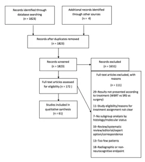
TABLES
Table 1. Search Strategies
| PUBMED (NLM), searched on February 3-4, 2016: |
| Step 1: Brain Neoplasms [Mesh] |
| Step 2: (brain OR brainstem OR intracranial) AND (cancer OR tumor* OR tumour* OR neoplasm*) [TIAB] |
| Step 3: #1 OR #2 |
| Step 4: Neoplasm Metastasis [Mesh] |
| Step 5: (brain OR brainstem OR intracranial) AND (Metastas*) [TIAB] |
| Step 6: #4 OR #5 |
| Step 7: #3 AND #6 |
| Step 8: Brain neoplasms/secondary [Mesh] |
| Step 9: #7 OR #8 |
| Step 10: Cranial irradiation [Mesh] |
| Step 11: WBRT [TIAB] |
| Step 12: “whole brain” [TIAB] AND (radiotherap* OR radiation OR radiation therap* OR irradiation) [TIAB] |
| Step 13: #10 OR #11 OR #12 |
| Step 14: #9 AND #13 |
| Step 15: #14 AND English [Lang] |
| Step 16: (animals [MeSH] NOT humans [MeSH]) OR case reports [PT] OR review [PT] OR comment [PT] OR letter [PT] OR editorial [PT] OR addresses [PT] OR news [PT] OR “newspaper article” [PT] |
| Step 17: #15 NOT #16 |
| Step 18: #17 AND (“1990/10/01″[PDAT] : “2015/12/31″[PDAT]) |
| Embase, searched on February 3-4, 2016: |
| Step 1: ‘Brain tumor’/exp |
| Step 2: ((brain OR brainstem OR intracranial) NEAR/3 (cancer OR tumor* OR tumour* OR neoplasm*)):ab, ti |
| Step 3: #1 OR #2 |
| Step 4: ‘brain metastasis’/exp |
| Step 5: ((brain OR brainstem OR intracranial) NEXT/3 metastas*):ab,ti |
| Step 6: #4 OR #5 |
| Step 7: #3 AND #6 |
| Step 8: ‘brain radiation’/exp |
| Step 9: WBRT:ab,ti |
| Step 10: (‘whole brain’ NEXT/3 (radiation OR radiotherapy* OR irradiation)):ab,ti |
| Step 11: #8 OR #9 OR #10 |
| Step 12: #7 AND #11 |
| Step 13: Limits: English, humans, 1990-2015, article OR conference paper NOT case report |
| COCHRANE, searched on February 3-4, 2016 : |
| Step 1: MeSH descriptor: [Brain Neoplasms] explode all trees |
| Step 2: ((brain OR brainstem OR intracranial) NEAR/3 (cancer OR tumor* OR tumour* OR neoplasm*)):ti,ab,kw |
| Step 3: #1 OR #2 |
| Step 4: MeSH descriptor: [Neoplasm Metastasis] explode all trees |
| Step 5: ((brain OR brainstem OR intracranial) NEAR/3 Metastas*):ti,ab,kw |
| Step 6: #4 OR #5 |
| Step 7: #3 AND #6 |
| Step 8: MeSH descriptor: [Brain neoplasms/secondary] |
| Step 9: #7 OR #8 |
| Step 10: MeSH descriptor: [Cranial irradiation] explode all trees |
| Step 11: WBRT:ti,ab,kw |
| Step 12: (‘whole brain’ NEXT/3 (radiation OR radiotherapy* OR irradiation)):ti,ab,kw |
| Step 13: #10 OR #11 OR #12 |
| Step 14: #9 AND #13 |
| Step 15: Filtered 1990-2015 |
Table 2. Outcomes of different dose/fractionation schedules of whole brain radiation therapy
| Author (Year) | Description of Study | Data Class | Conclusions |
| Sayed21 (2015) | Study descriptionProspective nonrandomized study at 1 center to compare 2 WBRT regimens for differences in response and overall survival.Patient population93 patients with MRI scan with >3 brain metastases, good performance status.Treatment regimenG1: 20 Gy in 4 Gy fractions (n = 54)G2: 30 Gy in 3 Gy fractions (n = 39) | III | ResultsMedian survivalG1: 9 monthsG2: 10 months(p = 0.02)MRI response at 3 months (partial response or stable)G1: 85%G2: 87%(p = NS)Author’s conclusionsNo significant difference in response or overall survival. Shorter fractionation beneficial to patients with RPA 2 (less time spent in treatment and little concern for late toxicity) and to radiation facilities (quicker throughput).Comments and conclusionsNo neurocognitive testing. Designated as Class III because it was a very small prospective study with “assignment” to 1 of 2 dose schedules. Statistical rationale for the accrual goal not given. |
| Saha et al20 (2014) | Study descriptionRCT in multiple centers comparing outcome of 2 WBRT regimens.Patient population56 patients with radiologic diagnosis of brain metastases on MRI, good performance statusTreatment regimenG1: 20 Gy in 4 Gy fractions (n = 26)G2: 30 Gy in 3 Gy fractions (n = 30) | III | ResultsMedian survivalG1: 26 weeksG2: 29 weeks(p = 0.955)MRI response at 3 months (complete or partial response or stable)G1: 81%G2: 93%Author’s conclusionsNo significant difference in response or overall survival. 20 Gy in 5 fractions recommended for patients with poor performance status, 30 Gy in 10 fractions for patients with good performance status.Comments and conclusionsNo neurocognitive testing. No significant difference in improvement in ADL between 2 arms, but ADL of both groups improved post-WBRT. Designated as Class III since the patient numbers are small and could account for the nonsignificant finding. Statistical rationale for the accrual goal not given. |
| Graham et al19 (2010) | Study descriptionRCT in cooperative group (ECOG) to compare intracranial control rate and QOL of 2 WBRT schemes.Patient population113 patients with good performance status; stable, absent, or concurrent presentation of extracranial diseaseTreatment regimenG1: 40 Gy in 2 Gy fx BID (n = 57)G2: 20 Gy in 5 Gy fx (n = 56) | I | ResultsMedian survivalG1: 6.1 monthsG2: 6.6 months(p = NS)Intracranial progressionG1: 44%G2: 64%(p = 0.03)Author’s conclusionsIntracranial disease control was improved and QOL maintained with 40 Gy in 20 twice-daily fractions. Authors recommend this dose/fractionation for patients with better prognosis.Comments and conclusionsThe dose/fractionation regimen was not a significant factor affecting overall survival on MVA. Significant factors for improved survival on MVA were resection, supratentorial location, absent extracranial metastases, younger age. QOL and cognitive function outcomes similar in both groups. Mean scores of QOL and cognitive function were stable to improved in most patients during the 6-9 months following treatment. |
| Davey et al5 (2008) | Study descriptionRCT at 2 centers to compare overall survival following accelerated and conventional hypofractionated WBRT.Patient population90 patients with radiologic features of brain metastases on CT or MRI. Good performance status, life expectancy >6 weeks.Treatment regimenG1: 20 Gy/5 daily fractions (n = 45)G2: 40 Gy/20 fractions/twice daily (n = 45) | I | ResultsMedian survivalG1: 19.1 weeksG2: 19.1 weeks(survival curves: log-rank; p = NS)Median time to treatment for intracranial relapseG1:14 weeksG2: 32 weeks(p = 0.03)Author’s conclusionsAlthough accelerated WBRT may improve intracranial control, this did not lead to improved overall survival.Comments and conclusionsNo QOL or neurocognitive testing. Favorable prognostic factors on MVA were low RPA class and colorectal pathology. |
| Murray et al9 (1997) | Study descriptionRCT by cooperative group (RTOG) comparing accelerated hyperfractionated WBRT with standard fractionation.Patient population429 patients with brain metastases measurable by CT or MRI scans, Karnofsky scale score ≥70, neurologic function class of 1-2.Treatment regimenG1: 30 Gy/10 fractions/daily (n = 213)G2: 54.4 Gy/34 fractions/twice daily (n = 216) | I | ResultsMedian survivalG1: 4.5 monthsG2: 4.5 months(p = NS)# pts with recurrence/progressionG1: 109/124 (88%)G2: 105/118 (89%)(p value not reported)Median time to recurrence / progressionG1: 11 weeksG2: 10 weeks(p value not reported)Author’s conclusions54.4 Gy in 34 fractions not recommended.Comments and conclusionsNo neurocognitive testing. 54.4 Gy delivered as 32 Gy in 20 fractions/twice daily followed by 24.4 Gy boost (visible lesion with 2-cm margin) in 14 fractions/twice daily. Age, performance status, extent of metastatic disease, and status of primary were prognostic factors. |
| Priestman et al10 (1996) | Study descriptionRCT at 25 institutions comparing 2 WBRT regimensPatient population544 patients with symptomatic brain metastases by CT scan or unequivocal radioisotope scan, or an intracranial biopsy. Required stable dose dexamethasone over week prior to randomization, WHO performance status of 0-3, neurologic status <4 by modified MRC scaleTreatment regimenG1: 12 Gy/2 fractions (n = 274)G2: 30 Gy/10 fractions (n = 270) | I | ResultsMedian survivalG1: 77 daysG2: 84 days(p = 0.04 for entire survival curve, no difference in median survival)Author’s conclusionsFor majority of patients, no advantage to longer courses of radiation therapy.Comments and conclusionsNo neurocognitive testing. Small improvement in survival with longer course but not thought by authors to be clinically meaningful. Might recommend longer course in small number of patients with good prognosis (female gender, age <60 years, breast primary, solitary brain metastasis, dexamethasone ≤8 mg/day, WHO performance status <3). |
| Chatani et al3 (1994) | Study descriptionRCT evaluating 2 different WBRT regimens in patients with normal (<250 U/L) vs high LDHPatient population162 patients with lung cancer (stratified for small vs nonsmall) with CT brain scan.Treatment regimenNormal LDH:G1: 30 Gy/10 fractions (n = 46)G2: 50 Gy/20 fractions with field reduction after 30 Gy if possible (n = 46)High LDH:G3: 30 Gy/10 fractions (n = 35)G4: 20 Gy/5 fractions (n = 35) | II | ResultsMedian survivalG1: 5.4 monthsG2: 4.8 months(p = NS)G3: 3.4 monthsG4: 2.4 months(p = NS)Author’s conclusionsLDH is important prognostic factor. 30 Gy/10 fractions recommended.Comments and conclusionsNo neurocognitive testing. RCT but designated as class II and the patient numbers were small, with no clear inclusion criteria beyond “lung cancer.” |
| Sause et al11 (1993) | Study descriptionCooperative group (RTOG) phase I/II trial of accelerated fractionationPatient populationPatients eligible had controlled or absent primary with metastases than brain stable, or only brain metastases with primary uncontrolled.Treatment regimenG1: 32 Gy in 1.6 Gy fractions + boost to 48.0 Gy] (n = 62)G2: 32 Gy in 1.6 Gy fractions + boost to 54.4 Gy] (n = 115)G3: 32 Gy in 1.6 Gy fractions + boost to 64.0 Gy] (n = 104)G4: 32 Gy in 1.6 Gy fractions + boost to 70.4 Gy] (n = 53)Fractions administered twice daily | II | ResultsMedian survivalG1: 4.2 monthsG2: 5.2 monthsG3: 4.8 monthsG4: 6.4 months(p = NS)Author’s conclusionsNonsignificant improvement in survival in higher dose arms was taken as an encouraging result.Comments and conclusionsNo neurocognitive testing. Used as basis for subsequent RTOG study.9 Designated as class II since it was a phase I/II randomized phase II study within cooperative group (RTOG). |
| Haie-Meder et al6 (1993) | Study descriptionRCT at 3 institutions comparing 2WBRT treatment regimensPatient population216 patients with lung, breast, head and neck, or unknown primaries. Diagnosed by CT scan. Age <71 years. Ineligible if Karnofisky scale score <20 or life expectancy <1 monthTreatment regimenG1: 18 Gy/3 fractions (n = 110)G2: 18 Gy/3 fractions; 4 weeks later a second identical course or 25 Gy/10 fractions (n = 106) | I | ResultsMedian survivalG1: 4.2 monthsG2: 5.3 months(p = NS)Author’s conclusionsNo difference in overall survival or neurologic response or incidence in complications. A radiation schedule as short as 18 Gy in 3 fractions as good as longer radiation schedules. No neurologic complications occurred among 45 patients living >12 monthsComments and conclusionsInvestigators could decide on whether G2 received 18 or 25 Gy in the second course- shortest regimen recommended if poor general or neurologic status. Methods of assessing neurocognitive function in follow-up were not clearly described. Two clinical factors predictive of poor survival were presence of multiple brain metastases and/or extracranial metastases. |
| Komarnicky et al7 (1991) | Study descriptionRCT by cooperative group (RTOG) evaluating role of misonidazole combined with WBRTPatient population859 patients with measurable disease on CT, 18-75 years of age, Karnofsky scale score ≥40, able to workTreatment regimenG1: 30 Gy/10 fractions (n = 193)G2: 30 Gy/6 fractions (n = 200)G3: 30 Gy/6 fractions + MISO (n = 196)G4: 30 Gy/10 fractions + MISO (n = 190) | I | ResultsMedian survivalG1: 4.5 monthsG2: 4.1 monthsG3: 3.1 monthsG4: 3.9 months(p = NS)# of pts retreated for BM after protocol therapyG1: 54/179 (30%)G2: 54/180 (30%)G3: 33/173 (19%)G4: 54/163 (33%)(p = NS)Author’s conclusionsRecommended treatment was 30 Gy in 10 fractions, without misonidazoleComments and conclusionsNo neurocognitive testing. Approximately one-third of patients died of uncontrolled metastases, suggesting the need for more effective therapy. |
| Chatani et al4 (1985) | Study descriptionRCT at a single institutionPatient population69 consecutive patients with metastases from lung cancerTreatment regimenG1: 30 Gy/10 fractions (n = 35)G2: 50 Gy in 20 fractions (n = 34) | II | ResultsMedian survivalG1: 4 monthsG2: 3 months(p = NS)Survival at 6 monthsG1: 42%G2: 14%(p < 0.05)Author’s conclusionsPerformance status and LDH were the factors influencing 6-month survivalComments and conclusionsNo neurocognitive testing. Designated as Class II due to small numbers and was limited to lung cancer. |
| Kurtz et al8 (1981) | Study descriptionRCT by cooperative group (RTOG)Patient population309 patients (255 evaluable) from 31 participating institutions. Ineligible if evidence of other sites of metastatic disease or progressive untreated primary, or poor neurologic functionTreatment regimenG1: 30 Gy/10 fractions (n = 130)G2: 50 Gy/20 fractions (n = 125) | I | ResultsMedian survivalG1: 18.2 weeksG2: 16.9 weeks(p = NS)# pts with recurrence/progression in patients with information availableG1: 109/124 (88%)G2: 105/118 (89%)(p value not reported)Author’s conclusions30 Gy in 10 fractions as effective as 50 Gy.Comments and conclusionsExcluded patients with evidence of extracranial metastases, uncontrolled primaries, or poor neurologic function. 21% of patients in 50 Gy arm unable to complete therapy. No neurocognitive testing. Authors recommended 20-30 Gy in 5-10 fractions. |
| Borgelt et al2 (1981) | Study descriptionTwo large (>900 patients in each study) national RCTs by cooperative group study (RTOG) with optional randomization to very short regimens at small number of institutions. This study is analysis of patients randomized at 4-6 centers that had very short regimens open.Patient populationIneligible if lesions too numerous or symptoms too vague to allow for adequate follow-up or assessment.First RCT: 155 patients randomized at 6 institutionsSecond RCT: 78 patients randomized at 4 institutionsTreatment regimenFirst RCT:30 Gy/10 fractions/2 wks (n = 233)30 Gy/15 fractions/3 wks (n = 217)40 Gy/15 fractions/3 wks (n = 233)40 Gy/20 fractions/4 wks (n = 227)10 Gy/single fraction: option in 6 institutions (n = 26)Second RCT:20 Gy/5 fractions/1 wk (n = 31)12 Gy in 2 fractions (n = 33)Analysis by groupFirst RCT:G1: 10 Gy/1 fractionG2: 30-40 Gy over 2-4 weeksSecond RCT:G3: 12 Gy/2 fractionsG4: 20 Gy over 1 week | I | ResultsMedian survivalFirst RCT:G1: 15 weeksG2: 21 weeks(survival curves: log-rank; p = NS)Second RCT:G3: 13 weeksG4: 12 weeks(survival curves: log-rank; p = NS)Median time to progression (measured by deterioration in neurologic function ):First RCT:Initial NF 1: G1: 9 wks; G2: 14 wksInitial NF 2: G1: 9 wks; G2: 10 wksInitial NF 3: G1: 7 wks; G2: 12 wks (Cox’s model; p = 0.07)Second RCT:Initial NF 1: G3: 9 wks; G4: 10 wksInitial NF 2: G3: 11 wks; G4: 8 wksInitial NF 3: G3: 3 wks; G4: 3 wks (Cox’s model; p = NS)Authors’ conclusionsResponse of patients receiving the ultra-rapid treatment (10-12 Gy in 1-2 fractions) as assessed by the percent who had improvement in neurologic function, was comparable to that of patients receiving the more protracted schedules. Promptness of neurologic function improvement, treatment morbidity, and median survival were also comparable to those of patients receiving the more protracted courses. However, the duration of improvement, time to progression of neurologic status and rate of complete disappearance of neurologic symptoms were generally less for patients treated with ultrarapid treatment. Ultrarapid treatment may not be as effective as higher dose schedules in the palliation of brain metastases.Comments and conclusionsNo neurocognitive testing. Large cooperative group RCT but relatively small numbers of patients in the second RCT testing ultrarapid treatment. |
| Borgelt et al33 (1980) | Study description2 RCT by cooperative group (RTOG) to study effectiveness of different WBRT dose fractionation schemes on palliation.Patient populationFirst RCT 993 (910 evaluable) and second RCT 1001(902 evaluable) patients with brain metastases established by clinical symptoms, EEG, radioisotope brain scan, arteriogram, pneumoencephalogram, or biopsy. Patients excluded if lesions too numerous or symptoms too vague to allow for adequate follow-up or assessment.Treatment regimenFirst RCT:G1: 30 Gy/10 fractions/2 wks (n = 233)G2: 30 Gy/15 fractions/3 wks (n = 217)G3: 40 Gy/15 fractions/3 wks (n = 233)G4: 40 Gy/20 fractions/4 wks (n = 227)Second RCT:G1: 20 Gy/5 fractions/ 1 wk (n = 447)G2: 30 Gy/10 fractions/ 2 wks (n = 228)G3: 40 Gy/15 fractions/ 3 wks (n = 227) | I | ResultsMedian survivalFirst RCT: 18 weeks. No significant difference between G1-4 (range 16-20 wks)Second RCT: 15 weeks. No significant difference between G1-3 (range 14-15 wks)Brain metastases as cause of deathFirst RCT: 49%. No significant difference between G1-4 (range 46-54%)Second RCT: 31% No significant difference between G1-3 (range 25-33%)Palliation of neurologic symptomsRelief in 60-90% of patients with no significant difference between studiesImprovement in neurologic function at 2 weeksFirst RCT:G1: 55%G2-4: 43%(p = 0.06)Second RCT:G1: 64%G2-3: 54%(p = 0.01)Author’s conclusionsAll treatment schedules were comparable with respect to frequency of improvement, duration of improvement, time to progression, survival, and palliation. Important prognosticators of response included initial neurologic function and general performance status. Administration of steroids during irradiation favored more rapid improvementComments and conclusionsThe administration of steroids was not controlled in either study. Results by treatment regimens not presented separately. Primary site (lung vs breast vs other) had no influence on palliative benefit of WBRT. Palliation reported sooner in shorter WBRT regimens but reporting bias suspected. Relatively small numbers of patients in the second RCT testing ultrarapid treatment. No neurocognitive testing. |
ADL, activities of daily living; BID, twice daily; CT, computed tomography; ECOG, Eastern Cooperative Oncology Group; Gy, Gray; LDH, lactate dehydrogenase; MRC, Medical Research Council; MRI, magnetic resonance imaging; MVA, multivariate analysis; QOL, quality of life; RCT, randomized controlled trial; RPA, recursive partitioning analysis; WBRT, whole brain radiation therapy; WHO, World Health Organization.
Table 3. Effect of histology of primary cancer on outcomes of whole brain radiation therapy
| Author (Year) | Description of Study | Data Class | Conclusions |
| Lee et al30 (2012) | Study descriptionSingle institution, retrospective review of impact of EGFR mutation in patients with NSCLC brain metastases treated with WBRT in terms of RPFS and OSPatient population43 patients with NSCLC (40 adenocarcinoma, 1 adenosquamous carcinoma, 2 poorly differentiated carcinoma)EGFR-positive: 30 patients with EGFR mutation (15 with exon 19 deletions, 15 with exon 21 L858R point mutation);EGFR-negative: 13 patients with EGFR wild-typeTreatment regimen43 patients underwent WBRT (30-40 Gy in 10-20 fractions, 40% of patients had additional local boost up to 50-60 Gy).EGFR tyrosine kinase inhibitor (TKI) given to 50% of EGFR-positive and 69% of EGFR-negative patients. | III | ResultsMedian follow-up 15 monthsRadiographic response to RTOverall 70% radiographic response rate to RTEGFR-positive: 80%EGFR-negative: 46 (p = 0.037)Multivariate analysis of radiographic responseEGFR mutation was only predictor for treatment response (odds ratio: 4.67, 95% CI; p = 0.032)Median intracranial RPFSOverall 18 months (95% CI: 8.33-27.68)EGFR-positive: 21 monthsEGFR-negative: 12 months (p = 0.009)Multivariate analysis for RPFSEGFR mutation (p = 0.025) and RPA class ( p = 0.026) were 2 predictors for longer RPFSOverall survivalMedian OS 15 months (95% CI: 9.61-20.39 months)Univariate analysis showed that EGFR mutations ( p = 0.061) and performance status (p = 0.076) had a trend to predict OS.Author’s conclusionMutant EGFR in NSCLC brain metastasis patients is an independent prognostic factor for better treatment response and longer intracranial RPFS following WBRTComments and conclusionsThis is a retrospective case series (class III) of patients with brain metastasis from NSCLC treated with WBRT, which found mutant EGFR as a positive prognostic factor for treatment response after WBRT. EGFR TKI given to more than half of these patients and difficult to know how this impacted results. EGFR TKI should not be given to patients known to be EGFR wild-type, since it has been shown in other settings to be associated with poor outcome. |
| Gow et al31 (2008) | Study descriptionSingle institution, retrospective case series of patients with brain metastases from lung adenocarcinoma treated with WBRT, evaluating the role of EGFR mutation status in response to WBRT and survivalPatient population63 patients patient with brain metastases from lung adenocarcinoma treated with WBRTEGFR-positive: Positive EGFR mutations (n = 46)EGFR-negative: Wild-type EGFR (n = 17)Treatment regimen63 patients with NSCLC brain metastases received WBRT (30-35 Gy in 15 to 18 fractions); 18 patients received gefitinib treatment (either before or during WBRT treatment).Pertinent methods of study techniqueUnivariate and logistic regression models were used to test predictive factors associated with clinical response; log-rank test and cox regression were used to identify factors affecting survival | III | ResultsClinical response to WBRTOverall response rate 46%EGFR-positive: 54%EGFR-negative: 24% (p = 0.045)Both EGFR expression and EGFR tyrosine kinase inhibitor administration were independently associated with response to WBRT (p = 0.034 and p = 0.029, respectively)Survival with WBRTMedian survival was 14.7 months (95% CI, 7.5-21.9 months)Better OS in responders vs nonresponders to WBRT (20.7 vs 6.6 months, p = 0.017).On univariate analysis, RPA class (p = 0.025), KPS (p = 0.013), and absence of extracranial metastases (p = 0.005) were significant prognosticators for overall survival.EGFR mutation (p = 0.131) and administration of EGFR TKI during WBRT (p = 0.121) showed a trend but no significant correlation with survival.Author’s conclusionEGFR mutation and EGFR TKI administration during WBRT are independent predictors of response to WBRT in brain metastases from lung adenocarcinoma.Comments and conclusionThis retrospective case series (class III) found mutant EGFR expression and TKI administration were predictive of improved response to WBRT, with a trend to improved overall survival but not statistically significant. All patients received WBRT but a small number also received systemic therapy with gefitinib, representing a heterogeneous treatment population. |
| Wolstenholm et al32 (2008) | Study descriptionSingle institution, retrospective case series examining the influence of HER2 status on outcome of patients with brain metastases from breast cancer who received WBRTPatient population181 patients with breast cancer metastasis and known HER2 status receiving WBRTHER2+ (n=88)HER2- (n=93)Treatment regimenWBRT regimens included 20 Gy in 5 fractions or 30 Gy in 10 fractions (5 and 2 patients in the HERR2+ and HER2- groups respectively received surgery as primary treatment followed by WBRT, and 11 and 1 patients in the HER2+ and HER2- groups respectively received stereotactic radio surgery (18-22 Gy at the 90-100%) in addition to WBRT.Heterogeneous chemotherapy regimens; trastuzumab treatment in 53 HER2+ patients. Pertinent methods of study techniqueUnivariate and multivariate Cox regression analysis of prognostic factors; Kaplan-Meier survival analysis with log-rank test | III | ResultsMedian survival after WBRTHER2-: 8 monthsHER2+: 4 monthsp=0.008Prognostic factors8 patients (4% of entire study population) had solitary brain metastases, with significantly improved survival compared to multiple brain metastases (p=0.005); 6 of these patients were HER2+On univariate analysis performance status was significant predictor of longer survival (p=0.01)On multivariate analysis HER2 status was an independent prognostic factor (p=.02)Author’s conclusionImproved median survival in patients with HER2+ status following WBRT, which could be attributed to a more aggressive approach to their management with combined cytotoxic chemotherapy and ongoing trastuzumab.Comments and conclusionsThis is a retrospective study (Class III) with no comparison group, with a heterogeneous mix of treatments in addition to WBRT and varied chemotherapy regimens, including use of trastuzumab in a portion of the HER2+ patients. |
| Sundstrom et al29 (1998) | Study descriptionSingle institution, retrospective review of patients treated with WBRT for brain metastases diagnosed by CT or MRI with minimum midline dose to the whole brain of at least 25 Gy. Patient populationBreast cancer (n=19)Lung cancer (n=35)Renal cell (n=9)Melanoma (n=6)Other (n=6)Extra-cranial metastasesBreast: 17/19Lung: 6/35Renal cell: 5/9Melanoma: 4/6Other: 5/6Treatment regimens WBRT mean dose 30 Gy (range 25–40 Gy) in 1.8– 3 Gy fractions | III | ResultsMedian survival by primary tumor typeBreast cancer: 7 months (range 1–62 months) Lung cancer: 4 months (range 1–21 months) Renal cell: 4 months (range 2–34 months)Melanoma: 3 months (range 1–6 months)Other: 4 months (range 1–9 months)Survival curves: P-value not reportedMedian time to recurrence of brain metastases Not reportedTumor control, functional performance, cause of death, adverse events Not reported by histologyAuthor’s conclusionsApproximately two-thirds of the patients experienced a relief in symptoms allowing a reduction in the dose of corticosteroid medication, which clearly supports the use of whole-brain radiotherapy as a palliative treatment.Comments and conclusionsDesignated Class III since numbers too small to allow meaningful statistical comparison between histologies. |
| Borgelt et al33 (1980) and Borgelt et al 2 (1981) | Study description2 RCT by cooperative group (RTOG) to study effectiveness of different WBRT dose fractionation schemes on palliation.Patient populationFirst RCT 993 (910 evaluable) and second RCT 1001 (902 evaluable) patients with brain metastases established by clinical symptoms, EEG, radioisotope brain scan, arteriogram, pneumoencephalogram, or biopsy.Stratified by site of primary lesion: lung vs breast vs other, and presence or absence of metastases to sites other than brain; pPatients excluded if lesions too numerous or symptoms too vague to allow for adequate follow-up or assessment.Treatment regimenFirst RCT:G1: 30 Gy/10 fractions/2 wks (n = 233)G2: 30 Gy/15 fractions/3 wks (n = 217)G3: 40 Gy/15 fractions/3 wks (n = 233)G4: 40 Gy/20 fractions/4 wks (n = 227)Second RCT:G1: 20 Gy/5 fractions/ 1 wk (n = 447)G2: 30 Gy/10 fractions/ 2 wks (n = 228)G3: 40 Gy/15 fractions/ 3 wks (n = 227) | II | ResultsMedian survivalFirst RCT: 18 weeks. No significant difference between G1-4 (range 16-20 wks)Second RCT: 15 weeks. No significant difference between G1-3 (range 14-15 wks)Brain metastases as cause of deathFirst RCT: 49%. No significant difference between G1-4 (range 46-54%)Second RCT: 31% No significant difference between G1-3 (range 25-33%)Primary site60% of patients had lung primaries.Lung cancer patients more likely to have brain as only site of metastases; primary site had no influence on response to WBRT.Time to progression longer for breast cancer patients.Median survival for breast cancer patients longer than for lung cancer patients (21 weeks vs 16 wks, p < 0.001). This survival difference between breast and lung cancer not seen in nonambulatory patients.Author’s conclusionsAll treatment schedules were comparable with respect to frequency of improvement, duration of improvement, time to progression, survival, and palliation. Important prognosticators of response included initial neurologic function and general performance status. Administration of steroids during irradiation favored more rapid improvementComments and conclusionsPrimary site (lung vs breast vs other) had no influence on palliative benefit of WBRT. The administration of steroids was not controlled in either study. Palliation reported sooner in shorter WBRT regimens but reporting bias suspected. Relatively small numbers of patients in the second RCT testing ultrarapid treatment. Designated class II since results by treatment regimens not presented separately by histology. |
EGFR, epidermal growth factor receptor; Gy, Gray; KPS, Karnofsky Performance Scale; NSCLC, non–small cell lung cancer; OS, overall survival; RCT, randomized controlled trial; RPFS, radiologic progression-free survival; TKI, tyrosine kinase inhibitor; WBRT, whole brain radiation therapy.
Table 4. Neurocognitive outcomes of prophylactic cranial irradiation versus no prophylactic cranial irradiation for patients without brain metastases
| Author and Year | Description of Study | Data Class | Conclusions |
| Wolfson et al43 (2011) | Study descriptionSecondary endpoint of multi-institutional phase II RCTSCLC histology (N = 264)Testing different PCI RT schedules for patients with SCLC in complete remission after induction therapy.Treatment regimensG1: 25 Gy in 10 fractions (n = 131)G2: 36 Gy in 18 fractions (n = 67)G3: 36 Gy in 24 fractions, 1.5 Gy BID (n = 66)Randomization to 25 Gy vs 36 Gy, then secondary randomization to G2 vs G3.ND defined as a significant decrease at 12 months in at least one neurocognitive test (HVLT, COWAT, or TMT-A and -B) from baseline regardless of brain metastases. | II | ResultsStatistically significant differences for COWAT (p = 0.03) and TMT-A (adjusted p = 0.03) testing at baseline among the 3 groups.Proportion with ND (regardless of brain metastases) at 12-months:G1: 62%G2: 85%G3: 89%Significant difference in ND between G1 and G2/3 (p = 0.03)Proportion with ND without brain metastases at 12-months:G1: 60%G2: 85%G3: 89%Significant difference in ND between G1 and G2/3 (p = 0.02)Logistic regression model for ND without brain metastases at 12 months showed significantly higher risk with 36 Gy (p = 0.03) and older age (p = 0.005)Author’s conclusionDue to increased risk of ND with 36 Gy PCI, 25 Gy PCI remains standard of care for this patient populationComments and conclusionsFormal neurologic testing within prospective trial indicating that ND increased with increasing WBRT dose, and there was no beneficial neurocognitive effect to BID fractionation. Designated as Class II since neurologic decline was a secondary endpoint. |
| Le Péchoux et al44 (2011) | Study descriptionSecondary endpoint of international multi-institutional phase III RCT for SCLC histology. Testing different PCI RT schedules for patients with limited SCLC in complete remission after induction therapyTreatment regimensG1: 25 Gy in 10 fractions (n = 360)G2: 36 Gy in 18 daily fractions or 24 fractions of 1.5 Gy BID (n = 360) | II | ResultsProportion of patients with abnormal QoL-cognitive functioning (scale <75) at baseline (N = 667 with baseline data available)G1: 23%G2: 25%Proportion of patients with abnormal QoL-cognitive functioning (scale <75) at 24-months (n = 140)G1: 41%G2: 46%Proportion of patients with abnormal LENT-SOMA intellectual functioning at 24-months (n = 144)G1: 20%G2: 28%G1 and G2 showed a similar, mild deterioration across time in communication deficit, weakness of legs, intellectual deficit and memory. This deterioration over time was statistically significant (p < 0.005).Author’s conclusion:Patients should be informed of the potential neurologic and neurocognitive deficits, as well as the benefit of PCI on survival and the incidence of brain metastases. 25 Gy remains the standard of care for PCI for limited SCLC.Comments and conclusionsLarge RCT in cooperative group using validated QOL tools. Designated as class II since neurologic decline was a secondary endpoint. |
| Sun et al45 (2011) | Study descriptionSecondary endpoint of US multi-institutional phase III RCT in NSCLC histology PCI vs no PCI for patients with stage IIIA/B NSCLC without disease progression after definitive therapy. Treatment regimensNo PCI (n= 163)PCI 30 Gy in 15 fractions (n = 177)Accrual was 340 eligible patients out of planned 1058 (trial closed early due to poor accrual). | III | ResultsBaseline neurocognitive results not reported.Baseline used for per patient measurement of declineProportion with significant deterioration in HVLT-IR at 1 year (n = 90)Control: 7% PCI: 26% (adjusted p = 0.03)Proportion with significant deterioration in HVLT-DR at 1 year (n = 90)Control: 5% PCI: 32% (adjusted p = 0.008)Proportion with deterioration in MMSE score as defined by reliable change index (n = 95)Control: 18% PCI: 23% (p = NS)Authors conclusionNo significant differences in global cognitive function (MMSE) or QOL after PCI, but there was a significant decline in memory (HVLT) at 1 year.Comments and conclusionsThis was designated as class III given that it closed with only approximately one third of planned accrual, perhaps accounting for the lack of significant differences. |
| Slotman et al46 (2009) | Study descriptionSecondary endpoint of European multi-institutional phase III RCTSCLC histology, extensive stage with response to induction therapyTreatment regimenPCI (n = 143)No PCI (n = 143)Most common PCI dose fractionation regimens:20 Gy in 5 fractions (62%)30 Gy in 10 fractions (16%)30 Gy in 12 fractions (6%)25 Gy in 10 fractions (5%)HRQOL measured with EORTC Quality of Life Questionnaire C30 (EORTC-QLQ-C30) and EORTC QLQ Brain Cancer Module (EORTC-QLQ-BN20)268 of 286 with baseline scores available. | II | ResultsProportion with worsened global health status (≥ 20-point decline) at 3 months (n = 188)PCI: 34.7% No PCI: 22.2% (p = NS)Proportion with worsened cognitive functioning (≥ 20-point decline) at 3 months (n = 188)PCI: 22.4% No PCI: 10% (p = NS)Mean difference in cognitive functioning score at 3 months between arms (No PCI – PCI) of 8.8 points (below significance definition of ≥10 points)Authors conclusions:PCI should be offered to all responding ED SCLC patients. Patients should be informed of the potential adverse effects from PCI. Comments and conclusionsThe largest mean difference between the 2 arms was observed for fatigue and hair loss. The impact of PCI on global health status as well as on neurocognitive functioning scores was more limited. Designated as Class II since change in cognitive function was a secondary endpoint. |
| Gregor et al47 (1997) | Study descriptionSecondary endpoint of UKCCCR and EORTC multi-institutional phase III RCT of SCLC histology.Included patients without brain metastases with complete remission after induction therapyTreatment regimenPCI (n = 120)No PCI (n = 194)Most common PCI regimens were 30 Gy in 10 fractions, 24 Gy in 12 fractions, and 36 Gy in 18 fractions.Initially 1:1 randomization to PCI:No PCI, then revised to 3:2 (PCI:No PCI)Neurocognitive portion of trial was optional.125 of 314 patients (40%) with baseline neurocognitive testing available.59 of 314 patients (19%) with 6-month testing results available. | III | ResultsNo significant difference on multiple neurocognitive tests between PCI and No PCI at 6-months and 1-year. Cognitive impairment on study entry was seen on study entry in up to 42% of patientsAuthors conclusion:In both groups, there was similar degree of impairment of cognitive function and QOL before PCI. No difference in neurocognitive detriment between PCI and control in this patient population without brain metastasesComments and conclusions:Used simple proportions to compare cognitive decline at each time point. Designated as class III since patient numbers were relatively small at all time points, and neurocognitive testing was only available on 40% of patients at baseline. |
| Arriagada et al48 (1995) | Study descriptionPrimary endpoint of multi-institutional French phase III RCTSCLC histologyIncluded patients with SCLC, without brain metastases, with complete remission after induction therapy.Treatment regimenPCINo PCIPCI was 24 Gy in 8 fractionsNeuropsychologic assessments performed by neurologists, N = 294 | II | Results41% of all patients did not have neurocognitive abnormalities at baselineNumber of patients free from any abnormalities at baseline:PCI: 50No PCI: 442-year cumulative incidence of negative change in cognitive “higher functions” 36% (control) vs 30% (PCI), p = NSPCI 30%No PCI 36%, p = NSAuthors conclusionProphylactic cranial irradiation given to patients with small cell lung cancer in complete remission decreases the risk of brain metastasis threefold without a significant increase in complications. No difference in neurocognitive detriment between PCI and control in this patient population without brain metastasesComments and conclusionsUsed cumulative incidence for cognitive dysfunction endpoint. Designated as class II since “higher functions” were not defined, in addition to the lack of definition of criteria used to define decline. |
BID, twice daily; COWAT, Controlled Oral Word Association Test; EORTC, European Organisation for Research and Treatment of Cancer; HRQOL, health-related quality of life; HVLT, Hopkins Verbal Learning Test; KPS, Karnofsky Performance Scale; MMSE, Mini-Mental State Examination; ND, neurocognitive decline; NS, not significant; PCI, prophylactic cranial irradiation; QOL, quality of life; RCT, randomized controlled trial; SCLC, small cell lung cancer; TMT, Trail Making Test.
Table 5. Neurocognitive outcomes of whole brain radiation therapy and local therapy versus local therapy only
| Author and Year | Description of Study | Data Class | Conclusions |
| Soffietti et al51 (2013) | Study descriptionSecondary endpoint of European multi-institutional phase III RCTPatient populationPatients with 1-3 brain metastasesTreatment regimenLocal only: local therapy alone with SRS or surgery (n = 179)Local + WBRT (n = 180)Local therapy either SRS (n = 199) or surgery (n = 160)HRQOL measured with the EORTC Quality of Life Questionnaire C30 and the EORTC QLQ Brain Cancer ModuleN = 341 with baseline HRQOL data | II | ResultsEORTC QLQ C30 cognitive functioning score mean difference at 12 monthsLocal vs local + WBRT mean difference = -10.8 points (p < 0.05)Mean EORTC QLQ C30 cognitive functioning score at 12 monthsLocal: 80.4 Local + WBRT: 69.7 (p = 0.05)Authors conclusionsAdjuvant WBRT after surgery or SRS of a limited number of brain metastases may negatively impact some aspects of HRQOL, including self-reported cognitive functioning.Comments and conclusionsOverall, patients treated with surgery or SRS only reported better HRQOL scores than did patients who also received WBRT. Most scores, which differed significantly during the first time points, had a tendency to recover. The positive effect of WBRT in decreasing the rate of intracranial progression and modestly improving the progression-free survival did not translate into an advantage in terms of HRQOL. Designated as class II since cognitive functioning was a secondary endpoint. |
| Chang et al50 (2009) | Study descriptionPrimary endpoint of single institutional phase III RCTPatient populationPatients with 1-3 brain metastasesTreatment regimenSRS alone (n = 30)SRS + WBRT (n = 28)WBRT dose: 30 Gy in 12 fractionsPrimary endpoint: significant deterioration of HTLV-R total recall at 4 months defined as ≥5 points drop from baseline.Bayesian analysisTrial enrollment stopped after 58 patients enrolled due to significant differences. | I | ResultsHTLV-R significant deterioration rates at 4 monthsTotal recall:SRS: 24%SRS + WBRT: 52%Delayed recall:SRS: 6%SRS + WBRT: 22%Delayed recognition:SRS: 0%SRS + WBRT: 11% Authors conclusionsPatients treated with SRS + WBRT were at a greater risk of a significant decline in learning and memory function by 4 months compared with the group that received SRS aloneComments and conclusionsSignificantly longer overall survival in patients treated with SRS alone as compared to SRS + WBRT. Given that this is a finding not found in other studies, thought to possibly be indicative of more favorable prognostic factors in SRS alone group. |
| Aoyama et al49 (2007) | Study descriptionSecondary endpoint of Japanese multi-institutional phase III RCTPatients with 1-4 brain metastasesTreatment regimenSRS alone (n = 67)SRS + WBRT (n = 65)WBRT dose: 30 Gy in 10 fractionsNeurocognition measured with MMSE.110 of 132 randomized patients (83%) had baseline MMSE scores available. | II | ResultsAverage baseline MMSE did not differ significantly between treatment groups (p = 0.47).Median MMSE score at 12 monthsSRS alone: 28SRS+WBRT: 27Actuarial rate of MMSE preservation (decline < 3 points) at 12 monthsSRS alone: 59.3%SRS+WBRT: 76.1% (p = NS)Actuarial rate of MMSE preservation (decline < 3 points) at 24 monthsSRS alone: 51.9%SRS+WBRT: 68.5% (p = NS)Average duration until MMSE deteriorationSRS alone: 7.6 monthsSRS+WBRT: 16.5 months (p = 0.05)Authors conclusionIntracranial control is the most important factor for stabilizing neurocognitive function. Addition of WBRT stabilized neurocognition in the intermediate term due to improved intracranial control, however WBRT may be associated with long-term adverse effects on neurocognition.Comments and conclusionsDesignated as class II since MMSE is a relatively insensitive measure of neurocognition and may miss more subtle changes. |
Table 6. Effect of pharmacologic agents or whole brain radiation therapy techniques on neurocognitive decline
| Author and Year | Description of Study | Data Class | Conclusions |
| Rapp et al53 (2015) | Study descriptionPrimary endpoint of multi-institutional phase III RCT of donepezil versus placebo. Patient eligibilityPatients with either primary or secondary brain tumors receiving partial brain (60%) or WBRT (40%) of at least 30 Gy ≥6 months before enrollment.27% metastatic brain tumors7% PCI66% primary brain tumorsTreatment regimensDonepezil: n = 99Placebo: n = 99Donepezil single daily 5-mg dose for 6 weeks, which was escalated to 10 mg per day for 18 weeks if well tolerated.Primary endpoint: overall cognitive performance after 24 weeks of therapy | II | Results24 week results:Patients in both groups showed improved cognitive function at 24 weeks, but there was no difference in overall cognitive composite score between arms (p = 0.48)No significant differences between groups except for memory recognition (p = 0.027), memory discrimination (p = 0.007), and motor speed and dexterity (p = 0.016)The benefits of donepezil greater for those who were more cognitively impaired at baseline.Author’s conclusions:Treatment with donepezil did not significantly improve the overall composite score, but it did result in modest improvements in several cognitive functions, especially among patients with greater pretreatment impairments.Comments and conclusions:Assigned class II since only 40% of patients received WBRT. Donepezil only started 6 months after radiation therapy, providing a source of bias. |
| Brown et al56 (2013) | Study descriptionPrimary endpoint of North American multi-institutional phase III RCT. Primary endpoint was decline in HVLT-R delayed recall at 24 weeks. Patient eligibilityPatients with brain metastases (number not limited)Treatment regimensWBRT + memantine: n = 278WBRT + placebo: n=276WBRT 37.5 Gy in 15 fractionsMemantine dosing, starting before or during WBRT:Week 1 5 mg qAMWeek 2 5 mg BIDWeek 3 10mg qAM / 5 mg qPMWeek 4-24 10 mg BIDN = 473 with baseline scores availableOnly 149 (53%) of 280 alive patients at 24 weeks had neurocognitive assessments and were analyzable. | I | ResultsMedian decline in HVLT-R delayed recall at 24 weeksWBRT + memantine: 0WBRT + placebo: -0.9 (p = 0.059, NS)Probability of cognitive failure at 24 weeks:WBRT + Memantine: 53.8% WBRT + placebo: 64.9% (p = 0.01)Authors’ conclusionsMemantine well tolerated. Although memantine was associated with less decline in the primary endpoint of delayed recall at 24 weeks, this lacked statistical significance possibly due to significant patient loss. Overall, patients treated with memantine had better cognitive function over time; specifically, memantine delayed time to cognitive decline and reduced the rate of decline in memory, executive function, and processing speed in patients receiving WBRT. |
| Gondi et al54 (2014) | Study descriptionPrimary endpoint of multi-institutional North American phase II single arm trial of hippocampal avoidance (HA). Results compared with historical control of control arm of previous phase III RCT. Primary endpoint was decline in HVLT-R delayed recall (DR) at 4 months as compared with standard arm of PCI-P-120-9801 phase III trial using WBRT 30 Gy in 10 fractions without HA. Patient eligibilityPatients with brain metastases outside a 5-mm margin around either hippocampus. Treatment regimentPatients treated with HA (n=113) during WBRT to 30 Gy in 10 fractions.Hippocampal D100 goal <9 Gy and max point dose goal <16 Gy100 patients with baseline scores available.42 patients with scores analyzable at 4 months. | II | Results:42 patients analyzable for primary endpoint at 4 months (71% of alive patients)Mean relative HVLT-R DR decline between baseline and 4 monthsHA: 7%Historical controls: 30% (p = 0.0003)Probability of HVLT-R total recall significant deterioration by 4 monthsHA: 19%Probability of HVLT-R DR significant deterioration by 4 monthsHA: 33%Intracranial progression within HA region5% of patients with intracranial progression3% of patients overallAuthors conclusionsConformal avoidance of the hippocampus during WBRT is associated with preservation of memory and QOL as compared with historical series.Comments and ConclusionsDesignated as Class II since it was a Phase II study. |
| Butler et al55 (2007) | Study descriptionSecondary endpoint of multi-institutional phase III RCT of d-threo-methylphenidate HCl (d-MPH) versus placebo. Primary endpoint was fatigue subscale of the FACIT-F.Patient eligibilityPatients with either primary brain tumors (n = 33) or brain metastases (n = 35) receiving partial brain RT or WBRT ≥25 GyTreatment regimensRT + d-MPH: n = 34RT + placebo: n = 34d-MPH or placebo started by day 5 of RT.Starting dose of d-MPH was 5 mg BID and was escalated by 5 mg BID to a maximum of 15 mg BID.Study drug continued for 8 weeks post-RT.QOL measured with FACT-Brain and FACIT-F and cognition measured with MMSE. Trial closed after accrual of 68 of planned 162 patients due to slow accrual and withdrawal of financial support. | II | ResultsFatigue:No difference in fatigue assessment at any time point up to 8 weeks post-RT between arms.Baseline MMSE scoreRT + d-MPH: 27.2 RT + placebo: 26.5 (p = NS)MMSE 8 weeks post-RTRT + d-MPH: 23.3 RT + placebo: 25.6 (p = NS)Authors conclusionsProphylactic use of d-MPH in brain tumor patients undergoing RT did not result in an improvement in QOL. Comments and conclusionsDesignated as Class II due to low patient accrual and reduced statistical power. Only a small number of patients receiving WBRT. |
Table 7. Intracranial progression-free survival and overall survival following local therapy (surgery or stereotactic radiosurgery) alone or local therapy with whole brain radiation therapy
| Author (Year) | Description of Study | Data Class | Conclusions |
| Kocher et al28 (2011) | Study descriptionRCT comparing WBRT to observation after SRS or surgical resection on duration of functional independence (WHO performance status)Patient population359 patients with 1-3 brain metastases with WHO performance status ≤2 who had previously undergone either surgical resection or SRS prior to randomized interventionTreatment regimenSRS + observation (n = 100)SRS + WBRT (n = 99)Surgery + observation (n = 79)Surgery + WBRT (n = 81)Local therapy + WBRT arm (180 total)Local therapy + Observation (179 total) WBRT 30 Gy in 10 fractions | II | ResultsSurvival with functional independence (time to WHO PS>2)Observation: 10 monthsWBRT: 9.5 months(HR = 0.96, p = 0.71)At 2 years, 22.3% and 22.6% were alive and independent in the observation and WBRT arms, respectively.Progression-free survivalObservation: 3.4 monthsWBRT: 4.6 months(p = 0.020)Overall survivalObservation: 10.9 monthsWBRT: 10.7 months(HR = 0.98, p = 0.89)Author’s conclusionsAfter surgery or SRS, WBRT reduces the probability of intracranial relapses from 80% to 50%, and is most pronounced after surgery. This is translated into a modest PFS, but no improvement in OS. There was no difference in functional independence between the 2 groups.Comments and conclusionsIn well-performing patients with otherwise stable systemic disease and 1-3metastases, who are initially treated with either radiosurgery or surgery, WBRT can be withheld if serial imaging for follow-up is performed. Regarding the patients undergoing resection of a single lesion, because adjuvant irradiation substantially reduces the risk of recurrence in the tumor bed, postoperative local irradiation should be an option that is investigated. Designated class II since the primary endpoint was functional independence, not PFS or OS. |
| Aoyama et al59 (2006) | Study descriptionRCT comparing patients with 1-4 brain metastases receiving either WBRT + SRS or SRS alone on overall survival, recurrence, function, and cause of death. Study closed early due to poor accrual.Patient population132 patients with 1-4 brain metastases (each <3 cm in diameter).No surgical resection performed prior to treatment.Treatment regimenWBRT + SRS (n = 65)SRS alone (n = 67)WBRT 30 Gy in 10 fractions | III | ResultsSurvival (median and 1-year actuarial survival rate)WBRT + SRS: 7.5 months and 38.5%SRS alone: 8.0 months and 28.4%(p = 0.42)Intracranial recurrence rate at 12 monthsWBRT + SRS: 46.8%SRS alone: 76.4%(p < 0.001)Salvage intracranial treatmentWBRT + SRS: 10 patientsSRS alone: 29 patients(p < 0.001)Cause of death: Neurological causesWBRT + SRS: 22.8%SRS alone: 19.3%(p = 0.64)Author’s conclusionsCompared to SRS alone, the use of WBRT + SRS did not improve survival for patients in this trial, but intracranial relapse occurred more frequently in those not receiving WBRT.Comments and conclusionsBetween both groups, there was no difference in OS, but higher rates of recurrence in the SRS only group lead to the more frequent need for salvage treatment. Assigned class III due to early closure of study due to poor accrual, resulting in lack of statistical power. |
| Patchell et al58 (1998) | Study descriptionRCT comparing patients with single brain metastases who underwent surgical resection followed by postoperative WBRT vs observation on tumor recurrence and survival.Patient population95 patients with single metastases to the brain treated with complete surgical resectionTreatment regimenSurgery + WBRT (n = 49)surgery + observation (n = 46)WBRT 50.4 Gy in 28 fractionsPrimary end point: intracranial recurrenceSecondary end points: Overall survival, cause of death, and preservation of ability to function independently | I | ResultsTumor recurrenceSurgery + WBRT: 18%surgery + observation: 70%(p < 0.001)WBRT prevented recurrence at the site of original metastases (10% vs. 46%, p < 0.001) as well as other sites (14% vs. 37%, p < 0.01) vs. observation, respectively.Death from neurological causesSurgery + WBRT: 14%surgery + observation: 44%(p = 0.003)Overall survivalSurgery + WBRT: 48 weekssurgery + observation: 43 weeks(p = 0.39)Author’s conclusionsPostoperative WBRT after complete surgical resection of a single metastasis results in better control of disease in the brain and a reduction in the number of deaths due to neurological causes.Due to the decreased death due to neurologic causes, the authors recommended routine postoperative WBRT.Comments and conclusionsDespite the reduction in brain recurrence rates and neurologic deaths, postoperative WBRT did not result in an increased survival or improvement in the length of time patients were able to function independently. |
Gy, Gray; HR, hazard ratio; OS, overall survival; PFS, progression-free survival; SRS, stereotactic radiosurgery; WBRT, whole brain radiation therapy; WHO, World Health Organization.
REFERENCES
1. Gaspar LE, Mehta MP, Patchell RA, et al. The role of whole brain radiation therapy in the management of newly diagnosed brain metastases: a systematic review and evidence-based clinical practice guideline. J Neurooncol. Jan 2010;96(1):17-32.
2. Borgelt B, Gelber R, Larson M, Hendrickson F, Griffin T, Roth R. Ultra-rapid high dose irradiation schedules for the palliation of brain metastases: final results of the first two studies by the Radiation Therapy Oncology Group. Int J Radiat Oncol Biol Phys. Dec 1981;7(12):1633-1638.
3. Chatani M, Matayoshi Y, Masaki N, Inoue T. Radiation therapy for brain metastases from lung carcinoma. Prospective randomized trial according to the level of lactate dehydrogenase. Strahlenther Onkol. Mar 1994;170(3):155-161.
4. Chatani M, Teshima T, Hata K, Inoue T, Suzuki T. Whole brain irradiation for metastases from lung carcinoma. A clinical investigation. Acta Radiol Oncol. Jul-Aug 1985;24(4):311-314.
5. Davey P, Hoegler D, Ennis M, Smith J. A phase III study of accelerated versus conventional hypofractionated whole brain irradiation in patients of good performance status with brain metastases not suitable for surgical excision. Radiother Oncol. Aug 2008;88(2):173-176.
6. Haie-Meder C, Pellae-Cosset B, Laplanche A, et al. Results of a randomized clinical trial comparing two radiation schedules in the palliative treatment of brain metastases. Radiother Oncol. Feb 1993;26(2):111-116.
7. Komarnicky LT, Phillips TL, Martz K, Asbell S, Isaacson S, Urtasun R. A randomized phase III protocol for the evaluation of misonidazole combined with radiation in the treatment of patients with brain metastases (RTOG-7916). Int J Radiat Oncol Biol Phys. Jan 1991;20(1):53-58.
8. Kurtz JM, Gelber R, Brady LW, Carella RJ, Cooper JS. The palliation of brain metastases in a favorable patient population: a randomized clinical trial by the Radiation Therapy Oncology Group. Int J Radiat Oncol Biol Phys. Jul 1981;7(7):891-895.
9. Murray KJ, Scott C, Greenberg HM, et al. A randomized phase III study of accelerated hyperfractionation versus standard in patients with unresected brain metastases: a report of the Radiation Therapy Oncology Group (RTOG) 9104. Int J Radiat Oncol Biol Phys. Oct 1 1997;39(3):571-574.
10. Priestman TJ, Dunn J, Brada M, Rampling R, Baker PG. Final results of the Royal College of Radiologists’ trial comparing two different radiotherapy schedules in the treatment of cerebral metastases. Clin Oncol (R Coll Radiol). 1996;8(5):308-315.
11. Sause WT, Scott C, Krisch R, et al. Phase I/II trial of accelerated fractionation in brain metastases RTOG 85-28. Int J Radiat Oncol Biol Phys. Jul 15 1993;26(4):653-657.
12. Bach F, Sorensen JB, Adrian L, et al. Brain relapses in chemotherapy-treated small cell lung cancer: a retrospective review of two time-dose regimens of therapeutic brain irradiation. Lung Cancer. Sep 1996;15(2):171-181.
13. Conill C, Jorcano S, Domingo-Domenech J, et al. Whole brain irradiation and temozolomide based chemotherapy in melanoma brain metastases. Clin Transl Oncol. Apr 2006;8(4):266-270.
14. Nieder C, Berberich W, Nestle U, Niewald M, Walter K, Schnabel K. Relation between local result and total dose of radiotherapy for brain metastases. Int J Radiat Oncol Biol Phys. Sep 30 1995;33(2):349-355.
15. Rades D, Schild SE, Lohynska R, Veninga T, Stalpers LJ, Dunst J. Two radiation regimens and prognostic factors for brain metastases in nonsmall cell lung cancer patients. Cancer. Sep 1 2007;110(5):1077-1082.
16. Rades D, Haatanen T, Schild SE, Dunst J. Dose escalation beyond 30 grays in 10 fractions for patients with multiple brain metastases. Cancer. Sep 15 2007;110(6):1345-1350.
17. Rades D, Bohlen G, Dunst J, et al. Comparison of short-course versus long-course whole-brain radiotherapy in the treatment of brain metastases. Strahlenther Onkol. Jan 2008;184(1):30-35.
18. Nieder C, Nestle U, Niewald M, Schnabel K. Accelerated radiotherapy for brain metastases. Radiother Oncol. Oct 1997;45(1):17-22.
19. Graham PH, Bucci J, Browne L. Randomized comparison of whole brain radiotherapy, 20 Gy in four daily fractions versus 40 Gy in 20 twice-daily fractions, for brain metastases. Int J Radiat Oncol Biol Phys. Jul 1 2010;77(3):648-654.
20. Saha A GS, Roy C, Kayal P. Treatment outcomes in patients with multiple brain
metastases: A prospective randomized study. . Clinical Cancer Investigation Journal. 2014;3(4):269-275.
21. Sayed M. Different fractionation in whole brain irradiation for multiple brain metastases
Middle East Journal of Cancer. 2015;6(2):85-90.
22. Fowler J. Practical time-dose evaluations, or how to stop worrying and learn to love linear quadratics. In: Purdy JA PC, Vijayakumar S, ed. Technical Basis of Radiation Therapy: Practical Clinical Applications . 4 ed: Springer Berlin Heidelberg; 2006:3-31.
23. Jones B, Dale RG, Deehan C, Hopkins KI, Morgan DA. The role of biologically effective dose (BED) in clinical oncology. Clin Oncol (R Coll Radiol). 2001;13(2):71-81.
24. Caravatta L, Deodato F, Ferro M, et al. Results of a phase II study of Short-course Accelerated Radiation Therapy (SHARON) for multiple brain metastases. Am J Clin Oncol. Aug 2015;38(4):395-400.
25. Rodrigues G, Yartsev S, Tay KY, Pond GR, Lagerwaard F, Bauman G. A phase II multi-institutional study assessing simultaneous in-field boost helical tomotherapy for 1-3 brain metastases. Radiation oncology (London, England). 2012;7:42.
26. Levegrun S, Pottgen C, Wittig A, Lubcke W, Abu Jawad J, Stuschke M. Helical tomotherapy for whole-brain irradiation with integrated boost to multiple brain metastases: evaluation of dose distribution characteristics and comparison with alternative techniques. Int J Radiat Oncol Biol Phys. Jul 15 2013;86(4):734-742.
27. Lee DS, Kim YS, Lee CG, et al. Early volumetric change and treatment outcome of metastatic brain tumors after external beam radiotherapy: differential radiotherapy for brain metastasis. Clin Transl Oncol. Nov 2013;15(11):889-896.
28. Kocher M, Soffietti R, Abacioglu U, et al. Adjuvant whole-brain radiotherapy versus observation after radiosurgery or surgical resection of one to three cerebral metastases: results of the EORTC 22952-26001 study. J Clin Oncol. Jan 10 2011;29(2):134-141.
29. Sundstrom JT, Minn H, Lertola KK, Nordman E. Prognosis of patients treated for intracranial metastases with whole-brain irradiation. Ann Med. Jun 1998;30(3):296-299.
30. Lee HL, Chung TS, Ting LL, et al. EGFR mutations are associated with favorable intracranial response and progression-free survival following brain irradiation in non-small cell lung cancer patients with brain metastases. Radiation oncology (London, England). Oct 30 2012;7:181.
31. Gow CH, Chien CR, Chang YL, et al. Radiotherapy in lung adenocarcinoma with brain metastases: effects of activating epidermal growth factor receptor mutations on clinical response. Clin Cancer Res. Jan 1 2008;14(1):162-168.
32. Wolstenholme V, Hawkins M, Ashley S, Tait D, Ross G. HER2 significance and treatment outcomes after radiotherapy for brain metastases in breast cancer patients. Breast. Dec 2008;17(6):661-665.
33. Borgelt B, Gelber R, Kramer S, et al. The palliation of brain metastases: final results of the first two studies by the Radiation Therapy Oncology Group. Int J Radiat Oncol Biol Phys. Jan 1980;6(1):1-9.
34. Sperduto PW, Kased N, Roberge D, et al. Effect of tumor subtype on survival and the graded prognostic assessment for patients with breast cancer and brain metastases. Int J Radiat Oncol Biol Phys. Apr 1 2012;82(5):2111-2117.
35. Andrews DW, Scott CB, Sperduto PW, et al. Whole brain radiation therapy with or without stereotactic radiosurgery boost for patients with one to three brain metastases: phase III results of the RTOG 9508 randomised trial. Lancet. May 22 2004;363(9422):1665-1672.
36. Solomon BJ, Cappuzzo F, Felip E, et al. Intracranial Efficacy of Crizotinib Versus Chemotherapy in Patients With Advanced ALK-Positive Non-Small-Cell Lung Cancer: Results From PROFILE 1014. J Clin Oncol. Aug 20 2016;34(24):2858-2865.
37. Schuler M, Wu YL, Hirsh V, et al. First-Line Afatinib versus Chemotherapy in Patients with Non-Small Cell Lung Cancer and Common Epidermal Growth Factor Receptor Gene Mutations and Brain Metastases. J Thorac Oncol. Mar 2016;11(3):380-390.
38. Iuchi T, Shingyoji M, Sakaida T, et al. Phase II trial of gefitinib alone without radiation therapy for Japanese patients with brain metastases from EGFR-mutant lung adenocarcinoma. Lung Cancer. Nov 2013;82(2):282-287.
39. Selek U, Chang EL, Hassenbusch SJ, 3rd, et al. Stereotactic radiosurgical treatment in 103 patients for 153 cerebral melanoma metastases. Int J Radiat Oncol Biol Phys. Jul 15 2004;59(4):1097-1106.
40. Liew DN, Kano H, Kondziolka D, et al. Outcome predictors of Gamma Knife surgery for melanoma brain metastases. Clinical article. Journal of neurosurgery. Mar 2011;114(3):769-779.
41. Mori Y, Kondziolka D, Flickinger JC, Kirkwood JM, Agarwala S, Lunsford LD. Stereotactic radiosurgery for cerebral metastatic melanoma: factors affecting local disease control and survival. Int J Radiat Oncol Biol Phys. Oct 1 1998;42(3):581-589.
42. Fogarty GB, Hong A, Dolven-Jacobsen K, et al. First interim analysis of a randomised trial of whole brain radiotherapy in melanoma brain metastases confirms high data quality. BMC Res Notes. May 08 2015;8:192.
43. Wolfson AH, Bae K, Komaki R, et al. Primary analysis of a phase II randomized trial Radiation Therapy Oncology Group (RTOG) 0212: impact of different total doses and schedules of prophylactic cranial irradiation on chronic neurotoxicity and quality of life for patients with limited-disease small-cell lung cancer. Int J Radiat Oncol Biol Phys. Sep 1 2011;81(1):77-84.
44. Le Pechoux C, Laplanche A, Faivre-Finn C, et al. Clinical neurological outcome and quality of life among patients with limited small-cell cancer treated with two different doses of prophylactic cranial irradiation in the intergroup phase III trial (PCI99-01, EORTC 22003-08004, RTOG 0212 and IFCT 99-01). Ann Oncol. May 2011;22(5):1154-1163.
45. Sun A, Bae K, Gore EM, et al. Phase III trial of prophylactic cranial irradiation compared with observation in patients with locally advanced non-small-cell lung cancer: neurocognitive and quality-of-life analysis. J Clin Oncol. Jan 20 2011;29(3):279-286.
46. Slotman BJ, Mauer ME, Bottomley A, et al. Prophylactic cranial irradiation in extensive disease small-cell lung cancer: short-term health-related quality of life and patient reported symptoms: results of an international Phase III randomized controlled trial by the EORTC Radiation Oncology and Lung Cancer Groups. J Clin Oncol. Jan 1 2009;27(1):78-84.
47. Gregor A, Cull A, Stephens RJ, et al. Prophylactic cranial irradiation is indicated following complete response to induction therapy in small cell lung cancer: results of a multicentre randomised trial. United Kingdom Coordinating Committee for Cancer Research (UKCCCR) and the European Organization for Research and Treatment of Cancer (EORTC). Eur J Cancer. Oct 1997;33(11):1752-1758.
48. Arriagada R, Le Chevalier T, Borie F, et al. Prophylactic cranial irradiation for patients with small-cell lung cancer in complete remission. J Natl Cancer Inst. Feb 1 1995;87(3):183-190.
49. Aoyama H, Tago M, Kato N, et al. Neurocognitive function of patients with brain metastasis who received either whole brain radiotherapy plus stereotactic radiosurgery or radiosurgery alone. Int J Radiat Oncol Biol Phys. Aug 1 2007;68(5):1388-1395.
50. Chang EL, Wefel JS, Hess KR, et al. Neurocognition in patients with brain metastases treated with radiosurgery or radiosurgery plus whole-brain irradiation: a randomised controlled trial. The Lancet. Oncology. Nov 2009;10(11):1037-1044.
51. Soffietti R, Kocher M, Abacioglu UM, et al. A European Organisation for Research and Treatment of Cancer phase III trial of adjuvant whole-brain radiotherapy versus observation in patients with one to three brain metastases from solid tumors after surgical resection or radiosurgery: quality-of-life results. J Clin Oncol. Jan 1 2013;31(1):65-72.
52. Brown PD, Jaeckle K, Ballman KV, et al. Effect of Radiosurgery Alone vs Radiosurgery With Whole Brain Radiation Therapy on Cognitive Function in Patients With 1 to 3 Brain Metastases: A Randomized Clinical Trial. Jama. Jul 26 2016;316(4):401-409.
53. Rapp SR, Case LD, Peiffer A, et al. Donepezil for Irradiated Brain Tumor Survivors: A Phase III Randomized Placebo-Controlled Clinical Trial. J Clin Oncol. May 20 2015;33(15):1653-1659.
54. Gondi V, Pugh SL, Tome WA, et al. Preservation of memory with conformal avoidance of the hippocampal neural stem-cell compartment during whole-brain radiotherapy for brain metastases (RTOG 0933): a phase II multi-institutional trial. J Clin Oncol. Dec 1 2014;32(34):3810-3816.
55. Butler JM, Jr., Case LD, Atkins J, et al. A phase III, double-blind, placebo-controlled prospective randomized clinical trial of d-threo-methylphenidate HCl in brain tumor patients receiving radiation therapy. Int J Radiat Oncol Biol Phys. Dec 1 2007;69(5):1496-1501.
56. Brown PD, Pugh S, Laack NN, et al. Memantine for the prevention of cognitive dysfunction in patients receiving whole-brain radiotherapy: a randomized, double-blind, placebo-controlled trial. Neuro Oncol. Oct 2013;15(10):1429-1437.
57. Auperin A, Arriagada R, Pignon JP, et al. Prophylactic cranial irradiation for patients with small-cell lung cancer in complete remission. Prophylactic Cranial Irradiation Overview Collaborative Group. N Engl J Med. Aug 12 1999;341(7):476-484.
58. Patchell RA, Tibbs PA, Regine WF, et al. Postoperative radiotherapy in the treatment of single metastases to the brain: a randomized trial. Jama. Nov 04 1998;280(17):1485-1489.
59. Aoyama H, Shirato H, Tago M, et al. Stereotactic radiosurgery plus whole-brain radiation therapy vs stereotactic radiosurgery alone for treatment of brain metastases: a randomized controlled trial. Jama. Jun 7 2006;295(21):2483-2491.
60. Sahgal A, Aoyama H, Kocher M, et al. Phase 3 trials of stereotactic radiosurgery with or without whole-brain radiation therapy for 1 to 4 brain metastases: individual patient data meta-analysis. Int J Radiat Oncol Biol Phys. Mar 15 2015;91(4):710-717.
61. Rades D, Hornung D, Veninga T, Schild SE, Gliemroth J. Single brain metastasis: radiosurgery alone compared with radiosurgery plus up-front whole-brain radiotherapy. Cancer. Jun 1 2012;118(11):2980-2985.
62. McPherson CM, Suki D, Feiz-Erfan I, et al. Adjuvant whole-brain radiation therapy after surgical resection of single brain metastases. Neuro Oncol. Jul 2010;12(7):711-719.
63. Yamamoto M, Serizawa T, Shuto T, et al. Stereotactic radiosurgery for patients with multiple brain metastases (JLGK0901): a multi-institutional prospective observational study. Lancet Oncol. Apr 2014;15(4):387-395.
64. Chang WS, Kim HY, Chang JW, Park YG, Chang JH. Analysis of radiosurgical results in patients with brain metastases according to the number of brain lesions: is stereotactic radiosurgery effective for multiple brain metastases? Journal of neurosurgery. Dec 2010;113 Suppl:73-78.
65. Bhatnagar AK, Flickinger JC, Kondziolka D, Lunsford LD. Stereotactic radiosurgery for four or more intracranial metastases. Int J Radiat Oncol Biol Phys. Mar 1 2006;64(3):898-903.
66. Gaspar L, Scott C, Rotman M, et al. Recursive partitioning analysis (RPA) of prognostic factors in three Radiation Therapy Oncology Group (RTOG) brain metastases trials. Int J Radiat Oncol Biol Phys. Mar 1 1997;37(4):745-751.
67. Banfill KE, Bownes PJ, St Clair SE, Loughrey C, Hatfield P. Stereotactic radiosurgery for the treatment of brain metastases: impact of cerebral disease burden on survival. British journal of neurosurgery. Oct 2012;26(5):674-678.
68. Mulvenna P, Nankivell M, Barton R, et al. Dexamethasone and supportive care with or without whole brain radiotherapy in treating patients with non-small cell lung cancer with brain metastases unsuitable for resection or stereotactic radiotherapy (QUARTZ): results from a phase 3, non-inferiority, randomised trial. Lancet. Oct 22 2016;388(10055):2004-2014.
Source: Neurosurgery
4. The Use of Stereotactic Radiosurgery in the Treatment of Adults with Metastatic Brain Tumors
Sponsored by: The Congress of Neurological Surgeons and the Section on Tumors
Affirmation of Educational Benefit by: The Congress of Neurological Surgeons and the American Association of Neurological Surgeons
Jerome J. Graber, MD, MPH,1 Charles S. Cobbs, MD,2 Jeffrey J. Olson, MD3
- Ben and Catherine Ivy Center for Advanced Brain Tumor Treatment, Department of Neurology, Swedish Neuroscience Institute; University of Washington Department of Neurology, Alvord Brain Tumor Center, Seattle, Washington, USA
- Ben and Catherine Ivy Center for Advanced Brain Tumor Treatment, Swedish Neuroscience Institute, Department of Neurosurgery, Seattle, Washington, USA
- Department of Neurosurgery, Emory University School of Medicine, Atlanta, Georgia, USA
Correspondence:
Jerome J. Graber, MD, MPH
Ben and Catherine Ivy Center for Advanced Brain Tumor Treatment
Swedish Neuroscience Institute
Department of Neurology
550 17th Avenue
Suite 540
Seattle, Washington 98122
Email: jgraber@uw.edu
Disclaimer of Liability
This clinical systematic review and evidence-based guideline was developed by a multidisciplinary physician volunteer task force and serves as an educational tool designed to provide an accurate review of the subject matter covered. These guidelines are disseminated with the understanding that the recommendations by the authors and consultants who have collaborated in their development are not meant to replace the individualized care and treatment advice from a patient’s physician(s). If medical advice or assistance is required, the services of a competent physician should be sought. The proposals contained in these guidelines may not be suitable for use in all circumstances. The choice to implement any particular recommendation contained in these guidelines must be made by a managing physician in light of the situation in each particular patient and on the basis of existing resources.
Keywords:
Brain metastases, cerebral metastases stereotactic radiosurgery, radiation
Abbreviations
SRS: Stereotactic radiosurgery
WBRT: Whole brain radiation therapy
GPA: Graded Prognostic Assessment
CNS: Central Nervous System
KPS: Karnofsky Performance Scale
MMSE: Mini Mental Status Examination
EGFR: Epidermal Growth Factor Receptor
ALK: Anaplastic Lymphoma Kinase
HER2: Human Epidermal Growth Factor Receptor-2
NSCLC: Non-Small Cell Lung Cancer
ABSTRACT
Target Population: These recommendations apply to adult patients with new or recurrent solitary or multiple brain metastases from solid tumors as detailed in each section.
Question 1: Should patients with newly diagnosed metastatic brain tumors undergo stereotactic radiosurgery (SRS) compared with other treatment modalities?
Recommendations:
Level 3: SRS is recommended as an alternative to surgical resection in solitary metastases when surgical resection is likely to induce new neurological deficits and tumor volume and location are not likely to be associated radiation-induced injury to surrounding structures.
Level 3: Stereotactic radiosurgery should be considered as a valid adjunctive therapy to supportive palliative care for some patients with brain metastases when it might be reasonably expected to relieve focal symptoms and improve functional quality of life in the short term if this is consistent with the overall goals of the patient.
Question 2: What is the role of SRS after open surgical resection of brain metastasis?
Recommendation:
Level 3: After open surgical resection of a solitary brain metastasis, SRS should be used to decrease local recurrence rates.
Question 3: What is the role of SRS alone in the management of patients with 1 to 4 brain metastases?
Recommendations:
Level 3: For patients with solitary brain metastasis, SRS should be given to decrease the risk of local progression.
Level 3: For patients with 2 to 4 brain metastases, SRS is recommended for local tumor control, instead of whole brain radiation therapy, when their cumulative volume is <7 ml.
Question 4: What is the role of SRS alone in the management of patients with more than 4 brain metastases?
Recommendation:
Level 3: The use of stereotactic radiosurgery alone is recommended to improve median overall survival for patients with more than 4 metastases having a cumulative volume <7 ml.
INTRODUCTION
Brain metastases from systemic cancers are by far the most common cause of malignant central nervous system (CNS) tumors in adults, and the majority of these derive from systemic breast or lung cancers. Historically, these patients lived on average 2 to 7 months from the time of their diagnosis; however, the last 2 decades have seen significant advances in the diagnosis, prognosis, and treatment of patients with brain metastases. 1 There has remained considerable debate regarding the relative benefits in terms of survival, cancer control, and preservation of function and quality of life using stereotactic radiosurgery (SRS) or whole brain radiation (WBRT) in this population. No Class I evidence was available in this review to establish whether SRS is recommended over other treatment options, alone or in combination, for adults with brain metastases. Prior major trials addressing this question usually include mixed populations of adult patients with different histologies that were stratified based on the previously described Recursive Partitioning Analysis prognostic factors of age, number of metastases, and functional status. 2 Most of these trials only address WBRT or SRS as solitary interventions at a single time point, under the assumption that prior benefits of surgical interventions were independent and that subsequent treatments had no influence on these outcomes. 3 , 4
Newer information and possibly more effective modalities force re-interpretation of the prior data on this topic, especially based on the diagnosis-specific Graded Prognostic Assessment. Total tumor volume has emerged as an important prognostic factor for outcomes and complications of SRS.5 It is also now apparent that patients with different histologies and molecular subtypes of the same histologies (HER2Neu-positive breast cancer, epidermal growth factor receptor [EGFR] mutant lung cancer) have very different prognoses, and some common subsets of adult patients have significant CNS responses to systemic therapies alone or in combination with radiation therapy. 6 , 7 The American Society of Clinical Oncology published a Clinical Practice Guideline specifically for brain metastases from HER2-positive breast cancer, recognizing the different behavior of these tumors and the need for an approach that recognizes this. 8
There is also no gold standard for leptomeningeal disease, which can mimic solitary or multiple brain metastases, especially in the posterior fossa, so misdiagnosis of leptomeningeal disease at the initial diagnosis or recurrence may also be a common factor confounding study populations. It should also be noted that no gold standard exists to differentiate necrotic pseudoprogression from recurrent tumor growth, so that studies reporting intracranial recurrence may also be hampered by misdiagnosis, especially because this phenomenon is dose-dependent and more common with sequential or additive radiation treatments. Few of these studies have used truly rigorous measures of cognitive outcomes or patient reported outcomes on quality of life. Mini-Mental Status Exam (MMSE) is relatively insensitive to the predominantly subcortical deficits commonly seen after WBRT, so assessments of cognitive outcomes from studies only using MMSEs are likely to under report cognitive decline. Many of the available studies did not control or track subsequent treatments, and because single or multiple rounds of SRS are commonly given at recurrence, the main question is which sequential treatments may be best for patients at both initial diagnosis and with changing circumstances at recurrence. It is also recognized that in terms of cognitive outcomes, systemic therapies, including both chemotherapy and hormonal therapy, can affect cognition independent of radiation. The relative safety and feasibility of various surgical and focal radiation interventions depend on the precise size and location of the target tumor also cannot be reduced into a general guideline or adequately described in the context of a large clinical trial. Other anatomic factors may also play an important role in treatment decisions and are rarely captured in the context of large studies. Large cystic and necrotic lesions may present their own particular challenges, due to their higher local recurrence rate, especially when they co-exist with other solid metastases. 5 Studies of SRS versus fractionated radiotherapy for arteriovenous malformations showed that SRS has a higher toxicity rate when applied to deep gray matter and brainstem, as well as cranial nerves II and VIII. 9 Patient treatment must be more individualized and requires multi-disciplinary decision-making with the input of neurosurgeons, radiation oncologists, neurologists and neuro-oncologists, medical oncologists, neuroradiologists, and neuropathologists.
For the above reasons, the levels of evidence of the recommendations in this updated guideline were substantially downgraded from the previous guideline.10 Despite the study type (randomized control trials), there are serious design flaws that limit their application to individual patients. New prognostic factors and effective treatment modalities must now be accounted for in these treatment decisions. For example, even for the largest, most commonly included patient group, non-small cell lung cancer (NSCLC), it is now recognized that EGFR and anaplastic lymphoma kinase status can significantly affect CNS prognosis, as well as response to both radiation and systemic treatments and may have led to unrecognized imbalance and bias between randomized groups. 6 , 11-14
Rationale
The main focus of this guideline is on intracranial metastases from solid malignances in adults >18 years of age. There continues to be no clear consensus on which patients are most appropriate for SRS, WBRT, surgical resection, chemotherapy, or palliative care, and when these modalities should be combined. Since the last guideline was published in 2010, there is greater recognition of distinct subtypes of patients with different prognoses and responses to therapy that suggest significant possible bias, which force a reinterpretation of the previously available data. Therefore, the majority of prior evidence available on these topics has been downgraded to Class III evidence because these are now considered to have major flaws in design that introduce significant possible bias and limit the interpretation and confident application of the available evidence to patients, as well as new prognostic factors and changing effectiveness of other treatment modalities that must be considered.
Objectives
To critically re-evaluate the previously available evidence on the use of SRS in adults with metastatic brain tumors in light of the emerging and evolving data on individualized diagnosis-specific prognosis for patients with brain metastases and other changes in therapeutic options since the previous guideline published in 2010.
METHODS
Writing Group and Question Establishment
The authors represent a multi-disciplinary panel of clinical experts, including neurosurgeons, radiation oncologists, and neuro-oncologists. Multiple disciplines interact in decision-making for these patients and individual practitioners, as well as expertise from neuroradiologists, neuropathologists, medical oncologists, and hospice and palliative care teams for overall assessments of prognosis and quality of life. Questions were developed by the collective clinical guidelines task force.
Search Method
The following electronic databases were searched for the period of January 1, 1990, through December 31, 2015: PubMed, Embase, and Cochrane Central. The searches extended prior to the end date of the previously published guideline to account for the significant change in the questions related to SRS in this new guideline. An additional bibliography search of these candidate papers revealed an additional study. The search strategies for each question can be found in Appendix A.
Study Selection and Eligibility Criteria
Eligibility Criteria
1. Peer-reviewed publications
2. Patients with any number of brain metastases. A small number of older studies that mixed primary and secondary brain tumors in the same patient population were excluded. Studies that mixed hematologic (e.g., lymphoma), small cell lung cancer brain metastases and leptomeningeal tumor were excluded unless these patient populations could be analyzed separately. Studies that included spinal metastases were also excluded unless the brain population could be analyzed separately.
3. More than 10 patients included
4. Adult patients, usually defined as 18 years of age
5. Study full results available in English language. Studies with only abstracts in English were not included.
Data Collection Process
Citations were independently reviewed and included if they met the a priori criteria for relevance. Corresponding full-text PDFs were obtained for all citations meeting the criteria and were reviewed. Articles that did not meet the selection criteria were removed. Full-text manuscripts were more carefully reviewed to make sure there were no discrepancies in study eligibility. Data were extracted and compiled into evidence tables. The evidence tables and data were reviewed by all authors.
Evidence Classification and Recommendation Levels
The search generated a list of abstracts that were screened. Articles that addressed the identified questions underwent full-text independent review by the authors. Reviewers were critical in their assessment of trial design, including whether the study was retrospective, study size, randomization of treatment, baseline characteristics between study groups that could account for survivorship bias, blindness, selection bias, and appropriate statistical analyses of reported data. Studies were also evaluated as single surgeon experiences, single institution, or multi-institution studies. Studies were rated on the quality of the published evidence and the factors mentioned above.
Only therapeutic studies were included to establish levels of evidence, which were evaluated based on the CNS Guideline Methodology, which have been updated since the previous guideline on this topic (here .) “While no uniform methodology exists for evaluating and classifying [meta-analysis and systematic reviews], in general, the Class of Evidence provided by these reports can be no better than the preponderance of the class of evidence in the individual papers that have been used” to generate them. Therefore, high-quality relevant meta-analysis were included.
Level 1 recommendations are based on well-designed randomized controlled trials ascertained to have limited bias. Level 2 recommendations are based on randomized controlled trials with design flaws leading to potential bias limiting interpretation and broad application, non-randomized cohort studies and case-control studies. Level 3 recommendations were based on randomized studies with significant design flaws hampering interpretation and application to all patients, single institution case series, and comparative studies based on historical controls. The methodological quality of randomized controlled trials and the risk of bias were assessed using the following 6 criteria: treatment group allocation and concealment, blinding, complete reporting of outcome data without selective reporting and other potential threats to validity. The majority of trials conducted did not have blinding or concealment and did have other potential threats to validity (heterogeneous composition of patient groups). For these reasons, the majority of recommendations are classified as Level 2 or Level 3. Additional information on the method of data classification and translation can be found here .
Assessment for Risk of Bias
The authors critically evaluated the studies based on randomization procedures, stratification procedures possibly affecting study outcomes, retrospective or prospective nature, study size, potential bias and single or multi-site study. It is important to note that geographic locations of studies and predominant ethnic background of patient populations must be taken into account, as various molecular subtypes of breast and lung cancers that influence outcomes and make up the majority of study populations can be substantially different (eg, higher incidence of EGFR mutant lung cancers and HER2neu-postivie breast cancers in various countries).
RESULTS
Study Selection and Characteristics
The search yielded 1,780 unique articles. After reviewing the titles and abstracts, the authors excluded 997 articles based on the criteria above (pediatric patients, <10 patients, etc.), as well as articles that did not directly address clinical outcomes of stereotactic radiosurgery for brain metastases or relevant prognostic information for patients with brain metastases that impacted the interpretation of prior studies, which left us with 783 articles. Of these, 31 studies met the defined criteria for inclusion (Figure 1). The authors considered therapeutic studies and did not include reviews, meta-analyses, or small case studies.
Summary of Prior Recommendations
One of the major differences in the current guideline compared with the previous version of this guideline is a downgrading of the level of several recommendations. The prior version of this guideline 10 concluded that SRS along with WBRT leads to: significantly longer survival compared to WBRT alone for solitary brain metastases in patients with KPS score ≥70 (Level 1 recommendation) and 2 to 3 brain metastases (Level 3 recommendation); and superior local control and maintaining function for patients with 1 to 4 brain metastases and KPS score ≥70 (Level 2 recommendation). Later studies found that WBRT added after SRS worsened quality of life and cognitive outcomes, compared with SRS alone without improving overall survival. 15 The prior version of this guideline also concluded that SRS alone was superior to WBRT for survival of patients with 1 to 3 brain metastases (Level 3 recommendation), but that both modalities were effective.
Question 1: Should patients with newly diagnosed metastatic brain tumors undergo stereotactic radiosurgery compared with other treatment modalities?
Results of Individual Studies, Discussion of Study Limitations and Risk of Bias
No available Class I evidence exists to establish whether SRS should be preferred over surgical resection, alone or in combination. A single Class III study examined the addition of WBRT versus observation after either non-randomized surgical resection or SRS for 1 to 3 brain metastases and found no impact on functional independence based on the initial SRS versus resection.16 Most outcomes of this study compared the secondary randomization to WBRT versus observation. Several Class III retrospective single center uncontrolled studies compared surgical resection versus SRS prior to WBRT in patients with single brain metastasis of mixed histologies (primarily lung), and were mostly conducted before the modern chemotherapeutic era. 17-21 Only 1 study suggested improved survival in the surgical resection group, suggesting that, in general, the 2 modalities have similar efficacy in terms of overall survival for most patients. 20
However, there is an overt bias in uncontrolled studies of this nature, such that when physicians could freely choose to perform either surgery or SRS, they likely did so in an educated manner. Numerous complex factors determine whether a particular patient may be better served by SRS or surgical resection. Whether patients with newly diagnosed metastatic brain tumors should undergo SRS versus attempted surgical resection depends on whether surgical tissue is needed for diagnostic and therapeutic purposes, the overall surgical risk for the patient, surgical accessibility, radiation risk to adjacent structures, total tumor volume (and the degree it might be improved by resection), and whether surgical resection may provide more immediate relief of severe or life-threatening neurologic symptoms due to tumor (eg, herniation, obstructive hydrocephalus). It should be noted that in patients with known systemic disease that is unlikely to produce CNS metastases, or with a remote history of systemic disease without recent active systemic tumor, it is often prudent to obtain new diagnostic tissue to verify the histologic diagnosis and tumor marker expression, which can change with time and in different organ sites, and may have important impacts on therapeutic and prognostic decisions (especially for breast and lung primaries wherein different molecular subtypes have different prognoses and therapeutic options, including in the CNS).
In a patient with multiple metastases who may be an appropriate candidate for SRS, it should be considered whether debulking of a particular metastasis, even if it cannot achieve gross total resection, might make SRS more feasible by creating space from radiosensitive structures or reducing the total tumor volume needing treatment, which is a better predictor of outcome than the overall number of metastases. Patients with overt leptomeningeal disease may be less appropriate candidates for resection, except when resection is needed for urgent symptomatic or obstructive relief. Recovery time from surgery should be considered in patients with actively symptomatic systemic disease who have a highly beneficial systemic therapy option, especially if it may also be effective for CNS disease.
SRS or WBRT alone should be favored over WBRT + SRS for most patients, suggesting a detrimental effect of the combination on cognitive function and quality of life (Hasan et al 15). Prior Class III evidence had suggested a possible improvement in median overall survival (mOS) for SRS + WBRT and other studies had reported improvements in intracranial recurrence, which is a less relevant clinical outcome than measures like mOS, functional independence, quality of life and rigorously tested cognitive function. 22-24
There is no available Class I evidence on whether patients with newly diagnosed metastatic brain tumors should undergo SRS versus WBRT. Factors that favor SRS or WBRT based on available Class III studies depend on total tumor volume and location, diagnosis-specific GPA and patient-specific molecular histology and radiosensitivity, status of systemic disease and systemic therapeutic options, patient performance status and overall prognosis, and consideration of the possibility of occult or impending diffuse leptomeningeal involvement. Kocher et al. studied the addition of WBRT after either surgical resection or SRS for 1 to 3 brain metastases and found no impact on mOS. 16
No higher-class evidence yet exists on whether patients with newly diagnosed metastatic brain tumors should undergo SRS versus or in addition to systemic or intrathecal chemotherapy. This decision should primarily depend on whether systemic therapy is also necessary and likely to be effective for systemic and CNS disease. Class III data suggests that patients with EGFR mutant NSCLC and HER2-positive breast cancer may have a significant and durable response to systemic tyrosine kinase inhibitors with CNS penetrance, so these tumors in particular may be more amenable to systemic therapy than other cancers and their use as adjunctive therapy after SRS should be considered, but there are not yet available studies directly comparing these therapies to SRS. 7 , 25 In NSCLC unselected by molecular subtype, the addition of temozolomide or erlotinib to WBRT in combination with SRS appeared to worsen survival, so these should only be considered when the actionable mutation is present. 26 Studies of combination systemic and radiation treatment for brain metastases are ongoing. Patients with overt leptomeningeal disease with an effective chemotherapeutic option should be considered for SRS mainly when there is a relatively small total volume of symptomatic lesions that are not amenable to surgical resection. 7 , 26
No higher-level evidence exists on which patients should receive SRS versus supportive palliative care only. Because SRS can rapidly reduce focal neurology symptoms in a significant portion of patients and is generally safe and well-tolerated, SRS should be considered as a possible palliative intervention in these patients, based on the nature of their focal symptoms and overall function and quality of life, and how much SRS may be expected to improve and maintain these, depending on tumor histology, volume and location in relation to focal symptoms. 27 Symptomatic response to and tolerance of corticosteroids, which are the mainstay of symptomatic management in patients with brain metastases, should also be considered and radiation may variably increase or decrease corticosteroid needs.27
Synthesis of Results
SRS is a valid option compared to surgical resection in solitary metastases when surgical risks are high, and tumor volume and location are acceptable for employment of SRS.
SRS alone is preferred to WBRT + SRS for most patients due to increased cognitive consequences with WBRT + SRS, without an improvement in other patient-relevant outcomes.
SRS should be compared to WBRT on an individual patient basis using total tumor volume, disease-specific GPA and tumor histology and molecular status, as well as other factors, in deciding between the two.
SRS is a valid adjunctive therapy option to supportive palliative care and can improve patient symptoms and quality of life.
Question 2: What is the role of stereotactic radiosurgery after open surgical resection of brain metastasis?
Based on Class III evidence, after open surgical resection of a solitary brain metastasis, SRS should be considered to decrease local recurrence rates depending on the presence of residual tumor, radiation risk of adjacent structures, and sensitivity to radiation versus systemic therapeutic options in the CNS based on molecular histology. 28 , 29 No higher class studies have compared whether SRS should be used instead of WBRT after resection, but Class III evidence from retrospective studies suggests a higher intracranial recurrence rate after SRS versus WBRT without a notable difference in OS. 30 Some studies have observed a high rate of leptomeningeal recurrence (especially in breast cancer patients) and postulated that surgical resection may increase the risk of this phenomenon. 31 It should be noted that association does not imply causation, and that some histologies and locations have a high risk of leptomeningeal spread before any surgery has occurred, or after multifocal SRS or even WBRT, and that leptomeningeal disease can radiographically mimic a solitary parenchymal metastasis, especially in the cerebellar folia. Hopefully, ongoing studies comparing WBRT to SRS will help verify risk factors for leptomeningeal relapse and establish whether WBRT can prevent or delay this occurrence in high risk patients. A single observational study using neoadjuvant SRS prior to planned resection of 1 to 3 metastases found no cases of postoperative leptomeningeal recurrence, so this may be another strategy to address at risk patient populations once they are better defined. 32 Cystic and necrotic metastases are at higher risk of rapid recurrence and may be a particular population to evaluate, although there are no high-quality data on this particular topic.
Synthesis of Results
SRS is a valid option after open resection of solitary brain metastases to decrease the risk of local recurrence. SRS should be compared to WBRT after resection of 1 or multiple brain metastases in patients with multiple brain metastases depending on residual total tumor volume, diagnosis-specific GPA and tumor histology.
Question 3: What is the role of stereotactic radiosurgery alone in the management of patients with 1 to 4 brain metastases?
Class III evidence supports the statement that patients with solitary brain metastasis can mostly be treated with SRS with equivalent or possibly improved outcomes and side effects compared to WBRT. 27 , 33-37 It should be again noted that tumor size, total volume and location may not always make SRS feasible.
Class III evidence suggests that SRS should be compared to WBRT for patients with 2 to 4 brain metastases (and possibly more), depending on total tumor volume, diagnosis-specific GPA and patient-specific molecular histology and radiosensitivity, status of systemic disease and systemic therapeutic options, and consideration of the possibility of occult or impending diffuse leptomeningeal involvement. 7 , 26,38, 39 Total tumor volume appears to be more important than tumor number. 32-35 , 37,40, 41 A prospective study of SRS for 1 to 10 brain metastases found no difference in mOS for patients with 2 to 4 versus 5 to 10 brain metastases. 40
Synthesis of Results
SRS alone is an appropriate treatment option when total tumor volume is “low” (generally <7 cc, but up to 13 cc). However, other patient-specific factors must be considered on an individual patient basis using total tumor volume, disease-specific GPA and tumor histology and molecular status, as well as other factors in deciding between SRS and WBRT.
SRS alone is preferred to WBRT + SRS for most patients, due to increased cognitive consequences with WBRT + SRS without an improvement in measured outcomes.33-37
Question 4: What is the role of stereotactic radiosurgery alone in the management of patients with more than 4 brain metastases?
Several Class III studies have addressed the use of SRS alone in patients with >4 brain metastases and confirmed that overall survival is not different for patients with >4 brain metastases compared with 1 or 2 to 4 metastases when total tumor volume was <13 cc, and no single metastasis was >3 cc in volume. 40 , 42, 43 Patients with total tumor volumes >7 cc or >15 metastases had higher intracranial recurrence rates, but appear to have similar overall survival. 42 , 44, 45
Synthesis of Results
SRS alone is an appropriate treatment option when total tumor volume is “low” (generally <7 cc but <13 cc), however other patient-specific factors must be considered.
DISCUSSION
The ongoing intergroup trial (RTOG 1270 NCCTG N107C) randomizes patients with 1 to 4 brain metastases to WBRT or SRS in a non-blinded fashion. 46 Primary outcome measures are both overall survival at 6 months and neurocognitive outcome at 6 months, measured by the Hopkins Verbal Learning Test, with delayed recall and recognition, Controlled Oral Word Association Test and Trail Making. Secondary measures include outcomes up to 5 years, quality of life measurements, intracranial failure rates and biomarkers that attempt to identify patients at greater risk of neurocognitive decline after radiation. Patients are stratified based on age, histology (lung, radioresistant sarcoma, melanoma or renal, or “other”), and number of metastases (1 or 2 to 4). Hopefully, a parallel study of 5 or greater metastases stratified by tumor volume and different histologies will eventually provide higher quality evidence to guide individual patient care decisions. A meta-analysis of 3 randomized controlled trials of SRS versus WBRT, not included as evidence for recommendations in this guideline, suggested a survival advantage of SRS (10 vs 8 months) for patients younger than 50 with <5 brain metastases. 47
Post-hoc analysis of data from the randomized phase 3 trials with retroactive application of the diagnosis-specific GPA may provide some insight to aid decisions. Two such analyses support the conclusion that WBRT + SRS provided improved OS versus SRS or WBRT alone in non-breast brain metastases (mostly non-small cell lung cancer) with 1 to 3 or 4 brain metastases and a “good” diagnosis-specific GPA score (2.5 or 3.5 to 4.0). 24 , 37 However, adding WBRT to SRS increases cognitive side effects, so treatment should be individualized for each patient, using known prognostic information, such as total tumor volume and histology-specific prognosis to weigh competing risks of cognitive consequences versus short-term risk of mortality and morbidity from systemic and intracranial disease. One major study on this topic was published after the cut-off date for the literature search for this systematic review, but is included in this discussion, due to its high quality and relevance to the guidelines. 48 This study randomized 213 patients with 1 to 3 brain metastases (two-thirds from lung cancer) to SRS alone versus SRS plus WBRT and found more cognitive deterioration and lower quality of life at 3 months with SRS plus WBRT without any significant differences in functional independence or overall survival, although time to intracranial failure was shorter with SRS alone. Notably, cognitive deterioration was still less at 12 months in the SRS alone group. This study suffered from the common biases affecting others in this field (mainly heterogeneous and uncontrolled histologies among the groups, lack of blinding except for cognitive testing), which could have affected survival but theoretically should not affect cognitive and functional deterioration due to radiation. However, tumor progression could vary by these factors and also commonly affects cognitive and functional outcomes. This study would therefore meet Class II criteria that SRS should not be combined with WBRT as upfront therapy in patients with 1 to 3 brain metastases, though there may be some reasonable exceptions depending on individual patient factors. This study confirmed the findings of the Hasan et al meta-analysis published in 2014.
If the recently initiated phase 3 trial of memantine and hippocampal avoidance with WBRT 49 shows a significant decrease in long-term neurocognitive consequences, as suggested by phase 2 studies, the cognitive consequences of WBRT may decrease for a substantial number of patients, thereby influencing treatment choices in favor of WBRT in some cases. If the benefits are substantial and sustained, it may even re-open the question of whether some patients might be best served by upfront SRS together with WBRT, because the cognitive consequences and impairment of functional independence (seen in Brown et al 48) are the main reason to avoid this currently.
Another complicating factor is the expanding landscape of treatment options that confound imaging interpretation. Immunotherapies can provoke inflammatory responses around CNS metastases that mimic progressive disease, and anti-angiogenic agents can mimic response, so that interpretation of imaging regarding disease “progression” and “response” are more complicated than in the past, and may even be disparate in different lesions from the same patient. The Radiologic Assessment in Neuro-Oncology group has proposed a set of guidelines on interpreting imaging for brain metastases. 50
CONCLUSION AND KEY ISSUES FOR FUTURE INVESTIGATIONS
While high-quality evidence is lacking, participation in well-designed clinical trials that will provide answers to these important and common dilemmas is encouraged. In the meantime, a rational application of the available data to each particular patient is the best approach. This field will rapidly evolve if improvements in the reduction of neurocognitive consequences of WBRT are confirmed, and more effective systemic treatments improve both systemic and intracranial prognosis for patients with brain metastases, depending on their molecular histology.
Future investigations should stratify patients by new prognostic criteria, especially tumor histology and molecular type, and account for difficulties in interpretation of imaging. In addition, more rigorous assessment of cognitive outcomes and patient-reported quality of life are needed to weigh the various therapeutic options. As alternate effective therapies emerge, future investigations should follow sequential therapies to determine the best order of employment of the various therapeutic options.
Potential Conflicts of Interest
The Brain Metastases Guideline Update Task Force members were required to report all possible conflicts of interest (COIs) prior to beginning work on the guideline, using the COI disclosure form of the AANS/CNS Joint Guidelines Review Committee, including potential COIs that are unrelated to the topic of the guideline. The CNS Guidelines Committee and Guideline Task Force Chair reviewed the disclosures and either approved or disapproved the nomination. The CNS Guidelines Committee and Guideline Task Force Chair are given latitude to approve nominations of task force members with possible conflicts and address this by restricting the writing and reviewing privileges of that person to topics unrelated to the possible COIs. The conflict of interest findings are provided in detail in the companion introduction and methods manuscript (here).
Disclosures
These evidence-based clinical practice guidelines were funded exclusively by the Congress of Neurological Surgeons and the Tumor Section of the Congress of Neurological Surgeons and the American Association of Neurological Surgeons, which received no funding from outside commercial sources to support the development of this document.
ACKNOWLEDGEMENTS
The authors acknowledge the CNS Guidelines Committee for its contributions throughout the development of the guideline and the AANS/CNS Joint Guidelines Review Committee for its review, comments, and suggestions throughout peer review, as well as Trish Rehring, MPH, CHES, CNS Guidelines Senior Manager, and Mary Bodach, MLIS, Senior Guidelines Specialist, for their assistance. Throughout the review process, the reviewers and authors were blinded from one another. At this time, the guidelines task force would like to acknowledge the following individual peer reviewers for their contributions: Manish Aghi, MD, PhD, Manmeet Ahuwalia, MD, Sepideh Amin-Hanjani, MD, Edward Avila, MD, Maya Babu, MD, MBA, Kimon Bekelis, MD, Priscilla Brastianos, MD, Paul Brown, MD, Andrew Carlson, MD, MS, Justin Jordan, MD, Terrence Julien, MD, Cathy Mazzola, MD, Adair Prall, MD, Shayna Rich, MD, PhD, Arjun Sahgal, MD, Erik Sulman, MD, May Tsao, MD, Michael Voglebaum, MD, Stephanie Weiss, MD, and Mateo Ziu, MD.
Figure 1. PRISMA diagram showing flow of study evaluation for inclusion
Table 1. Should patients with newly diagnosed metastatic brain tumors undergo stereotactic radiosurgery compared with other treatment modalities?
| Author and Year | Description of Study | Data Class | Conclusions |
| Kocher et al16 (2011) | RCTMultiple institutions1-3 BMsSRS ± WBRT (n = 199 then WBRT n = 99) vs surgery ± WBRT (n = 160 then WBRT n = 81)53% lung 12% breast(brainstem excluded) | II | Most outcomes reported compared WBRT vs observation after either SRS or surgery, not initial randomization to SRS vs surgery. |
| Kim et al25 (2009) | Retrospective reviewSingle InstitutionNewly diagnosed asymptomatic brain metastases from lung adenocarcinomas in nonsmokers given erlotinib or gefitinib (n = 23) | III | CNS response rate of 73.9%, median time to WBRT was 19.3 months. |
| Kano et al27 (2009) | Retrospective reviewSingle institutionvarious BMs invading cavernous sinus (n = 37), 29 of 37 had failed fractionated RT, chemotherapy, or both | III | 35.3% of patients showed improvement in neurologic symptoms after SRS. |
| Andrews et al23 (2004); secondary analysis by Sperduto et al24 (2014) | RCTMultiple institutionsWBRT (n = 167) vs WBRT + SRS (n = 163) for 1 (56%) or 2 to 3 BM (44%)63% lung, 10% breastSecondary analysis, n = 252 (84% lung) | III | WBRT + SRS > WBRT alone for patients with 1 BM (6.5 vs 4.9 months, p = .039)WBRT + SRS also favored for subgroups with RPA class 1, largest tumor >2 cm, and lung primary.No difference in OS for 2-3 BM or total pooled patient population.KPS and steroid use were also more likely to be stable or improved in the WBRT + SRS group for the 50% of patients surviving at 6 months.Secondary analysis found WBRT + SRS vs SRS mOS 21 vs 10 months) in patients with DS-GPA 3.5-4.0“ Mixed histologies included with highly varying prognoses were well balanced but no molecular subtypes known, limits application of results to individual patients.” |
| O’Neill et al21 (2003) | ObservationalSingle CenterRetrospectiven = 97 solitary BMs treated with SRS (n = 23) vs resection (n = 74) ± WBRT | III | SRS = surgery for mOS (p = .15) and 1-year survival rate (56% vs 62%). SRS > surgery for local failure (0% vs 58%) |
| Sanghavi et al22 (2001) | Retrospective cohort vs historical controlsMultiple institutionsWBRT (n = 1200) vs WBRT + SRS (n = 502)~60% lung, 13% breast, 22% melanoma in WBRT + SRS vs 0% melanoma in WBRT historical cohort | III | WBRT + SRS superior OS across RPA classes [RPA I 16 vs 7 months; RPA II 10 vs 4 months; RPA III 9 vs 2 months ( p < .05)]Mixed histologies, especially disparity in melanoma cases. |
| Schoggl et al19 (2000) | Case-controlSingle Center Retrospectiven = 133 patients treated with SRS (n = 67) vs “microsurgery” (n = 66) ± WBRT | III | SRS = “microsurgery” for mOS (12 months vs 9 months p = .19)SRS > microsurgery for local control (p < .05), especially for “radioresistant” metastases ( p < .005)Critique: SRS group had smaller tumor volume compared with microsurgery group. |
| Garell et al17 (1999) | ObservationalSingle CenterRetrospectiven = 45 patients with solitary BMs treated with surgery + WBRT (n = 37) vs SRS + WBRT (n = 8) | III | mOS = 8 months (surgery + WBRT) vs 12.5 months (SRS + WBRT) not significantly different.Critique: Small SRS group size, mixed histologies |
| Auchter et al18 (1996) | ObservationalMulticenterRetrospectiven = 122 (48% NSCLC)SRS + WBRT for newly diagnosed resectable solitary BMs | III | Survival comparable to historical controls treated with surgical resection followed by WBRTKPS (p < .0001) and non-CNS metastasis ( p = .02) were significant prognostic factors for survival. |
| Bindal et al20 (1996) | ObservationalSingle CenterRetrospectiven = 75 BMs treated with SRS (n = 31) vs resection (n = 62) ± WBRT ± chemotherapy | III | Surgery > SRS for mOS (p = .0009)Critique: Significant difference in chemotherapy between groups, small SRS group, mixed histologies |
BM, brain metastasis; CNS, central nervous system; DS-GPA, diagnosis-specific Graded Prognostic Assessment; KPS, Karnofsky Performance Scale; mOS, median overall survival; NSCLC, non–small cell lung cancer; RPA, recursive partitioning analysis; RT, radiation therapy; SRS, stereotactic radiosurgery; WBRT, whole brain radiation therapy.
id=”chapter4Table2″Table 2. What is the role of stereotactic radiosurgery after open surgical resection of brain metastasis?
| Author and Year | Description of Study | Data Class | Conclusions |
| Brennan et al28 (2014) | ObservationalSingle CenterSRS after resection (n = 49) | III | Local and regional failure highest for superficial dural/pial tumors, infratentorial, >3 cm |
| Patel et al30 (2014) | Observational Retrospective Single CenterSurgery followed by WBRT (n = 36) or SRS (n = 96) | III | 1-year survival 56% vs 55% (p = .64)leptomeningeal relapse at 18 months after WBRT 13% vs SRS 31% (p = .045)Uncontrolled, mixed histologies |
| Asher et al32 (2014) | ObservationalSingle Center n = 23 retrospective and n = 24 prospectiveNeoadjuvant preoperative SRS prior to resection of 1-3 BMs; 37.25% NSCLC, 23.5% breast, and 20% melanoma | III | 0/47 cases had leptomeningeal failureTumor volume >10 cc had lower OS (p = .0021) |
| Atalar et al31 (2013) | ObservationalRetrospective Single CenterSRS after resection of BMsn = 175 resection cavities in 165 patients 43% NSCLC, 15% breast, and 14% melanoma | III | Risk of leptomeningeal relapse was higher in breast cancer compared with other histologies (24% at 1 year vs 9%, p = .004) |
| Choi et al29 (2012) | ObservationalRetrospective Single CenterSurgery followed by SRS without (n = 54) or with (n = 58) a 2-mm margin 43% NSCLC, 16% breast, and 16% melanoma | III | Local failure at 12 months was lower with a 2-mm margin (3% vs 16%, p = .042)Melanoma histology or >1 metastasis had higher distant failure (p = .038 and .0097) |
BM, brain metastasis; OS, median overall survival; NSCLC, non–small cell lung cancer; SRS, stereotactic radiosurgery; WBRT, whole brain radiation therapy.
id=”chapter4Table3″Table 3. What is the role of stereotactic radiosurgery alone in the management of patients with 1 to 4 brain metastases?
| Author and Year | Description of Study | Data Class | Conclusions |
| Asher et al32 (2014) | Observationalsingle center (n = 23) retrospective and (n = 24) prospectiveNeoadjuvant preoperative SRS prior to resection of 1-3 BMs 37.25% NSCLC, 23.5% breast, and 20% melanoma | III | 0/47 cases had leptomeningeal failureTumor volume >10 cc had lower OS (p = .0021) |
| Yamamoto et al40 (2014) | Prospective single arm studyMulticenter1-10 brain BMs (total volume <15 mL) treated with SRS alone n = 1194, 76% lung and 10% breast | III | No difference in mOS for patients with 2-4 vs 5-10 BM ( p = .0001)Total cumulative tumor volume had to be <15 mL for patients to be included. |
| Sperduto et al26 (2013) | Prospective randomized controlled trialMulticenter1-3 BMs from NSCLCArm 1: WBRT + SRS, (n = 44)Arm 2: WBRT + SRS + temozolomide, (n = 40)Arm 3: WBRT + SRS + erlotinib, (n = 41) | II | mOS Arm 1 = 13.4 months,Arm 2 = 6.3 months, Arm 3 = 6.1 months (p = .93)Performance status decline at 6 months Arm 1 = 52.5%, Arm 2 = 85.7%, Arm 3 = 85.7% (p = .002)Systemic chemotherapy with temozolomide or erlotinib should NOT be added to WBRT + SRS in an unselected patient population. |
| Bachelot et al7 (2013) | Prospective single arm studyMulticenter≥1 unresectable BMs >1.0 cm from her2neu+ breast cancer without prior SRS or WBRT treated with upfront lapatinib and capecitabine(n = 45) | III | 5% complete response and 52% partial response by RECIST82% received some form of radiation at a median of 8.3 monthsmOS = 17.0 monthsShows efficacy of systemic therapy alone prior to any form of radiation in BMs. |
| Banfill et al41 (2012) | Single institution retrospective review of various brain metastases (≥1) patients treated with SRS alone, before or after failure of WBRT(n = 58) | III | Total tumor volume is a strong predictor of prognosis (<5 cc vs >10 cc) or largest single tumor <5 ccMixed population of histologies and mix of SRS alone, before or after failure of WBRT. |
| Kano et al27 (2009) | Single institution retrospective review various BMs invading cavernous sinus, (n = 37), 29 of 37 had failed fractionated RT, chemotherapy, or both | III | 35.3% of patients showed improvement in neurologic symptoms after SRS. |
| Muacevic et al36 (2008) | RCTMultiple CenterSRS (n = 31) vs resection + WBRT (n = 33) for single BM <3 cm | III | mOS 10.3 mos with SRS and 9.5 mos with WBRTTrial was stopped early for poor accrual, mixed histologiesBecause this study was stopped for poor accrual, and the accrual that did occur had diverse histologies impairing the data analysis further, the data yielded are evidence class III. |
| Aoyama et al34 (2006) and Aoyama et al 37 (2015) | RCTMultiple SRS (n = 67) vs SRS + WBRT (n = 65) for patients with 1-4 BMs <3 cc each 67% lung included in 2015 secondary analysis based on new DS-GPA | III | Adding WBRT to SRS decreased brain recurrence rate, but did not improve overall survival, functional preservation, or MMSE at 12 months.Secondary analysis found better mOS in NSCLC patients with DS-GPA of 2.5 to 4.0 with SRS + WBRT vs SRS alone (17 vs 11 months).Mixed population of histologies, single-institution, nonblinded. |
| Rades et al35 (2007) | RetrospectiveSingle CenterWBRT (n = 91) or SRS (n = 95) for 1-3 BMs in RPA class 1 or 2 patients (37% lung, 17% breast, and 46% other; 53% solitary metastases) | III | mOS not significantly differentlocal control and brain control possibly improved with SRS vs WBRTmixed histologies without molecular subtypes or tumor volumes accounted for |
| Li (2000) | Prospective RCTSingle Center1 BM <4.5 cmSRS (n = 23) vs WBRT (n = 19) vs WBRT+ SRSSCLC and NSCLC | III | SRS vs WBRT mOS 9 vs 6 months. Inclusion of SCLC with high rate of leptomeningeal spread |
BM, brain metastasis; DS-GPA, diagnosis-specific Graded Prognostic Assessment; MMSE, Mini-Mental State Examination; mOS, median overall survival; NSCLC, non–small cell lung cancer; RCT, randomized controlled trial; SRS, stereotactic radiosurgery; WBRT, whole brain radiation therapy.
id=”chapter4Table4″Table 4. What is the role of stereotactic radiosurgery alone in the management of patients with more than 4 brain metastases?
| Author and Year | Description of Study | Data Class | Conclusions |
| Yamamoto et al40 (2014) | Prospective single arm studyMulticenter1-10 BMs (total volume <15 mL) treated with SRS alone (n = 1194), 76% lung and 10% breast | III | No difference in mOS for patients with 2-4 vs 5-10 brain metastases (p = .0001)Total cumulative tumor volume had to be <15 mL for patients to be included. |
| Chang et al42 (2010) | Single institution retrospective review of various BMs (≥4) patients treated with SRS alone, together with WBRT or after failure of WBRT(n = 323) | III | >15 metastases had higher intracranial recurrence than <15, but similar survivalMixed population of histologies and mix of SRS alone, SRS + WBRT, and SRS given at recurrence after WBRT. |
| Bhatnagar et al44 (2006) and Bhatnagar et al 45 (2007) | Single institution retrospective review of various BMs (≥4) patients treated with SRS alone, together with WBRT, or after failure of WBRT(n = 205) | III | Total tumor volume is a strong predictor of prognosis, <7 cc and 4-6 total metastasesMixed population of histologies and mix of SRS alone, SRS + WBRT, and SRS given at recurrence after WBRT. |
BM, brain metastasis; mOS, median overall survival; SRS, stereotactic radiosurgery; WBRT, whole brain radiation therapy.
id=”chapter4Table5″Table 5. Factors influencing prognosis and treatment options for patients with brain metastases
| Factor | Favors SRS | Favors WBRT |
| Total tumor volume | Low (<7-13 cc)* | High (>7-13 cc)* |
| DSGPA/RPA Prognosis | “Good”@ | “Poor”@ |
| Tumor radiosensitivity | Radioresistant$ | Radiosensitive |
| Tumor number | 1-2 | ≥5* |
| Chemotherapy efficacy in CNS | Effective# | Ineffective# |
| Leptomeningeal Risk | “Low”^ | “High”^ |
*Most studies support total tumor volume as more predictive than total tumor number, but varying cut off volumes and dose levels were found in different studies, generally between 5-10 cc
@Brainmetgpa.com
$Relatively radioresistant tumors would include melanoma, thyroid, renal, most sarcoma and squamous histologies
#Low quality data to support, but EGFR mutant lung cancer and Her2Neu positive breast cancer, possibly BRAF mutant melanoma. SCLC and lymphoma can be very responsive to systemic chemotherapy, but also have a high likelihood of widespread dissemination with leptomeningeal involvement and are radiosensitive. Early studies suggest some targeted agents may be given together with radiation and potentially improve its efficacy (erlotinib, lapatinib, tyrosine kinase inhibitors for renal clear cell). Durable responses to immunotherapies in the CNS have been reported in a subset of patients. Some have postulated that radiation-induced apoptosis might theoretically increase immunogenic stimulation prior to immunotherapies.
^Breast, especially triple negative and small cell lung cancer. Infratentorial tumor location and superficial dural/pial involvement may also confer higher risk.
id=”chapter4Table6″Table 6. SRS after WBRT
In patients with recurrent brain metastases after receiving WBRT, studies support possible benefit of SRS, which also varies based on factors including recurrent tumor total volume (more than number), tumor histology, KPS, and systemic control (Caballero et al IJROBP 2012). 51
| Factor | Favors SRS | Favors Resection |
| Other accessible diagnostic source | Yes# | No# |
| Surgical risk | High | Low |
| Radiation risk of adjacent structures | Low | High |
| Total tumor volume | Low (<10 cc) | High (>10 cc) |
| Tumor radiosensitivity | Radiosensitive$ | Radioresistant$ |
| Tumor number | 1-2 | ≥5 |
#Several studies have documented that molecular markers relevant for treatment may differ systemically and intracranially, and in comparison to markers obtained systemically prior to cranial involvement (e.g. her2neu status of breast adenocarcinoma). In addition, patients with prior histories of treated and controlled systemic cancers may present with second primaries of different histology.
$ relatively radioresistant tumors would include melanoma, thyroid, renal, most sarcoma and squamous histologies.
Appendix A Search Strategies
Pubmed search
- Brain Neoplasms [Mesh]
- (brain OR brainstem OR intracranial) AND (cancer OR tumor* OR tumour* OR neoplasm*) [TIAB]
- #1 OR #2
- Neoplasm Metastasis [Mesh]
- (brain OR brainstem OR intracranial) AND (Metastas*) [TIAB]
- #4 OR #5
- #3 AND #6
- Brain neoplasms/secondary [Mesh]
- #7 OR #8
- Radiosurgery [Mesh]
- Radiosurg* [TIAB] OR radio-surg* [TIAB] OR radio surg* [TIAB] OR SRS [TIAB] OR “gamma knife” [TIAB]
- #10 OR #11
- #9 AND #12
- #13 AND English [Lang]
- (animals [MeSH] NOT humans [MeSH]) OR case reports [PT] OR review [PT] OR comment [PT] OR letter [PT] OR editorial [PT] OR addresses [PT] OR news [PT] OR “newspaper article” [PT]
- #14 NOT #15
- #16 AND (“1990/01/01″[PDAT] : “2015/12/31″[PDAT])
Embase Search
- ‘Brain tumor’/exp
- ((brain OR brainstem OR intracranial) NEAR/3 (cancer OR tumor* OR tumour* OR neoplasm*)):ab,ti
- #1 OR #2
- ‘brain metastasis’/exp
- ((brain OR brainstem OR intracranial) NEXT/3 metastas*):ab,ti
- #4 OR #5
- #3 AND #6
- ‘Radiosurgery’/exp
- ‘Stereotaxic surgery’/exp
- ‘gamma knife’/exp
- radiosurg*:ab,ti OR ‘radio surg*’:ab,ti OR ‘radio-surg*’:ab,ti OR srs:ab,ti OR ‘gamma knife’:ab,ti
- #8 OR #9 OR #10 OR #11
- #7 AND #12
- #13 AND ([article]/lim OR [article in press]/lim OR [conference paper]/lim) AND [embase]/lim AND [humans]/lim AND [english]/lim AND [1990-2015]/py
- #14 NOT ‘case report’/de
Cochrane central Search
- MeSH descriptor: [Brain Neoplasms] explode all trees
- ((brain OR brainstem OR intracranial) NEAR/3 (cancer OR tumor* OR tumour* OR neoplasm*)):ti,ab,kw
- #1 or #2
- MeSH descriptor: [Neoplasm Metastasis] explode all trees
- ((brain OR brainstem OR intracranial) NEAR/3 Metastas*):ti,ab,kw
- #4 OR #5
- #3 AND #6
- MeSH descriptor: [Brain neoplasms/secondary]
- #7 OR #8
- MeSH descriptor: [Radiosurgery] explode all trees
- (Radiosurg* OR radio-surg* OR radio surg* OR SRS OR “gamma knife”):ti,ab,kw
- #10 OR #11
- #9 AND #12
Publication year from 1990 to 2015, in Trials
REFERENCES
1. Lin X, DeAngelis LM. Treatment of Brain Metastases. J. Clin. Oncol. Oct 20 2015;33(30):3475-3484.
2. Sperduto PW, Kased N, Roberge D, et al. Summary report on the graded prognostic assessment: an accurate and facile diagnosis-specific tool to estimate survival for patients with brain metastases. J. Clin. Oncol. Feb 01 2012;30(4):419-425.
3. Sahgal A. Point/Counterpoint: Stereotactic radiosurgery without whole-brain radiation for patients with a limited number of brain metastases: the current standard of care? Neuro-oncology. Jul 2015;17(7):916-918.
4. Mehta MP. The controversy surrounding the use of whole-brain radiotherapy in brain metastases patients. Neuro Oncol. Jul 2015;17(7):919-923.
5. Rodrigues G, Bauman G, Palma D, et al. Systematic review of brain metastases prognostic indices. Pract Radiat Oncol. Apr-Jun 2013;3(2):101-106.
6. Luo S, Chen L, Chen X, Xie X. Evaluation on efficacy and safety of tyrosine kinase inhibitors plus radiotherapy in NSCLC patients with brain metastases. Oncotarget. 2015;6(18):16725-16734.
7. Bachelot T, Romieu G, Campone M, et al. Lapatinib plus capecitabine in patients with previously untreated brain metastases from HER2-positive metastatic breast cancer (LANDSCAPE): a single-group phase 2 study. Lancet Oncol. Jan 2013;14(1):64-71.
8. Ramakrishna N, Temin S, Chandarlapaty S, et al. Recommendations on disease management for patients with advanced human epidermal growth factor receptor 2-positive breast cancer and brain metastases: American Society of Clinical Oncology clinical practice guideline. J. Clin. Oncol. 2014;32(19):2100-2108.
9. Flickinger JC, Kondziolka D, Lunsford LD, et al. Development of a model to predict permanent symptomatic postradiosurgery injury for arteriovenous malformation patients. Arteriovenous Malformation Radiosurgery Study Group. Int. J. Radiat. Oncol. Biol. Phys. Mar 15 2000;46(5):1143-1148.
10. Linskey ME, Andrews DW, Asher AL, et al. The role of stereotactic radiosurgery in the management of patients with newly diagnosed brain metastases: a systematic review and evidence-based clinical practice guideline. J. Neurooncol. Jan 2010;96(1):45-68.
11. Johung KL, Yeh N, Desai NB, et al. Extended Survival and Prognostic Factors for Patients With ALK-Rearranged Non-Small-Cell Lung Cancer and Brain Metastasis. J. Clin. Oncol. Jan 10 2016;34(2):123-129.
12. Sperduto PW, Yang TJ, Beal K, et al. The Effect of Gene Alterations and Tyrosine Kinase Inhibition on Survival and Cause of Death in Patients With Adenocarcinoma of the Lung and Brain Metastases. Int. J. Radiat. Oncol. Biol. Phys. Oct 01 2016;96(2):406-413.
13. Welsh JW, Komaki R, Amini A, et al. Phase II trial of erlotinib plus concurrent whole-brain radiation therapy for patients with brain metastases from non-small-cell lung cancer. J. Clin. Oncol. Mar 01 2013;31(7):895-902.
14. Dempke WC, Edvardsen K, Lu S, Reinmuth N, Reck M, Inoue A. Brain Metastases in NSCLC – are TKIs Changing the Treatment Strategy? Anticancer Res. Nov 2015;35(11):5797-5806.
15. Hasan S, Shah AH, Bregy A, et al. The role of whole-brain radiation therapy after stereotactic radiation surgery for brain metastases. Pract Radiat Oncol. Sep-Oct 2014;4(5):306-315.
16. Kocher M, Soffietti R, Abacioglu U, et al. Adjuvant whole-brain radiotherapy versus observation after radiosurgery or surgical resection of one to three cerebral metastases: results of the EORTC 22952-26001 study. J. Clin. Oncol. Jan 10 2011;29(2):134-141.
17. Garell PC, Hitchon PW, Wen BC, Mellenberg DE, Torner J. Stereotactic radiosurgery versus microsurgical resection for the initial treatment of metastatic cancer to the brain. Journal of Radiosurgery. 1999;2(1):1-5.
18. Auchter RM, Lamond JP, Alexander E, et al. A multiinstitutional outcome and prognostic factor analysis of radiosurgery for resectable single brain metastasis. Int. J. Radiat. Oncol. Biol. Phys. Apr 1 1996;35(1):27-35.
19. Schoggl A, Kitz K, Reddy M, et al. Defining the role of stereotactic radiosurgery versus microsurgery in the treatment of single brain metastases. Acta Neurochir. (Wien.). 2000;142(6):621-626.
20. Bindal AK, Bindal RK, Hess KR, et al. Surgery versus radiosurgery in the treatment of brain metastasis. J. Neurosurg. May 1996;84(5):748-754.
21. O’Neill BP, Iturria NJ, Link MJ, Pollock BE, Ballman KV, O’Fallon JR. A comparison of surgical resection and stereotactic radiosurgery in the treatment of solitary brain metastases. International Journal of Radiation Oncology Biology Physics. 2003;55(5):1169-1176.
22. Sanghavi SN, Miranpuri SS, Chappell R, et al. Radiosurgery for patients with brain metastases: a multi-institutional analysis, stratified by the RTOG recursive partitioning analysis method. Int. J. Radiat. Oncol. Biol. Phys. Oct 1 2001;51(2):426-434.
23. Andrews DW, Scott CB, Sperduto PW, et al. Whole brain radiation therapy with or without stereotactic radiosurgery boost for patients with one to three brain metastases: phase III results of the RTOG 9508 randomised trial. Lancet. May 22 2004;363(9422):1665-1672.
24. Sperduto PW, Shanley R, Luo X, et al. Secondary analysis of RTOG 9508, a phase 3 randomized trial of whole-brain radiation therapy versus WBRT plus stereotactic radiosurgery in patients with 1-3 brain metastases; poststratified by the graded prognostic assessment (GPA). Int. J. Radiat. Oncol. Biol. Phys. Nov 1 2014;90(3):526-531.
25. Kim JE, Lee DH, Choi Y, et al. Epidermal growth factor receptor tyrosine kinase inhibitors as a first-line therapy for never-smokers with adenocarcinoma of the lung having asymptomatic synchronous brain metastasis. Lung Cancer. Sep 2009;65(3):351-354.
26. Sperduto PW, Wang M, Robins HI, et al. A phase 3 trial of whole brain radiation therapy and stereotactic radiosurgery alone versus WBRT and SRS with temozolomide or erlotinib for non-small cell lung cancer and 1 to 3 brain metastases: Radiation Therapy Oncology Group 0320. International journal of radiation oncology, biology, physics. Apr 1 2013;85(5):1312-1318.
27. Kano H, Niranjan A, Kondziolka D, Flickinger JC, Lunsford LD. The role of palliative radiosurgery when cancer invades the cavernous sinus. Int. J. Radiat. Oncol. Biol. Phys. Mar 1 2009;73(3):709-715.
28. Brennan C, Yang TJ, Hilden P, et al. A phase 2 trial of stereotactic radiosurgery boost after surgical resection for brain metastases. Int. J. Radiat. Oncol. Biol. Phys. Jan 1 2014;88(1):130-136.
29. Choi CY, Chang SD, Gibbs IC, et al. Stereotactic radiosurgery of the postoperative resection cavity for brain metastases: prospective evaluation of target margin on tumor control. Int. J. Radiat. Oncol. Biol. Phys. Oct 1 2012;84(2):336-342.
30. Patel KR, Prabhu RS, Kandula S, et al. Intracranial control and radiographic changes with adjuvant radiation therapy for resected brain metastases: whole brain radiotherapy versus stereotactic radiosurgery alone. J. Neurooncol. Dec 2014;120(3):657-663.
31. Atalar B, Modlin LA, Choi CY, et al. Risk of leptomeningeal disease in patients treated with stereotactic radiosurgery targeting the postoperative resection cavity for brain metastases. Int. J. Radiat. Oncol. Biol. Phys. Nov 15 2013;87(4):713-718.
32. Asher AL, Burri SH, Wiggins WF, et al. A new treatment paradigm: neoadjuvant radiosurgery before surgical resection of brain metastases with analysis of local tumor recurrence. International journal of radiation oncology, biology, physics. Mar 15 2014;88(4):899-906.
33. Li B, Yu J, Suntharalingam M, et al. Comparison of three treatment options for single brain metastasis from lung cancer. Int. J. Cancer. Feb 20 2000;90(1):37-45.
34. Aoyama H, Shirato H, Tago M, et al. Stereotactic radiosurgery plus whole-brain radiation therapy vs stereotactic radiosurgery alone for treatment of brain metastases: a randomized controlled trial. JAMA. Jun 7 2006;295(21):2483-2491.
35. Rades D, Pluemer A, Veninga T, Hanssens P, Dunst J, Schild SE. Whole-brain radiotherapy versus stereotactic radiosurgery for patients in recursive partitioning analysis classes 1 and 2 with 1 to 3 brain metastases. Cancer. Nov 15 2007;110(10):2285-2292.
36. Muacevic A, Wowra B, Siefert A, Tonn JC, Steiger HJ, Kreth FW. Microsurgery plus whole brain irradiation versus Gamma Knife surgery alone for treatment of single metastases to the brain: a randomized controlled multicentre phase III trial. J. Neurooncol. May 2008;87(3):299-307.
37. Aoyama H, Tago M, Shirato H. Stereotactic Radiosurgery With or Without Whole-Brain Radiotherapy for Brain Metastases: Secondary Analysis of the JROSG 99-1 Randomized Clinical Trial. JAMA Oncol. Jul 2015;1(4):457-464.
38. Grubb CS, Jani A, Wu CC, et al. Breast cancer subtype as a predictor for outcomes and control in the setting of brain metastases treated with stereotactic radiosurgery. Journal of neuro-oncology. Mar 2016;127(1):103-110.
39. Johnson MD, Avkshtol V, Baschnagel AM, et al. Surgical Resection of Brain Metastases and the Risk of Leptomeningeal Recurrence in Patients Treated With Stereotactic Radiosurgery. International journal of radiation oncology, biology, physics. Mar 1 2016;94(3):537-543.
40. Yamamoto M, Serizawa T, Shuto T, et al. Stereotactic radiosurgery for patients with multiple brain metastases (JLGK0901): a multi-institutional prospective observational study. Lancet Oncol. Apr 2014;15(4):387-395.
41. Banfill KE, Bownes PJ, St Clair SE, Loughrey C, Hatfield P. Stereotactic radiosurgery for the treatment of brain metastases: impact of cerebral disease burden on survival. British journal of neurosurgery. Oct 2012;26(5):674-678.
42. Chang WS, Kim HY, Chang JW, Park YG, Chang JH. Analysis of radiosurgical results in patients with brain metastases according to the number of brain lesions: is stereotactic radiosurgery effective for multiple brain metastases? J. Neurosurg. Dec 2010;113 Suppl:73-78.
43. Nichol A, Ma R, Hsu F, et al. Volumetric Radiosurgery for 1 to 10 Brain Metastases: A Multicenter, Single-Arm, Phase 2 Study. Int. J. Radiat. Oncol. Biol. Phys. Feb 1 2016;94(2):312-321.
44. Bhatnagar AK, Flickinger JC, Kondziolka D, Lunsford LD. Stereotactic radiosurgery for four or more intracranial metastases. Int. J. Radiat. Oncol. Biol. Phys. Mar 1 2006;64(3):898-903.
45. Bhatnagar AK, Kondziolka D, Lunsford LD, Flickinger JC. Recursive partitioning analysis of prognostic factors for patients with four or more intracranial metastases treated with radiosurgery. Technol Cancer Res Treat. Jun 2007;6(3):153-160.
46. RTOG Foundation I. RTOG 1270 Protocol Information. 2011; https://www.rtog.org/ClinicalTrials/ProtocolTable/StudyDetails.aspx?study=1270 . Accessed June 28, 2017.
47. Sahgal A, Aoyama H, Kocher M, et al. Phase 3 trials of stereotactic radiosurgery with or without whole-brain radiation therapy for 1 to 4 brain metastases: Individual patient data meta-analysis. International Journal of Radiation Oncology Biology Physics. 2015;91(4):710-717.
48. Brown PD, Jaeckle K, Ballman KV, et al. Effect of Radiosurgery Alone vs Radiosurgery With Whole Brain Radiation Therapy on Cognitive Function in Patients With 1 to 3 Brain Metastases: A Randomized Clinical Trial. JAMA. Jul 26 2016;316(4):401-409.
49. Memantine Hydrochloride and Whole-Brain Radiotherapy With or Without Hippocampal Avoidance in Reducing Neurocognitive Decline in Patients With Brain Metastases. 2015; https://clinicaltrials.gov/ct2/show/NCT02360215?term=NCT02360215&rank=1 . Accessed June 28, 2017.
50. Lin NU, Lee EQ, Aoyama H, et al. Response assessment criteria for brain metastases: proposal from the RANO group. Lancet Oncol. Jun 2015;16(6):e270-278.
51. Caballero JA, Sneed PK, Lamborn KR, et al. Prognostic factors for survival in patients treated with stereotactic radiosurgery for recurrent brain metastases after prior whole brain radiotherapy. International journal of radiation oncology, biology, physics. May 1 2012;83(1):303-309.
Source: Neurosurgery
5. The Role of Chemotherapy in the Management of Adults with Newly Diagnosed Metastatic Brain Tumors
Sponsored by: The Congress of Neurological Surgeons and the Section on Tumors
Affirmation of Educational Benefit by: The Congress of Neurological Surgeons and the American Association of Neurological Surgeons
Jonathan H. Sherman, MD,1 Simon S. Lo, MD,2 Tom Harrod, MS, MLS,3 Alia Hdeib, MD,4 Yiping Li, MD,5 Timothy Ryken, MD, MS6 and Jeffrey J. Olson, MD 7
- Department of Neurosurgery, The George Washington University, School of Medicine and Health Sciences, Washington, DC, USA
- Department of Radiation Oncology, University of Washington School of Medicine, Seattle, Washington, USA
- Himmelfarb Health Sciences Library, The George Washington University, School of Medicine and Health Sciences, Washington, DC, USA
- Department of Neurological Surgery, Case Western Reserve University, Cleveland, Ohio, USA
- Department of Neurosurgery, University of Wisconsin, Madison, Wisconsin, USA
- Section of Neurosurgery, Dartmouth-Hitchcock Medical Center, Lebanon, New Hampshire, USA
- Department of Neurosurgery, Emory University, Atlanta, Georgia, USA
Correspondence:
Jonathan H. Sherman, MD
Department of Neurosurgery
George Washington University Medical Center
2150 Pennsylvania Avenue, NW Suite 7-408
Washington, DC 20037, USA
Email: jsherman0620@gmail.com
All authors contributed equally to this work.
Disclaimer of Liability
This clinical systematic review and evidence-based guideline was developed by a multidisciplinary physician volunteer task force and serves as an educational tool designed to provide an accurate review of the subject matter covered. These guidelines are disseminated with the understanding that the recommendations by the authors and consultants who have collaborated in their development are not meant to replace the individualized care and treatment advice from a patient’s physician(s). If medical advice or assistance is required, the services of a competent physician should be sought. The proposals contained in these guidelines may not be suitable for use in all circumstances. The choice to implement any particular recommendation contained in these guidelines must be made by a managing physician in light of the situation in each particular patient and on the basis of existing resources.
Keywords:
Brain metastases, cerebral metastases, chemotherapy, practice guideline, stereotactic radiosurgery, systematic review, whole brain radiation therapy
ABSTRACT
Question 1: Should patients with brain metastases receive chemotherapy in addition to whole brain radiation therapy (WBRT) for the treatment of their brain metastases?
Target population: This recommendation applies to adult patients with newly diagnosed brain metastases amenable to both chemotherapy and radiation treatment.
Recommendations
Level 1: Routine use of chemotherapy following WBRT for brain metastases is not recommended.
Level 3: Routine use of WBRT plus temozolomide is recommended as a treatment for patients with triple negative breast cancer.
Question 2: Should patients with brain metastases receive chemotherapy in addition to stereotactic radiosurgery (SRS) for the treatment of their brain metastases?
Recommendations
Level 1: Routine use of chemotherapy following SRS is not recommended.
Level 2 : SRS is recommended in combination with chemotherapy to improve overall survival and progression free survival in lung adenocarcinoma patients.
Question 3: Should patients with brain metastases receive chemotherapy alone?
Recommendation
Level 1: Routine use of cytotoxic chemotherapy alone for brain metastases is not recommended as it has not been shown to increase overall survival.
INTRODUCTION
Rationale
Brain metastases commonly present in patients with systemic malignancy. As systemic treatment has improved, and patients are displaying an increase in overall survival, the incidence of brain metastases has also increased, ranging between 20 and 40%.1, 2 Traditional cytotoxic chemotherapeutics have been proven to provide limited efficacy to intracranial pathology, secondary to their inability to cross the blood-brain barrier (BBB). The increase in the incidence of brain metastases with new chemotherapeutics corresponds with this finding. Despite disruption of the BBB with the growth of these tumors, they do not appear to receive a cytotoxic level of the drugs.3 There are limited data regarding the response of certain cancer subtypes to a higher degree with regard to brain metastases. These include subtypes of breast carcinoma and lung carcinoma among others. In an effort to circumvent the issue presented by the BBB, new treatment strategies are being developed that can treat both the primary and metastatic systemic malignancy as well as the metastatic intracranial malignancy. These treatment modalities include such areas as targeted agents to key receptors involved with tumor progression (ie, epidermal growth factor receptor, HER-2) and immunotherapy. Despite these promising new treatment options, the mainstay of treatment for brain metastases is radiation therapy. There exists a growing body of literature discussing the increasing efficacy of stereotactic radiosurgery (SRS) and delaying whole brain radiation therapy (WBRT).4
Objectives
No definitive trial has established a routine role for chemotherapy for the treatment of brain metastasis, either as stand-alone treatment for brain metastases or in combination. This literature review sought to identify evidence-based guidelines for the use of chemotherapy as a stand-alone treatment for brain metastases or in combination with either WBRT or SRS. This review serves as an update to the review by Mehta et al. 2010 2. In the original review, the primary question involved the efficacy of WBRT used in addition to chemotherapy based on literature from 1990 to September 2008. The current review looked at literature published since the prior report to answer this question. In addition, the authors sought to address the efficacy of chemotherapy in addition to SRS, as well as the role of chemotherapy as a stand-alone treatment. In an effort to address these 3 questions, the authors identified 8 primary categories in the published literature:
1. Chemotherapy vs chemotherapy + WBRT
2. WBRT vs WBRT + chemotherapy
3. Chemotherapy first, followed by WBRT vs WBRT first, followed by chemotherapy
4. Sequential or concomitant chemotherapy + WBRT
5. WBRT + 2 concurrent chemotherapeutic regimens
6. Chemotherapy vs chemotherapy + WBRT vs chemotherapy + SRS
7. Chemotherapy + WBRT vs chemotherapy + WBRT + SRS
8. Chemotherapy vs chemotherapy + SRS
METHODS
Search Strategy
To answer the questions stated above, a comprehensive systematic review of the literature was performed. The following electronic databases were searched from 1990 to December 31, 2015: MEDLINE, Cochrane Database of Systematic Reviews, Cochrane Controlled Trials Registry, and Cochrane Database of Abstracts of Reviews of Effects. A broad search strategy using a combination of subheadings and text words was employed. The bibliography of included studies were also reviewed.
For inclusion in this analysis, the following criteria had to be met:
• Published in English with a publication date of 1990 forward for the SRS search.
• Published in English with a publication date of 2008 forward for the WBRT search.
• Patients with newly diagnosed brain metastases.
• Fully published peer-reviewed primary comparative studies (all comparative study designs for primary data collection included; eg, RCT, non-randomized trials, cohort studies or case-control studies).
• Any comparative studies evaluating chemotherapy alone or in combination with other treatment modalities for the treatment of newly diagnosed brain metastases.
• Number of study participants with newly diagnosed brain metastases > or = 5 per study arm for at least two of the study arms.
Study Selection and Quality Assessment
Studies that met the eligibility criteria had their data extracted by one reviewer, and the extracted information was checked by a second reviewer.
Evidence Classification and Recommendation Levels
Both the quality of the evidence and the strength of the recommendations were graded according to the American Association of Neurological Surgeons (AANS)/Congress of Neurological Surgeons (CNS) criteria. These criteria are provided in the methodology paper for this guideline series and can also be found at here
Guideline Development Process
The Joint Tumor Section of the AANS/CNS convened a multi-disciplinary panel of clinical experts to develop a series of evidence-based practice guidelines on the management of brain metastases based on a systematic review of the literature conducted in collaboration with the CNS Guidelines Office and local university reference librarians.
Risk of Bias across Studies
Bias was assessed across all studies. The primary form of bias involved the assessment of selective reporting across studies.
RESULTS
The literature search resulted in the identification of 3,170 citations, of which 3,146 were eliminated at abstract review as not having relevance to the specific questions. The remaining 24 studies were subject to full-text screening, and 7 were excluded because the data were heavily weighted toward targeted agents and not cytotoxic chemotherapeutics. Seventeen eligible studies5-21 were therefore fully reviewed and form the basis for this report (see Tables 1-3; Figure 1).
These 17 studies were assigned to the eight primary categories above as follows:
1. Chemotherapy vs chemotherapy + WBRT: 4 studies.13-15, 21
2. WBRT vs WBRT + chemotherapy: 7 studies.5-7, 11, 12, 17, 20
3. Chemotherapy first, followed by WBRT vs WBRT first, followed by chemotherapy: 1 study.9
4. Sequential or concomitant chemotherapy + WBRT: 1 study.19
5. WBRT + 2 concurrent chemotherapeutic regimens: 1 study.16
6. Chemotherapy vs chemotherapy + WBRT vs chemotherapy + SRS: 1 study. 8
7. WBRT + SRS vs Chemotherapy + WBRT + SRS vs TT + WBRT + SRS: 1 study. 18
8. Chemotherapy vs chemotherapy + SRS: 1 study.10
Through this exhaustive search of the literature, it is readily apparent that a limited number of randomized controlled trials have been performed that address this topic of interest. Consequently, the overall applicability of the recommendations generated from this search is limited. The original guidelines that were published in 20102 only addressed the efficacy of chemotherapy in combination with WBRT. Interestingly, the number of studies addressing this same question since the original guidelines were developed is far greater than those involving SRS specifically for cytotoxic chemotherapy. The majority of studies that combined SRS with a therapeutic agent involved either a targeted agent or an immunotherapy drug and was eliminated for evaluation in this manuscript. These studies are addressed in a manuscript specifically looking at emerging therapies for patients with brain metastases (please refer to Tables 1-3 for details of the included studies). A combination of these 8 categories were used to answer the 3 primary questions such that articles fitting categories 1- 5 were used to address the efficacy of WBRT, articles fitting categories 6-8 were used to address the efficacy of SRS, and categories 1 and 6 were used to address the efficacy of chemotherapy alone.
Should patients with brain metastases receive chemotherapy in addition to whole brain radiation therapy (WBRT)?
(1) Chemotherapy vs chemotherapy + WBRT
Four studies13-15, 21 met the inclusion criteria for this category. Three of the 4 studies provided Class I evidence (phase III randomized controlled trials), and the fourth was a retrospective cohort study, and provided Class II evidence.
Neuhaus et al.21 performed a phase III randomized controlled trial in 2009 studying the role of chemotherapy plus WBRT (G1) versus chemotherapy alone (G2) in patients with lung metastases. The authors evaluated 96 adult patients with both small cell lung carcinoma (SCLC) and nonSCLC (NSCLC). The patients received WBRT at a dose of 2 Gy per treatment for a total of 40 Gy. The chemotherapeutic drug was topotecan given at a dose of 0.4 mg/m2/day for 5 days over 4 weeks and 2 hours prior to WBRT. The G1 group enrolled 47 patients and the G2 group enrolled 49 patients. The non-hematologic toxicities were evenly distributed among groups. With regard to hematologic events, 24 occurred in the G1 group and 1 occurred in the G2 group. Neither OS (p = .43) nor PFS (p = .89) differed significantly between groups. This was true for both the SCLC and NSCLC cohort. These results and an overall slow recruitment resulted in termination of the study following interim analysis.
Mornex et al.13 published the results of a prospective randomized phase III trial in 2003 comparing fotemustine plus WBRT (n = 37) and fotemustine alone (n = 39) in patients with cerebral metastases from malignant melanoma. The main objectives were objective response and time to brain progression. Patients with histologically confirmed malignant melanoma with at least one non-resectable metastasis and who fit other parameters were included. It was required that patients had received no chemotherapy in the prior 4 weeks, no previous nitrosourea-based chemotherapy, and no previous brain radiotherapy. Both arms were well balanced with regard to the number of brain metastases, extent of visceral disease, and age. Patients in the fotemustine alone arm had worse baseline performance status (ECOG 2-3 54% compared with 30% in the fotemustine plus WBRT arm) and had been treated with more chemoimmunotherapy in a higher proportion (59% vs 32% respectively). Furthermore, the median time intervals between the primary diagnosis and the onset of brain metastases were different between the two arms (550 days for the fotemustine alone arm vs 1131 days for the fotemustine + WBRT arm). The prescribed dose of WBRT was 37.5 Gy in 15 fractions over 3 weeks. Fotemustine was given intravenously at 100 mg/m2 on days 1, 8, and 15, followed by a 5-week rest period, and then every 3 weeks in non-progressing patients. Although the patients who had fotemustine alone had worse prognostic factors, there was no difference in brain response or local control or in overall survival (86 days in the fotemustine arm vs 105 days in the combined modality arm). There was a statistically significant difference in the time interval to brain progression, favoring the WBRT + fotemustine group (p = .028) (median time to objective brain progression of 56 days compared with 49 days in the chemotherapy alone arm).
Postmus et al15 reported the results of a phase III randomized study in 2000 comparing teniposide (arm 1) versus teniposide with WBRT (arm 2) in patients with brain metastases from SCLC. The stated aim of the trial was to evaluate the role of WBRT in SCLC patients with brain metastases. The primary end point was survival. Teniposide was administered intravenously at 120 mg/m2 on days 1, 3, and 5 every 3 weeks up to a maximum of 12 courses, or until disease progression either intra- or extra-cranially. WBRT, delivering 30 Gy in 10 fractions over 2 weeks, had to be started within 3 weeks of the start of treatment with teniposide. Dexamethasone dosing parameters were outlined. Among the 134 patients randomized, 120 were eligible with 60 in each group. The groups were well matched for age, ECOG performance status, neurologic function, and number of brain metastases. Despite the higher response rate of 57% (vs 22%) in the combined modality group (arm 2) compared to the teniposide alone group (arm 1), this did not translate into a prolongation of overall survival. This was thought to be due to progression of disease outside the brain (3.2 months in Arm1 and 3.5 months in arm 2). Time to progression in the brain was evaluated using CT scan instead of MRI in this European study and was significantly longer in the combined modality group.
In a retrospective cohort study by Moscetti et al14 in 2007, 110 patients (cohort 1) with newly diagnosed NSCLC with brain metastases receiving upfront platinum-based chemotherapy were compared with 46 patients who receiving WBRT followed by chemotherapy (cohort 2). The investigators attempted to analyze the process by which six oncology centers guided the pattern of care. In this survey of unselected patients, the choice of treatment appeared to be guided by presence of neurologic symptoms from the brain metastases. The response rate in the brain was 27.3% in cohort 1 and 34.8% in cohort 2 with no significant difference in median time to progression in the brain (6 months for both cohorts). With regard to the first treatment option, the median survival was 10 months for cohort 1 and 14 months for cohort 2.
Risk of bias across studies
In the Postmus et al.15 study, time to progression in the brain was evaluated using CT scan instead of MRI which may generate bias. Although the study by Moscetti et al. 14 showed that some patients with brain metastases from NSCLC will respond initially to platinum-based chemotherapy, there were too many confounding factors that hamper unbiased evaluation, rendering it very difficult to derive any meaningful data for recommendation.
(2) WBRT vs WBRT + chemotherapy
Seven studies5-7, 11, 12, 17, 20 met the inclusion criteria for this category. Five of the 7 studies provided Class I evidence (three are phase II randomized controlled trials and two are meta-analyses). Two studies provided Class II evidence (one is a prospective cohort study and one is a non-randomized phase II controlled trial). Cao et al.5 performed a phase II randomized controlled trial in 2015 studying the role of WBRT alone (G1) versus chemotherapy plus WBRT (G2) in patients with breast carcinoma. The authors evaluated 100 adult patients. WBRT was provided at a dose of 3 Gy in 10 fractions for a total of 30 Gy. The chemotherapeutic drug was temozolomide at a dose of 75 mg/m2/day for a total of 14 days. Each group initially enrolled 50 patients. Of the patients, 40 displayed ER, PR positive and Her-2 negative, 33 specimens displayed ER, PR, and Her-2 negative, and 19 specimens displayed HER-2 positive. For final endpoint analysis, the G1 group had 47 patients and the G2 group had 37 patients.
The relative response rate was 36% in the G1 group and 30% in the G2 group. The median overall survival (OS) was 11.1 months in the G1 group and 9.4 months in the G2 group. The median progression-free survival (PFS) was 7.4 months in the G1 group and 6.8 months in the G2 group. For patients with HER2 positive tumors, the OS was 16.1 months in the G1 group and 20.2 months in the G2 group. The PFS was 15 months in the G1 group and 13.1 months in the G2 group. For patients with ER, PR positive, Her-2 negative tumors, the OS was 9.3 months in the G1 group and 9.4 months in the G2 group. The PFS was 6.7 months in the G1 group and 5.1 months in the G2 group. In patients with triple negative tumors, the OS was 4.9 months in the G1 group and 9.2 months in the G2 group. The PFS was 2.8 months in the G1 group and 8.0 months in the G2 group. Overall, there was no significant difference in either OS or PFS between groups. While the power was too small for statistical analysis for the subgroups, the patients with triple negative tumors displayed a noticeable difference in both OS and PFS after receiving temozolomide plus WBRT as compared to the patients receiving WBRT alone.
Chua et al.6 performed a phase II randomized open-label trial in 2010 studying the role of chemotherapy plus WBRT (G1) versus placebo plus WBRT (G2) in patients with brain metastases from NSCLC. The study enrolled 95 patients from 35 sites in 14 countries. The KPS was > or = 70 in all patients. The patients received WBRT with a total of 30 Gy given in 10 fractions over 2 weeks. Temozolomide was given as the chemotherapeutic drug at a dose of 75 mg/m2 for 21 days in group G1 versus placebo in group G2.
Systemic disease was stable in 52% of patients at the time of enrollment. More patients in the G1 group had brain metastases at presentation as compared to the G2 group (30% vs 13%) (p < .47). In addition, more patients in the G1 group received prior chemotherapy as compared to the G2 group (81% versus 51%) (p < .025). Treatment compliance was 91% in the G1 group and 96% in the G2 group. Median OS was 4.4 months in the G1 group and 5.7 months in the G2 group (p = .59). Median CNS PFS was 3.1 months in the G1 group and 3.8 months in the G2 group (p = .95). Overall, this study displayed no difference with regard to OS or PFS between treatment groups.
Gamboa-Vignolle et al7 performed a phase II randomized controlled trial in 2012 studying the role of chemotherapy plus WBRT (G1) versus WBRT alone (G2) in patients with brain metastases. The study enrolled 55 adult patients with KPS ≥ 50. The patients received WBRT at a total dose of 30 Gy in 10 fractions over 2 weeks. Temozolomide was the chemotherapeutic agent and was given at a dose between 200-300 mg/m 2 1 hour prior to WBRT. The study enrolled 28 patients in the G1 group and 27 patients in the G2 group. The Overall Response Rate (ORR) was 78.6% in the G1 group and 48.1% in the G2 group (p = .019). The median PFS was 11.8 months in the G1 group and 5.6 months in the G2 group (p = .014). The median OS was 8 months in the G1 group and 8.1 months in the G2 group (p = .84). Approximately 50% of the G1 group had grade 3 or 4 lymphopenia. Overall, the G2 group displayed a significantly better response rate and median PFS; however, the median OS was not significantly different between the groups.
Schild et al.17 performed a prospective cohort study in 2010 studying the role of chemotherapy plus WBRT (G1) as compared to a retrospective cohort in patients with brain metastases from melanoma. The retrospective cohort included a chemotherapy plus WBRT group (G2) and a WBRT alone group (G3). The study enrolled 7 adult patients in the G1 group and retrospectively analyzed 14 patients in the G2 group and 39 patients in the G3 group. WBRT was given for a total dose of 3750 cGy x 15 fractions. Temozolomide was given as the chemotherapeutic at a dose of 200 mg/m 2 for 5 days every 4 weeks for up to 8 cycles. The study closed the prospective cohort as rapid systemic progression occurred in 5 of 7 patients. Median survival was 3.6 months for the G1 group, 3.8 months for the G2 group and 4.3 months for the G3 group (p = 0.5). PFS was 3 months in the G2 group and 5 months in the G3 group (p = .1). Median time to systemic failure was 3 months in the G2 group and 12 months in the G3 group (p = .4). Hematologic toxicity was seen in 64% of the G2 group and 3% of the G3 group (p < .0001). Overall, the prospective arm was closed at an early stage without efficacy. The retrospective cohort displayed no overall survival benefit with a significant increase in toxicity in the patients receiving temozolomide.
Ge et al.20 performed a phase II nonrandomized prospective trial in 2013 studying the role of chemotherapy plus WBRT (G1) versus WBRT alone (G2) in patients with brain metastases from non-small cell and small-cell lung carcinoma. The study enrolled 76 adult patients with KPS > or = to 60. The patients received a total of 40 Gy in 20 fractions. Patients received a local boost increase to 56-60 Gy. Topotecan was the chemotherapeutic with a dose of 1.75 mg/m2 given 4-6 times over 4-6 weeks. The study enrolled 38 patients in each group. The median PFS and 1- and 2-year PFS rates in the G1 group and the G2 group were 6 months, 42.8%, 21.6% and 3 months, 11.6%, 8.7%, respectively (p = .014). The 1-year intracranial control rate was 75.9% in the G1 group and 65.2% in the G2 group. The 2-year intracranial control rates were 41.6% in the G1 group and 31.2% in the G2 group (p = .049). The 1-year OS was 50.8% in the G1 group and 37.9% in the G2 group. The 2-year OS was 40.4% in the G1 group and 16.5% in the G2 group (p = .178). Bone marrow suppression was seen in 68.42% in the G1 group and 50% in the G2 group. GI toxicity was seen in 63.15% in the G1 group and 44.73% in the G2 group. Overall, despite the significant increase in PFS in the topotecan group, the study displayed no significant difference in overall survival, with an increase in toxicity in the toptecan group as compared the WBRT alone group.
Risk of bias across studies
No clear bias was noted in these studies
(3) Chemotherapy first, followed by WBRT vs WBRT first, followed by chemotherapy
One study9 met the inclusion criteria this category and provided Class I evidence. Lee et al9 performed a randomized controlled trial in 2008 evaluating the role of chemotherapy before or after WBRT in patients with NSCLC. The authors evaluated 48 adult patients with clinically silent brain metastases. WBRT was provided at a total dose of 30 Gy given in 10 fractions over 12 days. The chemotherapeutic regimen included gemcitabine at a dose of 900 mg/m2 and vinorelbine at a dose of 25 mg/m2. The G1 group received up to 6 cycles of chemotherapy prior to WBRT and enrolled 25 patients. The G2 group received WBRT and 2 weeks of rest prior to chemotherapy and enrolled 23 patients. Median follow-up was 40 months across the two groups. There was no difference in ORR between treatment groups. The PFS was 3.6 months in group G1 and 4.4 months in group G2 (p= .62). The median OS was 9.1 months for group G1 and 9.9 months for group G2 (p = .61). Overall, timing of WBRT did not produce a significant difference in survival between treatment groups.
Risk of bias across studies
No clear bias was noted in this study.
(4) Sequential or concomitant chemotherapy + WBRT
One study19 met the inclusion criteria for this category and provided Class I evidence. Liu et al19 performed a randomized controlled trial in 2010 studying the role of sequential or concomitant chemotherapy in combination with WBRT in patients with SCLC. The authors evaluated 39 adult patients. The total radiation dose was 36 Gy given over 4 weeks. The chemotherapeutic regimen included teniposide (Vm26) 60 mg/m2 and cisplatin (DDP) 20 mg/m2. The concomitant group (G1) enrolled 19 patients and the sequential group (G3) enrolled 19 patients. In the G1 group, systemic chemotherapy was initiated two weeks after WBRT. The RR was 70.0% for the G1 group and 78.9% for the G2 group (p = .52). The median OS was 10 months in the G1 group and 11 months in the G2 group (p > .05). The incidence of grade III-IV leukopenia was 5% in the G1 group and 42.11% in the G2 group (p < .05). Overall, the study displayed no survival benefit between groups with a significant increase in toxicity in the group receiving chemotherapy after WBRT.
Risk of bias across studies
No clear bias was noted in this study.
(5) WBRT + 2 concurrent chemotherapeutic regimens
One study16 met the inclusion criteria for this category and provided Class I evidence. Quantin et al16 performed a randomized phase II controlled trial in 2010 studying the role of two concurrent chemotherapeutic regimens with WBRT in patients with NSCLC. The authors evaluated 70 adult patients. The patient received WBRT with a dose of 1.8 Gy per fraction in 30 fractions. Group G1 was given the regimen of vinorelbine, ifosfamide, uromitexan, cisplatin, methylprednisolone and enrolled 37 patients. Group G2 was given the regimen of ifosfamide, urometixan, methylprednisolone and enrolled 33 patients.
The ORR was seen in 17 patients (45.9%) in the G1 group and in 11 patients (33.3%) in the G2 group (p = .28). The median OS was 8.5 months in the G1 group and 5.7 months in the G2 group (p = .82). The PFS at 6 months was 58% and at 12 months was 19.3% in the G1 group. The PFS at 6 months was 30% and at 12 months 10% in the G2 group. Overall, there was no significant difference between treatment groups with regard to OR or PFS in this study.
Risk of bias across studies
No clear bias was noted in this study.
Synthesis of Results
The primary aim of this project was to address the efficacy of chemotherapy in the treatment of brain metastases in combination with WBRT, SRS, or in isolation. The authors showed that routine use of chemotherapy following WBRT for brain metastases has not been shown to increase survival and is not recommended via a Level 1 recommendation. Ten Class I studies, including two meta-analyses, examined the role of temozolomide, vinorelbine, ifosfamide, cisplatin, topotecan, and gemcitabine. Three Class II studies examined the role of temozolomide, topotecan, teniposide, and cisplatin. All studies displayed no survival benefit. The Class I studies displayed a predominance of breast and NSCLC patients, which limits the value of this recommendation that should be applicable to all patients with brain metastases. In addition, the 2 meta-analyses only provided response rates and did not evaluate overall survival or quality of life parameters, limiting the value of these studies. The authors also showed with a Level 3 recommendation that on subgroup analysis for patients with breast cancer, patients with triple negative tumors displayed increased overall survival and increased progression-free survival for those receiving WBRT plus temozolomide as compared to WBRT alone. Statistical significance was not assessed because this subgroup analysis was not powered enough to detect any difference. Patients within subgroups of breast carcinoma are encouraged to participate in future clinical trials.
Should patients with brain metastases receive chemotherapy in addition to stereotactic radiosurgery (SRS)?
(6) Chemotherapy vs chemotherapy + WBRT vs chemotherapy + SRS
One study8 met the inclusion criteria for this category and provided Class III evidence. Kim et al8 performed a retrospective cohort study in 2010 studying the role chemotherapy (G1) versus chemotherapy plus WBRT (G2) versus chemotherapy plus SRS (G3) in patients with NSCLC. The authors evaluated 129 adult patients. All patients receiving SRS received Gamma Knife radiosurgery. SRS was given at a dose of 20 Gy for lesions with a greatest diameter of 2 cm and 18 Gy for lesions with a greatest diameter between 2-3 cm. For patients receiving WBRT, 20 Gy in 5 fractions was given to 17 patients and 30 Gy in 10 fractions was given to 10 patients. The G1 group enrolled 78 patients, the G2 group enrolled 27 patients and the G3 group enrolled 24 patients. No patients received WBRT and SRS.
For patients that initially were in the G1 group, 41 (52.5%) patients developed CNS progression, of which 14 received subsequent SRS and 27 received subsequent WBRT. Median follow-up was 30 months across treatment groups. There existed a trend toward prolonged survival for the G3 group (22.4 months) versus the G1 group (13.9 months) versus the G2 group (17.7 months) (p = .86). There was no difference in PFS with the G3 group (6.3 months) versus the G1 group (5.7 months) versus the G2 group (6.9 months) (p = .68). On subgroup analysis of 110 adenocarcinoma patients, the G3 group displayed significantly favorable survival when compared with G2 group (29.3 months versus 17.7 months; p = .01) as well as when compared to the G1 group (29.3 months versus 14.6 months; p = .04). Overall, the entire group did not display a significant difference in survival between treatment groups.
Risk of bias across studies
No clear bias was noted in this study.
(7) WBRT + SRS vs chemotherapy + WBRT + SRS vs TT + WBRT + SRS
One study18 met the inclusion criteria for this sub-question and provided Class I evidence. Sperduto et al18 performed a randomized phase III controlled trial in 2013 studying the role of SRS plus WBRT (G1) versus SRS plus WBRT plus chemotherapy (G2) versus SRS plus WBRT plus targeted therapy (TT) in patients with NSCLC. The authors evaluated 125 adult patients from 28 institutions with 1-3 brain metastases < 4 cm in greatest diameter. The KPS of patients ranged between 70-100, and all patients displayed stable systemic disease. The G1 group enrolled 44 patients, the G2 group enrolled 40 patients, and the G3 group enrolled 41 patients. The chemotherapeutic was temozolomide and the TT was Erlotinib. SRS was given 14 days after the completion of WBRT. SRS was given based on the size of the lesion such that lesions < 2 cm were given 24 Gy, lesions 2.1 – 3 cm were given 18 Gy, and lesions 3.1 – 4 cm were given 15 Gy in one fraction. Temozolomide was given at a dose of 75 mg/m2 /day for 21 days with WBRT. It could then be discontinued or given at a dose of 150 mg/m2/day, 5dys/month for 6 months. Erotinib was given at a dose of 150 mg/day with WBRT or after radiation and could be continued up to 6 months.
The median follow-up was 33.6 months in this study. Median survival time for G1 was 13.4 months, for G2 was 6.3 months, and for G3 was 6.1 months. CNS progression rates were 16% for G1, 29% for G2, and 20% for G3. Median CNS PFS was 8.1 months for G1, 4.6 months for G2, and 4.8 months for G3. Deterioration rate of performance status at 6 months was 53% for G1, 86% for G2, and 86% for G3. Rate of death from neurologic cause was 17% for G1, 15% for G2, and 19% for G3. Serious grade 3-5 toxicities were 11% in G1, 41% in G2, and 49% in G3. Overall, neither the addition of temozolomide nor Erlotinib to WBRT and SRS resulted in an improvement in OS or time to CNS progression compared with WBRT and SRS alone. However, patients displayed a significant increase in toxicity with the addition of either drug.
Risk of bias across studies
No clear bias was noted in this study.
(8) Chemotherapy vs chemotherapy + SRS
One study10 met the inclusion criteria for this sub-question and provided Class I evidence. Lim et al10 performed a randomized phase III controlled trial in 2015 studying the role of SRS plus chemotherapy (G1) versus chemotherapy alone (G2) in patients with brain metastases. The authors evaluated 105 adult patients with 1-4 brain metastases < 3 cm in greatest diameter with brain edema grade 0-1. The ECOG performance status was 0-1 in all patients and all patients displayed stable systemic disease. Both treatment groups included 49 patients with > 80% of patients in each group harboring adenocarcinoma. Patients received chemotherapy within 3 weeks of SRS, and Gamma Knife radiosurgery was performed in all patients. The chemotherapeutic regimens included one of the following: (1) 60 mg/m2 cisplatin on day 1 plus 1000 mg/m 2 gemcitabine on days 1 and 8, (2) 70 mg/m2 cisplatin on day 1 plus 500 mg/m2 pemetrexed, (3) 75 mg/m2 docetaxel on day 1. (4) 60 mg/m2 cisplatin plus 175 mg/m 2 paclitaxel on day 1, (5) 60 mg/m2 cisplatin on day 1 plus 100 mg/m2 etoposide on days 1-3. In addition, cisplatin could be replaced by carboplatin.
The G1 group displayed a statistically higher number of patients with > 2 extracranial metastases (p = .026). The median follow-up was 43 months among both groups. Chemotherapy regimen 1 was the most commonly used regimen among groups. The median overall survival (OS) was 14.6 months for the G1 group and 15.3 months for the G2 group (hazard ratio 1.2). The median progression-free survival (PFS) was 9 months for the G1 group and 6.6 months for the G2 group (p = .248). The median PFS for extracranial disease was 5.4 months for both groups (p = .824). The median local PFS was not reached in the G1 group and was 10.4 months in the G2 group (p < .001). The median distal PFS was 11.9 months for the G1 group and 8.7 months for the G2 group (p = .247). The overall response rate (ORR) for cranial disease was 57% in the G1 group and 37% in the G2 group (p = .011). ORR for extracranial disease was 43% in the G1 group and 40% in the G2 group. Progressive symptomatic brain metastases 9 (18.4%) in the G1 group and 13 (26.5%) in the G2 group. Salvage SRS, chemotherapy or WBRT was higher in the G2 group but was used in both groups (p = .157). While the ORR was significantly higher in the SRS plus chemotherapy group as compared to the chemotherapy alone group, there was no statistical difference in either OS or PFS between treatment groups.
Risk of bias across studies
No clear bias was noted in this study.
Synthesis of Results
The authors showed that routine use of chemotherapy following SRS has not been shown to increase survival and is not recommended (Level 1 recommendation). Two Class I studies examined the role of cisplatin, gemcitabine, pemetrexed, docetaxel, paclitaxel, etoposide, and temodar in patients with non-small cell lung carcinoma. One Class II study examined the role of platinum-based chemotherapy in combination with either gemcitabine, docetaxel, or paclitaxel in patients with non-small cell lung carcinoma. Both the Class I and the Class II studies displayed no difference in either overall survival or progression-free survival between treatment groups as a whole. The Class II study displayed a significant difference with regard to overall and progression-free survival in lung adenocarcinoma patients receiving SRS in combination with chemotherapy as compared to other groups, from which a Level 2 recommendation based on subgroup analysis was developed. This patient population may benefit from the combination treatment, but this is to be validated in a randomized phase III trial.
Should patients with brain metastases receive chemotherapy alone?
(1) Chemotherapy vs chemotherapy + WBRT
As previously stated, four studies13-15, 21 met the inclusion criteria for this category. Three of the 4 studies provided Class I evidence (phase III randomized controlled trials). The fourth is a retrospective cohort study, providing class II evidence. Neuhaus et al. 21 performed a phase III randomized controlled trial where neither OS (p = .43) nor PFS (p = .89) differed significantly between groups. Mornex et al.13 performed a prospective randomized phase III trial and displayed no difference in brain response or local control or in overall survival between groups. Postmus et al.15 performed a phase III randomized study and displayed an improved response rate in the WBRT plus chemotherapy group with a difference in overall survival between groups. Moscetti et al.14 performed a retrospective cohort study and displayed no significant difference in progression-free or overall survival between groups. The issue of bias for each study was discussed previously for each of these studies.
(6) Chemotherapy vs chemotherapy + WBRT vs chemotherapy + SRS
Kim et al8 provided Class II evidence via a retrospective cohort study. The study displayed no difference in PFS between groups with a trend toward significance in the overall survival in the group receiving chemotherapy and SRS. The discussion of bias was performed previously.
Synthesis of Results
The authors displayed, with a Level 1 recommendation, that routine use of cytotoxic chemotherapy alone for brain metastases has not been shown to increase survival and is not recommended. Two Class I studies examined the role of fotemustine and teniposide and two Class II studies examined the role of platinum-based chemotherapy. Both of the Class I studies displayed a significantly higher median time to intracranial tumor progression in patients receiving WBRT plus chemotherapy as compared to chemotherapy alone. No difference was noted on tumor response or overall survival. Both of the Class II studies displayed no difference in overall survival in patients receiving chemotherapy alone as compared to combination treatments. This recommendation is not for emerging targeted and immune therapies addressed in the Emerging Therapy section.
CONCLUSIONS AND DISCUSSION
The use of chemotherapy in the treatment of brain metastases has been explored in a limited number of randomized controlled trials, limiting the utility of the recommendations generated by this review. Furthermore, the majority of studies included primarily patients with either breast or lung carcinoma. The studies analyzing the role of stereotactic radiosurgery included only patients with NSCLC. In addition, many of the trials do not account for prior treatment with chemotherapy. Primary endpoints also varied between trials, such that some studies looked at overall and progression-free survival while other studies looked at response rate. Statistically significant results were also not found in many of the trials, limiting the value of the acquired data. This is especially true in quantifying the value of subgroup analysis. While definitive treatment decisions are difficult to provide using the available literature, the conclusions are presented in an effort to help clinicians make informed decisions for their patients. The sub-question analysis was essentially used to answer three primary questions. These include the role of chemotherapy alone, chemotherapy plus WBRT, and chemotherapy plus SRS for the treatment of patients with brain metastases. Major conclusions from these studies include:
1. The lack of clear and robust survival benefit with cytotoxic chemotherapeutic agents alone.
2. The lack of clear and robust survival benefit with the addition of cytotoxic chemotherapy to WBRT.
3. The lack of clear and robust survival benefit with the addition of cytotoxic chemotherapy to SRS.
4. In terms of secondary endpoints, such time to neurologic progression, the data and results are mixed and do not permit robust conclusions.
5. In one trial, patients with brain metastases from lung adenocarcinoma displayed a statistically significant survival benefit with the addition of chemotherapy to SRS as compared to chemotherapy alone.
6. In one trial, patients with triple negative breast carcinoma displayed a survival benefit with the combination of chemotherapy to WBRT as compared to WBRT alone, although statistical significance was not addressed.
7. Two trials provide evidence that outcome is similar between patients receiving chemotherapy before, after, or in concomitant fashion with WBRT. However, the data remains too limited to support definitive recommendations for the delay of radiation therapy.
Potential Conflicts of Interest
The Brain Metastases Guideline Update Task Force members were required to report all possible conflicts of interest (COIs) prior to beginning work on the guideline, using the COI disclosure form of the AANS/CNS Joint Guidelines Review Committee, including potential COIs that are unrelated to the topic of the guideline. The CNS Guidelines Committee and Guideline Task Force Chair reviewed the disclosures and either approved or disapproved the nomination. The CNS Guidelines Committee and Guideline Task Force Chair are given latitude to approve nominations of task force members with possible conflicts and address this by restricting the writing and reviewing privileges of that person to topics unrelated to the possible COIs. The conflict of interest findings are provided in detail in the companion introduction and methods manuscript (here).
Disclosures
These evidence-based clinical practice guidelines were funded exclusively by the Congress of Neurological Surgeons and the Tumor Section of the Congress of Neurological Surgeons and the American Association of Neurological Surgeons, which received no funding from outside commercial sources to support the development of this document.
ACKNOWLEDGEMENTS
The authors acknowledge the CNS Guidelines Committee for its contributions throughout the development of the guideline and the AANS/CNS Joint Guidelines Review Committee for its review, comments, and suggestions throughout peer review, as well as Trish Rehring, MPH, CHES, CNS Guidelines Senior Manager, and Mary Bodach, MLIS, Senior Guidelines Specialist, for their assistance. Throughout the review process, the reviewers and authors were blinded from one another. At this time, the guidelines task force would like to acknowledge the following individual peer reviewers for their contributions: Manish Aghi, MD, PhD, Manmeet Ahuwalia, MD, Sepideh Amin-Hanjani, MD, Edward Avila, MD, Maya Babu, MD, MBA, Kimon Bekelis, MD, Priscilla Brastianos, MD, Paul Brown, MD, Andrew Carlson, MD, MS, Justin Jordan, MD, Terrence Julien, MD, Cathy Mazzola, MD, Adair Prall, MD, Shayna Rich, MD, PhD, Arjun Sahgal, MD, Erik Sulman, MD, May Tsao, MD, Michael Voglebaum, MD, Stephanie Weiss, MD, and Mateo Ziu, MD.
FIGURE 1. PRISMA diagram showing flow of study evaluation for inclusion
Table 1 . Chemotherapy
| Author, Year | Description of Study | Data Class | Conclusions |
| Mornex et al,13 2003 | Study description:Phase III RCT comparing Chemotherapy vs chemotherapy + WBRTPatient population:Pts with BM frommelanomaTreatment regimen:G1: Fotemustine (n = 39)G2: Fotemustine + WBRT(n = 37) | I | Median survival:G1: 86 daysG2: 105 days (survivalcurves: log-rank; p = NS)Tumor response:Response rate in brain at day 50(by ITT)G1: OR 5.1% (CR 0/39, PR 2/39)G2: OR 8.1% (CR 0/37, PR 3/37) (p = NS)Median time to progression/ recurrenceMedian time to progression in brain:G1: 49 daysG2: 80 days (BM progression-free curves; Wilcoxon: p = .03; log-rank:p = .069) |
| Postmus et al,15 2000 | Study description:Phase III RCT comparing Chemotherapy vs chemotherapy + WBRTPatient population:Pts with BM fromsmall cell lung cancerTreatment regimen:G1: Teniposide (n = 60)G2: Teniposide + WBRT (n = 60) | I | Median survival:G1: 3.2 monthsG2: 3.5 months (survivalcurves: log-rank; p = NS)Tumor response:Response rate in brain: (by ITT)G1: OR 22% (CR 5/60, PR 8/60)G2: OR 57% (CR 18/60, PR 16/60) (P= .001)Response rate outside the brain:G1: OR 20% (CR 3/60, PR 9/60)G2: OR 33% (CR 6/60, PR 14/60) (p = NS)Median time to progression/ recurrenceMedian time to progression in brain:NRSignificant difference in favor of G2(BM progression-free curves: log-rank; p = .005) |
| Moscetti et al,14 2007 | Study description:Retrospective cohort study comparing Chemotherapy vs chemotherapy + WBRTPatient population:Pts with BM from non-small cell lung cancerTreatment regimen:G1: Platinum-based chemotherapy(n = 110)G2: WBRT+ chemotherapy (n = 46) | II | Median survival:G1: 10 monthsG2: 14 months (survivalcurves: test not specified;p = .07; NS)Tumor response:Response rate in brain: (OR byITT)G1: OR 27.3% (CR 15/107, PR 15/107)G2: OR 34.8% (CR 2/46, PR 14/46) (p = NS)Extra-cranial response rate:G1: OR 34.5% (CR 0/107, PR 38/107)G2: OR 41.3% (CR 0/46, PR 19/46) (p = NS)Median time to progression/ recurrenceMedian progression-free survival:G1: 6 monthsG2: 6 months (Progression-freecurves: test not specified; p = NS) |
| Kim et al,8 2010 | Study description:Retrospective cohort study comparing Chemotherapy vs chemotherapy + WBRT vs chemotherapy + SRSPatient population:Pts with BM from non-small cell lung carcinomaTreatment regimen:G1: Chemotherapy(n = 78)G2: WBRT+ chemotherapy (n = 27)G3: SRS + chemotherapy (n = 24) | II | Median survival:OverallG1: 13.9 monthsG2: 17.7 monthsG3: 22.4 months (p = .86)AdenocarcinomaG1: 14.6 monthsG2: 17.7 monthsG3: 29.3 monthsG1 vs G3 (p = .04)G2 vs G3 (p = .01)Progression-free survival:G1: 5.7 monthsG2: 6.9 monthsG3: 6.3 months(p = .68) |
BM, brain metastases; NS, not significant; Pts, patients; PFS, progression-free survival; RCT, randomized controlled trial; SRS, stereotactic radiosurgery; WBRT, whole brain radiation therapy
id=”chapter5Table2″Table 2 . Whole Brain Radiotherapy
| Author, Year | Description of Study | Data Class | Conclusions |
| Cao et al,5 2015 | Study description:Phase II RCT comparing chemotherapy + WBRT vs WBRTPatient population:Pts with BM form breast carcinomaTreatment regimen:G1: WBRT (n = 47)G2: Temozolomide + WBRT (n = 37) | I | Response rate:G1: 36%G2: 30% (p = NS)Median survival:G1: 11.1 monthsG2: 9.4 months (p = NS)Median PFS:G1: 7.4 monthsG2: 6.8 months (p = NS)Tumor subtypes:HER2+ survivalG1: 16.1 monthsG2: 20.2 monthsHER2+ PFSG1: 15 monthsG2: 13.1 monthsHormone + survivalG1: 9.3 monthsG2: 9.4 monthsHormone + PFSG1: 6.7 monthsG2: 5.1 monthsTriple – survivalG1: 4.9 monthsG2: 9.2 monthsTriple – PFSG1: 2.8 monthsG2: 8.0 months |
| Quantin et al,16 2010 | Study description:Phase II RCT comparing 2 concurrent chemoradiotherapy regimensPatient population:Pts with BM from NSCLCTreatment regimen:G1:WBRT + ifosfamide, vinorelbine, and cisplatin (n = 37)G2: WBRT + ifosfamide (n = 33) | I | Response rate:G1: 45.9%G2: 33.3% (p = .28)Survival:G1: 8.5 monthsG2: 5.7 months (p = .82)PFS:G1: 6 months 58% 12 months 19.3%G2: 6 months 30% 12 months 10% (p = NS) |
| Chua et al,6 2010 | Study description:Phase II RCT comparing chemotherapy + WBRT vs placebo + WBRTPatient population:Pts with BM from NSCLCTreatment regimenG1: temozolomide + WBRT (n = 47)G2: placebo + WBRT (n = 48) | I | Number pts with BM at presentation:G1: 30%G2: 13% (p < .47)Median survival:G1: 4.4 monthsG2: 5.7 months (p = .59)Median CNS PFS:G1: 3.1 monthsG2: 3.8 months (p = .95) |
| Gamboa-Vignolle et al,7 2012 | Study description:Phase II RCT comparing chemotherapy + WBRT vs WBRTPatient populationPts with BMTreatment regimen:G1: temozolomide + WBRT (n = 28)G2: WBRT (n = 27) | I | Breast Carcinoma:G1: 71%G2: 52%Response rate:G1: 78.6%G2: 48.1% (p = .019)Median PFS:G1: 11.8 monthsG2: 5.6 months (p = .014)Median survival:G1: 8 monthsG2: 8.1 months (p = .84)Approximately 50% of G1 had grade 3 or 4 lymphopenia |
| Ge et al,20 2013 | Study description:Prospective phase II nonrandomized trial comparing chemotherapy + WBRT vs WBRTPatient population:Pts with BM from lung carcinomaTreatment regimen:G1: topotecan + WBRT (n = 38)G2: WBRT (n = 38) | II | PFS:G1: 6 monthsG2: 3 months1-year PFS:G1: 42.8%G2: 11.6%2-year PFS:G1: 21.6%G2: 8.7% (p = .014)1-year intracranial control rate:G1: 75.9%G2: 65.2%2-year intracranial control rate:G1: 41.6%G2: 31.2% (p = .049)1-year survival:G1: 50.8%G2: 37.9%2-year survival:G1: 40.4%G2: 16.5% (p = .178)Bone marrow suppression:G1: 68.42%G2: 50%Gastrointestinal toxicity:G1: 63.15%G2: 44.73% |
| Lee et al,9 2008 | Study description:RCT comparing WBRT before or after chemotherapyPatient population:Pts with BM from NSCLCTreatment regimen:G1: Chemotherapy (up to 6 cycles prior) + WBRT (n = 25)G2: WBRT (2 weeks of rest prior) + chemotherapy (n = 23) | I | Response rate:G1 = G2PFS:G1: 3.6 monthsG2: 4.4 months (p = .62)Median survival:G1: 19.1 monthsG2: 9.9 months (p = .61) |
| Liu et al,19 2010 | Study description:RCT comparing sequential or concomitant chemotherapy + WBRTPatient population:Pts with BM from SCLCTreatment regimen:G1: Chemotherapy at same time as WBRT (n = 19)G2: Chemotherapy 2 weeks after WBRT (n = 20) | I | Response rate:G1: 70.0%G2: 78.9% (p = .52)Median survival:G1: 10 monthsG2: 11 months (p > .05)Grade III-IV leukopeniaG1: 5%G2: 42.11% (p < .05) |
| Neuhaus et al,21 2009 | Study population:Phase III RCT comparing chemotherapy + WBRT vs chemotherapyPatient population:Pts with BM from lung carcinomaTreatment regimen:G1: Topotecan + WBRT (n = 47)G2: WBRT (n = 49) | I | Neither overall survival (p = .43) or PFS (p = .89) differed significantly between groupsTrue for both NSCLC and SCLCStudy stopped at interim analysis |
| Schild et al,17 2010 | Study population:Prospective cohort study comparing chemotherapy + WBRT vs WBRTPatient population:Pts with BM from melanomaTreatment regimen:G1: Prospective group temozolomide + WBRT (n = 7)G2: Retrospective group temozolomide + WBRT (n = 14)G3: Retrospective group WBRT (n = 39) | II | Median survival:G1: 3.6 monthsG2: 4.3 monthsG3: 3.8 months (p = NS)Study closed for G1 as rapid systemic progression in 5/7 ptsPFS:G2: 3 monthsG3: 5 months (p = .1)Median systemic failure G2: 3 monthsG3: 12 months (p = .4)Toxicity:G2: 64%G3: 3% (p < .0001) |
| Sperduto et al,18 2013 | Study description:Phase III RCT comparing chemotherapy + WBRT vs chemotherapy + WBRT + SRSPatient population:Pts with BM from NSCLCTreatment regimen:G1: WBRT + SRS(n = 44)G2: WBRT+ SRS + temozolomide (n = 40)G3: WBRT + SRS + erlotinib (n = 41) | I | Median survival:G1: 13.4 monthsG2: 6.3 monthsG3: 6.1 months (p = NS)Median progression-free survival:G1: 8.1 monthsG2: 4.6 monthsG3: 4.8 monthsDeterioration rate of performance status at 6 months:G1: 53%G2: 86% G3: 86%G1 vs G2 (p = .002)G1 vs G3 (p < .001)Rate of death of neurologic cause:G1: 17% G2: 15% G3: 19% (p = NS)Serious grade 3-5 toxicities:G1: 11%G2: 41%G3: 49% (p < .001) |
| Kim et al,8 2010 | Study description:Retrospective cohort study comparing chemotherapy vs chemotherapy + WBRT vs chemotherapy + SRSPatient population:Pts with BM from NSCLCTreatment regimen:G1: Chemotherapy (n = 78)G2: WBRT + chemotherapy (n = 27)G3: SRS + chemotherapy (n = 24) | II | Median survival:OverallG1: 13.9 monthsG2: 17.7 monthsG3: 22.4 months (p = .86)AdenocarcinomaG1: 14.6 monthsG2: 17.7 monthsG3: 29.3 monthsG1 vs G3 (p = .04)G2 vs G3 (p = .01)ORRG1: 53.7%G2: 58.6%G3:68.8% (p = .05) |
BM, brain metastases; DDP, cisplatin; NSCLC, non-small cell lung carcinoma; NS, not significant; ORR, objective response rate; Pts, patients; PFS, progression-free survival; RCT, randomized controlled trial; SCLC, small cell lung carcinoma; SRS, stereotactic radiosurgery; Vm26, teniposide; WBRT, whole brain radiation therapy
id=”chapter5Table3″Table 3 . Stereotactic Radiosurgery
| Author, Year | Description of Study | Data Class | Conclusions |
| Lim et al,10 2015 | Study description:Phase III RCT comparing Chemotherapy vs chemotherapy + SRSPatient population:105 adult patients with BM from NSCLCTreatment regimen:G1: Chemotherapy + SRS (n = 49)G2: Chemotherapy (N=49)ChemotherapyCisplatin + GemcitabineCisplatin + PemetrexedDocetaxelCisplatin + PaclitaxelCisplatin + Etoposide | I | G1: Greater patients with > 2 metastases (p = .026)Median survival:G1: 14.6 monthsG2: 15.4 months (p = NS)Median PFS:G1: 9.0 monthsG2: 6.6 months (p = .248)Median local PFS:G1: Not reachedG2: 10.5 months (p < .001)Median distal PFS:G1: 11.9 monthsG2: 8.7 months (p = .247)ORR cranial disease:G1: 57%G2: 37% (p = .011)ORR extracranial disease:G1: 43%G2: 40% (p = NS) |
| Sperduto et al,18 2013 | Study description:Phase III RCT comparing Chemotherapy + WBRT vs chemotherapy + WBRT + SRSPatient population:Pts with BM from NSCLCTreatment regimen:G1: WBRT + SRS(n = 44)G2: WBRT+ SRS + temozolomide (n = 40)G3: WBRT + SRS + erlotinib (n = 41) | I | Median survival:G1: 13.4 monthsG2: 6.3 monthsG3: 6.1 months (p = NS)Median progression-free survival:G1: 8.1 monthsG2: 4.6 monthsG3: 4.8 monthsDeterioration rate of performance status at 6 months:G1: 53%G2: 86% G3: 86%G1 vs G2 (p = .002)G1 vs G3 (p < .001)Rate of death of neurologic cause:G1: 17% G2: 15% G3: 19% (p = NS)Serious grade 3-5 toxicities:G1: 11%G2: 41%G3: 49% (p < .001) |
| Kim et al,8 2010 | Study description:Retrospective cohort study comparing Chemotherapy vs chemotherapy + WBRT vs chemotherapy + SRSPatient population:Pts with BM from NSCLCTreatment regimen:G1: Chemotherapy (n = 78)G2: WBRT+ chemotherapy (n = 27)G3: SRS + chemotherapy (n = 24) | III | Median survival:OverallG1: 13.9 monthsG2: 17.7 monthsG3: 22.4 months (p = .86)AdenocarcinomaG1: 14.6 monthsG2: 17.7 monthsG3: 29.3 monthsG1 vs G3 (p = .04)G2 vs G3 (p = .01)ORRG1: 53.7%G2: 58.6%G3:68.8% (p = .05) |
BM, brain metastases; NSCLC, non-small cell lung carcinoma; NS, not significant; ORR, objective response rate; RCT, randomized controlled trial; SRS, stereotactic radiosurgery; WBRT, whole brain radiation therapy
REFERENCES
1. Gavrilovic IT, Posner JB. Brain metastases: epidemiology and pathophysiology. J Neurooncol. Oct 2005;75(1):5-14.
2. Mehta MP, Paleologos NA, Mikkelsen T, et al. The role of chemotherapy in the management of newly diagnosed brain metastases: a systematic review and evidence-based clinical practice guideline. J Neurooncol. Jan 2010;96(1):71-83.
3. Gerstner ER, Fine RL. Increased permeability of the blood-brain barrier to chemotherapy in metastatic brain tumors: establishing a treatment paradigm. J Clin Oncol. Jun 1 2007;25(16):2306-2312.
4. Halasz LM, Uno H, Hughes M, et al. Comparative effectiveness of stereotactic radiosurgery versus whole-brain radiation therapy for patients with brain metastases from breast or non-small cell lung cancer. Cancer. Jul 01 2016;122(13):2091-2100.
5. Cao KI, Lebas N, Gerber S, et al. Phase II randomized study of whole-brain radiation therapy with or without concurrent temozolomide for brain metastases from breast cancer. Ann Oncol. Jan 2015;26(1):89-94.
6. Chua D, Krzakowski M, Chouaid C, et al. Whole-brain radiation therapy plus concomitant temozolomide for the treatment of brain metastases from non-small-cell lung cancer: a randomized, open-label phase II study. Clin Lung Cancer. May 2010;11(3):176-181.
7. Gamboa-Vignolle C, Ferrari-Carballo T, Arrieta O, Mohar A. Whole-brain irradiation with concomitant daily fixed-dose temozolomide for brain metastases treatment: a randomised phase II trial. Radiother Oncol. Feb 2012;102(2):187-191.
8. Kim KH, Lee J, Lee JI, et al. Can upfront systemic chemotherapy replace stereotactic radiosurgery or whole brain radiotherapy in the treatment of non-small cell lung cancer patients with asymptomatic brain metastases? Lung Cancer. May 2010;68(2):258-263.
9. Lee DH, Han JY, Kim HT, et al. Primary chemotherapy for newly diagnosed nonsmall cell lung cancer patients with synchronous brain metastases compared with whole-brain radiotherapy administered first : result of a randomized pilot study. Cancer. Jul 1 2008;113(1):143-149.
10. Lim SH, Lee JY, Lee MY, et al. A randomized phase III trial of stereotactic radiosurgery (SRS) versus observation for patients with asymptomatic cerebral oligo-metastases in non-small-cell lung cancer. Ann Oncol. Apr 2015;26(4):762-768.
11. Liu R, Wang X, Ma B, Yang K, Zhang Q, Tian J. Concomitant or adjuvant temozolomide with whole-brain irradiation for brain metastases: a meta-analysis. Anticancer Drugs. Jan 2010;21(1):120-128.
12. Liu WJ, Zeng XT, Qin HF, Gao HJ, Bi WJ, Liu XQ. Whole brain radiotherapy plus chemotherapy in the treatment of brain metastases from lung cancer: a meta-analysis of 19 randomized controlled trails. Asian Pac J Cancer Prev. 2012;13(7):3253-3258.
13. Mornex F, Thomas L, Mohr P, et al. A prospective randomized multicentre phase III trial of fotemustine plus whole brain irradiation versus fotemustine alone in cerebral metastases of malignant melanoma. Melanoma Res. Feb 2003;13(1):97-103.
14. Moscetti L, Nelli F, Felici A, et al. Up-front chemotherapy and radiation treatment in newly diagnosed nonsmall cell lung cancer with brain metastases: survey by Outcome Research Network for Evaluation of Treatment Results in Oncology. Cancer. Jan 15 2007;109(2):274-281.
15. Postmus PE, Haaxma-Reiche H, Smit EF, et al. Treatment of brain metastases of small-cell lung cancer: comparing teniposide and teniposide with whole-brain radiotherapy–a phase III study of the European Organization for the Research and Treatment of Cancer Lung Cancer Cooperative Group. J Clin Oncol. Oct 1 2000;18(19):3400-3408.
16. Quantin X, Bozonnat MC, Pujol JL. Recursive Partitioning Analysis Groups II-III brain metastases of non-small cell lung cancer: a phase II randomized study comparing two concurrent chemoradiotherapy regimens. J Thorac Oncol. Jun 2010;5(6):846-851.
17. Schild SE, Behl D, Markovic SN, et al. Brain metastases from melanoma: is there a role for concurrent temozolomide in addition to whole brain radiation therapy? Am J Clin Oncol. Dec 2010;33(6):633-636.
18. Sperduto PW, Wang M, Robins HI, et al. A phase 3 trial of whole brain radiation therapy and stereotactic radiosurgery alone versus WBRT and SRS with temozolomide or erlotinib for non-small cell lung cancer and 1 to 3 brain metastases: Radiation Therapy Oncology Group 0320. Int J Radiat Oncol Biol Phys. Apr 1 2013;85(5):1312-1318.
19. Liu M ZY, Han Q, Gao T, Luo Z, Wang W. Whole brain radiotherapy concomitant or sequential Vm26/DDP in treating small cell lung cancer patients with brain metastases. Chinese-German Journal of Clinical Oncology. 2010;9(1):17-21.
20. Ge XH, Lin Q, Ren XC, et al. Phase II clinical trial of whole-brain irradiation plus three-dimensional conformal boost with concurrent topotecan for brain metastases from lung cancer. Radiat Oncol. 2013;8:238.
21. Neuhaus T, Ko Y, Muller RP, et al. A phase III trial of topotecan and whole brain radiation therapy for patients with CNS-metastases due to lung cancer. Br J Cancer. Jan 27 2009;100(2):291-297.
APPENDIX A
Search 1: BNC_SRS
The Cochrane Databases of Systematic Reviews
- MeSH descriptor [Brain Neoplasms] explode all trees
- ((brain or brainstem or intracranial or posterior fossa) and (cancer* or carcinoma* or tumor* or tumour* or neoplasm*))
- MeSH descriptor [Neoplasm Metastasis] explode all trees
- metastas*
- (1 or 2)
- (3 or 4)
- (5 and 6)
- MeSH descriptor [Drug Therapy] explode all trees
- drug therap*
- chemotherap*
- antineoplastic
- anticancer drug*
- (8 or 9 or 10 or 11 or 12)
- MeSH descriptor [Radiosurgery] explode all trees
- radiosurg*
- stereotactic radiotherap*
- stereotactic surger*
- stereotaxic technique*
- SRS
- (14 or 15 or 16 or 17 or 18 or 19)
- (7 and 13 and 20)
Publication Year from 2008 to 2015, in Cochrane Reviews (Reviews and Protocols), Other Reviews, Trials, Methods Studies, Technology Assessments, Economic Evaluations and Cochrane Groups
Results: 43
PubMed (MEDLINE)
- exp Brain Neoplasms/
- ((brain or brainstem or intracranial or posterior fossa) and (cancer* or carcinoma* or tumor* or tumour* or neoplasm*)).mp.
- exp Neoplasm Metastasis/
- metastas*.mp.
- (1 or 2)
- (3 or 4)
- (5 and 6)
- exp Drug Therapy/
- drug therap*.mp.
- Chemotherap*.mp.
- antineoplastic.mp.
- anticancer drug*.mp.
- (8 or 9 or 10 or 11 or 12)
- exp Radiosurgery/
- Radiosurg*.mp.
- Stereotactic radiotherap*.mp.
- stereotactic surger*.mp.
- stereotaxic technique*.mp.
- SRS.mp.
- (14 or 15 or 16 or 17 or 18 or 19)
- (7 and 13 and 20)
- limit 21 to (english language and humans and yr=”1990-2015”)
- limit 22 to (case reports or letter or “review”)
- (22 not 23)
Results: 192
Search 2: BNC_WBRT
The Cochrane Databases of Systematic Reviews
- MeSH descriptor [Brain Neoplasms] explode all trees
- ((brain or brainstem or intracranial or posterior fossa) and (cancer* or carcinoma* or tumor* or tumour* or neoplasm*))
- MeSH descriptor [Neoplasm Metastasis] explode all trees
- metastas*
- (1 or 2)
- (3 or 4)
- (5 and 6)
- MeSH descriptor [Drug Therapy] explode all trees
- drug therap*
- chemotherap*
- antineoplastic
- anticancer drug*
- (8 or 9 or 10 or 11 or 12)
- MeSH descriptor [Radiotherapy] explode all trees
- MeSH descriptor [Radiotherapy, Adjuvant] explode all trees
- radiotherap*
- radiat*
- stereotactic radiotherap*
- whole brain irradiation
- whole brain radiotherap*
- WBI
- WBRT
- whole brain radiation therap*
- (14 or 15 or 16 or 17 or 18 or 19 or 20 or 21 or 22 or 23)
- (7 and 13 and 24)
Publication Year from 2008 to 2015, in Cochrane Reviews (Reviews and Protocols), Other Reviews, Trials, Methods Studies, Technology Assessments, Economic Evaluations and Cochrane Groups
Results: 203
PubMed (MEDLINE)
- exp Brain Neoplasms/
- ((brain or brainstem or intracranial or posterior fossa) and (cancer* or carcinoma* or tumor* or tumour* or neoplasm*)).mp.
- exp Neoplasm Metastasis/
- metastas*.mp.
- (1 or 2)
- (3 or 4)
- (5 and 6)
- exp Drug Therapy/
- drug therap*.mp.
- Chemotherap*.mp.
- antineoplastic.mp.
- anticancer drug*.mp.
- (8 or 9 or 10 or 11 or 12)
- exp Radiotherapy/
- exp Radiotherapy, Adjuvant/
- radiotherap*.mp.
- radiat*.mp.
- Stereotactic radiotherap*.mp.
- Whole brain irradiation.mp.
- Whole brain radiotherap*.mp.
- WBI.mp.
- WBRT.mp.
- Whole brain radiation therap*.mp.
- (14 or 15 or 16 or 17 or 18 or 19 or 20 or 21 or 22 or 23)
- (7 and 13 and 24)
- limit 25 to (english language and humans and yr=”2008-2015”)
- limit 26 to (case reports or letter or “review”)
- (26 not 27)
Results: 483
Search 3: BNC
The Cochrane Databases of Systematic Reviews
- MeSH descriptor [Brain Neoplasms] explode all trees
- ((brain or brainstem or intracranial or posterior fossa) and (cancer* or carcinoma* or tumor* or tumour* or neoplasm*))
- MeSH descriptor [Neoplasm Metastasis] explode all trees
- metastas*
- (1 or 2)
- (3 or 4)
- (5 and 6)
- MeSH descriptor [Drug Therapy] explode all trees
- drug therap*
- chemotherap*
- antineoplastic
- anticancer drug*
- (8 or 9 or 10 or 11 or 12)
- (7 and 13)
Publication Year from 1990 to 2015, in Cochrane Reviews (Reviews and Protocols), Other Reviews, Trials, Methods Studies, Technology Assessments, Economic Evaluations and Cochrane Groups
Results: 572
PubMed (MEDLINE)
- exp Brain Neoplasms/
- ((brain or brainstem or intracranial or posterior fossa) and (cancer* or carcinoma* or tumor* or tumour* or neoplasm*)).mp.
- exp Neoplasm Metastasis/
- metastas*.mp.
- (1 or 2)
- (3 or 4)
- (5 and 6)
- exp Drug Therapy/
- drug therap*.mp.
- Chemotherap*.mp.
- antineoplastic.mp.
- anticancer drug*.mp.
- (8 or 9 or 10 or 11 or 12)
- (7 and 13)
- limit 14 to (english language and humans and yr=”1990-2015”)
- limit 15 to (case reports or letter or “review”)
- (15 not 16)
Results: 1959
Source: Neurosurgery
6. Treatment Options for Adults with Multiple Metastatic Brain Tumors
Sponsored by: The Congress of Neurological Surgeons and the Section on Tumors
Affirmation of Educational Benefit by: The Congress of Neurological Surgeons and the American Association of Neurological Surgeons
Mario Ammirati, MD, MBA,1 Brian V. Nahed, MD, MSc,2 David Andrews, MD,3 Clark C. Chen, MD, PhD,4 and Jeffrey J. Olson, MD5
- Department of Neurosurgery, St. Rita Medical Center, Lima, Ohio, USA; Department of Biology, College of Science and Technology and Sbarro Health Research Organization, Temple University, Philadelphia, Pennsylvania, USA
- Department of Neurosurgery, Massachusetts General Hospital, Boston, Massachusetts, USA
- Department of Neurosurgery, Thomas Jefferson University, Philadelphia, Pennsylvania, USA
- Department of Neurosurgery, University of Minnesota Medical School, Minneapolis, Minnesota, USA
- Department of Neurosurgery, Emory University School of Medicine, Atlanta, Georgia, USA
Correspondence:
Mario Ammirati, MD, MBA
Department of Neurosurgery
St. Rita Medical Center
770 W. High Street, Suite 220
Lima, Ohio 45801
lemoko60@me.com
Disclaimer of Liability
This clinical systematic review and evidence-based guideline was developed by a multidisciplinary physician volunteer task force and serves as an educational tool designed to provide an accurate review of the subject matter covered. These guidelines are disseminated with the understanding that the recommendations by the authors and consultants who have collaborated in their development are not meant to replace the individualized care and treatment advice from a patient’s physician(s). If medical advice or assistance is required, the services of a competent physician should be sought. The proposals contained in these guidelines may not be suitable for use in all circumstances. The choice to implement any particular recommendation contained in these guidelines must be made by a managing physician in light of the situation in each particular patient and on the basis of existing resources.
Keywords:
Brain metastases, cerebral metastases, multiple metastases, radiotherapy, resection, whole brain radiation therapy
Abbreviations
COI: Conflict of interest
GKR: Gamma knife radiosurgery
MVA: Multivariate analysis
OS: Overall survival
RCT: Randomized controlled trial
RT: Radiation therapy
SRS: Stereotactic radiosurgery
WBRT: Whole brain radiation therapy
ABSTRACT
Target population: These recommendations apply to adult patients newly diagnosed with multiple (more than one) brain metastases.
Question 1: In what circumstances should whole brain radiation therapy be recommended to improve tumor control and survival in patients with multiple brain metastases?
Recommendation:
Level 2: It is recommended that whole brain radiation therapy can be added to stereotactic radiosurgery to improve local and distant control, keeping in mind the potential for worsened neurocognitive outcomes and that there is unlikely to be a significant impact on overall survival.
Question 2: In what circumstances should stereotactic radiosurgery be recommended to improve tumor control and survival in patients with multiple brain metastases?
Recommendations:
Level 1: In patients with 2 to 3 brain metastases not amenable to surgery, the addition of stereotactic radiosurgery to whole brain radiation therapy is not recommended to improve survival beyond that obtained with whole brain radiation therapy alone.
Level 3: The use of stereotactic radiosurgery alone is recommended to improve median overall survival for patients with more than 4 metastases having a cumulative volume <7 cc.
Question 3: In what circumstances should surgery be recommended to improve tumor control and survival in patients with multiple brain metastases?
Recommendation:
Level 3: In patients with multiple brain metastases, tumor resection is recommended in patients with lesions inducing symptoms from mass effect that can be reached without inducing new neurologic deficit and who have control of their cancer outside the nervous system.
INTRODUCTION
Rationale
Multiple brain metastases are found in up to 61% of patients at diagnosis.1 With the widespread use of 3T magnetic resonance imaging, it is likely that the incidence of multiple brain metastases will increase.2 In general, the presence of multiple brain metastases per se is not an indicator of an adverse prognosis compared to a single brain metastasis. Some randomized controlled trials (RCTs) show, with different degrees of robustness linked to primary endpoint selection and sample size, that overall survival is not affected by 1 versus >1 (<4) metastases.3-5 Alternatively, a prospective observational non-inferiority study showed that patients with 1 metastasis survive longer than those with 2 to 10 metastases.6 Rather, the activity of systemic disease and its propensity to be controlled represent in many studies a significant factor linked to survival.3 In many studies reporting the cause of death, systemic causes of death trump neurological causes of death.4 The goal of treatment of a patient with brain metastases, either single or multiple, is that of palliating and/or preventing neurologic symptoms, while also maintaining a good quality of life. In this context, surgery and radiation (focal or otherwise) have represented the mainstay of treatment. Lately, targeted therapies for some cancers have shown central nervous system activity, to a degree, making them a useful adjunct in the treatment of brain metastases. 7, 8
Treatment of brain metastases needs to be individualized while relying as much as possible on evidence- based guidelines. Unfortunately, Class I evidence is very rare, likely due to multiple factors, including inherent clinician bias favoring one treatment versus the other.
Objectives
With these limitations in mind, the authors undertook the task of looking at the available evidence in guiding treatment for patients with multiple brain metastases to better define the relative indications of stereotactic radiosurgery (SRS), whole brain radiation therapy (WBRT), and surgery.
METHODS
Writing Group and Question Establishment
The task force represents a multi-disciplinary panel of clinical experts encompassing neurosurgery, neurooncology, and radiation oncology. Together, participants were recruited to develop these evidence-based practice guidelines for patients with metastatic brain tumors. Questions were developed following suggestions on salient clinical questions from the collective clinical task force.
Search Method
The following electronic databases were searched for the period of January 1, 2000, to December 31, 2015: PubMed, Embase, and Cochrane CENTRAL. The search strategies used for each question can be found in Tables 1 and 2.
Study Selection and Eligibility Criteria
Eligibility Criteria
- Peer-reviewed publications,
- Patients with >1 brain metastases representing either the whole subject of the study or a subgroup of the study population, if actionable information could be extracted from it,
- Each study had >10 subjects,
- Patients >18 years of age. Studies with mixed adult and child populations were included if the adult cohorts could be isolated and analyzed separately
- Publications in English,
- Excluded radiosensitive tumor histologies (small cell lung cancer, lymphoma, and multiple myeloma).
Data Collection Process
Citations were independently reviewed and included if they met the a priori criteria for relevance. Corresponding full-text manuscripts were obtained for all citations meeting the criteria, and reviewed. Articles that did not meet the selection criteria were removed. Full-text manuscripts were more carefully reviewed to make sure there were no discrepancies in study eligibility. Data were extracted and compiled into evidence tables. The evidence tables and data were reviewed by all authors.
Evidence Classification and Recommendation Levels
The search generated a list of abstracts, which were screened. Those articles that addressed the identified questions underwent full-text independent review by the authors. Reviewers were critical in their assessment of trial design, including whether the study was retrospective, study size, randomization of treatment, baseline characteristics between study groups which could account for survivorship bias, blindness, selection bias, and appropriate statistical analyses of reported data. Studies were also evaluated as single physician experiences, single institution, or multi-institution studies. Studies were rated on the quality of the published evidence and the factors mentioned above. Level 1 recommendations were based on well-designed randomized controlled studies with clear mechanisms to limit bias. Level 2 recommendations were based on studies that were randomized control studies with design flaws, leading to bias that limited the paper’s conclusions, non-randomized cohort studies, and case-control studies. Level 3 recommendations were based on single physician, single institutional case series, comparative studies with historical control, and randomized studies with significant flaws related to under-powered studies and statistical analysis. Additional information on the method of data classification and translation to recommendation level can be found at here .
Assessment for Risk of Bias
The authors critically evaluated the studies design in terms of:
- retrospective/prospective nature,
- study size,
- randomization,
- characteristics of studies that could be related to survivorship bias or, selection bias such as single versus different primary cancer,
- appropriate statistical analysis including clear endpoint specification,
- single versus multi institutions accrual.
- Level I was reserved for well-designed randomized controlled studies with clear mechanisms to limit bias. Level II recommendations described studies that were randomized control studies with design flaws leading to bias that limits the paper’s conclusions, non-randomized cohort studies, and case-control studies. Level III recommendations were reserved for single surgeon, single institutional case series, comparative studies with historical control, and randomized studies with significant flaws related to under-powered studies and statistical analysis. Additional information on study classification and recommendation development can be found at here
RESULTS
Study Selection and Characteristics
The literature search yielded 4,228 unique articles. By reviewing the titles and/or abstracts, the authors excluded, among others, all articles referring to case reports, pediatric patients, those dealing predominantly with chemotherapy or with <10 patients, as well as articles dealing with lymphoma, small cell cancer, or myelomas. The authors were then left with 964 publications, whose abstracts/full texts were reviewed by 2 authors independently. Of these, 13 studies met the defined criteria for inclusion. Figure 1 depicts the number of studies in each part of the selection and review process.
Summary of prior recommendations
In 2009, Videtic et al9 reported on the American College of Radiology appropriateness criteria on multiple brain metastases. Videtic et al9 concluded that “WBRT is an effective palliative treatment for patients with multiple brain metastases. Approximately half of these patients experience an improvement in their neurological symptoms. However, a majority of them do not achieve local control and frequently die of progressive brain disease. Any perceived benefit from surgery needs verification in prospective, randomized, phase III clinical trials. The effectiveness of SRS for patients with multiple metastases may be primarily a function of proper patient selection but it probably cannot replace the benefits of WBRT, as demonstrated in the Aoyama trial.”
In 2012, Tsao et al10 published an updated Cochrane Review on WBRT for the treatment of newly diagnosed multiple brain metastases. The authors reported that “none of the RCTs with altered WBRT dose-fractionation schemes as compared to standard (3000 cGy in 10 daily fractions or 2000 cGy in 4 or 5 daily fractions) found a benefit in terms of overall survival, neurologic function, or symptom control. The use of radiosensitizers or chemotherapy in conjunction with WBRT remains experimental. Radiosurgery boost with WBRT may improve local disease control in selected participants as compared to WBRT alone, although survival remains unchanged for participants with multiple brain metastases. The addition of WBRT to radiosurgery improves local and distant brain control but there is no difference in overall survival. Patients treated with radiosurgery alone were found to have better neurocognitive outcomes in one trial, as compared to patients treated with WBRT and radiosurgery. The benefit of WBRT, as compared to supportive care alone, has not been studied in RCTs. It may be that supportive care alone, without WBRT, is appropriate for some participants, particularly those with advanced disease and poor performance status.”
Tsao et al11 in 2012 reported on radiotherapeutic and surgical management for newly diagnosed brain metastasis(es) in the American Society for Radiation Oncology evidence-based guideline. Tsao et al11 concluded that “multiple brain metastases and good prognosis (expected survival 3 months or more): for selected patients with multiple brain metastases (all less than 3 to 4 cm), radiosurgery alone, WBRT and radiosurgery, or WBRT alone should be considered, based on level 1 evidence. Safe resection of a brain metastasis or metastases causing significant mass effect and postoperative WBRT may also be considered (level 3).
Patients with poor prognosis (expected survival of less than 3 months): patients with either single or multiple brain metastases with poor prognosis should be considered for palliative care with or without WBRT (level 3). It should be recognized, however, that there are limitations in the ability of physicians to accurately predict patient survival. Prognostic systems such as recursive partitioning analysis, and diagnosis-specific graded prognostic assessment may be helpful.”
Sahgal et al in 201512 published a meta-analysis of phase III trials of SRS with or without WBRT for 1 to 4 brain metastases. Using individual patient data, the meta-analysis was performed on 3 prospective randomized trials comparing SRS or surgery + WBRT versus SRS/surgery alone. The authors concluded that “for patients ≤ 50 years of age, SRS alone favored survival, in addition, the initial omission of WBRT did not impact distant brain relapse rates. SRS alone may be the preferred treatment for this age group.”
In a late secondary analysis of the population from Aoyama et al 13 in 2006, it was observed that for patients with a favorable prognosis determined by high diagnosis-specific Graded Prognostic Assessment scores who had 1-4 metastases from non-small cell carcinoma of the lung the addition of WBRT to SRS resulted in a clear improvement to overall survival.
In a point-counterpoint setting published in 2015, 2 radiation oncologists gave opposing recommendations on the use of WBRT in patients with a limited number of brain metastases. Sahgal et al 14 favored withholding WBRT, while Mehta et al15 developed the opposite recommendation.
Question 1: In what circumstances should whole brain radiation therapy be recommended to improve tumor control and survival in patients with multiple brain metastases?
Class II evidence
There is 1 prospective randomized study analyzing patients with 1 to 3 brain metastases allocated to WBRT + SRS or SRS alone, designed to evaluate neurocognition (a primary endpoint).16 In this study, withholding WBRT in favor of radiosurgery alone was associated with improved neurocognition and increased survival, but decreased local and distant control. These findings need to be interpreted carefully considering that in this study WBRT was not implemented using hippocampal sparing that has been suggested in a phase II study, to reduce the neurocognitive deleterious effects of WBRT.17
Another prospective randomized study analyzed patients with 1 to 4 brain metastases treated with WBRT + SRS versus SRS alone. The primary endpoint was overall survival (OS). WBRT + SRS was no better than SRS alone in terms of OS. Local and distant failures, as well as salvage treatment were significantly less in WBRT + SRS than SRS alone. However, no difference in the cause of death between the 2 groups was detected. Multivariate analysis (MVA) showed that the presence of multiple metastases did not affect OS or the development of non-original failure in the 2 treatment groups. Overall survival was affected by age (<65 years old), primary tumor status, and extracranial disease status (MVA). Distant, non-original site metastases were affected by extracranial disease status. One observation from this publication is that the local control rate was higher in WBRT + SRS, despite the SRS dose being 30% lower in this group.4 This suggests that the dosing of SRS with and without WBRT has not been optimized yet.
Another Class II study looked at patients with 1 to 3 brain metastases treated with surgery or SRS + WBRT versus surgery or SRS alone. There was no difference in functional independence (primary endpoint). WBRT significantly decreased local failure, significantly decreased neurological death and significantly increased distant control, but had no significant effect on OS. In MVA, the number of brain metastases was not related to the primary endpoint (functional independence). Withholding WBRT does not affect functional independence.5
Although these studies are good quality, prospective, randomized studies, they are deemed to be Class II evidence, supporting Level II recommendations because they lump together single and multiple brain metastases, and because they were not designed to specifically address the value of WBRT in patients with multiple brain metastases. However, useful and actionable information may be extracted from these studies to answer the question.
Class III evidence
Multiple Class III studies, mainly observational retrospective cohort studies, suggest that radiosurgery is an effective modality to treat multiple brain metastases.18, 19 Other Class III studies suggest that WBRT is an effective tool to treat multiple brain metastases. 20, 21 As there is no preponderance of Class III evidence for SRS or WBRT alone over a broad range of circumstances no specific recommendation based on this information has been formulated.
Synthesis of Results
Class III data shows that for 2 to 4 metastases SRS can be used instead of WBRT depending on tumor volume, location, and histology and on patient functional status. For >4 metastases SRS is an option, especially when the overall volume of the lesions is clinical determined to be small. Class II data suggest that WBRT can be added in cases of multiple metastases to improve local and distant central nervous system control but may have an adverse effect on neurocognitive function and is unlikely to improve overall survival.
Question 2: In what circumstances should stereotactic radiosurgery be recommended to improve tumor control and survival in patients with multiple brain metastases?
Class I evidence
There is 1 Class I study showing that in patients with 1 to 3 brain metastases not amenable to surgery, the addition of stereotactic radiosurgery to WBRT does not improve survival compared to WBRT alone, both in the whole group or in the group with 2 to 3 brain metastases.3
Class II evidence
There is 1 prospective randomized study showing that SRS + WBRT is superior to WBRT alone in patients with 2 to 4 brain metastases in terms of local control (primary endpoint). However, this study is underpowered, and its findings relating to local control have never been replicated. 22 Although outside of the planned period of literature search, this study is included here for historical perspective from the prior sets of guidelines. Additional detail is available in the evidence tables.
Class III evidence
The results of a multi-institutional prospective observational non-inferiority study show that in patients with 2 to 4 versus 5 to 10 metastases treated with SRS, OS is the same (primary endpoint). Neurologic death, neurologic deterioration, local recurrence, and distant failure are the same in the 2 to 4 metastases group versus the 5 to 10 metastases group. The authors of the study advocate SRS, rather than WBRT, as the primary treatment for patients with <10 brain metastases.6
A single-institution retrospective cohort study identified among patients with ≥4 brain metastases treated with radiosurgery a subgroup of patients with overall combined metastatic volume of <7cc and 4 to 6 metastases with a favorable survival compared to patients with overall metastatic volume of ≥7 cc and/or ≥7 metastases.23
Two observational cohort studies, 1 retrospective and 1 prospective, have been reported in the time period examined showing that SRS is a valid treatment modality for patients with multiple brain metastases.18, 19
Synthesis of Results
A synthesis of the available data shows that it is safe and effective to use focal radiation therapy to improve local control, but not extend overall survival, in the treatment of patients with multiple brain metastases.
Question 3: In what circumstances should surgery be recommended to improve tumor control and survival in patients with multiple brain metastases?
Class III evidence
Bindal et al24 reported on a retrospective case series of 56 patients with multiple brain metastases treated with surgery and WBRT. Surgery involved resection of all metastases in 26 patients and resection of some metastases in 30 patients. The authors concluded that “…surgical removal of all lesions in selected patients with multiple brain metastases results in significantly increased survival time and gives a prognosis similar to that of patients undergoing surgery for a single metastasis.” Although outside of the planned period of literature search, and therefore not used for preparation of the recommendation, this study is included here for historical perspective from the prior sets of guidelines. Additional detail is available in the evidence tables.
Iwadate et al25 investigated, in a retrospective cohort, the role of surgery and WBRT in the treatment of 138 patients with single and multiple brain metastases. Median survival times were 8.7 months for patients with single metastases and 9.2 months for patients with multiple metastases, showing no significant difference.
Pollock et al26 reported on a retrospective case series of 52 patients with multiple brain metastases treated with a combination of WBRT, surgery, and SRS. Five patients (10%) underwent multiple simultaneous craniotomies and resection of large, symptomatic, surgically accessible metastases, while 16 patients (30%) underwent resection of only 1 metastasis. The authors concluded that “well-selected patients with multiple brain metastases appear to benefit from surgery and SRS compared to historical controls of patients treated with WBRT alone. An approach to good prognosis patients with multiple brain metastases utilizing surgical resection, SRS, and WBRT, may improve survival for this difficult patient group.”
Synthesis of Results
The use of surgery in treating multiple brain metastases may be beneficial in patients with accessible symptomatic lesions, and controlled or treatable primary disease.
DISCUSSION
Surgery may be of benefit in patients with multiple brain metastases with accessible lesions and neurological symptoms that would benefit from decompression in the context of treatable and/or controllable primary disease. Otherwise, WBRT or SRS should both be considered as valid primary therapies depending on the clinical setting and goals of therapy. They are also useful therapeutic modalities after-surgical resection.
SRS may have an advantage versus WBRT when neurocognition is assessed, although the role of the primary tumor burden on declining neurocognition may be relevant.4
Moreover, the newer WBRT delivery techniques using hippocampal avoidance may lessen the SRS advantage regarding neurocognition.17 Targeted systemic therapies are another variable to consider when individualizing therapy in patients with multiple brain metastases.7, 8
In summary, it is mandatory that the clinical team treating patients with brain metastases always be cognizant of the palliative nature of brain metastases treatment and of the paramount importance of preserving good quality life in the context of preventing, as much as possible, neurologic death.
Key Issues for Future Investigation
There is a need for robust Class I studies addressing the necessity of WBRT and the value of focal therapy (SRS and/or surgery) in patients with multiple brain metastases. Similarly, the value of targeted systemic therapy will need to be assessed, especially in patients with small and/or non-symptomatic multiple brain metastases. Regarding hippocampal avoidance WBRT, NRG CC001 is a National Cancer Institute-approved phase III trial ( https://www.nrgoncology.org/Clinical-Trials/NRG-CC001 ) that will evaluate the potential combined neuroprotective effects of hippocampal avoidance in addition to prophylactic memantine during WBRT for brain metastases.
Conflict of Interest
The Brain Metastases Guideline Update Task Force members were required to report all possible COI prior to beginning work on the guideline, using the COI disclosure form of the AANS/CNS Joint Guidelines Review Committee, including potential COIs that are unrelated to the topic of the guideline. The CNS Guidelines Committee and Guideline Task Force Chair reviewed the disclosures and either approved or disapproved the nomination. The CNS Guidelines Committee and Guideline Task Force Chair are given latitude to approve nominations of task force members with possible conflicts and address this by restricting the writing and reviewing privileges of that person to topics unrelated to the possible COIs. The conflict of interest findings are provided in detail in the companion introduction and methods manuscript (here).
Disclosures
These evidence-based clinical practice guidelines were funded exclusively by the Congress of Neurological Surgeons and the Tumor Section of the Congress of Neurological Surgeons and the American Association of Neurological Surgeons, which received no funding from outside commercial sources to support the development of this document. The authors have no personal, financial, or institutional interest in any of the drugs, materials, or devices described in this article.
ACKNOWLEDGEMENTS
The authors acknowledge the Congress of Neurological Surgeons Guidelines Committee for its contributions throughout the development of the guideline and the American Association of Neurological Surgeons/Congress of Neurological Surgeons Joint Guidelines Review Committee for its review, comments, and suggestions throughout peer review, as well as Trish Rehring, MPH, CHES, CNS Guidelines Senior Manager, and Mary Bodach, MLIS, for her assistance with the literature searches. Throughout the review process, the reviewers and authors were blinded from one another. At this time, the guidelines task force would like to acknowledge the following individual peer reviewers for their contributions: Manish Aghi, MD, PhD, Manmeet Ahuwalia, MD, Sepideh Amin-Hanjani, MD, Edward Avila, MD, Maya Babu, MD, MBA, Kimon Bekelis, MD, Patricia Brastianos, MD, Paul Brown, MD, Andrew Carlson, MD, MS, Justin Jordan, MD, Terrence Julien, MD, Cathy Mazzola, MD, Adair Prall, MD, Shayna Rich, MD, PhD, Arjun Sahgal, MD, Erik Sulman, MD, May Tsao, MD, Michael Voglebaum, MD, Stephanie Weiss, MD, and Mateo Ziu, MD.
Figure 1. PRISMA Flow Chart
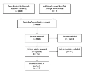
Table 1. Search Strategies for Multiple Metastases and WBRT
| PUBMED, searched on April 19, 2016-April 20, 2016 |
| Step 1: Brain Neoplasms [Mesh] |
| Step 2: (brain [TIAB] OR brainstem [TIAB] OR intracranial [TIAB]) AND (cancer [TIAB] OR tumor* [TIAB] OR tumour* [TIAB] OR neoplasm* [TIAB]) |
| Step 3: Step #1 OR Step #2 |
| Step 4: Neoplasm Metastasis [Mesh] |
| Step 5: (brain [TIAB] OR brainstem [TIAB] OR intracranial [TIAB]) AND (Metastas*) [TIAB] |
| Step 6: Step #4 OR Step #5 |
| Step 7: Step #3 and Step #6 |
| Step 8: Brain neoplasms/secondary [Mesh] |
| Step 9: Step #7 OR Step #8 |
| Step 10: Cranial irradiation [Mesh] |
| Step 11: WBRT [TIAB] |
| Step 12: “whole brain” [TIAB] AND (radiotherap* [TIAB] OR radiation [TIAB] OR radiation therap* [TIAB] OR irradiation [TIAB]) |
| Step 13: Step #10 OR Step #11 OR Step #12 |
| Step 14: Step #9 AND Step #13 |
| Step 15: Step #14 AND English [Lang] |
| Step 16: (animals [MeSH] NOT humans [MeSH]) OR case reports [PT] OR review [PT] OR comment [PT] OR letter [PT] OR editorial [PT] OR addresses [PT] OR news [PT] OR “newspaper article” [PT] |
| Step 17: Step #15 NOT Step #16 |
| Step 18: Step #17 AND (“2000/01/01″[PDAT] : “2015/12/31″[PDAT]) |
| Total: 1212 results |
| EMBASE, searched on April 19, 2016-April 20, 2016 : |
| Step 1: ‘Brain tumor’/exp |
| Step 2: ((brain OR brainstem OR intracranial) NEAR/3 (cancer OR tumor* OR tumour* OR neoplasm*)):ab,ti |
| Step 3: Step #1 OR Step #2 |
| Step 4: ‘brain metastasis’/exp |
| Step 5: ((brain OR brainstem OR intracranial) NEXT/3 metastas*):ab,ti |
| Step 6: Step #4 OR Step #5 |
| Step 7: Step #3 AND Step #6 |
| Step 8: ‘brain radiation’/exp |
| Step 9: WBRT:ab,ti |
| Step 10: (‘whole brain’ NEXT/3 (radiation OR radiotherapy OR irradiation)):ab,ti |
| Step 11: Step #8 OR Step #9 OR Step #10 |
| Step 12: Step #7 AND Step #11 |
| Step 13: Step #12 AND ([article]/lim OR [conference paper]/lim) AND [humans]/lim AND [english]/lim AND [embase]/lim AND [2000-2015]/py |
| Step 14: #13 NOT ‘case report’/de |
| Total: 1060 results |
| COCHRANE, searched on April 19, 2016-April 20, 2016 : |
| Step 1: MeSH descriptor: [Brain Neoplasms] explode all trees |
| Step 2: ((brain OR brainstem OR intracranial) NEAR/3 (cancer OR tumor* OR tumour* OR neoplasm*)):ti,ab,kw |
| Step 3: Step #1 OR Step #2 |
| Step 4: MeSH descriptor: [Neoplasm Metastasis] explode all trees |
| Step 5: ((brain OR brainstem OR intracranial) NEAR/3 Metastas*):ti,ab,kw |
| Step 6: Step #4 OR Step #5 |
| Step 7: Step #3 AND Step #6 |
| Step 8: MeSH descriptor: [Brain neoplasms/secondary] |
| Step 9: Step #7 OR Step #8 |
| Step 10: MeSH descriptor: [Cranial irradiation] explode all trees |
| Step 11: WBRT:ti,ab,kw |
| Step 12: (‘whole brain’ NEXT/3 (radiation OR radiotherapy OR irradiation)):ti,ab,kw |
| Step 13: Step #10 OR Step #11 OR Step #12 |
| Step 14: Step #9 and Step #13 |
| Step 16: Filtered for publication year from 2000 to 2015 |
| Total: 100 results |
| Summary of Primary SearchCombined from 3 database searched, de-duplicated, and non-English articles removed for total of 1,535 candidate articles |
Table 2 . Search Strategies for Multiple Metastases and Focal Therapy
| PUBMED, searched on May 3, 2016-May 4, 2016 : |
| Step 1: Brain Neoplasms [Mesh] |
| Step 2: (brain [TIAB] OR brainstem [TIAB] OR intracranial [TIAB]) AND (cancer [TIAB] OR tumor* [TIAB] OR tumour* [TIAB] OR neoplasm* [TIAB]) |
| Step 3: Step #1 OR Step #2 |
| Step 4: Neoplasm Metastasis [Mesh] |
| Step 5: (brain [TIAB] OR brainstem [TIAB] OR intracranial [TIAB]) AND (Metastas*) [TIAB] |
| Step 6: Step #4 OR Step #5 |
| Step 7: Step #3 and Step #6 |
| Step 8: Brain neoplasms/secondary [Mesh] |
| Step 9: Step #7 OR Step #8 |
| Step 10: Radiosurgery [Mesh] OR Neurosurgical Procedures [Mesh] |
| Step 11: Radiosurg* [TIAB] OR radio-surg* [TIAB] OR radio surg* [TIAB] OR SRS [TIAB] |
| Step 12: Surg*[TIAB] OR resect*[TIAB] OR excision [TIAB] OR operati*[TIAB] OR neurosurg* [TIAB] |
| Step 13: Step #10 OR Step #11 OR Step #12 |
| Step 14: Step #9 AND Step #13 |
| Step 15: Step #14 AND English [Lang] |
| Step 16: (animals [MeSH] NOT humans [MeSH]) OR case reports [PT] OR review [PT] OR comment [PT] OR letter [PT] OR editorial [PT] OR addresses [PT] OR news [PT] OR “newspaper article” [PT] |
| Step 17: Step #15 NOT Step #16 |
| Step 18: Step #17 AND (“2000/01/01″[PDAT] : “2015/12/31″[PDAT]) |
| Total: 2624 results |
| EMBASE, searched on May 3, 2016-May 4, 2016 : |
| Step 1: ‘Brain tumor’/exp |
| Step 2: ((brain OR brainstem OR intracranial) NEAR/3 (cancer OR tumor* OR tumour* OR neoplasm*)):ab,ti |
| Step 3: Step #1 OR Step #2 |
| Step 4: ‘brain metastasis’/exp |
| Step 5: ((brain OR brainstem OR intracranial) NEXT/3 metastas*):ab,ti |
| Step 6: Step #4 OR Step #5 |
| Step 7: Step #3 AND Step #6 |
| Step 8: ‘Radiosurgery’/exp |
| Step 9: ‘Stereotaxic surgery’/exp |
| Step 10: ‘Neurosurgery’/exp |
| Step 11: (Radiosurg* OR radio surg* OR SRS):ab,ti |
| Step 12: (Surg* OR resect* OR excision OR operati* OR neurosurg*):ab,ti |
| Step 13: Step #8 OR Step #9 OR Step #10 OR Step #11 OR Step #12 |
| Step 14: Step #7 AND Step #13 |
| Step 15: Step #14 AND ([article]/lim OR [conference paper]/lim) AND [humans]/lim AND [english]/lim AND [embase]/lim AND [2000-2015]/py |
| Step 16: Step #15 NOT ‘case report’/de |
| Total: 1060 results |
| COCHRANE, searched on May 3, 2016-May 4, 2016 : |
| Step 1: MeSH descriptor: [Brain Neoplasms] explode all trees |
| Step 2: ((brain OR brainstem OR intracranial) NEAR/3 (cancer OR tumor* OR tumour* OR neoplasm*)):ti,ab,kw |
| Step 3: Step #1 OR Step #2 |
| Step 4: MeSH descriptor: [Neoplasm Metastasis] explode all trees |
| Step 5: ((brain OR brainstem OR intracranial) NEAR/3 Metastas*):ti,ab,kw |
| Step 6: Step #4 OR Step #5 |
| Step 7: Step #3 AND Step #6 |
| Step 8: MeSH descriptor: [Brain neoplasms/secondary] |
| Step 9: Step #7 OR Step #8 |
| Step 10: MeSH descriptor: [Radiosurgery] explode all trees |
| Step 11: MeSH descriptor: [Neurosurgical Procedures] explode all trees |
| Step 12: (Radiosurg* OR radio-surg* OR radio surg* OR SRS):ti,ab,kw |
| Step 13: (Surg* OR resect* OR excision OR operati* OR neurosurg*):ti,ab,kw |
| Step 14: Step #10 OR Step #11 OR Step #12 or Step #13 |
| Step 15: Step #9 AND Step #14 |
| Step 16: Filtered for publication year from 2000 to 2015 |
| Total: 100 results |
| Summary of Primary SearchCombined from 3 database searched, de-duplicated, and non-English articles removed for total of 3,698 candidate articles |
Table 3. Evidence
| Cho et al,19 2015 | Retrospective cohort Single institution817 patients with brain metastases from NSCLC treated with SRS270 (33%) had single brain metastasis 547 (67%) > 1 metastasisEndpoints: OS, PFS, salvage treatment-free survival | III | OS was 13 months (median): age (<65 vs ≥65), sex (male vs female), lower RPA, DS-GPA score, adenocarcinoma vs squamous cell carcinoma, synchronous vs methachronous, number × volume of tumors were associated with longer survival in MVA.· Conclusions: “Intracranial tumor burden, reflecting the combined impact of the number of lesions and the cumulative tumor volume, is a more significant prognostic factor than tumor volume or tumor number alone. However, further studies confirming this prognostic factor should be performed. Although the cause of death was not progression of brain lesions in the majority of our patients, the brain lesions tended to be persistently progressive in most of these patients, despite repeated salvage treatment. LMS, in addition to local progression or development of new lesions, is an important pattern of failure and a neurological cause of death.” |
| Oehlke et al,20 2015 | Prospective, nonrandomized cohort Single institution 20 patients with >1 brain metastasis treated with HA WBRT Number of brain metastases (median) 5; range 2-13Endpoints: OS, PFS | III | OS was 71.5 weeks (median); PFS (intracranial) was 40 weeks (median)Conclusions: “Whole brain irradiation with hippocampal sparing (SIP) and dose escalation (SIB) on multiple brain metastases is a safe and tolerable treatment regime and may provide an important improvement of tumor control compared to WBRT alone. At the same time, HA-WBRT bears the potential to minimize the treatment-related side-effect of cognitive deterioration, which cannot be reliably assessed from retrospective chart review. Accordingly, the hypothesized beneficial effect on cognition is currently under investigation in a prospective randomized phase II trial led by one of the authors.” |
| Yamamoto et al,6 2014 | Prospective observational; noninferiority study; multiple institutionsPatients with 1-10 brain metastases 1 metastasis (n = 455) 2-4 metastases (n = 531) 5-10 metastases (n = 208) Treatment: SRSPrimary endpoint: OS in patients with 5-10 versus patients with 1-4 metastases | II | · Primary endpoint: OS better for 1 metastasis but no different between 2-4 and 5-10 metastases· Survival in patients with 5-10 metastases is not inferior to that of patients with 2-4 metastases (p < .0001) Neurologic death (10-14% in all 3 groups), neurologic deterioration, local recurrence, distant metastases were the same in the 2-4 vs 5-10 metastases groups; LMD more in 5-10 than in 2-4 groupConclusions:· “To our knowledge, our study of 1194 patients is the first sufficiently powered prospective observational investigation to examine whether stereotactic radiosurgery without whole-brain radiotherapy (WBRT) as the initial treatment for patients with five to ten brain metastases is non-inferior to that for patients with two to four brain metastases in terms of overall survival. Our results show the non-inferiority of stereotactic radiosurgery without WBRT for patients with five to ten brain metastases as compared with those with two to four tumours. This result challenges the practice of inconsistent use of stereotactic radiosurgery for patients with five or more brain metastases, in whom most treatment guidelines still strongly recommended WBRT, and provides evidence in favour of offering stereotactic radiosurgery to patients with multiple brain metastases. Existing treatment guidelines for the management of patients with brain metastases might need to be revised in the near future.”Authors’ Comments:· Does not demonstrate that SRS is associated with longer survival than WBRT for patients with multiple metastases· Nonrandomized study: referral bias· Nonhomogeneous group; for example, 76% of patients had lung cancerPatients with 5-10 metastases had a range of volume from 0.02-3.90, so some of these tumors were very small.· 70% of patients did not have neurologic symptomsThe usual factors affect survival in MVA, such asKPSAge· Extracranial disease statusNeurologic symptoms1 vs 2-4 metastases |
| Zhou et al,21 2014 | Retrospective Single Institution 29 NSCLC patients with 87 brain metastases treated with WBRT + SIB no. of brain metastases (mean) 3 15 patients (52%) <3 metastases 14 patients (48%) ≥3 metastasesEndpoints: OS, PFS (intracranial) | III | · OS: 10 months (median)· PFS (intracranial): 10 months (median)· Male vs female, adenocarcinoma vs nonadenocarcinoma, history of EGFR-TKI treatment vs non–EGFR-TKI treatment were all associated with increased OS, both in UVA and MVA.Conclusions:· “WBRT plus SIB with IG-IMRT is a tolerable and effective treatment for NSCLC patients with inoperable brain metastases, especially for those with SIR score >5, number of intracranial lesions <3, and history of EGFR-TKI treatment.” |
| Grandhi et al,18 2012 | Retrospective cohort Single Institution 61 patients with ≥10 brain metastases treated with SRS7 (11.5%) had no prior therapy 8 (13.1%) had prior SRS 22 (36.1%) had prior WBRT 16 (26.2%) had prior SRS + WBRT 8 (13.1%) had prior craniotomyPrimary endpoint: OS | III | · Primary endpoint: <14 metastases, nonmelanoma primary, controlled systemic disease, KPS ≥90, lower RPA class associated with longer survival both in UVA and MVA· OS was 4 months (median) 6.6 months (mean) and 0.25-24 months (range)Conclusions:· “Our findings support a role for the use of SRS in treating select patients with extensive intracranial metastatic disease. Gamma Knife surgery, because of its minimal invasiveness and single-fraction approach, may be of particular value in this population given its limited life expectancies.” |
| Kocher et al,5 2011 | Prospective randomized Multiple institutionsPatients with 1-3 brain metastases Treatment groups SRS (32 with multiple mets)/surgery (2 with multiple mets) + WBRT SRS (29 with multiple mets) /surgery (5 with multiple mets) + observationOverall, 68 of 347 patients (19%) had multiple metastasesPrerandomization stratification for single vs 2-3 mets presence vs absence of extracranial disease Surgery vs SRS WHO PSPrimary endpoint: functional independence measured as WHO PS of ≤2 | II | · Primary endpoint: no difference among different treatments· MVA of primary endpoint: Only pretreatment WHO PS of ≤2 and absence of extracranial disease related to primary endpoint. Number of brain metastases and lung vs nonlung histology not related to primary endpoint.· WBRT decreased local failure, increased distal control and decreased neurologic death. No effect on OS.Conclusions:· This study shows that after radiosurgery or surgery of a limited number of brain metastases (1-3 metastases) in patients with stable or asymptomatic solid tumor outside the brain, standard adjuvant WBRT reduces the probability of intracranial relapses from nearly 80% to approximately 50%. This effect is most pronounced after surgery, where the frequency of recurrence in the resection bed is reduced from 60% to <30%. Although it translated into a modest increase in PFS, the increased intracranial tumor control did not translate into a prolonged survival time with functional independence or into a prolonged OS time.Authors’ Comments:· This study demonstrates that WBRT decreases intracranial failure but this does not translate into longer independence or in longer OS.· The data from patient with 2-3 metastases were not consistently separated from those with 1 metastasis. Additionally the small number of patients with 2 or 3 metastases limits statistical power for reaching conclusions on outcomes. For these reasons this manuscript is downgraded to class II for the purposes of this guideline. |
| Chang et al,16 2009 | Prospective randomized Single institutionPatients with 1-3 brain metastases Treatment groups SRS (n = 28) 40% had multiple brain metastases SRS + WBRT (n = 30) 40% had multiple brain metastasesPrimary endpoint: Neurocognition: HVLT at 4 months | II | · Interim analysis stopped trial because there was a significant probability (52 vs 24%; 96% confidence interval) SRS + WBRT patients had impairment of HVLT at 4 months vs SRS alone patients· OS higher in SRS alone group (p = .003)· Neurologic death not statistically different in the 2 groups while systemic death higher in SRS + WBRT ( p = .013)· Local and distant control at 1 year higher for SRS + WBRT group (p = .01 and .02).· Combined brain control higher for SRS + WBRT (p = .0003)Study Conclusions:· “…memory as assessed by HVLT–R total recall is more likely to be preserved with initial SRS alone than SRS plus WBRT… This study provides Class I evidence to support the use of SRS alone in the initial management of patients newly diagnosed with one to 3 brain metastases. Authors recommend that initial SRS alone combined with close clinical monitoring should be the preferred treatment strategy for such patients. Surgical salvage should be used for local failures, and SRS or WBRT for distant failures as indicated. This strategy is consistent with the trend towards personalized medicine and tailoring therapies, rather than applying the “one size fits all” approach of giving WBRT to all patients with brain metastasis.”Authors’ Comments:· Very strong study demonstrating that neurocognition is negatively affected by WBRT. However, newer ways of delivering WBRT, such as hippocampal avoidance, were not tested in the study and this is a clear limitation.· Good data on better intracranial control when WBRT is added and good data on cause of death.· If WBRT is better for ICC but patients live less (yet both groups are stratified for RPA, number of metastases and radioresistant histology) and die more frequently of systemic disease, maybe WBRT has negative systemic effects.· The data from patients with 2 or 3 metastases were not consistently separated from those with 1 metastasis. As the information on disease control and neurocognition cannot be separated, this manuscript is downgraded to class II for the purposes of this guideline. |
| Bhatnagar et al,23 2007 | Retrospective cohortSingle institution205 patients (189 evaluable) with ≥4 metastases treated with GKRS as sole management (17% of patients), in combination with WBRT (46%), or after failure of WBRT (38%).Primary endpoint: OS | III | Patients with total treatment volume <7 cc and 4-6 metastases had longer OS than patients with treatment volume <7 cc and >6 metastases or patients with treatment volume ≥7 cc (13 vs 6 months; p < .00005) Authors’ Conclusions: It is possible to develop a MM-RPA classification in patients with >4 metastases based on total treatment volume (and no. of metastases) |
| Aoyama et al,4 2006 | Prospective randomized Multiple institutionsPatients with 1-4 brain metastases Treatment groups SRS (n = 67) 34 (51%) had multiple mets WBRT + SRS (n = 65) 34 (52%) had multiple metsPrerandomization stratification for single vs 2-4 mets stable vs nonstable extracranial disease lung vs nonlung primaryPrimary endpoint: OS | II | · OS: no difference· Cause of death: no difference· Functional preservation: No difference· Brain tumor recurrence (local and distant sites) less ( p < .001) in WBRT + SRS, both local and distant· Salvage therapy less (p < .001) in WBRT + SRS· Toxic effects of radiation SameConclusions:· SRS alone without upfront WBRT was associated with increased brain tumor recurrence; however, it did not result in either worsened neurologic function or increased risk of neurologic death. With respect to patient survival, the control of systemic cancer might outweigh the frequent recurrence of brain tumors. Therefore, SRS alone could be a treatment option, provided that frequent monitoring of brain tumor status is conducted.· The local control rate was significantly higher in the WBRT + SRS group than in the SRS alone group, despite the fact that in the WBRT + SRS group the SRS dose was 30% less. This observation lends merit to the value of fractionation, which might help overcome some radiation resistance mechanisms, such as hypoxia.Authors’ Comments:· Presence of multiple mets did not affect OS (MVA) or development of nonoriginal mets (MVA)· OS was affected by age <65, primary tumor status and extracranial disease status (MVA)· Distant, and nonoriginal site mets were affected by extracranial disease status (MVA)· The data from patients with 2 to 4 metastases were not consistently separated from those with one metastasis. As the information on disease control could not be separated, this manuscript is downgraded to class II for the purposes of this guideline. |
| Andrews et al,3 2004 | Prospective randomized Multiple institutionPatients with 1-3 nonoperable brain metastases Treatment groups WBRT (n = 164) 73 with multiple metastases WBRT + SRS (n = 167) 72 with multiple metastasesPrimary endpoint: OS | I | · No difference in OSMVA significant· RPA1 vs RPA2 survival· Squamous/non–small cell cancer vs others survivalUVA significant· WBRT + SRS superior for patients with 1 metastasis survival· WBRT + SRS have better KPS and less steroids at 6 monthsAuthors’ Comments:This is a good study suggesting that “radiosurgery boost after WBRT is better than WBRT alone for surgically unresectable single brain metastasis. Because of improved performance in all patients who had radiosurgery boost. WBRT and stereotactic radiosurgery should also be considered for patients with 2 or 3 brain metastases” |
| Pollock et al,26 2003 | Retrospective cohort Single Institution 52 patients with >1 brain met treated with combination of WBRT/SRS/surgery5 patients (10%) underwent multiple simultaneous craniotomies and resection of multiple mets 16 patients (30%) underwent single craniotomy and SRS 31 patients (60%) had SRS alonePrimary endpoint: OS | III | · Primary endpoint: RPA class 1 patients have a median survival of 19 months, class 2 of 13 and class 3 of 8 monthsAuthors’ Conclusions:· “At our center, management of patients with multiple brain metastases is based primarily on three factors: extent of systemic disease, performance status, and size and number of brain tumors. Briefly, patients with progressive systemic disease or poor performance status are generally recommended to have WBRT alone unless they have symptomatic mass effect from a tumor. In those cases, patients generally undergo surgical resection followed by WBRT. Alternatively, patients with stable systemic disease and a good performance status were considered candidates for aggressive management and comprise the patients in this series. The decision as to whether a particular tumor was resected was based on tumor size and a patient’s symptoms. Patients with larger tumors and symptomatic mass effect underwent tumor resection; patients with smaller tumors not causing symptomatic mass effect had radiosurgery. Diabetic patients were given special consideration for tumor resection in order to simplify their postoperative care by minimizing the need for corticosteroids. Patients with multiple large tumors underwent simultaneous craniotomies to resect separate metastases to relieve mass effect. Patients with multiple small tumors were recommended to undergo radiosurgery”· “Well-selected patients with multiple brain metastases appear to benefit from surgery and SRS compared to historical controls of patients treated with WBRT alone. An approach to good prognosis patients with multiple brain metastases utilizing surgical resection, SRS, and WBRT, may improve survival for this difficult patient group”Comments:· This is a descriptive paper. Presence of multiple brain metastases is not an absolute contraindication to surgery· The authors describe their individualized treatment of patients with multiple brain metastases.· This is a paper describing a treatment “philosophy” |
| Iwadate et al,25 2000 | Retrospective cohort Single Institution77 patients with single metastasis 61 patients with >1 brain metastasis Group A: patients with single metastasis who underwent total or subtotal resection. Group B: patients with multiple metastases who underwent total/subtotal resection and had residual tumor < 2 cm Group C: patients with single metastasis who underwent partial resection Group D: all other patients with multiple metastases not falling in Group BAll patients underwent WBRTPrimary endpoint: OS | III | · Primary endpoint: OS statistically longer for A/B groups vs C/D groups No difference in OS between patients with single or multiple metastasesAuthors conclusions· “Surgical reduction of tumor volume which is approximately larger than 2 cm improves the efficacy of adjuvant radiation therapy and contributes to survival even in the patients with multiple brain metastases”Authors’ Comments:· Presence of multiple brain metastases is not an absolute contraindication to surgery |
| Kondziolka et al,22 1999 | Prospective randomized Single institutionPatients with 2-4 brain metastases Treatment groups WBRT (n = 14) WBRT + GKRS (n = 13)Primary end point: Control of brain disease | II | · Local control at 1 year 0% with WBRT alone and 92% with WBRT + GKRS (p = .0016)· Time to failure anywhere in the brain better for WBRT + GKRS than WBRT alone (p = .002)· Trial stopped at 60% accrual because of interim analysis resultsAuthors’ Comments:· “Combined WBRT and radiosurgery for patients with two to four brain metastases significantly improves control of brain disease”Critique· Very few patients. Excellent (92%) local control not replicated in other studies |
| Bindal et al,24 1993 | Retrospective cohort single institution56 patients with >1 brain met Group A: 30 patients who had 40 lesions removed via single/multiple craniotomy at the same setting. Some lesions left unresected. Group B: 26 patients who had 55 lesions removed via single/multiple craniotomy at the same setting. No lesion left unresected. Group C: 26 patients with single lesion resected to serve as a control for group BAll patients underwent WBRTPrimary endpoint: OS | III | · Primary endpoint: OS statistically longer for groups B and C compared with group A. No difference in OS between Groups B and C· MVA of Survival: only group status and systemic disease significantAuthors conclusions:· “The authors conclude that surgical removal of all lesions in selected patients with multiple brain metastases results in significantly increased survival time and gives a prognosis similar to that of patients undergoing surgery for a single metastasis.”· “Our guidelines for the management of patients with multiple brain metastases begin with an evaluation of the extent of systemic disease in the patient. Those patients not expected to survive for longer than 3 months due to their systemic cancer are not considered surgical candidates. Radiation therapy can palliate symptoms for this length of time and is, therefore, recommended for these patients. Patients with limited or controlled systemic cancer in whom resection of all lesions is possible are considered excellent surgical candidates. Even patients in whom all lesions cannot be removed are considered surgical candidates under certain circumstances. If one or two lesions are life-threatening or highly symptomatic, surgical removal may provide the patient an increased life span or an improved quality of life beyond that achievable by radiation therapy alone. In general, the need for multiple craniotomies should not be an important deterrent to the decision to operate.”Authors’ Comments:· Presence of multiple brain metastases is not an absolute contraindication to surgery· Status of systemic disease is paramount in the decision-making progress |
DS-GPA, diagnosis specific graded prognostic assessment; EGFR-TKI, epidermal growth factor receptor-tyrosine kinase inhibitor; GKRS, Gamma Knife radiosurgery; HA, hippocampal avoidance; HVLT, Hopkins Verbal Learning Test; IG-IMRT, image-guided intensity-modulated radiotherapy; KPS, Karnofsky performance status; LMD, leptomeningeal disease; LMS, leptomeningeal spread; MM, multiple metastases; MVA, multivariate analysis; NSCLC, non–small cell lung cancer; OS, overall survival; PFS, progression-free survival; PS, performance status; RPA, recursive partitioning analysis; RPA 1, recursive partitioning analysis class 1; RPA 2, recursive partitioning analysis class 2; SIB, simultaneous integrated boost; SIP, simultaneous integrated protection; SIR, score index for radiosurgery in brain metastases; SRS, stereotactic radiosurgery; UVA, univariate analysis; WBRT, whole brain radiation therapy; WHO, World Health Organization.
REFERENCES
1. Nayak L, Lee EQ, Wen PY. Epidemiology of brain metastases. Current oncology reports. Feb 2012;14(1):48-54.
2. Tabouret E, Chinot O, Metellus P, Tallet A, Viens P, Goncalves A. Recent trends in epidemiology of brain metastases: an overview. Anticancer research. Nov 2012;32(11):4655-4662.
3. Andrews DW, Scott CB, Sperduto PW, et al. Whole brain radiation therapy with or without stereotactic radiosurgery boost for patients with one to three brain metastases: phase III results of the RTOG 9508 randomised trial. Lancet (London, England). May 22 2004;363(9422):1665-1672.
4. Aoyama H, Shirato H, Tago M, et al. Stereotactic radiosurgery plus whole-brain radiation therapy vs stereotactic radiosurgery alone for treatment of brain metastases: a randomized controlled trial. Jama. Jun 7 2006;295(21):2483-2491.
5. Kocher M, Soffietti R, Abacioglu U, et al. Adjuvant whole-brain radiotherapy versus observation after radiosurgery or surgical resection of one to three cerebral metastases: results of the EORTC 22952-26001 study. Journal of clinical oncology : official journal of the American Society of Clinical Oncology. Jan 10 2011;29(2):134-141.
6. Yamamoto M, Serizawa T, Shuto T, et al. Stereotactic radiosurgery for patients with multiple brain metastases (JLGK0901): a multi-institutional prospective observational study. The Lancet. Oncology. Apr 2014;15(4):387-395.
7. Renfrow JJ, Lesser GJ. Molecular subtyping of brain metastases and implications for therapy. Current treatment options in oncology. Dec 2013;14(4):514-527.
8. Lin NU. Targeted therapies in brain metastases. Current treatment options in neurology. Jan 2014;16(1):276.
9. Videtic GM, Gaspar LE, Aref AM, et al. American College of Radiology appropriateness criteria on multiple brain metastases. International journal of radiation oncology, biology, physics. Nov 15 2009;75(4):961-965.
10. Tsao MN, Lloyd N, Wong RK, et al. Whole brain radiotherapy for the treatment of newly diagnosed multiple brain metastases. The Cochrane database of systematic reviews. Apr 18 2012(4):Cd003869.
11. Tsao MN, Rades D, Wirth A, et al. Radiotherapeutic and surgical management for newly diagnosed brain metastasis(es): An American Society for Radiation Oncology evidence-based guideline. Practical radiation oncology. 2012;2(3):210-225.
12. Sahgal A, Aoyama H, Kocher M, et al. Phase 3 trials of stereotactic radiosurgery with or without whole-brain radiation therapy for 1 to 4 brain metastases: Individual patient data meta-analysis. International Journal of Radiation Oncology Biology Physics. 2015;91(4):710-717.
13. Aoyama H, Tago M, Shirato H. Stereotactic Radiosurgery With or Without Whole-Brain Radiotherapy for Brain Metastases: Secondary Analysis of the JROSG 99-1 Randomized Clinical Trial. JAMA oncology. Jul 2015;1(4):457-464.
14. Sahgal A. Point/Counterpoint: Stereotactic radiosurgery without whole-brain radiation for patients with a limited number of brain metastases: the current standard of care? Neuro-oncology. Jul 2015;17(7):916-918.
15. Mehta MP. The controversy surrounding the use of whole-brain radiotherapy in brain metastases patients. Neuro Oncol. Jul 2015;17(7):919-923.
16. Chang EL, Wefel JS, Hess KR, et al. Neurocognition in patients with brain metastases treated with radiosurgery or radiosurgery plus whole-brain irradiation: a randomised controlled trial. The Lancet. Oncology. Nov 2009;10(11):1037-1044.
17. Gondi V, Pugh SL, Tome WA, et al. Preservation of memory with conformal avoidance of the hippocampal neural stem-cell compartment during whole-brain radiotherapy for brain metastases (RTOG 0933): a phase II multi-institutional trial. Journal of clinical oncology : official journal of the American Society of Clinical Oncology. Dec 1 2014;32(34):3810-3816.
18. Grandhi R, Kondziolka D, Panczykowski D, et al. Stereotactic radiosurgery using the Leksell Gamma Knife Perfexion unit in the management of patients with 10 or more brain metastases. Journal of neurosurgery. Aug 2012;117(2):237-245.
19. Cho KR, Lee MH, Kong DS, et al. Outcome of gamma knife radiosurgery for metastatic brain tumors derived from non-small cell lung cancer. Journal of neuro-oncology. Nov 2015;125(2):331-338.
20. Oehlke O, Wucherpfennig D, Fels F, et al. Whole brain irradiation with hippocampal sparing and dose escalation on multiple brain metastases: Local tumour control and survival. Strahlentherapie und Onkologie : Organ der Deutschen Rontgengesellschaft … [et al]. Jun 2015;191(6):461-469.
21. Zhou L, Liu J, Xue J, et al. Whole brain radiotherapy plus simultaneous in-field boost with image guided intensity-modulated radiotherapy for brain metastases of non-small cell lung cancer. Radiation oncology (London, England). 2014;9:117.
22. Kondziolka D, Patel A, Lunsford LD, Kassam A, Flickinger JC. Stereotactic radiosurgery plus whole brain radiotherapy versus radiotherapy alone for patients with multiple brain metastases. International journal of radiation oncology, biology, physics. Sep 01 1999;45(2):427-434.
23. Bhatnagar AK, Kondziolka D, Lunsford LD, Flickinger JC. Recursive partitioning analysis of prognostic factors for patients with four or more intracranial metastases treated with radiosurgery. Technology in cancer research & treatment. Jun 2007;6(3):153-160.
24. Bindal RK, Sawaya R, Leavens ME, Lee JJ. Surgical treatment of multiple brain metastases. Journal of neurosurgery. Aug 1993;79(2):210-216.
25. Iwadate Y, Namba H, Yamaura A. Significance of surgical resection for the treatment of multiple brain metastases. Anticancer research. Jan-Feb 2000;20(1b):573-577.
26. Pollock BE, Brown PD, Foote RL, Stafford SL, Schomberg PJ. Properly selected patients with multiple brain metastases may benefit from aggressive treatment of their intracranial disease. Journal of neuro-oncology. 2003;61(1):73-80.
Source: Neurosurgery
7. The Role of Steroids in the Treatment of Adults with Metastatic Brain Tumors
Sponsored by: The Congress of Neurological Surgeons and the Section on Tumors
Affirmation of Educational Benefit by: The Congress of Neurological Surgeons and the American Association of Neurological Surgeons
Timothy C. Ryken, MD, MS,1 John S. Kuo, MD, PhD,2 Roshan S. Prabhu, MD, MS,3 Jonathan H. Sherman, MD,4 Steven N. Kalkanis, MD,5 Jeffrey J. Olson, MD6
- Section of Neurosurgery, Dartmouth-Hitchcock Medical Center, Lebanon, New Hampshire
- Department of Neurosurgery and Mulva Clinic for the Neurosciences, Dell Medical School, University of Texas at Austin, Austin, Texas
- Southeast Radiation Oncology Group, Levine Cancer Institute, Carolinas Healthcare System, Charlotte, North Carolina
- Department of Neurosurgery, The George Washington University, Washington, DC
- Department of Neurosurgery, Henry Ford Health System, Detroit, Michigan
- Department of Neurosurgery, Emory University School of Medicine, Atlanta, Georgia
Correspondence:
Timothy C. Ryken, MD, MS
Section of Neurosurgery
Dartmouth-Hitchcock Medical Center
One Medical Center Drive
Lebanon, NH 03756
Email: rykent@me.com
Disclaimer of Liability
This clinical systematic review and evidence-based guideline was developed by a multidisciplinary physician volunteer task force and serves as an educational tool designed to provide an accurate review of the subject matter covered. These guidelines are disseminated with the understanding that the recommendations by the authors and consultants who have collaborated in their development are not meant to replace the individualized care and treatment advice from a patient’s physician(s). If medical advice or assistance is required, the services of a competent physician should be sought. The proposals contained in these guidelines may not be suitable for use in all circumstances. The choice to implement any particular recommendation contained in these guidelines must be made by a managing physician in light of the situation in each particular patient and on the basis of existing resources.
Keywords:
Brain metastases, cerebral metastases, corticosteroids, neurologic symptoms, practice guideline, steroid dosage, systematic review
ABSTRACT
Questions:
Do steroids improve neurologic symptoms and/or quality of life in patients with metastatic brain tumors compared to supportive care only or other treatment options?
If steroids are given, what dose should be used?
Target population: These recommendations apply to adults diagnosed with brain metastases.
Recommendations
Steroid therapy versus no steroid therapy
Asymptomatic brain metastases patients without mass effect
Insufficient evidence exists to make a treatment recommendation for this clinical scenario.
Brain metastases patients with mild symptoms related to mass effect
Level 3: Corticosteroids are recommended to provide temporary symptomatic relief of symptoms related to increased intracranial pressure and edema secondary to brain metastases. It is recommended for patients who are symptomatic from metastatic disease to the brain that a starting dose of 4–8 mg/day of dexamethasone be considered.
Brain metastases patients with moderate to severe symptoms related to mass effect
Level 3: Corticosteroids are recommended to provide temporary symptomatic relief of symptoms related to increased intracranial pressure and edema secondary to brain metastases. If patients exhibit severe symptoms consistent with increased intracranial pressure, it is recommended that higher doses such as 16 mg/day or more be considered.
Choice of Steroid
Level 3: If corticosteroids are given, dexamethasone is the best drug choice given the available evidence.
Duration of Corticosteroid Administration
Level 3: Corticosteroids, if given, should be tapered as rapidly as possible but no faster than clinically tolerated, based upon an individualized treatment regimen and a full understanding of the long-term sequelae of corticosteroid therapy.
Given the very limited number of studies (two) which met the eligibility criteria for the systematic review, these are the only recommendations that can be offered based on this methodology. Please see “Discussion” and “Summary” section for additional details.
INTRODUCTION
Rationale
Steroids have been used to assist in controlling peritumoral intracerebral edema in the care of patients with newly diagnosed metastatic brain disease.1-12 Dexamethasone has been the steroid most commonly used due to its minimal mineralocorticoid effect. Steroids have been used for palliative care, and, in combination with surgery and radiation, to reduce treatment-related toxicity. Historically, the majority of patients treated with an initial dose of 4 to 8 mg/day responded within 24 to 72 hours. 13 Toxicity and side effects from steroids occur frequently and contribute to the overall morbidity and mortality in this often-tenuous patient population. However, as previously described, a review of the literature continues to demonstrate a lack of well-controlled studies addressing this topic and significant variability in the dosing and administration of steroids in both the symptomatic and asymptomatic patient. 11
Objectives
This updated systematic review addresses the role of corticosteroids in the treatment of metastatic brain disease with the following overall objectives:
1. To systematically review and update the evidence available addressing the use of corticosteroids in the management of patients with brain metastases since the previous review of 2010, 11 again addressing the following questions:
· Do steroids improve neurologic symptoms in patients with metastatic brain tumors compared to no treatment?
· If steroids are given, what dose should be used?
2. To make recommendations based on this evidence for the role of corticosteroids in the management of these patients.
METHODS
Writing Group and Question Establishment
The writers represent a multi-disciplinary panel of clinical experts encompassing neurosurgery and radiation oncology. Together, they were recruited to develop these evidence-based practice guidelines for surgery for metastatic brain tumors. Questions were developed by group consensus recognizing the questions used in the prior guidelines published on this topic and taking into account current salient concerns over the use of steroids in metastatic brain tumor management.
Search Method
The PubMed online database was searched for the period of October 1, 2008 through December 31, 2015, using the following queries in all fields: steroids and brain metastases, and dexamethasone and brain metastases. The results from each search were downloaded into an Endnote library. The libraries were merged and duplicate entries were eliminated. This inclusive search strategy was designed to capture all manuscripts pertaining to brain metastases and steroids for manual review and to determine if any more recent articles had been missed in the prior update. The reference lists of the most relevant and most recent articles were also reviewed, and additional articles selected for initial review.
Study Selection and Eligibility Criteria:
The following inclusion criteria were used for manual review of studies:
1. Published in English with a publication date prior to December 31, 2015.
2. Included only patients with brain metastases.
3. Published in a peer-reviewed journal with comparative data pertaining to the use of steroids in patients with brain metastases.
The search strategy was purposefully as broad as possible given the limited number of relevant articles found in the previous guideline.
Data Collection Process
The initial screening and evaluation of the initial search-returned citations using pre-determined criteria for relevance (initial screen via title/abstract, with a secondary full-text review of potentially relevant manuscripts) was performed by the primary author with additional input from the author group. Data from studies meeting eligibility criteria was then extracted by a single reviewer and checked by a minimum of two additional reviewers.
Assessment for Risk of Bias
Studies selected for full-text review were evaluated, in addition to the overall quality of the study design, for specific issues of bias. Particular attention was paid to potential bias related to selective case choice and reporting, publication bias, bias related to change in treatment methods over time, hidden agenda bias when perceived, and variability due to inconsistencies in data entry and oversight. When encountered concerns about specific examples of bias in the published data were noted in the evidentiary tables. The class of data and subsequent level of recommendation was then adjusted accordingly.
Description of the Data Classification System and Recommendation Formulation
The quality of each study regarding metastases-specific data and the strength of the recommendations within this work were graded according to the American Association of Neurological Surgeons (AANS)/Congress of Neurological Surgeons (CNS) criteria ( here ).
RESULTS
Study Selection and Characteristics
In the 2010 guideline, 11 despite the widespread use of steroids in the management of brain metastases, only 2 publications met the stated eligibility criteria. 13 , 14 Given the limited data yielded by the literature search, additional searches were undertaken by reviewing the bibliographies of relevant recent publications and additional review of any relevant published literature addressing the treatment of metastatic brain disease for references to steroid administration. These articles are summarized in the discussion below. The publications that report the Quality of Life after Treatment for Brain Metastases (QUARTZ) trial 1 , 8,12, 15 were summarized in the publication by Mulvenna et al 1 , 12 in 2016. Mulvenna et al 12 was published after the literature review for this guideline was performed, but is referenced for completeness, even though it was not included as evidence to support the recommendations of this guideline update.
Results of Individual Studies, Discussion of Study Limitations, and Risk of Bias
All of the following studies were graded as Class 3 evidence. Two studies were included in the original 2010 guideline.11 Vecht et al 13 conducted a randomized study of 4, 8, and 16 mg/day dosing of dexamethasone and demonstrated no advantage to higher dosing in patients without symptomatic intracranial hypertension. Two consecutive double-blind randomized trials in patients with brain metastases and Karnofsky performance scores (KPS) <80 were designed to evaluate the minimum effective dose of oral dexamethasone. Initially, a dexamethasone dosage of 8 mg/day (group 1) was compared to 16 mg/day (group 2), followed by a comparison of 4 mg/day (group 3) versus 16 mg/day (group 4). The outcomes of interest were alteration in KPS and the frequency of side effects at days 0, 7, 28, and 56.
Both groups showed improvement, but there was no significant difference in KPS improvement comparing the 8-mg group versus the 16-mg group at day 7 (mean 8.0 ± 10.1 versus 7.3 ± 14.2).
In the second trial conducted by Vecht et al 13, both groups showed improvement. There was no significant difference between the 4 mg and 16 mg groups, comparing 6.7 ± 11.3 points at day 7 and 7.1 ± 18.2 points at day 28 versus 9.1 ± 12.4 and 5.6 ± 18.5 points, respectively. Side effects were more frequent in the 16 mg/day versus the 4 mg/day group at day 28 (combined frequency 91% versus 46%, p<.03).
The authors concluded that the lower doses of 4 and 8 mg dexamethasone per day had an equivalent effect on improving neurologic performance when compared with a dose of 16 mg/day at 1 and 4 weeks of treatment, in moderately symptomatic patients without signs of impending herniation. The dosing recommendation from this study was 4 mg/day dosage with a dose taper for 28 days in patients with no symptoms of mass effect.
Wolfson et al 14 randomized 12 patients undergoing whole brain radiation therapy following an initial dose of 24 mg dexamathasone into a group receiving 4 mg every 6 hours during the radiotherapy versus no additional steroids. Although more patients were improved in the steroid group (29% versus 0%), the small size and complete lack of statistical analysis resulted in this study being excluded as evidence in the previous report.
Given the extremely limited number of studies that satisfied the conditions of inclusion, an additional discussion of published literature on the subject of corticosteroids in metastatic brain disease is provided to offer a larger context for this topic. While the following studies were not part of the body of evidence considered in formulating treatment recommendations in this evidence-based guideline, they do highlight areas of interest where clinical trials are still required to answer important steroid-related questions.
A series of authors have published contemporary updates, reviews, and consensus documents that recommend steroid therapy in the management of CNS metastatic disease, with no additional data provided. 3 , 4, 7 , 9,10, 15-18 For example the systematic review by Tsao et al provides little data on how the actual review was conducted. 9
The series of articles published describing the QUARTZ study compare palliative whole brain radiation therapy versus supportive care with steroids, and are significant in that they appear to establish the role of steroids as a baseline of care for the symptomatic patient with central nervous system metastasis. 1 , 8,12, 15 This study provides randomized data on the comparison of whole brain radiotherapy versus steroids alone but provides no comparative data on dosing or the comparison of no steroid versus steroid. It appears that this issue has been assumed to be adequately addressed with clinical practice, because no comparative studies addressing this issue have appeared in >20 years.
Although they do not provide comparative data, several additional studies are noted as they include issues related to steroid use in this population. Not mentioned in the 2010 guideline, 11 Sturdza et al19 studied steroid prescribing practices and patient side effects in 88 patients identified in the Palliative Radiation Oncology database. Forty-five percent of physicians used a dexamethasone dose of 4 mg 4 times daily (16mg/day) with 60% using a 4-week taper. The most common side effects were increased appetite or weight gain (46%), insomnia (24%), gastrointestinal symptoms (20%), and proximal muscle weakness (28%). The authors concluded that considerable variation in the prescribing practices existed even within a single institution, with many patients receiving high doses of steroids for considerable periods of time and developing related side effects, and they propose a prospective study to standardize dosing and taper practices to optimize management and minimize toxicity.
Pulenzas et al 6 surveyed a cohort of patients undergoing whole brain radiation therapy for changes in fatigue scores using a broad panel of outcome measures, including the Edmonton Symptom Assessment System, the Brain Symptom and Impact Questionnaire, the Spitzer Questionnaire, the European Organization for Research and Treatment of Cancer (EORTC) Quality of Life Questionnaire, the EORTC brain module, the EORTC Quality of Life Questionnaire Core 15 Palliative, and the Functional Assessment of Cancer Therapy-General. The authors concluded that fatigue was significantly increased and quality of life significantly reduced over the first month in all patients. Increased fatigue was significantly related with decreased overall QOL. Interestingly, for all groups, there was no significant difference in fatigue scores or quality of life with or without the addition of dexamethasone.
Alan et al5 studied the impact of preoperative steroids on 30-day morbidity and mortality of >4000 patients undergoing craniotomy for resection of malignant brain tumors (metastatic brain tumors 37.5% (n = 1611) and primary malignant gliomas 62.5% (n = 2796). Approximately 23% of patients received perioperative steroids (n = 1009). Logistic regression was used to assess the association between preoperative steroid use and perioperative complications before and after 1:1 propensity score matching. In the unmatched cohort (n = 1009), steroid use was associated with decreased length of hospitalization (odds ratio 0.7; 95% confidence interval 0.6-0.8). In this same group, the incidence of readmission (odds ratio 1.5; 95% confidence interval 1.2-1.8) was increased. In the matched cohort (n = 465), steroid use was not statistically associated with any adverse outcomes. As an independent risk factor, preoperative steroid use was not associated with any observed perioperative complications.
The authors concluded that preoperative steroids do not independently compromise the short-term outcome of craniotomy for resection of malignant brain tumors. Separating out the metastatic versus the primary tumor patients is difficult from the data presented and limits the ability to formulate recommendations.
Synthesis of Results
Vecht et al13 continues to provide the most convincing data on the role for steroids in patients with brain metastases and for the choice of dosing. Based on their observations of improvement in all groups treated with steroids, Level 3 recommendations were made in the 2010 Guideline. 11 The results of this guideline confirms the validity of those recommendations, because no novel evidence has been published on this topic since 2010.
Given the very limited number of studies only two of which met the eligibility criteria for the systematic review, these are the only recommendations that can be offered based on this methodology. Please see the Discussion section for additional details.
DISCUSSION
Although comparative studies addressing various steroid dosing regimens are generally lacking, studies addressing additional topics of interest have been published in recent years. 1 , 2,5,6, 12 A better understanding of the toxicity related to routine steroid use continues to develop, and this research would support the principle of using the lowest effective steroid dose. 6 , 19 The design of large clinical trials in which a steroid treatment-only groups are considered the “best supportive care” group underlines the conviction most physicians hold for the critical role of steroids in managing the patient with symptomatic central nervous system metastatic disease. 1 , 8, 15
The issue of dosing regimen is problematic to address based on the evidence available. The study noted by Vecht et al 13 used only 4 times daily dosing and does not address alternative dosing regimens. Therefore, only recommendations on total amount per day have been formulated. It is recognized as common practice that alternative dosing, such as twice daily, is acceptable practice.
In addition, the ability of steroids to reduce the likelihood of treatment-related toxicity, either following surgery or radiotherapy, continues to be of interest and warrants additional study at least as a component of the data collection process in clinical trials. 3 , 5
CONCLUSION AND KEY ISSUES FOR FUTURE INVESTIGATIONS
It is clear from this review of the literature that steroids are a mainstay of treatment for patients with metastatic brain disease despite the relative lack of high-quality evidence supporting any specific therapy. Based on the literature available for this guideline update, larger prospective or carefully planned retrospective studies should be considered to clarify more specific patient-dependent dosing. Complications related to steroid use, including adrenal insufficiency with tapering, should continue to be monitored, and perhaps alternative approaches to reducing peritumoral edema could be explored to eliminate the unwanted but common side effects of steroid therapy entirely.
Potential Conflicts of Interest
The Brain Metastases Guideline Update Task Force members were required to report all possible conflicts of interest (COIs) prior to beginning work on the guideline, using the COI disclosure form of the AANS/CNS Joint Guidelines Review Committee, including potential COIs that are unrelated to the topic of the guideline. The CNS Guidelines Committee and Guideline Task Force Chair reviewed the disclosures and either approved or disapproved the nomination. The CNS Guidelines Committee and Guideline Task Force Chair are given latitude to approve nominations of task force members with possible conflicts and address this by restricting the writing and reviewing privileges of that person to topics unrelated to the possible COIs. The conflict of interest findings are provided in detail in the companion introduction and methods manuscript (here).
Disclosures
These evidence-based clinical practice guidelines were funded exclusively by the Congress of Neurological Surgeons and the Tumor Section of the Congress of Neurological Surgeons and the American Association of Neurological Surgeons, which received no funding from outside commercial sources to support the development of this document.
ACKNOWLEDGEMENTS
The authors acknowledge the CNS Guidelines Committee for its contributions throughout the development of the guideline and the AANS/CNS Joint Guidelines Review Committee for its review, comments, and suggestions throughout peer review, as well as Trish Rehring, MPH, CHES, CNS Guidelines Senior Manager, and Mary Bodach, MLIS, Senior Guidelines Specialist, for their assistance. Throughout the review process, the reviewers and authors were blinded from one another. At this time, the guidelines task force would like to acknowledge the following individual peer reviewers for their contributions: Manish Aghi, MD, PhD, Manmeet Ahuwalia, MD, Sepideh Amin-Hanjani, MD, Edward Avila, MD, Maya Babu, MD, MBA, Kimon Bekelis, MD, Priscilla Brastianos, MD, Paul Brown, MD, Andrew Carlson, MD, MS, Justin Jordan, MD, Terrence Julien, MD, Cathy Mazzola, MD, Adair Prall, MD, Shayna Rich, MD, PhD, Arjun Sahgal, MD, Erik Sulman, MD, May Tsao, MD, Michael Voglebaum, MD, Stephanie Weiss, MD, and Mateo Ziu, MD.
Figure 1. PRISMA Flowchart
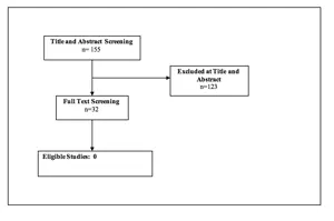
REFERENCES
1. Mulvenna P, Nankivell M, Barton R, et al. Dexamethasone and supportive care with or without whole brain radiotherapy in treating patients with non-small cell lung cancer with brain metastases unsuitable for resection or stereotactic radiotherapy (QUARTZ): results from a phase 3, non-inferiority, randomised trial. Lancet. Sep 2 2016.
2. Agar M, Koh ES, Gibbs E, et al. Validating self-report and proxy reports of the Dexamethasone Symptom Questionnaire -Chronic for the evaluation of longer-term corticosteroid toxicity. Support Care Cancer. Mar 2016;24(3):1209-1218.
3. Tsao MN. Brain metastases: advances over the decades. Ann Palliat Med. Oct 2015;4(4):225-232.
4. Pruitt AA. Medical management of patients with brain tumors. Continuum (Minneap Minn). Apr 2015;21(2 Neuro-oncology):314-331.
5. Alan N, Seicean A, Seicean S, Neuhauser D, Benzel EC, Weil RJ. Preoperative steroid use and the incidence of perioperative complications in patients undergoing craniotomy for definitive resection of a malignant brain tumor. J Clin Neurosci. Sep 2015;22(9):1413-1419.
6. Pulenzas N, Khan L, Tsao M, et al. Fatigue scores in patients with brain metastases receiving whole brain radiotherapy. Support Care Cancer. Jul 2014;22(7):1757-1763.
7. Expert Panel on Radiation Oncology-Brain M, Lo SS, Gore EM, et al. ACR Appropriateness Criteria(R) pre-irradiation evaluation and management of brain metastases. J Palliat Med. Aug 2014;17(8):880-886.
8. Langley RE, Stephens RJ, Nankivell M, et al. Interim data from the Medical Research Council QUARTZ Trial: does whole brain radiotherapy affect the survival and quality of life of patients with brain metastases from non-small cell lung cancer? Clin Oncol (R Coll Radiol). Mar 2013;25(3):e23-30.
9. Tsao MN, Khuntia D, Mehta MP. Brain metastases: what’s new with an old problem? Curr Opin Support Palliat Care. Mar 2012;6(1):85-90.
10. Pruitt AA. Medical management of patients with brain tumors. Curr Treat Options Neurol. Aug 2011;13(4):413-426.
11. Ryken TC, McDermott M, Robinson PD, et al. The role of steroids in the management of brain metastases: a systematic review and evidence-based clinical practice guideline. J Neurooncol. Jan 2010;96(1):103-114.
12. Mulvenna P, Nankivell M, Barton R, et al. Dexamethasone and supportive care with or without whole brain radiotherapy in treating patients with non-small cell lung cancer with brain metastases unsuitable for resection or stereotactic radiotherapy (QUARTZ): results from a phase 3, non-inferiority, randomised trial. Lancet. Oct 22 2016;388(10055):2004-2014.
13. Vecht CJ, Hovestadt A, Verbiest HB, van Vliet JJ, van Putten WL. Dose-effect relationship of dexamethasone on Karnofsky performance in metastatic brain tumors: a randomized study of doses of 4, 8, and 16 mg per day. Neurology. Apr 1994;44(4):675-680.
14. Wolfson AH, Snodgrass SM, Schwade JG, et al. The role of steroids in the management of metastatic carcinoma to the brain. A pilot prospective trial. Am J Clin Oncol. Jun 1994;17(3):234-238.
15. Mulvenna PM. The management of brain metastases in patients with non-small cell lung cancer-is it time to go back to the drawing board? Clin Oncol (R Coll Radiol). Jun 2010;22(5):365-373.
16. Roth P, Wick W, Weller M. Steroids in neurooncology: actions, indications, side-effects. Curr Opin Neurol. Dec 2010;23(6):597-602.
17. Nguyen TD, DeAngelis LM. Brain metastases. Neurol Clin. Nov 2007;25(4):1173-1192, x-xi.
18. Soffietti R, Cornu P, Delattre JY, et al. EFNS Guidelines on diagnosis and treatment of brain metastases: report of an EFNS Task Force. Eur J Neurol. Jul 2006;13(7):674-681.
19. Sturdza A, Millar BA, Bana N, et al. The use and toxicity of steroids in the management of patients with brain metastases. Support Care Cancer. Sep 2008;16(9):1041-1048.
Source: Neurosurgery, January 9, 2019
8. The Role of Prophylactic Anticonvulsants in the Treatment of Adults with Metastatic Brain Tumors
Sponsored by: The Congress of Neurological Surgeons and the Section on Tumors
Affirmation of Educational Benefit by: The Congress of Neurological Surgeons and the American Association of Neurological Surgeons
Clark C. Chen, MD, PhD1, Robert C. Rennert, MD2, Jeffrey J. Olson, MD3
- Department of Neurosurgery, University of Minnesota Medical School, Minneapolis, Minnesota, USA
- Department of Neurosurgery, University of California, San Diego, LaJolla, California, USA
- Department of Neurosurgery, Emory University School of Medicine, Atlanta, Georgia, USA
Correspondence:
Clark C. Chen, MD, PhD
Lyle A. French Chair in Neurosurgery
Professor and Department Head of Neurosurgery
University of Minnesota Medical School
D429 Mayo Memorial Building
420 Delaware Street S.E., MMC 96
Minneapolis, MN 55455
E-mail: ccchen@umn.edu
Tel: 612-626-5767
Fax: 612-624-0644
Disclaimer of Liability
This clinical systematic review and evidence-based guideline was developed by a multidisciplinary physician volunteer task force and serves as an educational tool designed to provide an accurate review of the subject matter covered. These guidelines are disseminated with the understanding that the recommendations by the authors and consultants who have collaborated in their development are not meant to replace the individualized care and treatment advice from a patient’s physician(s). If medical advice or assistance is required, the services of a competent physician should be sought. The proposals contained in these guidelines may not be suitable for use in all circumstances. The choice to implement any particular recommendation contained in these guidelines must be made by a managing physician in light of the situation in each particular patient and on the basis of existing resources.
Keywords:
Brain metastases, cerebral metastases, anti-epileptic drugs, seizure, craniotomy
Abbreviations
AED: Anti-epileptic drug
HGG: High-grade glioma
PHT: Phenytoin
ABSTRACT
Target Populations: Adults with solid brain metastases who have not experienced a seizure.
Question 1: Do prophylactic anti-epileptic drugs (AEDs) decrease the risk of seizures in non-surgical patients with brain metastases who are otherwise seizure free?
Recommendation:
Level 3: Prophylactic AEDs are not recommended for patients with brain metastases who did not undergo surgical resection and are otherwise seizure-free.
Question 2: Do prophylactic AEDs decrease the risk of seizures in patients with brain metastases and no prior history of seizures in the postoperative setting?
Recommendation:
Level 3: Routine post-craniotomy anti-epileptic drug use for seizure-free patients with brain metastases is not recommended.
INTRODUCTION
Rationale
Brain metastases occur in <30% of patients with systemic cancers. 1 Seizures are a common sequela of brain metastases due to disruption of neurotransmitters by cancer cells, general cerebral edema, or local mass effect.2, 3 Anti-epileptic drug (AED) use after seizure occurrence is well-established in patients with brain metastases. 3 However, the efficacy of prophylactic AEDs for patients with brain metastases who do not undergo surgery remains an unresolved issue.
Another issue in the management of patients with brain metastases involves whether prophylactic AED use is warranted in the postoperative setting for patients without a prior history of seizures. Surgical resection is frequently performed for symptomatic brain metastases.4 Surgical manipulation of the cerebrum has also been associated with increase seizure risk.5 In this context, it remains unclear whether prophylactic AED use is appropriate in the postoperative setting for patients with brain metastases who are otherwise seizure-free. Previous guidelines from 2010 do not recommend prophylactic AED use for patients with brain metastases without a seizure history. These guidelines are based on a single study of largely non-surgical patients.6
Objectives
The objective of this guideline is to address the role of AED prophylaxis in patients with brain metastases without prior seizures in the 1) non-surgical and 2) postoperative settings.
METHODS
Writing group and question establishment
The Joint Tumor Section of the American Association of Neurological Surgeons (AANS) and the Congress of Neurological Surgeons (CNS) identified an update of the metastatic brain tumor guidelines as a topic worthy of guideline development. Members of the Tumor Section, as well as other neurosurgeons and members of other specialties commonly involved in the management of metastatic brain tumors were identified to form the Metastatic Brain Tumor Evidence-Based Practice Guideline Task Force (ie, the “task force”). The writers were then divided up into topic sections and developed pertinent questions for those topics. These were circulated among the entire task force, modified, and agreed upon. With these questions in hand, the literature searches, such as the one described below, were executed. Additional details regarding the literature search and review methodology can be found in the introductory section of this set of guidelines. This guideline was then developed using multiple iterations of written review conducted by the authors, then by members of the task force, and finally by AANS/CNS Joint Guidelines Review Committee (JGRC).
Search strategy
The PubMed online database was searched from January 1, 1990, through December 31, 2015, using the following query: (craniotomy OR postcraniotomy OR post-craniotomy OR brain metastasis OR brain metastases OR central nervous system metastasis OR central nervous system metastases OR metastatic brain cancer OR brain mets OR metastatic cancer brain OR secondary brain neoplasm OR brain neoplasm OR brain tumor OR brain tumour OR cerebral tumor OR cerebral tumour OR cerebral metastasis OR cerebral metastases) AND (anticonvulsant OR seizure OR seizures OR antiepileptic drug OR anti-epileptic drug OR antiepileptic OR anti-epileptic OR anti-convulsant OR AED OR seizure prophylaxis). This inclusive search strategy was designed to capture all manuscripts pertaining to brain metastases and seizures for manual review.
Study selection and eligibility criteria
The following inclusion criteria were used for manual review of studies:
1. Published in English with a publication date of January 1, 1990, through December 31, 2015.
2. Included patients with brain metastases.
3. Published in a peer-reviewed journal with comparative data pertaining to seizure risk including patients with brain metastases (study designs for primary data collection included randomized controlled trials, non-randomized trials, cohort studies, case–control studies, and observational or retrospective studies).
4. Number of study subjects with brain metastases ≥10.
5. Provided information regarding use of AEDs and the relationship of seizures to surgical intervention for study participants with brain metastases.
6. Provided a comparison cohort to brain metastases patients without prophylactic AED treatment.
Studies reporting data solely on stereotactic radiosurgery or experimental drug therapies for metastases were excluded. Two reviewers evaluated search-returned citations via an initial title/abstract screen for relevance based on the above pre-determined criteria.
Data collection process
A secondary full-text review of potentially relevant manuscripts was conducted by two reviewers with detailed attention to the above inclusion/exclusion criteria. Data from studies meeting eligibility criteria were data extracted by a single reviewer and checked by a second reviewer.
Assessment for risk of bias
Included studies were assessed for potential bias upon full-text review by two reviewers. Specific areas of bias assessed for included selective data reporting/analysis regarding metastases subgroups, loss of data points over time, ascertainment bias, early termination/power biases, and retrospective data/causation bias.
Evidence classification and recommendation levels
The quality of each study regarding metastases-specific data and the strength of the recommendations within this work were graded according to the American Association of Neurological Surgeons (AANS)/Congress of Neurological Surgeons (CNS) criteria (here.). In brief, Class I evidence is from well-designed randomized controlled clinical trials, Class II evidence is from case-control and cohort studies, and Class III evidence is provided by expert opinion, case series, or studies with historical controls. Level 1 recommendations reflect a high degree of clinical certainty generally based on Class I data, Level 2 recommendations reflect clinical certainty based generally on Class II data, and Level 3 recommendations reflect clinical uncertainty based on inconclusive or conflicting evidence.
RESULTS
Study selection and characteristics
The literature search yielded 8167 studies that met the above criteria, of which, 8127 studies were excluded during title/abstract review. The remaining 40 studies underwent full-text review, and 35 studies were excluded due to limited metastases-specific data, a lack of information regarding use of AEDs, lack of information on surgical intervention, lack of a comparison cohort without AED prophylaxis, or other predefined exclusion criteria. Five studies met all inclusion criteria (Figure 1).
Results of individual studies, discussion of study limitations and risk of bias
Seizure prophylaxis in patients with brain metastases who did not undergo surgery
Two studies met inclusion criteria and provided information pertaining to the use of prophylactic AEDs in patients with brain metastases 1) who did not have prior histories of seizure and 2) the majority of whom did not undergo surgery (Table 1).
Forsyth et al7 randomized 100 brain tumor patients with no prior history of seizure to receive either 1) AED for 3 months, in the form of phenytoin (PHT) or phenobarbital, or 2) no AED. Sixty of the 100 (60%) patients suffered from brain metastases (92% of the remaining 40 patients suffered from high-grade glioma [HGG]). Fifty-two of the brain metastases patients (87%) did not undergo surgical intervention of any form. The primary study endpoint was seizure occurrence (<3 months), and a secondary endpoint was adverse drug reactions. Baseline characteristics of the 2 arms were generally comparable. The overall study showed that seizures occurred in 26% of patients treated with AEDs and 15% in the non-AED group (p= .98). Minor adverse events (nausea and rash) occurred in 7 AED-treated patients (16%), and major adverse events (myelosuppression and ataxia) occurred in 3 AED-treated patients (6%). Of the patients with brain metastases, 26 were treated with AEDs and 34 were not. Notably, ~30% of patients with brain metastases died prior to the 3-month follow-up. Within the brain metastases subgroup, there was no significant difference in 3-month seizure rates between groups (10% vs 13%, p = .90). Notably, the same results were observed for patients with HGG. The trial was terminated early based on the observed seizure frequency of ~10%, suggesting a high probability of data insufficiency for the primary endpoint. The data pertaining to metastases in this study were deemed to be Class III due to early study termination and a power bias to identify only large reductions in seizure rates.
Glantz et al8 randomized 74 brain tumor patients with no prior history of seizures to receive either 1) valproate (VAL), or 2) placebo for the duration of the study. Fifty-nine of these patients (80%) suffered from brain metastases (9 of 74 patients [12%] were diagnosed with glioblastoma). Sixty-six percent of the randomized patients did not undergo surgery of any form. The primary study endpoints were new onset seizure or death within 12 months of enrollment. Baseline characteristics of the 2 arms were generally comparable. The overall study showed that seizures occurred in 35% of patients treated with VAL and 24% in the placebo group (p = .3). By the end of the study, 26 of 37 (70%) VAL-treated patients had died, and 27 of 37 (73%) placebo-treated patients died. Insufficient data were included in the published article to allow post-hoc analysis specific to the 77% of the patients who suffered from brain metastases. The data pertaining to metastases in this study were deemed to be Class III due to a lack of subgroup analysis bias for this population.
Synthesis of results
Of the 2 studies identified pertaining to the use of prophylactic AEDs in patients with brain metastases, neither found a beneficial effect of AEDs on seizure rates. However, 1 study was terminated early because the proposed sample size was underpowered to detect lowered seizure risk attributable to AEDs, and the other study enrolled a similar number of patients but did not provide metastases-specific analysis. These studies are Class III evidence,9 leading to the Level 3 recommendation that the use of prophylactic AEDs is not justified for patients with brain metastases who did not undergo surgical resection and are otherwise seizure-free.
Results of individual studies, discussion of study limitations and risk of bias
Seizure prophylaxis in patients with brain metastases in the postoperative setting
Three studies met the inclusion criteria and provided information pertaining to the use of post-craniotomy prophylactic AEDs in brain tumor patients without preoperative seizures (Table 2).
Ansari et al10 retrospectively reviewed AED use and seizure frequency in 202 patients with no previous seizure history who underwent craniotomies for intra-axial brain tumors. Eighty-six of the 202 patients (43%) in this study suffered from brain metastases (36% of 202 patients suffered from glioblastoma). The overall study showed that 22.8 % of patients had postoperative seizures (median follow-up 321 days). No significant difference was found in patients who received prophylactic AEDs administered based on surgeon preference when compared with patients who were not taking prophylactic AEDs (p = .2867). Nineteen percent of patients with metastases experienced seizures. Insufficient data were provided to complete a post-hoc metastases specific analysis. The metastases data in this study were deemed to be Class III due a selective case/data reporting bias from a lack of subgroup analysis for this patient population.
Wu et al.,11 randomized 123 brain tumor patients with no prior seizure history to PHT or no AED in the postoperative setting for a 7-day duration. Seventy-seven of 123 patients (62%) in this study suffered from brain metastases (80% of the remaining 46 patients suffered from HGG). The primary study endpoint was seizure occurrence (<30 days), and a secondary endpoint was the occurrence of adverse reactions to phenytoin. Baseline characteristics of the 2 arms were generally comparable. The overall study results showed that 24% of the PHT group suffered seizures within 30 days of surgery while 18% of the no AED cohort suffered seizure within 30 days (p = .51). Significantly more adverse events were noted with PHT versus no AEDs (14% [n = 18] vs 0%, p = .01). Of the patients with brain metastases, 5% and 3% suffered seizures within 30 days in the no AED and PHT groups, respectively (p = .62). Thirteen percent and 8% suffered seizures that occurred after 30 days in the no AED and PHT groups, respectively (p =.71). Seizure onset within 30 days of surgery was not associated with seizure after 30 days. Seven PHT-treated metastases patients (9%) suffered adverse events. Similar results were observed for the HGG patients. The trial was terminated early based on an independent interim data analysis suggesting that a significantly larger sample size would be required to adequately address the hypothesis. The data pertaining to metastases in this study were deemed to be Class III due to early study termination and a power bias to identify only large reductions in seizure rates.
Franceschetti et al12 randomized 63 brain tumor patients without prior history of seizures to AEDs (phenobarbital or PHT, n = 41) or no AED (n = 22) in the postoperative setting. Anti-epileptic drugs were maintained for the duration of the study. Thirteen of 63 (21%) patients in this study suffered from brain metastases (23 of 63 patients [37%] suffered from HGG; 27 of 63 patients [42%] were afflicted with meningiomas). The end points assessed were early (<1 week) and late (>1 week) postoperative seizures. Baseline characteristics of the 2 arms were generally comparable. The overall study results showed that early postoperative seizures occurred in 7% of the AED cohort and 18% of the non-AED cohort (p = .23). Late postoperative seizures occurred in 12% of the AED cohort and 21% of the non-AED cohort (p = .64). Seizure <1 week was not associated with seizure >1 week. Overall, the study found increased risk of seizure without prophylactic AED. However, this risk was not statistically significant. Insufficient data were included in the published article to allow post-hoc analysis specific to the 21% of the patients who suffered from brain metastases. Data pertaining to metastases from this study were deemed to be Class III due to a lack of subgroup analysis bias for this population.
Synthesis of results
Of the 3 studies identified analyzing the use of prophylactic AEDs in patients with brain metastases in the postoperative setting, none found a beneficial effect of AEDs on postoperative seizure rates. One study was nonetheless terminated early when the proposed sample size was found to be underpowered for detection of lowered seizure risk from AEDs. The remaining 2 studies enrolled/reviewed a similar number of patients as the terminated study, but did not provide metastases-specific analyses. These works are Class III evidence,9 leading to the Level 3 recommendation that routine use of prophylactic postoperative AEDs is not justified for patients with brain metastases who are otherwise seizure-free.
DISCUSSION
While the studies do not conclusively resolve the issue of AED use in seizure-free patients with brain metastases, they do provide estimates of the seizure risk in this population. The risk of seizure in these cohorts who largely did not undergo surgical intervention ranged from 26% to 35%. 7, 8 The 3 studies of post-craniotomy patients with brain metastases also provide estimates of seizure risk in this setting. In the immediate postoperative period, the risk of seizure is ~5% within the first 30 days and 8% to 13% after the first 30 days. Early seizure onset (< 30 days) is not associated with an increased risk of late onset seizure (>30 days). The cumulative seizure risk for patients with brain metastases in the postoperative setting (<30 plus >30 days) is remarkably similar to those reported in the non-surgical patients, suggesting that surgical resection does not significantly increase the risk of seizure. In the 2 studies with data sufficiently granular as to allow for comparison of brain metastasis and HGG,7, 11 the seizure risk in these patient populations appears similar.
The authors’ interpretation of the available data is that if AEDs reduce seizure risk in seizure-free patients with brain metastases in the non-surgical or surgical setting, the effect is unlikely to be a dramatic one. As such, the task force cautions against chronic AED prophylaxis in these populations, particularly given that adverse effects are reported in 10% to 22% of the cohorts with chronic AED use. Insufficient data were presented in the article to allow metastases-specific sub-group analysis in a post-hoc manner. In this context, the task force cautions against routine AED prophylaxis for patients in the non-surgical or surgical setting with brain metastases, who are otherwise seizure-free (Level 3 recommendations).
CONCLUSION AND KEY ISSUES FOR FUTURE INVESTIGATIONS
Fundamentally, future studies of prophylactic AED use in patients with brain metastases can take on 1 of 2 forms. The first involves a cost-benefit analysis to define the level of efficacy that society, in general, is willing to accept for prophylactic AEDs and designing a study of appropriate sample size. For instance, cholesterol-lowering statins are commonly accepted as an efficacious medication for patients with elevated cardiac risk. Meta-analysis of the “overall net benefit” of statins suggest that treatment of patients with low cardiac risk with statins for 5 years will lower mortality risk by ~10%.13 If this 10% threshold is imposed as the acceptable efficacy threshold for prophylactic AEDs in the seizure-free brain metastasis population, then ~6200 patients would need to be enrolled to achieve a sample size sufficient to detect this effect size (assuming 20% of brain metastasis patients suffer seizures, an a of 0.05 (2-tailed) and a b of 0.8). Such a study would likely require a collaborative consortium. Alternatively, studies can be carried out to define “high-risk” brain metastasis patients who are more likely to suffer from seizures from brain metastasis (eg, brain metastases in epileptic areas of the cerebrum or with genomic profiles associated with epileptogenic processes).2 A randomized controlled trial with the appropriate sample size can then be designed to test the efficacy of prophylactic AEDs in these patient populations. Additional considerations in future studies on the topic include the dedicated study of AEDs for brain metastases (apart from gliomas), or a breakdown of data by tumor type in heterogeneous studies to allow for secondary metastases-specific analyses.
The available data do not suggest that routine AED use significantly reduces seizure risk in patients with brain metastases who were previously seizure-free. However, published studies suggest that seizure risk may be elevated after surgical manipulation of seizure-prone cerebrum. 5, 14 Moreover, the risk of adverse events related to a short course of AEDs is exceedingly low. In 1 study, the only adverse event attributable to a 7-day course of levetiracetam in the post-craniotomy setting was modest levels of somnolence in ~4% of treated patients; somnolence uniformly resolved after discontinuation of the AED. 15 Further studies with focus on these newer AED in BM patients deemed at risk for seizure should be considered. Until these studies are completed, prophylactic AED use in patients with brain metastases will remain more art than science.
Potential Conflicts of Interest
The Brain Metastases Guideline Update Task Force members were required to report all possible conflicts of interest (COIs) prior to beginning work on the guideline, using the COI disclosure form of the AANS/CNS Joint Guidelines Review Committee, including potential COIs that are unrelated to the topic of the guideline. The CNS Guidelines Committee and Guideline Task Force Chair reviewed the disclosures and either approved or disapproved the nomination. The CNS Guidelines Committee and Guideline Task Force Chair are given latitude to approve nominations of task force members with possible conflicts and address this by restricting the writing and reviewing privileges of that person to topics unrelated to the possible COIs. The conflict of interest findings are provided in detail in the companion introduction and methods manuscript (here).
Disclosures
These evidence-based clinical practice guidelines were funded exclusively by the Congress of Neurological Surgeons and the Tumor Section of the Congress of Neurological Surgeons and the American Association of Neurological Surgeons, which received no funding from outside commercial sources to support the development of this document.
ACKNOWLEDGEMENTS
The authors acknowledge the CNS Guidelines Committee for its contributions throughout the development of the guideline and the AANS/CNS Joint Guidelines Review Committee for its review, comments, and suggestions throughout peer review, as well as Trish Rehring, MPH, CHES, CNS Guidelines Senior Manager, and Mary Bodach, MLIS, Senior Guidelines Specialist, for their assistance. Throughout the review process, the reviewers and authors were blinded from one another. At this time, the guidelines task force would like to acknowledge the following individual peer reviewers for their contributions: Manish Aghi, MD, PhD, Manmeet Ahuwalia, MD, Sepideh Amin-Hanjani, MD, Edward Avila, MD, Maya Babu, MD, MBA, Kimon Bekelis, MD, Priscilla Brastianos, MD, Paul Brown, MD, Andrew Carlson, MD, MS, Justin Jordan, MD, Terrence Julien, MD, Cathy Mazzola, MD, Adair Prall, MD, Shayna Rich, MD, PhD, Arjun Sahgal, MD, Erik Sulman, MD, May Tsao, MD, Michael Voglebaum, MD, Stephanie Weiss, MD, and Mateo Ziu, MD.
Figure 1. PRISMA diagram showing flow of study evaluation for inclusion
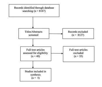
id=”chapter8Table1″Table 1. Summary of published data on prophylactic seizure medication in patients with brain metastases
| Author (Year) | Description of Study | Data Class | Conclusions |
| Forsyth et al,7 2003 | Multi-institutional, prospective, randomized controlled trial of PHT or phenobarbital vs no seizure prophylaxis (control) in patients with newly diagnosed cerebral metastases. Patients with systemic cancer (breast, lung, melanoma, and other) with typical radiographic appearance of brain metastases, recruited within 1 month of diagnosis, and no prior seizures. A total of 100 study patients enrolled, 52 with metastases (23 AED group, 29 no AED control); 8 additional patients randomized after diagnosis via craniotomy or biopsy. | III | Trial terminated early based on a high probability of data insufficiency. No significant difference in 3-month seizure rates between groups (10% with AEDs vs 13% without; p= .90). Class III data pertaining to metastases due to early study termination. |
| Glantz er al,8 1996 | Single-institution, prospective, randomized controlled trial of VAL vs placebo pill for length of study in patients with cerebral metastases (lung, breast, melanoma). Patients randomized within 14 days of diagnosis, and had no previous seizures, >1 supratentorial brain lesion, KPS ≥ 50%, and no previous anticonvulsant use or other brain disease. 74 total study patients enrolled, 57 with metastases (28 VAL, 29 placebo control). | III | Seizures occurred in 35% of all study patients treated with VAL and 24% of all patients in the placebo group (p= .3). Insufficient data published for subgroup analysis of patients with brain metastases (77% of total study patients). Class III data pertaining to metastases due to lack of subgroup analysis. |
AED, Anti-Epileptic Drug; KPS, Karnofsky Performance Scale; PHT, Phenytoin; VAL, Valproate.
Subgroup analysis not possible for patients with metastases.
id=”chapter8Table2″Table 2. Summary of published data on prophylactic seizure medications for post-craniotomy patients with brain metastases
| Author, Year | Description of Study | Data Class | Conclusions |
| Ansari et al,10 2014 | Single-institution, retrospective chart review of post-operative AED use vs no post-operative AED use in patients with cerebral metastases following initial craniotomy for tumor resection. Patients with no previous seizures or use of AEDs. 202 total study patients, 86 with metastases (53 prescribed AEDs, 33 not prescribed AEDs). | III | Overall, 22.8% of patients had a post-operative seizure (median follow-up 321 days). Prophylactic AEDs with a 1.62 times increased chance of seizures (p = .2867). A total of 19% of patients with metastases experienced seizures. Insufficient data provided for metastases specific sub-group analysis. Class III data pertaining to metastases due to lack of subgroup analysis. |
| Wu et al,11 2013 | Single-institution, prospective, randomized controlled trial of PHT for 7 days post-craniotomy vs no seizure prophylaxis (control) in patients with cerebral metastases undergoing craniotomy for tumor resection. Patients were previously untreated (except WBRT >1 month prior), with cerebral metastases from systemic cancer (lung, melanoma, renal, breast, and other), ≥8 years old, with a KPS ≥70, and no prior seizures or solely posterior fossa tumors. 123 total study patients, 77 with metastases (39 PHT, 38 controls). | III | Trial terminated early based on a high probability of data insufficiency. No significant difference in seizure rate between PHT and control groups (15 vs 13%, p = 1.00). The majority of seizures occurred >30 days post-operatively, with no significant difference across treatment groups for early (≤30 days; 3 vs 5%, p = .62) or late (>30 days; 13 vs 8%, p= .71) seizures. Significantly more adverse events noted with PHT (18 vs 0%, p= .01). Class III data pertaining to metastases due to early study termination. |
| Franceschetti et al,12 1990 | Single-institution, prospective, randomized controlled trial of post-operative AEDs (phenobarbital or PHT) vs placebo (control) in patients with cerebral metastases undergoing craniotomy. Patients had supratentorial tumors and no previous seizures. A total of 63 study patients (phenobarbital or PHT, n= 41) or no AED (n= 22), 13 with metastases (subgroup breakdown not provided). | III | Overall, early postoperative seizures occurred in 7% of the AED cohort and 18% of the non-AED cohort (p= .23). Late postoperative seizures occurred in 12% of the AED cohort and 21% of the non-AED cohort (p= .64). Insufficient data provided for metastases-specific sub-group analysis. Class III data pertaining to metastases due to lack of subgroup analysis. |
AED, antiepileptic drug; KPS, Karnofsky Performance Scale; PHT, Phenytoin; WBRT, Whole brain radiation therapy.
REFERENCES
1. Gavrilovic IT, Posner JB. Brain metastases: epidemiology and pathophysiology. J. Neurooncol. Oct 2005;75(1):5-14.
2. You G, Sha Z, Jiang T. The pathogenesis of tumor-related epilepsy and its implications for clinical treatment. Seizure. Apr 2012;21(3):153-159.
3. Maschio M. Brain tumor-related epilepsy. Curr. Neuropharmacol. Jun 2012;10(2):124-133.
4. Eichler AF, Loeffler JS. Multidisciplinary management of brain metastases. The oncologist. 2007;12(7):884-898.
5. Shaw MD, Foy PM. Epilepsy after craniotomy and the place of prophylactic anticonvulsant drugs: discussion paper. J. R. Soc. Med. Apr 1991;84(4):221-223.
6. Mikkelsen T, Paleologos NA, Robinson PD, et al. The role of prophylactic anticonvulsants in the management of brain metastases: a systematic review and evidence-based clinical practice guideline. J Neurooncol. Jan 2010;96(1):97-102.
7. Forsyth PA, Weaver S, Fulton D, et al. Prophylactic anticonvulsants in patients with brain tumour. Can. J. Neurol. Sci. May 2003;30(2):106-112.
8. Glantz MJ, Cole BF, Friedberg MH, et al. A randomized, blinded, placebo-controlled trial of divalproex sodium prophylaxis in adults with newly diagnosed brain tumors. Neurology. Apr 1996;46(4):985-991.
9. Robinson PD, Kalkanis SN, Linskey ME, Santaguida PL. Methodology used to develop the AANS/CNS management of brain metastases evidence-based clinical practice parameter guidelines. J. Neurooncol. Jan 2010;96(1):11-16.
10. Ansari SF, Bohnstedt BN, Perkins SM, Althouse SK, Miller JC. Efficacy of postoperative seizure prophylaxis in intra-axial brain tumor resections. J. Neurooncol. May 2014;118(1):117-122.
11. Wu AS, Trinh VT, Suki D, et al. A prospective randomized trial of perioperative seizure prophylaxis in patients with intraparenchymal brain tumors. J. Neurosurg. Apr 2013;118(4):873-883.
12. Franceschetti S, Binelli S, Casazza M, et al. Influence of surgery and antiepileptic drugs on seizures symptomatic of cerebral tumours. Acta Neurochir. (Wien.). 1990;103(1-2):47-51.
13. Abramson JD, Rosenberg HG, Jewell N, Wright JM. Should people at low risk of cardiovascular disease take a statin? BMJ (Clinical research ed ). 2013;347:f6123.
14. North JB, Penhall RK, Hanieh A, Frewin DB, Taylor WB. Phenytoin and postoperative epilepsy. A double-blind study. J. Neurosurg. May 1983;58(5):672-677.
15. Gokhale S, Khan SA, Agrawal A, Friedman AH, McDonagh DL. Levetiracetam seizure prophylaxis in craniotomy patients at high risk for postoperative seizures. Asian. J. Neurosurg. Oct 2013;8(4):169-173.
Source: Neurosurgery, January 9, 2019
Congress of Neurological Surgeons Systematic Review and Evidence-Based Guidelines Update for the Role of Emerging Therapies in the Management of Patients with Metastatic Brain Tumors
Sponsored by: Congress of Neurological Surgeons (CNS) and the Section on Tumors
Endorsed by: Reviewed for evidence-based integrity and endorsed by the American Association of Neurological Surgeons (AANS) and Congress of Neurological Surgeons (CNS)
Authors: Kristin Huntoon, PhD, DO1, J. Bradley Elder, MD2, Guilherme Finger MD MSc2, D. Ryan Ormond, MD, PhD3, Navid Redjal, MD4,5, Mark E. Linskey, MD6, Jeffrey J. Olson, MD 7
Departmental and institutional affiliations:
- Department of Neurosurgery at the University of Arizona and at Southern Arizona VA Health Care System, Tucson, AZ, USA
- Department of Neurological Surgery, The Ohio State University, Columbus, OH, USA
- Department of Neurosurgery, University of Colorado School of Medicine, Aurora, CO, USA
- Capital Institute for Neurosciences, Capital Health Pennington, NJ, USA
- Department of Neurosurgery, Cooper University Hospital, Cooper Medical School of Rowan University, Camden, NJ, USA
- Department of Neurological Surgery, University of California, Irvine, School of Medicine, Irvine, CA, USA
- Department of Neurosurgery, Emory University School of Medicine, Atlanta, Georgia, USA
Corresponding Author contact information:
Kristin Huntoon, PhD, DO
University of Arizona Department of Neurosurgery
PO Box 245070
1501 N Campbell Avenue, Room 4303
Tucson, Arizona, 85724-5070
Tel: (520) 694-8888
No part of this article has been published or submitted for publication elsewhere.
Keywords: Brain metastases, targeted therapy, immunotherapy, leptomeningeal, laser interstitial thermal therapy, radiation sensitizer
Abbreviations:
CNS = central nervous system
GKRS = Gamma Knife Radiosurgery
HIFU = magnetic resonance imaging–guided focused ultrasound
ICI = immune checkpoint inhibitor
IT = intrathecal
LITT = laser interstitial thermal therapy
LM = leptomeningeal metastasis
MBT = metastatic brain tumors
NSCLC = non–small-cell lung carcinoma
OS = overall survival
PD-L1 = programmed death-ligand 1
PFS = progression-free survival
SRS = stereotactic radiosurgery
TKI = tyrosine kinase inhibitor
TPS = Tumor Proportion Score
WBRT = whole brain radiation therapy
ABSTRACT
Background: Patients with metastatic brain tumors (MBTs) require a multidisciplinary team-based approach to select the best diagnostic, surgical, and radiation interventions.
Objective: The aim of this guideline is to provide an update of the evidence-based recommendations of the guideline produced in 2019 regarding the use of emerging therapies for adult patients with MBTs.
Methods: PubMed and Embase were searched from January 1, 2016 through May 3, 2022 using search strategies pertinent to the therapeutic topics: targeted agents, immune modulating agents, interstitial modalities, radiosensitizers, laser interstitial thermal therapy (LITT), and magnetic resonance imaging–guided focused ultrasound (HIFU). The search results were screened using preestablished exclusion/inclusion criteria. Evidence tables were constructed using these data and the recommendations from the 2019 version were left unchanged, updated or, where appropriate, new recommendations were formulated.
Results: Of 6403 qualifying abstracts, 162 met the inclusion criteria and were included in the evidence tables. They provided 8 class I recommendations, 3 class II recommendations, and 17 class III recommendations. In 3 instances there was insufficient evidence to support a recommendation. The proliferation of qualifying literature since the end of 2015 was greatest regarding the topics related to targeted therapy and immunotherapy of MBTs. Fewer were available for LITT and radiosensitizers, but enough information was available to formulate recommendations on these 2 topics. For interstitial modalities and HIFU, insufficient qualifying data was identified to create recommendations.
Conclusion: This systematic review provides evidence-based recommendations for adult patients with MBTs regarding the use of therapies beyond standard surgical, radiation, and cytotoxic chemotherapy.
PICO QUESTIONS AND RECOMMENDATIONS
Target Population: Adults with MBTs
Question 1
In patients with parenchymal brain metastases, does the use of molecular targeted agents provide benefit in terms of local control, overall survival (OS), progression-free survival (PFS), performance status, or reduction in central nervous system (CNS) side effects compared to standard management with chemotherapy, immune modulators, stereotactic radiosurgery (SRS), whole brain radiation therapy (WBRT), and surgical resection?
RECOMMENDATIONS
Unchanged Recommendation
Level I: The use of afatinib is not recommended in patients with brain metastasis due to breast cancer.
New Recommendations
Targeted Therapy for the Treatment of EGFR Mutant NSCLC Parenchymal Brain Metastases
Level I: In subjects with ≥3 untreated brain metastases from epidermal growth factor receptor (EGFR) mutant non–small-cell lung carcinoma (NSCLC), the use of icotinib and WBRT is recommended to improve intracranial PFS.
Level III: In subjects with brain metastases from EGFR mutant NSCLC, the addition of EGFR tyrosine kinase inhibitors to radiation therapy in the form of WBRT or SRS is suggested to improve OS, PFS, and intracranial PFS.
Targeted Therapy for the Treatment of ALK Mutation–Positive NSCLC Parenchymal Brain Metastases
Level I: In patients with ALK mutation-positive NSCLC with untreated brain metastases the use of alectinib is recommended to delay time to intracranial tumor progression.
Level II: In patients with untreated brain metastases from ALK mutation positive NSCLC lorlatinib is recommended to prolong intracranial tumor control and improve overall PFS.
Targeted Therapy for the Treatment of NSCLC Parenchymal Brain Metastases Not Assessed for EGFR and ALK Mutation Status
Level I: It is recommended that for patients with newly diagnosed brain metastases secondary to NSCLC not assessed for EGFR and ALK mutation status, and for whom WBRT is indicated, gefitinib be added to the treatment regimen to improve local tumor control and OS.
Level III: For individuals with brain metastases secondary to NSCLC not assessed for EGFR and ALK mutation status and for whom targeted therapy in the form of gefitinib or the combination of pemetrexed and platinum compounds are otherwise indicated, it is suggested that bevacizumab, when not contraindicated by other underlying medical conditions, be added to the treatment regimen to improve CNS control and to a lesser extent PFS and OS.
Targeted Therapy for the Treatment of EGFR Negative, ALK Negative NSCLC Parenchymal Brain Metastases
Level III: For individuals with brain metastases secondary to NSCLC that are EGFR and ALK mutation negative and for whom targeted therapy in the form of tyrosine kinase inhibitors (TKIs) are indicated, it is suggested that TKIs, when not contraindicated by other underlying medical conditions, be added to the treatment regimen, including radiation therapy, to improve CNS control and to a lesser extent PFS and OS.
Targeted Therapy for the Treatment of Melanoma Parenchymal Brain Metastases
Level I: It is recommended that for patients with newly diagnosed brain metastases secondary to melanoma that is BRAFV600E-positive, dabrafenib plus trametinib be added to the treatment regimen to obtain improved local tumor control.
Level III: For individuals with brain metastases secondary to BRAF-altered melanoma for whom targeted therapy in the form of BRAF inhibitors are indicated, it is suggested that immunotherapy, when not contraindicated by other underlying medical conditions, be added to the treatment regimen to improve CNS control and to a lesser extent PFS and OS.
Targeted Therapy for the Treatment of Breast Adenocarcinoma Parenchymal Brain Metastases
Level III: In adult patients with brain metastases from breast adenocarcinoma that are HER2-positive for whom radiation therapy is indicated, it is suggested that trastuzumab be added to the treatment regimen to improve PFS, median survival, and OS.
Level III: In adult patients with brain metastases from breast adenocarcinoma for whom SRS is indicated, it is suggested that lapatinib be added to that treatment to improve intracranial response rate and median survival.
Question 2
In patients with leptomeningeal brain metastases, does the use of molecular targeted agents provide benefit in terms of local control, OS, PFS, performance status, or reduction in CNS side effects compared to standard management with chemotherapy, immune modulators, SRS, WBRT, and surgical resection?
New Recommendations
Level III: In individuals with leptomeningeal disease from NSCLC with EGFR mutations, it is suggested that EGFR TKIs be utilized to increase median survival, specifically the third-generation TKI osimertinib for patients with EGFR-mutant NSCLC and the second-generation ALK-TKI alectinib for the treatment of leptomeningeal metastases (LMs) in ALK-positive NSCLC.
Level III: In individuals with LMs from Her2 positive breast cancer, it is suggested that intrathecal (IT) trastuzumab be utilized to increase median survival.
Question 3
In patients with parenchymal brain metastases, does the use of immune modulators provide benefit in terms of local control, OS, PFS, performance status, or reduction in CNS side effects compared to standard management with chemotherapy, molecular targeted agents, SRS, WBRT, and surgical resection?
New Recommendations
Level I: In individuals with active, untreated, asymptomatic parenchymal melanoma brain metastases (MBMs), ipilimumab plus nivolumab is recommended to increase median OS and be utilized without radiation to improve median OS.
Level III: In individuals with parenchymal brain metastases from NSCLC it is suggested that immune checkpoint inhibitors (ICIs) be utilized with radiation therapy to increase median survival, decrease incidence of local failure, increase intracranial PFS, and decrease distant intracranial failure.
Level III: In individuals with parenchymal brain metastases from NSCLC that are clinically stable for at least 4 weeks and with programmed death-ligand 1 (PD-L1) tumor proportion score (TPS) >50% it is suggested that ICIs be utilized without radiation to improve median OS.
Level III: In individuals with parenchymal brain metastases from breast cancer or colon carcinoma it is suggested that therapy with ICIs be considered alone or with radiation therapy to increase median survival and decrease incidence of local failure.
Question 4
In patients with leptomeningeal brain metastases, does the use of immune modulators provide benefit in terms of local control, OS, PFS, performance status, or reduction in CNS side effects compared to standard management with chemotherapy, molecular targeted agents, SRS, WBRT, and surgical resection?
New Recommendation
There is insufficient evidence to make a recommendation regarding the use of immune modulators for the therapy of leptomeningeal brain metastases.
Question 5
In patients with parenchymal brain metastases, does the use of interstitial modalities, in the form of interstitial chemotherapy or radiation (brachytherapy, intraoperative radiation therapy), provide benefit in terms of local control, OS, PFS, performance status, or reduction in CNS side effects compared to standard management with chemotherapy, immune modulators and molecular targeted agents, SRS, WBRT, and surgical resection?
Unchanged Recommendation
There is insufficient evidence to make a recommendation regarding the use of interstitial modalities in the form of interstitial chemotherapy or radiation.
Question 6
In patients with parenchymal brain metastases, does the use of radiosensitizers provide benefit in terms of local control, OS, PFS, performance status, or reduction in CNS side effects compared to standard management with chemotherapy, immune modulators and molecular targeted agents, SRS, WBRT, and surgical resection?
Unchanged Recommendations
Level I: The use of temozolomide as a radiation sensitizer is not recommended in the setting of whole-brain radiation therapy (WBRT) for patients with breast cancer brain metastases.
Level I: The use of chloroquine as radiation sensitizer is not recommended in the setting of WBRT for patients with brain metastases.
New Recommendations
Level II: When WBRT is utilized for brain metastases from NSCLC it is recommended that temozolomide be added to provide a smaller incidence of local failure, longer intracranial PFS, and longer OS.
Level III: For brain metastases from NSCLC with EGFR mutation positive status where WBRT or SRS is indicated, it is suggested that EFGR TKIs be added to that therapy to improved intracranial response rate and survival.
Question 7
In patients with parenchymal or leptomeningeal brain metastases, does the use of LITT provide benefit in terms of local control, OS, PFS, performance status, or reduction in CNS side effects compared to standard management with chemotherapy, immune modulators and molecular targeted agents, SRS, WBRT, and surgical resection?
New Recommendations
Level III: For adults who have undergone SRS for brain metastases with subsequent imaging progression due to tumor progression it is suggested that LITT be considered as equivalent to craniotomy in terms of PFS and OS and the choice of management should be individualized based on the unique characteristics of the tumor location and the subject’s clinical status.
Level III: For adults who have undergone SRS for brain metastases with subsequent imaging progression due to radiation necrosis it is suggested that LITT be considered as equivalent to medical management for radiation necrosis and the choice of management should be individualized based on the unique characteristics of the tumor location and the subject’s clinical status.
Question 8
In patients with parenchymal or leptomeningeal brain metastases, does the use of HIFU provide benefit in terms of local control, OS, PFS, performance status, or reduction in CNS side effects compared to standard management with chemotherapy, immune modulators and molecular targeted agents, SRS, WBRT, and surgical resection?
Unchanged Recommendation
There is insufficient evidence to make a recommendation regarding the use of HIFU for parenchymal and leptomeningeal brain metastases.
INTRODUCTION
Goals and Rationale
Advancements in the understanding of the biology of MBTs, the ability to create more sophisticated systemic treatments via improved pharmacologic chemistry, radiation therapy software and hardware advancements, and surgical equipment have yielded new information worthy of dissemination. As suggested by the Institute of Medicine, now the National Academy of Medicine, it is suggested that guidelines be updated in the range of every 5 years.1 Thus, interval updates of the guideline on emerging therapies for MBTs published by the American Association of Neurological Surgeons (AANS)/Congress of Neurological Surgeons (CNS) in 2018 were planned to update the information in that publication.2
Objectives
This document seeks to update the recommendations for molecular and targeted agents, immune modulating agents, interstitial modalities, radiosensitizers, intraoperative radiation therapy, laser interstitial thermal therapy, and HIFU published in the 2018 guideline on emerging therapies for MBTs.2 To accomplish this, the Joint Tumor Section of the AANS/CNS recruited representatives from the section to review and update the questions from the previous guideline to PICO (Patient \, Intervention, Comparison, Outcome) format, search the literature published regarding the items in each question since the search of the 2018 publication, and determine if that new information confirmed previous recommendations, required an update of previous recommendations, or new recommendations.
METHODOLOGY
Literature Search
To accomplish this update, new literature in PubMed and EMBASE from January 1, 2016 through May 3, 2022 was searched using the questions with their new PICO format, and data from the qualifying manuscripts for each topic was used to either confirm previous recommendations, update them, or create new ones. The search strategy used combinations of subheadings and key words and is documented in previous methodology papers. Search strategies for the root brain metastasis search as well as the 6 categories of emerging therapy (molecular and targeted agents, immune modulating agents, interstitial modalities, radiosensitizers, laser interstitial thermal therapy, and HIFU) can be found in Appendix I. Manuscripts selected for review upon screening of abstracts met the criteria described below. All citations were reviewed by 2 authors and acceptance or rejection recorded along with the reasons. When there was disagreement, the 2 reviewers met in live session to resolve the disagreement. The Guidelines Task Force used DistillerSR (which utilizes artificial intelligence) to cull, narrow, and aid its review of the relevant literature. All abstracts were reviewed, and relevant full text articles were retrieved and graded [by individuals on the Guidelines Task Force].
Inclusion/Exclusion Criteria
- Published in English
- Involves human patients with brain metastases
- Fully published primary study published between September 2008 and December 2015
- Paper evaluates ≥1 of the therapies in question:
- Molecular and targeted agents for parenchymal brain metastases
- Molecular and targeted agents for leptomeningeal brain metastases
- Immune modulating agents for parenchymal brain metastases
- Immune modulating agents for leptomeningeal brain metastases
- Interstitial modalities
- Radiosensitizers
- LITT
- HIFU
- Number of patients with brain metastases in the study ≥5 per study arm for ≥2 of the study arms for comparative studies, and ≥5 total patients if a noncomparative study
Data Collection Process
Manuscripts selected for review underwent full review by 2 authors to confirm that it met eligibility criteria; if not, the manuscript was rejected. As with the abstracts, when there was disagreement the 2 reviewers met in live session to resolve the disagreement. Data gleaned from the manuscript included type of study (e.g., phase 2 clinical trial, retrospective chart review, etc.), therapeutic agent evaluated, and the outcome measures and results yielded by the study.
Assessment for Risk of Bias
Each manuscript was evaluated by the writing group for bias, and the summation of different forms of bias are reflected in the data classification system. Inherent to emerging therapy agents, initial reports were noted to be in the form of small case series, anecdotal reports, and early phase clinical trials. As such, there is inevitable selection bias imposed by retrospective reviews and prospective studies with small numbers of patients. For example, patients selected for study, especially early phase trials, may have better medical status relative to patients not selected for study. In addition, small series of patients may have bias because of random variability. Our expectation is that the more promising techniques and agents mentioned in this guideline will be studied further as part of larger clinical trials which will eliminate some of the inherent bias of smaller, retrospective studies.
Rating Quality of Evidence and Recommendation Formulation
Each manuscript that met eligibility criteria and was found to have data relevant to the question was rated as providing class I, II, or III evidence based on the definitions provided in the AANS/CNS criteria. The pertinent classification levels and data for each paper were entered into an evidence table for each emerging therapy subtopic. The evidence tables were then validated among the writing group before the formulation of recommendations. The summation of the information from qualifying manuscripts was then synthesized and used to create level I, II, or III recommendations based on the classification of evidence on therapeutic effectiveness (Appendix II). An expanded description of the data classification system and translation to recommendation level designation is provided at Guideline Development Methodology – cns.org.
Revision Plans
In accordance with the National Academy of Medicine’s standards for developing clinical practice guidelines, the writers of the emerging therapies for MBTs task force will monitor related publications following the release of this document and will revise the entire document and/or specific sections “if new evidence shows that a recommended intervention causes previously unknown substantial harm; that a new intervention is significantly superior to a previously recommended intervention from an efficacy or harms perspective; or that a recommendation can be applied to new populations.”3 In addition, within 5 years from the date of publication the task force plans to assess the content this guideline to that it still reflects the clinical practice and treatment for patients with MBTs. In those cases where it does not, the recommendations will either be updated or new recommendations will be created.
SUMMARY OF PREVIOUS GUIDELINE
The prior version of the guidelines for the role of emerging and investigational therapy was written with one question: What evidence is available regarding emerging and investigational treatment options for MBTs?2 This was then applied over a range of topics in order of consideration at that time, including high intensity focused ultrasound (HIFU), laser interstitial thermal therapy (LITT), radiosensitizers, interstitial modalities, immune modulators, and molecular targeted agents.
Insufficient evidence was available to make a recommendation regarding the use of HIFU, LITT, interstitial modalities, and immune modulators.
For radiation sensitizers, two level I recommendations were derived, both of which were negative. The first stating temozolomide as a radiation sensitizer is not recommended in the setting of WBRT for patients with breast cancer brain metastases. The second stated chloroquine as a radiation sensitizer is not recommended in the setting of WBRT for patients with brain metastases. There is insufficient evidence to make a recommendation regarding the routine use of motexafin-gadolinium, sodium nitrite, temozolomide, or chloroquine in patients with brain metastases.
For molecular targeted agents, one level I recommendation was derived, again negative, stating that the use of afatinib is not recommended in patients with brain metastasis due to breast cancer. Evidence did not support making a recommendation regarding the use of epidermal growth factor inhibitors in non–small cell lung carcinoma (NSCLC), BRAF inhibitors for melanoma, HER2 inhibitors in breast cancer, or VEGF inhibitors in parenchymal brain metastases.
To facilitate comparison of the new recommendations with those from the previous version, a table displaying them side by side has been created (Table 1). An additional table of the recommendations enumerated in this document by treatment modality and then divided by tumor type and subdivided by molecular subtypes is provided (Table 2). This will facilitate access to recommendations for readers most interested in recommendations by tumor type.
RESULTS
Targeted Therapy for Brain Metastases
Targeted Therapy of Parenchymal Brain Metastases
Question 1: In patients with parenchymal brain metastases, does the use of molecular targeted agents provide benefit in terms of local control, OS, PFS, performance status, or reduction in CNS side effects compared to standard management with chemotherapy, immune modulators, SRS, WBRT, and surgical resection.
The literature search yielded 2634 abstracts. Task force members reviewed all titles and abstracts yielded from the literature search and identified the literature for full-text review and extraction, addressing the clinical questions, in accordance with the literature search protocol (Appendix I). Task force members identified the best research evidence available to answer the targeted clinical questions. When level I, II, and or III literature was available to answer specific questions, the task force did not review level IV studies.
The task force selected 263 full-text articles for full-text review. Of these, 216 were rejected for not meeting inclusion criteria or for being off topic. Forty-seven articles were selected for systematic review (Appendix III).
All 47 studies included in the qualitative analysis also met the criteria to be included in the quantitative analysis. Three studies were class I, 3 were class II, and 41 studies were class III. To ease the discussion of the targeted therapy of parenchymal brain metastases, the qualifying manuscripts are divided into those related to NSCLC, melanoma, and breast adenocarcinoma. The recommendations for the different tumor types are provided after the synthesis for each circumstance to ease understanding of where the recommendations apply.
NSCLC
Prognostic Studies
Gong et al1 performed a cross-section study collecting retrospective data of patients with NSCLC and BM treated between 2007 and 2012. The objective of this study was to observe the therapeutic efficacy and prognostic factors that influence survival rates of patients with NSCLC with multiple brain metastases (BMs; >3 and <10). A total of 209 patients were included, all of whom received WBRT. Two hundred patients received combined chemotherapy during the treatment process, 99 received targeted drug therapy, and 9 received only symptomatic and supportive treatment. The median survival time for all patients with BMs was 12.1 months (95% confidence interval [CI], 9.37-14.83). The 6-month and 1- and 2-year cumulative survival rates were 80%, 50.2%, and 10.7%, respectively. Based on univariate and multivariate analysis, the authors concluded that active treatment of NSCLC with multiple BMs was beneficial, and the patients’ cultural background had a strong influence on survival prognoses. Three or more cycles of chemotherapy (P = .001) combined with targeted drug therapy (P = .01) could increase the patients’ median and OS rates. Also, cultural backgrounds of the patients had significant effects on the patients’ survival prognoses. Though targeted therapy is mentioned in this article, the focus of this study was not targeted therapy. It provides class III prognostic data; additional confirming class III or better data is necessary before using this information to formulate a recommendation.
Targeted Therapy for the Treatment of NSCLC Parenchymal Brain Metastases
Most of the qualifying articles provided class III evidence (30 studies), 3 provided class II evidence, and 3 provided class I evidence. Details of the qualifying and informative manuscript data are available in the evidence table (Table 3). For better comprehension, this discussion is initially subdivided into groups of manuscripts that analyze different genetic profiles of the NSCLC: EGRF mutation status (present or absent) and/or ALK mutation status. Secondarily, the studies are grouped according to relatively common treatment themes for each of the groups. Patients were excluded if they discontinued TKI after RT or ever received surgery for BM. The objective of this study was to compare OS and intracranial PFS between these 2 groups.
Targeted Therapy for the Treatment of EGFR Mutant NSCLC Parenchymal Brain Metastases
The presence of EGFR mutation can be treated with tyrosine kinase inhibitors (TKIs) such as gefitinib, erlotinib, afatinib, and osimertinib. Huang et al4 published a study that investigated the efficacy of 3 different EGFR TKIs (gefitinib, erlotinib, and afatinib) in association with bevacizumab as the first-line treatment in patients with advanced EGFR mutant lung adenocarcinoma.4 A total of 36 patients were included in the final analysis. Three patients received gefitinib, 17 received erlotinib, and 16 received afatinib combined with bevacizumab as the first-line treatment. Since only 3 patients received gefitinib, the comparison analysis involved only erlotinib and afatinib. Regarding the use of different EGFR TKIs, the median PFS was 17.1 months in the erlotinib group and 21.6 months in the afatinib group (P = .617). In patients with brain metastasis at baseline, the median PFS was 18.9 months in the erlotinib group and 16.4 months in the afatinib group (P = .747). Among patients harboring an exon 19 deletion, the median PFS was 16.2 months in the erlotinib group and not reached in the afatinib group (P = .141). In patients with L858R mutation, the median PFS was 18.9 months in the erlotinib group and 16.2 months in the afatinib group (P = .481). Based on these results, the authors concluded that not only either erlotinib or afatinib combined with bevacizumab, as first-line treatment, provides solid clinical efficacy in patients with advanced EGFR mutant lung adenocarcinoma.
Wu et al5 designed a clinical trial to evaluate patients with advanced EGFR mutant NSCLC who experience disease progression with previous EGFR TKI treatment. The study compared osimertinib CNS efficacy with platinum-pemetrexed. Patients with asymptomatic, stable CNS metastases were eligible for enrollment and were randomly assigned 2:1 to osimertinib 80 mg once daily or platinum-pemetrexed. The primary objective for this analysis was CNS objective response rate (ORR). Of 419 patients randomly assigned to treatment, 116 had measurable and/or nonmeasurable CNS lesions, including 46 patients with measurable CNS lesions. At data cutoff (April 15, 2016), CNS ORR in patients with ≥1 measurable CNS lesions was 70% (21/30 [95% CI 51%-85%]) with osimertinib and 31% (5/16 [95% CI 11%-59%]) with platinum-pemetrexed (odds ratio [OR] 5.13 [95% CI 1.44-20.64]; P = .015); the ORR was 40% (30/75 [95% CI 29%-52%) and 17% (7/41 [95% CI 7%-32%]), respectively, in patients with measurable and/or nonmeasurable CNS lesions (OR 3.24 [95% CI 1.33-8.81]; P = .014). Median CNS duration of response in patients with measurable and/or nonmeasurable CNS lesions was 8.9 months (95% CI 4.3 months to not calculable) for osimertinib and 5.7 months (95% CI 4.4-5.7 months) for platinum-pemetrexed; median CNS PFS was 11.7 months and 5.6 months, respectively (hazard ratio [HR] 0.32 [95% CI 0.15-0.69]; P = .004). The authors concluded that osimertinib demonstrated superior CNS efficacy compared with PP.
Chiou et al6 performed retrospectively reviewed consecutive cases of EGFR mutant NSCLC with BM treated between 2012 and 2020. The goal of this study was to compare outcomes between EGFR TKE therapy alone (group I) and combined EGFR TKI therapy + SRS (group II) in patients with NSCLC with BMs and EGFR mutations. Tumor control (<10% increase in tumor volume) and OS rates were compared. The study cohort included 280 patients (n = 90 in group I and n = 190 in group II). Cumulative tumor control rates were higher in group II than in group I (79.8% vs 31.2% at 36 months, P < .0001). Cumulative OS rates were comparable between groups I and II (43.8% vs 59.4% at 36 months, P = .3203). Independent predictors of tumor control were older age (P < .01, HR 1.03), fewer BMs (P < .01, HR 1.09), lack of extracranial metastasis (P < .02, HR 0.70), and combined SRS and TKI therapy (P < .01, HR 0.25). Independent predictors of OS were fewer BMs (P < .01, HR 1.04) and a higher Karnofsky performance status score (P < .01, HR 0.97). Although the OS rate did not differ between TKI therapy with and without SRS, the addition of SRS to TKI therapy resulted in improvement of intracranial tumor control. The lack of effect on survival rate with the addition of SRS may be attributable to extracranial disease progression.
He et al7 performed a historic cohort comparing the efficacy of concurrent EGFR-TKI and whole-brain radiation therapy (WBRT) with EGFR TKI alone as a first-line therapy for advanced EGFR mutated NSCLC with brain metastases. One hundred four patients were included, 56 in the concurrent EGFR TKI group and 48 in the EGFR TKI alone group. The median follow-up of the sample was 23 months. Concurrent EGFR TKI and WBRT significantly improved the median intracranial PFS (iPFS) compared with EGFR TKI alone (17.7 vs 11.0 months, P = .015). Subgroup analysis showed that concurrent EGFR TKI and WBRT improved median iPFS compared with EGFR TKI alone in patients with >3 brain metastases (P = .001); however, no significant difference was observed between the 2 regimens in patients with ≤3 brain metastases (P = .526). Of note, the OS analysis was biased because 20 patients in the EGFR TKI alone group migrated to the other group with salvage WBRT upon brain metastasis progression.
He et al8 conducted a historic cohort aiming to compare erlotinib with pemetrexed as second-/third-line treatment in patients with lung adenocarcinoma with asymptomatic brain metastases. From January 2012 to June 2014, all patients with lung adenocarcinoma with asymptomatic brain metastases who received treatment with erlotinib or pemetrexed as second-/third-line treatment were retrospectively reviewed. Even though the EGFR mutation status was assessed in this study, patients were divided into 2 groups according to the therapeutic regimen received, not EGFR status. A total of 99 patients were included, 68 in the erlotinib-treated group and 31 in the pemetrexed-treated group. Among the 99 patients, 44 (44.4%) had EGFR mutated. Median PFS was not different between the groups treated with erlotinib and pemetrexed (4.2 vs 3.4 months; 95% CIs 2.01-6.40 vs 2.80-5.00, respectively; P = .635). A subanalysis based on EGFR status was performed by the authors. In the erlotinib-treated group, median PFS in EGFR mutation–positive patients was 8.0 months, whereas it was 1.3 months in EGFR mutation–negative patients (95% CI 5.85-10.15 vs 0.26-2.35; P < .001(Table 3). The median PFS in EGFR mutation–positive patients was 8.0 months in the erlotinib-treated group but was 3.9 months in the pemetrexed-treated group (95% CI 5.85-10.15 vs 1.25-6.55; P = .032). In conclusion, erlotinib and pemetrexed may be used as second-/third-line treatment in patients with lung adenocarcinoma with asymptomatic brain metastases, and detection of EGFR mutation status is particularly important in these patients. EGFR mutation–positive lung adenocarcinoma patients with asymptomatic brain metastases showed longer PFS when treated with erlotinib as opposed to pemetrexed.
Liu et al9 selected patients with NSCLC with brain metastases who had good responses to EGFR TKI to examine the role of early brain RT on intracranial disease control and survival. The selection method was not clearly outlined and introduces the risk of substantial bias into this article. All the patients were treated with EGFR TKI monotherapy (gefitinib 250 mg 4 times daily or erlotinib 150 mg 4 times daily or icotinib 125 mg 3 times daily). One hundred thirteen patients were included, 49 (43%) treated with brain radiation therapy within 4 weeks after EGFR TKI initiation and 64 (57%) were treated with EGFR TKI alone. Among the 64 patients in the second group, 27 received salvage brain radiation therapy and only 27 were truly treated only with EGFR TKI. When the 3 groups were compared at baseline, patients with early brain RT were more likely to be symptomatic from their BMs (80% early RT vs 13% EGFR TKI alone vs 33% salvage RT, P < .001). Besides, there were more patients with a less favorable prognosis in early RT group (DS-GPA of 0-2.0: 86% early RT vs 51% EGFR TKI vs 78% deferred RT, P = .002). The median iPFS of patients with early RT was significantly longer than those without early RT (21.4 vs 15.0 months, P = .001). The effect of early brain RT on iPFS remained significant on multivariate analysis (HR 0.34 [95% CI 0.19-0.61]; P < .001). However, after salvage brain RT, the iPFS did not differ significantly between the salvage RT and early RT groups (23.6 vs 21.4 months, P = .253). No significant difference of the IC-PFS was found between the patients with salvage RT and those with EGFR TKI alone (23.6 vs 24.4 months, P = .277). The median OS for early brain RT, EGFR TKI alone, and salvage brain RT groups was 28.1 months (95% CI 17.9-38.3), 24.5 months (95% CI 20.6-28.4), and 24.6 months (95% CI 19.0-30.1), respectively (P = .604). No significant difference in OS was observed between patients with early RT and those with salvage RT (28.1 vs 24.6 months, P = .385).
Huang et al10 retrospectively analyzed patients with EGFR mutant NSCLC treated in a single institution between January 2018 and December 2020 in order to compare the efficacy of 2 different EGFR TKI (osimertinib and afatinib) as first-line treatment in these patients. One hundred twenty-eight patients were selected for this study. The osimertinib group included 47 patients, while 81 patients received afatinib. The median PFS was 18.8 months and 13.1 months in the osimertinib and afatinib groups, respectively (HR 0.75 [95% CI 0.48-1.18]). The median OS was not reached in the osimertinib group and was 41.7 months in the afatinib group (HR 0.79 [95% CI 0.36-1.72]). In patients with brain metastasis at baseline, the median PFS was 22.1 months in the osimertinib group, and 10.9 months in the afatinib group (adjusted HR 0.45 [95% CI 0.21-0.96]). Their research demonstrates that there was no compelling evidence showing that patients taking osimertinib as first-line treatment experienced longer median PFS and OS than patients treated with afatinib. However, there was a statistical significance revealing that osimertinib provided better median PFS than afatinib in patients with brain metastasis at baseline.
Chiu et al11 designed a retrospective study to analyze patients with EGFR mutant NSCLC with brain metastasis who received first-line EGFR TKI (erlotinib or gefitinib) monotherapy or with bevacizumab. During the period of 2014 to 2019, 310 patients were identified and included in the study in which 267 (86.1%) patients received the treatment of single-agent EGFR TKI and 43 (13.9%) patients received the treatment of EGFR TKI plus bevacizumab. Patients receiving EGFR TKI and bevacizumab were significantly younger and had better performance status and with high incidence of brain metastasis (55.8%). In the propensity-score matched cohort, PFS (13.5 vs 13.7 months; log-rank P = .700) was similar between the 2 groups. The OS (61.3 vs 34.2 months; log-rank P = .010) and risk reduction of death (HR 0.42 [95% CI 0.20-0.85]; P = .017) were significantly improved in EGFR TKI plus bevacizumab group. Analysis of treatment by brain metastasis status demonstrated EGFR TKI plus bevacizumab in patients with brain metastasis was associated with significant OS benefit compared with other groups (log-rank P = .030) and these patients had lower early CNS and early systemic progressions.
Fan et al12 conducted a retrospective study to compare upfront RT combined with icotinib to icotinib alone as first-line therapies for patients with EGFR-mutant NSCLC and BM. A total of 152 patients with metastatic EGFR-mutant adenocarcinoma with BM who were diagnosed and received icotinib therapy between October 2011 and October 2014 were identified. Of these, 55 patients were excluded and 97 included. A total of 56 of 97 patients received RT for treatment of brain metastases, while 41 patients received TKI therapy alone. Surgical resection preceded RT in 9 patients. RT was delivered with either localized SRS (Gamma Knife) or WBRT. There was no difference in OS between the RT followed by icotinib group and the icotinib alone group (31.9 vs 27.9 months, P = .237), and similar results were found in the SRS subgroup (35.5 vs 27.9 months, P = .12). Intracranial PFS was improved in the patients who received RT followed by icotinib compared with those receiving icotinib alone (22.4 vs 13.9 months, P = .043).
Wang et al13 retrospectively reviewed patients with EGFR mutant patients treated in a single institution from January 2010 to December 2016 to compare the role of upfront RT in association to EGFR TKI versus target therapy alone. in these patients.13 Among the 93 patients included, 53 patients received upfront RT and TKI and 40 patients received TKI only. The upfront RT group showed lower intracranial progression risk with adjusted SHR 0.38 (95% CI 0.19-0.75, P = .006) and longer median time to sPFS (15.6 vs 8.9 months, P = .009). After the salvage RT, upfront RT did not prolong the median time to SST (23.6 vs 18.9 months, P = .862) and OS (median time, 35.4 vs 35.8 months, P = .695) compared with TKI alone. The authors concluded that compared with upfront intracranial RT, the salvage RT to oligo-progressive disease allowed patients receiving TKI to have similar time on initial TKI and OS despite worse iPFS. The best timing of intracranial RT remains to be further verified.
Jiang et al14 aimed to evaluate if EGFR TKI plus WBRT provide a better survival benefit than EGFR TKIs alone in patients with NSCLC with EGFR mutation and brain metastases. Two hundred thirty patients were included, 116 patients received EGFR TKIs alone (as first-line therapy in 91 cases) and 51 patients received EGFR TKIs plus WBRT therapy (as first-line treatment in 30 cases). Compared with TKIs alone, EGFR TKIs plus WBRT did not have superior intracranial PFS (6.9 vs 7.4 months, P = .232) and systemic PFS (7.5 vs 7.9 months, P = .546) but was associated with worse OS (21.6 vs 26.4 months, P = .049) in NSCLC with EGFR mutation and BM. The authors concluded that the addition of WBRT to EGFR TKIs did not appear to have survival benefit superior to that of EGFR TKIs alone in with EGFR mutant NSCLC with BM. WBRT also did not bring additional benefit to chemotherapy in patients with BM and EGFR of wild-type or unknown status.
Yang et al15 led a multicentered (17 institutions), open-label, randomized clinical trial including patients with EGFR mutated NSCLC and multiple brain metastases (≥3 lesions), who were naïve to treatment with EGFR TKI or radiation therapy. The participants were randomly assigned in a 1:1 ratio to either chemotherapy plus icotinib 125 mg orally (3 times per day) or chemotherapy plus WBRT (30 Gy in 10 fractions of 3 Gy). Groups were stratified by EGFR gene mutation status, treatment line (first or second), brain metastases only versus both intracranial and extracranial metastases, and presence or absence of symptoms of intracranial hypertension. Between December 2012 and June 2015, 176 participants were assigned to treatment: 85 to icotinib and 91 to WBRT. Median intracranial PFS was 10.0 months (95% CI 5.6-14.4) with icotinib versus 4.8 months (2.4-7.2) with WBRT (equating to a 44% risk reduction with icotinib for an event of intracranial disease progression or death; HR 0.56 [95% CI 0.36-0.90]; P = .014). The authors concluded that in patients with EGFR-mutant NSCLC and multiple brain metastases, icotinib was associated with significantly longer intracranial PFS than WBI plus chemotherapy, indicating that icotinib might be a better first-line therapeutic option for this patient population.
Chen et al16 proposed a study to compare the outcomes of first‐line EGFR TKI alone with EGFR TKI plus WBRT for the treatment of BM in patients with EGFR‐mutated lung adenocarcinoma. A total of 1665 patients were screened from 2008 to 2014, and 132 were enrolled in our study.16 None of the patients had received previous systemic therapy. All patients included in this analysis received 250 mg gefitinib or 150 mg erlotinib orally once daily. Among the 132 patients, 97 (73.5%) showed multiple intracranial lesions, and 67 (50.8%) had asymptomatic BM. Seventy‐nine patients (59.8%) were treated with EGFR TKI alone, 53 with concomitant WBRT. The intracranial objective response rate was significantly higher in the EGFR TKI plus WBRT treatment group (67.9%) compared with the EGFR TKI alone group (39.2%, P = .001). The median intracranial time to progression was 24.7 months (95% CI 19.5-29.9) in patients who received WBRT, which was significantly longer than in those who received EGFR TKI alone, with the median intracranial time to progression of 18.2 months (95% CI 12.5-23.9, P = .004). There was no significant difference in OS between WBRT and EGFR TKI alone groups (median 48.0 vs 41.1 months; P = .740). OS is significantly prolonged in patients who had an intracranial time to progression exceeding 22 months compared with those who developed intracranial progression <22 months after treatment (median 58.0 vs 28.0 months; P = .001). The authors concluded that for patients with EGFR mutated lung adenocarcinoma with BM treatment with concomitant WBRT achieved a higher response rate of BM and significant improvement in intracranial PFS compared with EGFR TKI alone.
Magnuson et al17 conducted a multi-institutional (6 institutions) analysis to determine the optimal management of patients with EGFR-mutant NSCLC who develop brain metastases and have not previously received EGFR-TKI. Patients were divided into 3 groups: SRS followed by EGFR-TKI, WBRT followed by EGFR-TKI, or EGFR-TKI followed by SRS or WBRT.17 Of the 351 patients, 131 (37%) received EGFR-TKI followed by SRS or WBRT at intracranial progression, 120 (34%) were treated with WBRT followed by EGFR-TKI, and 100 (29%) received SRS followed by EGFR-TKI. Baseline comparison among the 3 groups demonstrated that patients who received upfront EGFR-TKI were less likely to have symptomatic brain metastases (12% EGFR-TKI vs 51% WBRT and 49% SRS; P < .001) and were more likely to have brain metastases ≤1 cm (66% EGFR-TKI vs 35% WBRT and 44% SRS; P < .001). Patients who received upfront WBRT were more likely to have a less favorable prognosis (ds-GPA of 0-1.5; 75% WBRT vs 59% EGFR-TKI and 52% SRS; P = .001) and have >10 brain metastases (37% WBRT vs 15% EGFR-TKI and 7% SRS; P < .001). Patients treated with upfront EGFR-TKI and upfront WBRT were more likely to be stage IV at diagnosis (91% EGFR-TKI and 92% WBRT vs 80% SRS; P = .014). In terms of outcomes, the median OS for the upfront SRS, WBRT, and EGFR-TKI groups was 46 months (95% CI 37-57), 30 months (95% CI 27-38), and 25 months (95% CI 20-28), respectively (log-rank P < .001). OS at 2 years for the upfront SRS, WBRT, and EGFR-TKI groups was 78% (95% CI 66-85%), 62% (95% CI 52-70%), and 51% (95% CI 42-60%), respectively. After controlling for significant covariables in a multivariable model, upfront SRS was independently associated with improved OS relative to EGFR-TKI (adjusted HR 0.39 [95% CI 0.26-0.58]; P < .001). Upfront WBRT was also associated with improved OS relative to EGFR-TKI (adjusted HR 0.70 [95% CI 50-98%]; P = .039). They concluded that their analysis demonstrated that the use of upfront EGFR-TKI and deferral of radiation therapy is associated with inferior OS in patients with EGFR-mutant NSCLC who develop brain metastases. SRS followed by EGFR-TKI resulted in the longest OS and allowed patients to avoid the potential neurocognitive sequelae of WBRT. A prospective, multi-institutional randomized trial of SRS followed by EGFR-TKI versus EGFR-TKI followed by SRS at intracranial progression is urgently needed.
Cheng et al18 aimed to define the ideal EGFR TKI treatment when combined to SRS for patients with BM from NSCLC EGFR mutated tumors. The authors conducted a retrospective study comparing 3 EGFR‐TKIs (gefitinib, erlotinib, or afatinib) as first‐line therapy between January 2012 and October 2019. A total of 150 patients were enrolled during the period determined: 37 were treated with gefitinib, 76 with erlotinib, and 37 with afatinib. Significantly longer PFS was noted among those patients who received afatinib as first‐line therapy (gefitinib vs erlotinib vs afatinib: 8.4 vs 10.6 vs 12.1 months, P = .042). Afatinib or erlotinib as first‐line treatment significantly reduced mortality compared with gefitinib (HR 0.521, P = .004). The addition of local therapy with SRS provided patients with better outcomes (HR 0.531, P = .014), and patients treated with EGFR‐TKI plus SRS had increased median OS than those without SRS (39.4 vs 24.8 months; P = .002). Patients were divided into 2 groups to identify potential differences in the benefits of additional treatment (Lung‐mol GPA ≥3 and Lung‐mol GPA <3). The Lung-mol GPA system consists of 5 factors: age, Karnofsky performance status (KPS), extracranial metastases (ECM), number of BM, and gene status. The median OS for patients with Lung‐mol GPA ≥3 who received EGFR‐TKI plus SRS was longer than for those treated with EGFR‐TKI without SRS (44.9 vs 26.7 months, P = .005). However, no significant difference in OS was observed between patients with Lung‐mol GPA <3 who received EGFR‐TKI plus SRS and those who received EGFR‐TKI without SRS (30.2 vs 22.2 months, P = .309). Patients who received antiangiogenetic agents appeared to have longer OS than those without antiangiogenetic treatment in the univariate analysis (HR 0.454, P = .044). However, no significant difference in OS was observed after multivariate analysis (HR 0.579, P = .169). The authors demonstrated that patients with EGFR‐mutant NSCLC with BMs could be precisely treated with SRS according to Lung‐mol GPA ≥3. Sequential osimertinib was associated with prolonged survival, regardless of T790M status.
Wang et al19 assessed the factors that impact the prognosis of patients with EGFR-mutated NSCLC and BMs. The authors retrospectively reviewed the charts of consecutive patients with EGFR-mutated NSCLC diagnosed between January 2011 and December 2014 at a single institution. From 560 patients with NSCLC who underwent radical resection and EGFR mutation testing, 113 (20.2%) with exon 19 deletion and exon 21 L858R missense mutation of EGFR and developed BMs as the first progression were included in this study. All cases were adenocarcinomas. The proportion of patients with a complete or partial response after BM was significantly different across the treatment groups (P < .05). The proportion of CR + PR was 63.0% (17/27) for radiation therapy, 26.7% (4/15) for chemotherapy, 50.0% (7/14) for targeted therapy, and 89.7% (35/39) for targeted therapy combined with radiation therapy. The median survival of the 4 treatments was 20, 9, 12, and 25 months after BMs, respectively (P = .001). Multivariable analysis showed that <3 BMs (OR = 3.34 [95% CI 1.89-5.91], P < .001) and treatment after BMs (OR = 0.68 [95% CI 0.54-0.85], P = .001) were independently associated with better prognosis.
Synthesis for Targeted Therapy for EGFR-Mutant NSCLC
One class I study provided data that icotinib plus radiation for newly diagnosed with ≥3 brain metastases from EGFR-mutant NSCLC provided superior control of those metastases compared with icotinib plus cytotoxic chemotherapy. Though of interest, icotinib is not available in the United States. Similar second-generation agents, e.g., afatinib and dacomitinib, have not been assessed in the exact same manner but when done in less well-designed studies have not provided class I data. A series of qualifying articles provide class III data suggesting the addition of ≥1 EGFR TKIs to radiation in the form of WBRT or SRS provided survival and intracranial disease control benefits. The study populations, agents used, and radiation treatment paradigms used varied enough from study to study that a strong recommendation of one or another TKI or type of radiation could not be formulated. Studies comparing one targeted agent to another were unable to demonstrate superiority of any single agent. Taken together, these data support a level I recommendation that states that in subjects with ≥3 untreated brain metastases from EGFR mutant NSCLC, the use of icotinib and WBRT is recommended to improve intracranial PFS. Additionally, a level III recommendation can be formulated stating that in subjects with brain metastases from EGFR mutant NSCLC the addition of EGFR tyrosine kinase inhibitors to radiation therapy in the form of WBRT or SRS is suggested to improve OS, PFS, and intracranial PFS.
Recommendations
Level I: In subjects with ≥3 untreated brain metastases from EGFR mutant NSCLC the use of icotinib and WBRT is recommended to improve intracranial PFS.
Level III: In subjects with brain metastases from EGFR mutant NSCLC the addition of EGFR TKIs to radiation therapy in the form of WBRT or SRS is suggested to improve OS, PFS, and intracranial PFS.
Targeted Therapy for the Treatment of ALK Mutation Positive NSCLC Parenchymal Brain Metastases
The FDA has approved 5 ALK inhibitors for the treatment of ALK mutation positive NSCLC parenchymal brain metastases (alectinib, lorlatinib, brigatinib, ceritinib and crizotinib). Chen et al20 published a study to explore the relationship between ALK fusion status and metastasis sites. A total of 291 patients with advanced NSCLC (ALK+, n = 97; both ALK and EGFR–, n = 194) were enrolled. The occurrence of brain metastasis in patients with ALK‐positive NSCLC was significantly higher than double‐negative ones both at baseline (26.5% vs 16.5%, P = .038) and during treatment (25.8% vs 11.9%, P = .003), but opposite for pleural effusion (6.2% vs 26.9%, P < .001 at baseline; 3.1% vs. 10.3%, P = .031 during treatment). Among the 97 patients with ALK‐positive NSCLC, 53.6% used crizotinib, whereas 37.1% only received chemotherapy and 9% received supportive care. Usage of crizotinib prolonged PFS compared with chemotherapy in patients with ALK‐positive NSCLC (median PFS 17.6 m vs. 4.8 m, P < .001).
Yin et al21 performed a retrospective study to compare the effectiveness of alectinib or crizotinib, together with intracranial therapies in patients with untreated ALK+ NSCLC. A total of 34 patients with ≤3 intracranial metastases were included. Of these patients, 13 received oral alectinib 600 mg twice daily, and 21 received oral crizotinib 250 mg twice daily, until progressive disease, unacceptable toxicity, or death. All intracranial metastases were treated with craniotomy, CyberKnife, or both. Median overall PFS was 32.8 months (95% CI 24.4-41.2 months) in patients treated with alectinib and 8.0 months (95% CI 7.3-8.7 months) in patients treated with crizotinib (HR 0.007 [95% CI 0.000-0.258], P < .001). Median PFS of brain lesions was not yet reached with alectinib (95% CI 30.1 months-not estimated) and was 8.5 months (95% CI 7.2-12.3 months) with crizotinib (HR 0.007 [95% CI 0.000-0.558], P < .001). Median OS was not yet reached with alectinib (95% CI 31.0 months-not estimated) and 30.3 months (95% CI 27.3-37.1 months) with crizotinib (HR 0.141 [95% CI 0.032-0.625], P = .003). Compared with crizotinib, alectinib showed superior efficacy and lower toxicity in the treatment of ALK+ NSCLC and symptomatic and synchronic brain metastases. The inclusion of intracranial therapies such as craniotomy or CyberKnife further improved the brain PFS and OS of these patients.
Gadgeel et al22 also compared alectinib to crizotinib in patients with untreated ALK+ NSCLC. However, the authors aimed to analyze the outcomes, specifically in the CNS.22 Patients ≥18 years of age underwent 1:1 randomization to receive twice-daily doses of alectinib 600 mg or crizotinib 250 mg. Brain imaging was conducted in all patients at baseline and every subsequent 8 weeks. End points included PFS, CNS objective response rate (ORR), and time to CNS progression. In total, 122 patients were included (alectinib, n = 64; crizotinib, n = 58). Time to CNS progression was significantly longer with alectinib versus crizotinib (P < .0001) for patients with and without baseline CNS metastases. CNS ORR was 85.7% with alectinib versus 71.4% with crizotinib in patients who received prior radiation therapy and 78.6% versus 40.0%, respectively, in those who had not. The authors concluded that alectinib demonstrated superior CNS activity and significantly delayed CNS progression versus crizotinib in patients with previously untreated, advanced ALK+ NSCLC, irrespective of previous CNS disease or radiation therapy.
Thomas et al23 led a multi-institutional retrospective analysis aimed in comparing outcomes in patients with EGFR- or ALK-positive NSCLC who received CNS-penetrant TKI therapy alone versus in combination with radiation for new or progressing intracranial metastases. The 2 treatment groups were compared for both EGFR- and ALK-positive cohorts. Outcome variables included time to progression, time to intracranial progression, and time to treatment failure, measured from the date of initiation of CNS-penetrant TKI therapy. A total of 147 patients were included (EGFR n = 94, ALK n = 52, both n = 1). There were no significant differences between TKI and CNS radiation therapy plus TKI groups for any of the study outcomes, including time to progression (8.5 vs 6.9 mo, P = .13 [EFGR] and 11.4 vs 13.4 mo, P = .98 [ALK]), time to intracranial progression (14.8 vs 20.5 mo, P = .51 [EGFR] and 18.1 vs 21.8 mo, P = .65 [ALK]), or time to treatment failure (13.8 vs 8.6 mo, P = .26 [EGFR] and 13.5 vs 23.2 mo, P = .95 [ALK]). The authors concluded their data provides preliminary evidence that intracranial activity of CNS-penetrant TKIs may enable local radiation to be deferred in appropriately selected patients without negatively affecting progression.
Shaw et al24 conducted a multicentric (104 centers) randomized (in a 1:1 ratio) phase 3 trial comparing lorlatinib with crizotinib in patients with advanced ALK+ NSCLC who had received no previous systemic treatment for metastatic disease. Patients with asymptomatic treated or untreated CNS metastases were eligible. A total of 296 were randomized, 149 to the lorlatinib group and 147 to the crizotinib group. The percentage of patients who were alive without disease progression at 12 months was 78% (95% CI 70%-84%) in the lorlatinib group and 39% (95% CI 30%-48%) in the crizotinib group (HR for disease progression or death 0.28 [95% CI 0.19-0.41]; P < .001). An objective response occurred in 76% (95% CI 68%-83%) of the patients in the lorlatinib group and 58% (95% CI 49%-66%) of those in the crizotinib group; among those with measurable brain metastases, 82% (95% CI 57%-96%) and 23% (95% CI 5%-54%), respectively, had an intracranial response, and 71% of the patients who received lorlatinib had an intracranial complete response. In conclusion, for patients with previously untreated advanced ALK+ NSCLC, those who received lorlatinib had significantly longer PFS and a higher frequency of intracranial response than those who received crizotinib.
Synthesis for Targeted Therapy of ALK Mutation Positive NSCLC
Manuscripts providing class I and class II evidence support the use of alectinib in untreated brain metastases secondary to ALK mutation positive NSCLC. Additionally, there is class II evidence to support the use of lorlatinib in untreated brain metastases secondary to ALK mutation positive NSCLC. Qualifying manuscripts addressing other agents targeted to this mutation provide class III evidence their superiority over standard cytotoxic agents and suggest positive benefits in terms of systemic control but differences in study approach and data analysis do not clearly allow formulation of a recommendation. Based on this information, a level I recommendation stating that in patients with ALK mutation positive NSCLC with untreated brain metastases the use of alectinib be used to delay time to intracranial tumor progression. Also, a level II recommendation can be formulated stating that in patients with untreated brain metastases from ALK+ NSCLC lorlatinib can be used to prolong intracranial tumor control and improve overall PFS.
Recommendations
Level I: In patients with ALK mutation positive NSCLC with untreated brain metastases the use of alectinib is recommended to delay time to intracranial tumor progression.
Level II: In patients with untreated brain metastases from ALK mutation positive NSCLC lorlatinib is recommended to prolong intracranial tumor control and improve overall PFS.
Targeted Therapy for the Treatment of NSCLC Parenchymal Brain Metastases Not Assessed the EGFR and ALK Mutation Status
Yang et al25 performed a retrospective analysis of 228 patients treated between 2008 to 2014 who had previously been allocated into 1 of the following 3 groups: bevacizumab + gefitinib + WBRT (group 1), gefitinib + WBRT (group 2), and WBRT (group 3). The number of patients included in each group was 76, 77, and 65; the number of patients who were EGFR mutation negative was 43, 40, and 38 in groups 1, 2, and 3, respectively. The 3 groups were compared on the rate of partial response (71.1% vs 62.3% vs 38.7%, P < .05), progressive disease rate (3.9% vs 11.7% vs 26.7%, P < .05), response rate (80.3% vs 70.1% vs 44%, P < .05), and disease control rate (96.1% vs 83.1% vs 60%, P < .05). However, there was no difference in terms of complete response (P = .657) or disease stability (P = .843).
Tian et al26 performed a retrospective study including stage IV NSCLC with brain metastases that had been treated with pemetrexed-platinum (PP) alone or PP + bevacizumab as the first line of treatment. The EFGR status was evaluated, but no TKIs were prescribed as the first line of therapy.26 The authors found 71 patients eligible for the study in the period of 2013 to 2017. Twenty-six patients were allocated to the PP + bevacizumab group and 45 were allocated to the PP group. Regarding EGFR status, there was no difference in the rate of positive EGFR mutation between groups (P = .527). Overall response rates (ORRs), disease control rates (DCRs) of the thoracic tumors, and intracranial metastases and OS were not significantly different between the 2 groups. However, PFS and intracranial PFS were significantly prolonged in the PP + bevacizumab group compared with the PP group. The median PFS was 9.2 and 8.2 months, and the 1-year PFS rates were 47.1% and 15.9%, respectively, in the 2 groups (P = .029). The median intracranial PFS were 24.3 and 10.9 months, and the 1-year intracranial PFS rates were 80.1% and 40.1%, respectively, in the 2 groups (P = .008). The authors concluded that the addition of bevacizumab to the first-line pemetrexed and platinum significantly improved clinical outcomes of patients with advanced adenocarcinoma NSCLC and brain metastases.
Li et al27 performed a single-institution study to compare PP chemotherapy with or without bevacizumab as first-line treatment for patients with stages IIIB-IV NSCLC. The authors included patients with and without BM in the analysis. Data from 233 patients were revised and included in the analysis, with 136 patients in the PP group and 97 patients in the PP + bevacizumab group. The 2 groups had no difference in terms of sex, age, stage, performance status, and EGFR mutation status. However, they differed in the number and percentage of patients with brain metastases: n = 47 (34.46%) in the PP group and n = 15 (15.46%) in the PP + bevacizumab group. In the overall population, the median PFS was significantly longer in the PP + bevacizumab group than in the PP group (10.97 vs 6.67 months; P = .0002). Similarly, the ORR was improved in the PP + bevacizumab group (63.92% vs 20.74%; OR 7.63; P < .0001). Consistently, intracranial remission in patients with brain metastases was significantly improved in the PP + bevacizumab group, with a higher ORR (66.67% vs 22.22%; P = .0045). In the subgroup with brain metastases, the median PFS in the PP + bevacizumab group was 9.79 months compared with 6.21 months in the PP group (HR 0.569; P = .115). They concluded that their data further support the concept that PP + bevacizumab could be an effective and tolerable regimen in patients with advanced NS-NSCLC.
Li et al28 performed a single-institution prospective study of patients with multiple brain metastases from NSCLC who were admitted from December 2016 to October 2018. All patients were routinely given corresponding chemotherapy. However, the patients were treated either with radiation therapy alone (control group) or radiation therapy associated with EGFR TKI (gefitinib) and anti-VEGF (endostar, approved for use in China), which was the study group. A total of 87 patients were included, of which 40 patients were in the control and 47 patients were in the study group. The objective remission rate and the disease control rate in the research group were 46.81% and 89.36% respectively, which were significantly higher than those in the control group (22.50% and 67.50%, P < .050). Comparing the surviving patients in the 2 groups, there was no difference in survival at 6 months of follow-up (P > .050), but there was a significant higher survival in the study group at 12 months of follow-up (P < .050).
Sun et al delineated a randomized clinical trial of patients with NSCLC and brain metastasis and compared the outcomes of patients receiving chemotherapy and radiation therapy (control group) and patients receiving target therapy (gefitinib) and WBRT (study group).29 Fifty-eight patients were admitted between October 2016 and October 2017 were randomly divided into a control group and a study group, 29 cases in each group. The disease control rate of the study group was 68.97%, significantly higher than 41.38% of the control group (P < .05); the total incidence of adverse reactions in the study group was 6.90%, significantly lower than 24.14% of the control group (P < .05); the median survival time of the study group was (16.81 ± 5.32) months, significantly longer than that of the control group (9.76 ± 3.25 months). The 1- and 2-year survival rates in the study group were significantly higher than those in the control group (P < .05). They concluded that WBRT combined with targeted therapy is superior to concurrent radiation therapy and chemotherapy in the treatment of NSCLC with brain metastasis and has high safety. It can effectively prolong the life span of patients.
Cho et al30 performed a study aiming to evaluate the OS and complication rate of patients with NSCLC with BM that had been treated with Gamma Knife Radiosurgery (GKRS) alone or in association to immunotherapy or targeted therapy. The authors did not assess the EGFR or ALK mutation status. A retrospective review of NSCLC treated with GKRS identified a total of 488 patients that were enrolled in the study. Ninety patients were included in the immunotherapy group, receiving 1 of the following medications: nivolumab, pembrozilumab, atezolizumab, or durvalumab. Seventy-two patients included in the targeted therapy group received 1 of the following medications: erlotinib, gefitinib, afatinib, alectinib, crizotinib, osimertinib, nintedanib, brigatinib, or ceritinib. Twenty-four patients received a nonspecified combination of immunotherapy and targeted therapy and 286 received GKRS alone. After the first GKRS, the estimated median survival was 9.9 months (95% CI 8.3-11.4 months). Patients with concurrent immunotherapy or targeted therapy presented a significantly longer survival than patients with GKRS alone (P < .001). These significant differences in the survival remained after adjustment for KPS, recursive partitioning analysis (RPA) class, sex, and multiple BMs. Of note, there were no statistically significant differences among groups in the occurrence of radiation reaction, radiation necrosis, or intralesional hemorrhage.
Yomo et al31 conducted a study aiming to investigate the influence of EGFR TKI in terms of efficacy and toxicity for patients with NSCLC with BM that had been treated with GK. The authors reassessed a database that collected data about GK in patients with NSCLC with BM, in order to filter the patients that had been treated with EGFR TKI concurrently or during the post-GK clinical course. Among the 1194 patients registered in the primary database, 608 patients were lung adenocarcinoma and 238 of them had received EGFR TKI. The authors performed a propensity score matching to determine the impact of this therapy in the outcome. After performing this matching, there were 200 patient pairs with/without post-SRS EGFR-TKI use. When both groups were compared, EGFR-TKI use was associated with longer OS (median 25.5 vs 11.0 months, HR 0.60 [95% CI 0.48-0.75], P < .001). Distant intracranial recurrence was more likely in patients receiving EGFR-TKI (HR 1.45 [95% CI 1.12-1.89], P = .005). Neurological death, local recurrence, and SRS-related adverse event rates did not differ significantly between the 2 groups.
Chabot et al32 led a global phase 2, randomized, double blinded, multicenter study to evaluate WBRT in combination with veliparib or placebo in patients with brain metastases from NSCLC. The EGFR and ALK mutation status were not assessed in these patients. The patients were randomly assigned to 1 of 3 groups: WBRT + placebo (group 1), WBRT + veliparib 50 mg twice daily (group 2) or WBRT + veliparib 200 mg twice daily (group 3). In total, 307 patients were enrolled, 102 assigned to group 1, 103 to group 2, and 102 to group 3. Baseline characteristics of the patients were generally well balanced among the treatment groups. The median OS was 185 days for patients treated with WBRT plus placebo, 209 days for WBRT plus 50 mg veliparib (P = .927 vs placebo), and 209 days for WBRT plus 200 mg veliparib (P = .905 versus placebo). There was no significant difference in OS between either of the WBRT plus veliparib (50 or 200 mg) arms and the WBRT plus placebo arm. Evaluation of secondary end points (tumor response rate, time to clinical brain metastases progression, and time to intracranial radiographic progression), also did not identify any significant differences between either of the veliparib (50 mg vs 200 mg) plus WBRT arms and the placebo plus WBRT arm.
Synthesis for Targeted Therapy for NSCLC Not Assessed for EGFR and ALK Mutation Status
Class I evidence supports the use of gefitinib with radiation therapy over the use of cytotoxic chemotherapy with radiation in providing better OS. In support of this, class III data from studies designed in various manners also support gefitinib contributing positively to OS. Three class III studies suggest that the addition of bevacizumab to either targeted therapy in the form of gefitinib or the combination of pemetrexed and platinum compounds improves CNS control and to a lesser extent PFS and OS. Well-done studies, be they positive or negative, are of value and it should be noted that class I data suggest that veliparib contribute no value to survival or local disease control when added to radiation therapy. These data warrant a level I recommendation about the use of gefitinib for patients with newly diagnosed brain metastases secondary to NSCLC to improve tumor control in the brain and in survival. In addition, a level III recommendation can be formulated stating that in select circumstances bevacizumab may have a role in improving local tumor control and survival.
Recommendations
Level I: It is recommended that for patients with newly diagnosed brain metastases secondary to NSCLC not assessed for EGFR and ALK mutations, and for whom WBRT is indicated, gefitinib be added to the treatment regimen to obtain improved local tumor control and improved OS.
Level III: For individuals with brain metastases secondary to NSCLC not assessed for EGFR and ALK mutations and for whom targeted therapy in the form of gefitinib or the combination of pemetrexed and platinum compounds are indicated, it is suggested that bevacizumab, when not contraindicated by other underlying medical conditions, be added to the treatment regimen to improve CNS control and to a lesser extent PFS and OS.
Targeted Therapy for the Treatment of EGFR Mutation Negative, ALK Mutation Negative NSCLC Parenchymal Brain Metastases
He et al33 performed a case control study of patients with NSCLC with brain metastasis and non-EGFR/ALK/ROS1-TKIs indication. The groups analyzed were anlotinib + cranial radiation therapy versus cranial radiation therapy alone.33 During the period of 2016 to 2020, 73 patients were identified (45 patients received cranial radiation therapy alone, and 28 patients received cranial radiation therapy + anlotinib). There was no significant difference in clinical features between the 2 groups (P > .05). Compared with the cranial radiation therapy only group, the combined group had longer intracranial PFS (median 3.0 months vs 11.0 months, P = .048). However, there were no significant differences in OS, extracranial PFS, and systemic PFS. For clinical features, univariate and multivariate analysis showed that the anlotinib treatment was an independent advantage predictor of intracranial PFS (HR 0.51 [95% CI 0.27-0.95]; P = .04), and age ≥57 years (HR 1.04 [95% CI 1.01-1.08], P = .014) and KPS score ≤80 (HR 1.04 [95% CI 1.01-1.08], P = .014) were independent disadvantage predictors of OS (P < .05). The authors concluded that anlotinib can improve the intracranial lesion control and survival prognosis of NSCLC patients with cranial radiation therapy.
Ren et al34 retrospectively reviewed 34 patients with symptomatic multiple brain metastases from NSCLC (>4, and at least 1 measurable brain metastasis lesion with cerebral edema) in order to evaluate the effects and safety of apatinib (125 mg or 250 mg oral dose once daily) combined with WBRT in comparison to WBRT alone. Thirteen cases received apatinib combined with WBRT and 21 cases received chemotherapy combined with WBRT. The apatinib combination group reduced the volume of intracranial tumors, peritumoral brain edema and total steroid dosage used. It was associated with a better intracranial objective response rate (84.6% vs 47.6%, P = .067) and longer median intracranial PFS (6.97 vs 4.77 months; P = .014). There was no significant difference in median OS (7.70 vs 6.67 months; P = .14) between the 2 groups. Apatinib plus WBRT is well tolerated and may be a potential choice for patients with relapsed or drug-resistant advanced NSCLC with symptomatic multiple brain metastases and peritumoral brain edema.
Synthesis of Targeted Therapy for EGFR Mutation Negative, ALK Mutation Negative NSCLC
Two class III studies suggest that the addition of TKI to radiation therapy (eg, SRS or WBRT) improves CNS control and to a lesser extent OS. These data warrant a level I recommendation about the use of TKI for patients with newly diagnosed brain metastases secondary to NSCLC to improve tumor control in the brain and in survival. In addition, a level III recommendation can be formulated stating that in selected circumstances, TKI may have a role in improving local tumor control and survival.
Recommendations
Level III: For individuals with brain metastases secondary to NSCLC that are EGFR and ALK mutation negative and for whom targeted therapy in the form of TKI are indicated, it is suggested that TKI, when not contraindicated by other underlying medical conditions, be added to the treatment regimen, including radiation therapy, to improve CNS control and to a lesser extent PFS and OS.
Targeted Therapy for the Treatment of Melanoma Parenchymal Brain Metastases
The 9 qualifying articles all provided class III evidence, primarily because they were retrospective collections of cases with concurrent or historical controls, or no comparisons provided. Details of the qualifying and informative article data are available in the Evidence Table (Table 2). In an attempt to provide some cohesiveness, this discussion is subdivided into groups of articles with relatively common treatment themes.
All studies aimed to analyze the impact of adding BRAF kinase inhibitor (BRAFi) to SRS in patients with metastatic melanoma to the brain, evaluating different outcomes such as local control, response to SRS, development of new lesions in the brain, PFS, and OS.
Mastorakos et al35 published a multicentric retrospective study including 198 patients with brain metastatic melanoma, divided in 3 groups: group A patients had confirmed BRAFV600 mutation but did not receive BRAFi after diagnosis of BM.35 Group B patients had confirmed BRAFV600 mutation and were treated with therapeutic doses of BRAFi. All patients who received dabrafenib received adjuvant MEK inhibitor. Group C patients were those with wild-type BRAF protein status. All patients in this study were treated with SRS. There was no statistically significant difference in extracranial disease burden between groups. These authors concluded a BRAF mutation is an independent predictor of better prognosis in patients with melanoma BM that underwent BRAFi associated to SRS. Importantly, the effect of BRAFi was significantly affected by the timing of administration and appears to have optimal effect when treatment is initiated at least 1 week after SRS. They also found that the use of BRAFi may increase the risk of ICH. Furthermore, they found that the effectiveness of PD-1 inhibitors in patients with melanoma BM who undergo SRS was more pronounced in BRAF wild-type (negative) patients.
Xu et al36 performed a similar study, including 65 patients diagnosed with metastatic melanoma to the brain. All received SRS. Patients were divided into 3 groups: group A, those with mutant BRAF without BRAFi treatment (13 patients); group B, those with mutant BRAF with BRAFi treatment (17 patients); and group C, those with wild-type BRAF (35 patients).36 The objective of this study was to examine the impact of BRAF mutation status and use of BRAFi in conjunction with SRS in terms of median survival and local control. Median survival times after the diagnosis of melanoma BM and after SRS were favorable in patients with a BRAF mutation and treated with SRS in conjunction with BRAFi (group B) compared with the patients with wild-type BRAF (group C, 23 vs 8 months and 13 vs 5 months, respectively; P < .01, log-rank test). SRS provided a local tumor control rate of 89.4% in the entire cohort of patients. Furthermore, the local control rate was improved in the patients treated with SRS in conjunction with BRAFi (group B) compared with patients with wild-type (group C) or with BRAF
mutation but no BRAFi (group A) as an adjunct treatment for BMs. According to this author’s findings, BRAF mutation status plays a significant role as a potent prognostic factor in patients harboring melanoma BM. BRAFi in conjunction with SRS benefits this group of patients.
Gorka et al36 designed a case-controlled study, including 30 patients with BRAF mutated melanoma with brain metastasis diagnosed between 2014 to 2017 that were treated with BRAFi (dabrafenib) and compared with a control group of 204 patients treated with local radiation therapies (SRS or WBRT) and/or chemotherapy, between 2003 and 2015. The goal of this study was to compare OS and PFS between these 2 groups. Intracranial disease control rate (DCR) was 83% including 4 (13%) complete remissions (CRs), 9 (30%) partial remissions (PRs) and 12 (40%) stable diseases (SDs) in contrast to 5 (17%) progressive diseases (PDs). Median follow-up was 14 months, and median PFS and OS were 5.5 months and 8.8 months, respectively. If calculated from BM onset, the OS turned to be 11.8 months on the dabrafenib arm, while it was 6.0 months in the control arm (HR 0.45, P = .0014). The authors concluded that their analysis succeeded in confirming that dabrafenib had therapeutic effect on BM from melanoma in patients with BRAF mutation. Both PFS and OS improved with the use of dabrafenib, the significant OS improvement was demonstrated even by our comparative analysis versus local therapies and/or chemotherapy.
Wolf et al37 performed a study to compare the PFS and OS in patients with BRAF mutant versus BRAF wild type. All 35 patients in the BRAF mutant group received BRAFi (dabrafenib, vemurafenib, or dabrafenib/trametinib combination therapy) either before or after SRS.37 The 45 patients with BRAF-WT disease were treated most commonly with immunotherapies, other targeted therapies, or chemotherapy. The groups did not differ with respect to previous surgery or WBRT, RPA class, or extent of disease. As well, no significant differences were seen between groups for total number treated metastases, total number of SRS procedures, or total tumor volume. There was no significant difference in overall local control between BRAF-M on a BRAFi (94.6% ± 20.8%) and BRAF-WT (90.8% ± 25.2%) groups (P = .51). The time to progression/new metastasis was significantly longer for the patients with a BRAF-M treated with a BRAFi (median 3.9 months, range 0.8-16.6 months) compared with BRAF-WT patients (median 1.7 months, range 0.4-9.3 months) (P = .02). The median survival was 11.2 months (95% CI 5.3-17.0 months) for the BRAF-M group on an inhibitor and 4.5 months (95% CI 2.5-6.5 months) for the BRAF-WT group from the time of SRS (log-rank, P = .03). Thus, the authors concluded patients with BRAF-mutation treated with both SRS and BRAF inhibitors, at or after SRS, have increased OS from the time of SRS.
Forschner et al38 reported the outcomes OS and brain control (BC) in patients with metastatic brain melanoma (MBM) in 108 patients treated from 2010 to 2015. The authors included patients that received immunotherapy (anti-PD1 inhibitor or anti-CTLA4), targeted agents of the mitogen activated protein kinase (MAPK) pathway (BRAFi and/or MEKi) within 6 weeks of SRS and compared the outcomes of these 2 groups with patients that did not receive systemic therapy. The median OS were anti-CTLA4 7.5 months (95% CI 4.4-15.6), anti-PD1 20.4 months (95% CI 8.8-not available) and BRAFi + MEKi 17.8 months (95% CI 11.8-not available). Median BC for anti-CTLA4 was 7.5 months (95% CI 4.0-15.6), for anti-PD1 was 12.7 months (95% CI 5.5-not available) and for BRAFi/ MEKi was 12.7 months (95% CI 8.3-18.5). No statistical difference was noted among patients based on the type of systemic therapy received (P = .33). The median OS for those who did not receive any systemic drug therapy was 10.8 months. The OS was similar in patients with BRAF mutant melanoma who received BRAFi and those who did not. When authors analyzed the likelihood to die after SRS, patients who had immunotherapy were 49% less likely to die (HR 0.51 [95% CI 0.25-1.05]), and those who received BRAFi-based therapy were 70% less likely to die (HR 0.30 [95% CI 0.14-0.64]) compared with those who did not receive any systemic therapy. The likelihood to die difference was statistically significant favoring the BRAFi + MEKi group in a multivariate analysis (P = .0072). The authors conclude that in the setting of cerebral metastasis, patients treated with targeted therapy showed a longer median OS than patients treated with ipilimumab.
Acharya et al39 compared the local and distant intracranial failure rates of brain melanoma metastasis among patients who received SRS, SRS and target therapy, or SRS and immunotherapy. A total of 72 patients were included, with a median follow-up of 8.9 months. Among these 72 patients, 38 were in the SRS only groups, 18 were in the SRS and immunotherapy group, and 16 were in the SRS and target therapy group. These authors conclude that SRS with immunotherapy is associated with decreased distant and local intracranial failure compared with SRS alone.
Davies et al40 led a multicenter, multicohort, open-label, phase 2 study to evaluate oral dabrafenib (150 mg twice per day) plus oral trametinib (2 mg once per day) in 4 patient cohorts with MBMs: (A) BRAFV600E-positive, asymptomatic MBMs, with no previous local brain therapy, and an Eastern Cooperative Oncology Group (ECOG) performance status of 0 or 1; (B) BRAFV600E-positive, asymptomatic MBMs, with previous local brain therapy, and an ECOG performance status of 0 or 1; (C) BRAFV600D/K/R-positive, asymptomatic MBMs, with or without previous local brain therapy, and an ECOG performance status of 0 or 1; and (D) BRAFV600D/E/K/R-positive, symptomatic MBMs, with or without previous local brain therapy, and an ECOG performance status of 0, 1, or 2. Thirty-two institutions contributed to the study.40 Between February 28, 2014, and August 5, 2016, 125 patients were enrolled in the study: 76 patients in cohort A; 16 patients in cohort B; 16 patients in cohort C; and 17 patients in cohort D. Intracranial response was achieved in 44 (58% [95% CI 46-69]) of 76 patients in cohort A, 9 (56% [95% CI 30-80]) of 16 patients in cohort B, 7 (44% [95% CI 20-70]) of 16 patients in cohort C, and 10 (59% [95% CI 33-82]) of 17 patients in cohort D. They conclude that dabrafenib plus trametinib was active with a manageable safety profile in patients with BRAF V600–mutant MBMs, but that the median duration of response was relatively short.
Synthesis of Targeted Therapy for the Treatment of Melanoma Metastases
Class I evidence supports the use of dabrafenib plus trametinib providing better local control in brain metastases secondary to BRAFV600E-positive melanoma. In support of this, class III data from studies designed in various manners also support BRAFi (dabrafenib, vemurafenib, or dabrafenib/trametinib combination therapy) contributing positively to OS. Five class III studies suggest that the addition of SRS to BRAFi improves CNS control and to a lesser extent PFS and OS. Well-done studies, be they positive or negative, are of value, and it should be noted that class I data suggest that dabrafenib plus trametinib contributes no value to OS. These data warrant a level I recommendation about the use of dabrafenib plus trametinib for patients with newly diagnosed brain metastases secondary to BRAFV600E-altered melanoma to improve local control. In addition, a level III recommendation can be formulated stating that in selected circumstances, immunotherapy (eg, anti-PD-1) may have a role in improving local tumor control and survival with or without SRS.
Recommendations
Level I: It is recommended that for patients with newly diagnosed brain metastases secondary to BRAF V600E-positive melanoma dabrafenib plus trametinib be added to the treatment regimen to obtain improved local tumor control.
Level III: For individuals with brain metastases secondary to BRAF-altered melanoma for whom targeted therapy in the form of BRAF-inibitors are indicated, it is suggested that immunotherapy, when not contraindicated by other underlying medical conditions, be added to the treatment regimen to improve CNS control and to a lesser extent PFS and OS.
Targeted Therapy for the Treatment of Breast Adenocarcinoma Parenchymal Brain Metastases
The 4 qualifying articles all provided class III evidence, primarily because they were retrospective collections of cases with concurrent or historical controls, or no comparisons provided. Details of the qualifying and informative manuscript data are available in the Evidence Table (Table 2). In an attempt to provide some cohesiveness, this discussion is subdivided into groups of manuscripts with relatively common treatment themes.
All 4 studies evaluated the impact of HER2-blocking agents as an adjuvant therapy to brain radiation (SRS or WBRT). Three studies analyzed a single HER2-blocking medication (lapatinib or trastuzumab) and 1 study analyzed the dual HER2 blockage of trastuzumab and pertuzumab.
Bergen et al41 performed a retrospective study, including 252 nonconsecutive patients divided into 3 groups: dual HER2-blocking agents (trastuzumab and pertuzumab), single HER2-targeted therapy, and no HER2-targeted therapy. All patients also received systemic therapy and radiation therapy to the brain lesions (either SRS or WBRT). When the 3 groups were compared, the group that received trastuzumab and pertuzumab had a longest median OS when compared with the other groups (44 months vs 17 vs 3 months, P < .001, log-rank test).41
Zhang et al42 described a retrospective study of 60 HER2-positive breast cancer patients with BM after WBRT in combination with systemic treatments. Among them, 42 patients received systemic treatments while 18 patients did not receive it after WBRT.42 PFS was significantly longer in patients receiving trastuzumab therapy than in the nontrastuzumab group (HR 2.213 [P = .003] and HR 3.056 [P < .001], respectively) and significantly shorter in patients with ≥2 extracranial metastases (HR 0.417 [P = .002] and HR 0.317 [P < .001], respectively). Furthermore, patients on trastuzumab treatment experienced longer OS than patients in the nontrastuzumab group (HR 2.844 [P < .001] and HR 4.017 [P < .001], respectively.
Kim et al43 and Parsai et al44 analyzed the impact of single HER2 target therapy (lapatinib) versus no targeted therapy on brain local control and brain metastases response among patients with HER2-positive breast cancer with brain metastasis. Kim et al43 included 18 patients in the study group (apatinib and SRS) and 66 patients in the control group (SRS only) and concluded that the addition of concurrent lapatinib to SRS was associated with improved complete response rates among patients with HER2-positive brain metastases. Parsai et al44 included 126 patients divided into 3 groups: 24 with concurrent lapatinib and SRS, 23 with nonconcurrent lapatinib and SRS, and 79 who received SRS alone. This author concluded that for patients with HER2-positive breast cancer brain metastases, the use of lapatinib concurrently with SRS improved local failure rate at 12 months compared with nonconcurrent use (5.7% vs 15.1%, P < .01), without an increased rate of radiation necrosis. Concurrent lapatinib best augments the efficacy of SRS for lesions ≤1.10 cm3 in volume. Any use of lapatinib with SRS after development of brain metastasis improved median survival compared with SRS without lapatinib (27.3 vs 19.5 months, P = .03).
Synthesis Targeted Therapy for the Treatment of Breast Adenocarcinoma
The qualifying articles provide experience with the use of HER2-targeted agents in the form of trastuzumab or lapatinib with and without radiation in different sequences, all in a retrospective manner. The findings included improved OS, median survival, and PFS when trastuzumab is added to a treatment regimen. It was also noted that the addition of lapatinib to SRS improved CNS control and implied timing of administration may be important. This class III information warrants the formation of a recommendation suggesting the use of trastuzumab and radiation to improve PFS, median survival, and OS. The information also supports the ability of lapatinib in combination with SRS to improve intracranial response rate and median survival.
Recommendations
Level III: In adult patients with brain metastases from breast adenocarcinoma that are HER2-positive and for whom radiation therapy is indicated, it is suggested that trastuzumab be added to the treatment regimen to improve PFS, median survival, and OS.
Level III: In adult patients with brain metastases from breast adenocarcinoma for whom SRS is indicated, it is suggested that lapatinib be added to that treatment to improve intracranial response rate and median survival.
Targeted Therapy of Leptomeningeal Metastases
Question 2: In patients with leptomeningeal brain metastases, does the use of molecular targeted agents provide benefit in terms of local control, OS, PFS, performance status, or reduction in CNS side effects compared to standard management with chemotherapy, immune modulators, SRS, WBRT, and surgical resection.
The literature searches for targeted therapy and immunotherapy for parenchymal brain metastases resulted in some overlap in citations despite careful design of the search terminology. Therefore, the numerical results of the 2 searches are being combined to reflect that we reviewed and used citations from the results from both searches. The literature search yielded 3005 abstracts. Task force members reviewed all abstracts yielded from the literature searches and identified the literature for full-text review and extraction, addressing the clinical questions, in accordance with the Literature Search Protocol (Appendix I). Task force members identified the best research evidence available to answer the targeted clinical questions.
The task force selected 303 full-text articles for review. Of these, 292 were rejected for not meeting inclusion criteria or for being off-topic. Eleven were selected for systematic review. (Appendix III). In order to ease the discussion of the therapy of parenchymal brain metastases with leptomeningeal metastases, the qualifying articles are divided into those related to NSCLC or melanoma.
Targeted Therapy for the Therapy of NSCLC Leptomeningeal Metastases
Information on qualifying for this topic is presented in the Evidence Table (Table 4). Summaries of this information follow here. Zou et al,45 in a multicenter retrospective study of 65 patients with ALK-positive NSCLC with BMs or leptomeningeal metastases (LMs), found significant benefit of the second-generation ALK-TKI alectinib. In the patients treated with alectinib, CNS time to progression (TTP) for patients with LM was 408 days and 100% (8/8 patients) experienced significant improvement in CNS-related symptoms.
Yi et al46 retrospectively analyzed 27 patients with LMs with EGFR-mutant NSCLC receiving osimertinib with or without bevacizumab. The study showed improved survival of patients with LM patients in the osimertinib plus bevacizumab group, with a median OS of 18 months (n = 16) compared with 13.7 months with osimertinib alone (n = 11).
In a single-center retrospective series of 53 patients with EGFR-mutated NSCLC treated with EGFR-TK inhibitors, Li et al47 found patients who received osimertinib after developing LM (n = 35) had a significantly higher rate of LM disease control (P = .008) and significantly longer OS (15.0 vs 6.0 months; HR 2.4292 [95% CI 1.234-4.779]; P = .045) than those who received previous generations of EGFR TKIs or other localized therapies.
Similarly, Zhang et al48 in a retrospective study of 78 patients with EGFR-mutated NSCLC and
LM showed that osimertinib demonstrated significant efficacy against LM associated with NSCLC. Forty-four patients treated with osimertinib had an improved median OS of 13.15 months (95% CI [5.74-20.57]) and a median PFS of 9.50 months (95% CI [6.77-12.23]) when compared with patients treated with first- or second-generation EGFR-TKIs (median OS 3.00 months [95% CI 1.32-4.68] and median PFS 1.50 months [95% CI 0.00-3.14]).
Miyawaki et al49 found that patients treated with the third-generation TKI osimertinib had significantly better OS and TTF than those treated with first-/second-generation TKIs such as erlotinib. In the multivariate analysis for OS, treatment with osimertinib after onset of LM (HR 0.09 [95% CI 0.01-0.48]; P = .005) was associated with better OS.
In another retrospective study of 351 patients with EGFR-mutated NSCLC and cytologically confirmed LM, Lee et al50 showed that osimertinib was an effective treatment option for EGFR-mutated NSCLC with LM regardless of T790M mutational status. Patients treated with osimertinib had a superior OS of 17.0 months (95% CI 15.13-18.94) compared with those not treated with osimertinib who had a median OS of 5.5 months (95% CI 4.34-6.63) regardless of T790M mutational status (HR 0.36 [95% CI 0.28-0.47], P < .001).
Ahn et al51 found significant clinical benefit of osimertinib in 22 patients with LM with EGFR T790M-positive advanced NSCLC and progression after previous EGFR-TKI therapy. They found that of the 22 patients in the study, median LM PFS was 11.1 months (95% CI 4.6-NC) and median LM OS was 18.8 months (95% CI 6.3-NC).
Kwon et al retrospectively analyzed EGFR-mutant NSCLC with LM in 117 patients and found that therapeutic interventions including EGFR-TKIs, cytotoxic chemotherapy, or Ommaya reservoir, and good performance status were related to favorable survival outcomes.52 They found of the 62 patients treated with EGFR-TKIs, the 11 patients who received third-generation EGFR- TKIs had survival outcomes that were significantly longer than with other treatments, including first-generation EGFR-TKIs and cytotoxic chemotherapy.
Nosakiet al53 in a single-center phase II trial evaluating the efficacy of erlotinib for patients with NSCLC with LMs found that median OS was significantly longer in patients with mutant EGFR (P = .0113 and P < .0054). The median OS was 3.4 months with median values of 4.0 months for patients with EGFR mutations and 1.2 months for patients with wild-type EGFR.
In a multicenter retrospective study of 92 patients with EGFR-mutated NSCLC and LMs, Flippot et al54 found clinical benefit of patients rechallenged with TKI. Among 87 patients with TKI failure, patients rechallenged with TKI had a median LM OS of 7.6 months (95% CI 5.7-10.9) compared with 4.2 months (95% CI 1.6-6.7) in patients without further therapy. Of note, all 4 patients who received osimertinib after first- and second-generation TKIs experienced clinical benefit.
Wu et al55 in a retrospective, single-institution study of 29 patients with advanced NSCLC and LMs receiving effective first-generation EGFR TKI treatment found no significant difference in OS in patients who continued first-generation EGFR TKI after LM compared with patients who stopped treatment. Of note, this study did not examine the third-generation TKI osimertinib. In their study, OS after LM did not differ between the patients who continued erlotinib treatment versus those who did not (5.3 months vs 4.0 months, respectively; P = .941).
Yan et al56 reported after multivariate analysis that EGFR-TKIs (HR 0.507 [95% CI 0.283-0.908]; P = .022) after LM diagnosis was an independent favorable predictor of survival in a retrospective, single-institution study of 156 patients with pathology-proven NSCLC with either positive cerebrospinal fluid cytology or leptomeningeal enhancement by MRI. Of note, they found that the 30 patients who received WBRT plus EGFR TKIs achieved longer survival than those who only received WBRT (median 13.6 vs 8.8 months; P = .027), but did not add any survival benefit than those only received EGFR TKIs (median 13.6 vs 13.9 months; P = .352). This study did not list specific TKI treatments and grouped them together and, therefore, only provides a general observation in regard to TKIs in LM.
In a single-institution retrospective study, Choi et al57 examined subjects with leptomeningeal disease secondary to EGFR-mutant NSCLC treated with or without pemetrexed. Survival after diagnosis with leptomeningeal disease was significantly longer with pemetrexed use (median 13.7 months) than without pemetrexed use (median 4.0 months; P = .008).
Li et al58 Click or tap here to enter text.found that EGFR TKIs were the optimal treatment for LM, and active treatment with WBRT did not prolong OS for patients with EGFR-mutated disease. Among the 109 patients with common EGFR mutations, the 88 patients who received TKI therapy demonstrated longer OS than those who did not (10.0 months vs 3.3 months [P < .001]). This is another study that did not list specific TKI treatments and grouped them together and, therefore, only provides a general observation in regard to TKIs in LM.
Xu et al59 demonstrated that of the 108 patients who had been diagnosed with LM from NSCLC, the 42 patients who were treated with EGFR-TKIs after being diagnosed with LM had prolonged survival (11.1 vs 4.4 months, P < .01). In addition, patients who received concomitant WBRT and EGFR-TKIs had the longest median survival time (12.3 months).
Synthesis
Fifteen retrospective studies provide class III evidence examining the role of TKIs in the treatment of patients with EGFR-mutant NSCLC with LM. The articles provide strong evidence for the use of the third-generation TKI osimertinib for patients with EGFR-mutant NSCLC, while the evidence for first- and second-generation TKIs is not as robust. In addition, for ALK-mutated NSCLC, there is evidence for the benefit of second-generation ALK-TKI alectinib for the treatment of LM in ALK-positive NSCLC. Given the studies only provided level III evidence, this warrants a level III recommendation stating that in individuals with leptomeningeal disease from NSCLC with EGFR mutations it is suggested that EGFR TKIs be used to increase median survival, specifically the third-generation TKI osimertinib for patients with EGFR-mutant NSCLC and the second-generation ALK-TKI alectinib for the treatment of LM in ALK-positive NSCLC.
Targeted Therapy for Breast Cancer LMs
One study by Figura et al,60 as noted in the Evidence Table (Table 5), retrospectively studied 56 patients with LM in breast cancer, 18 of whom were treated with IT trastuzumab. They found that this therapy provided superior PFS and OS when compared with IT cytotoxic chemotherapy or WBRT and particularly improved survival for LM in Her2+ disease. As this is a retrospective study, it provides class III data.
Synthesis
The 1 study meeting inclusion criteria provided class III data suggesting IT trastuzumab is useful in LMs from breast cancer. It warrants a level III recommendation stating that in individuals with leptomeningeal disease from Her2+ breast cancer, it is suggested that IT trastuzumab be used to increase median survival. Of note, a recent phase I/II study published in 2023 specifically examined the role of IT trastuzumab for HER2-positive breast cancer with LD. The study could not be included in the evidence table since it did not make our initial search criteria (search interval for this update are January 1, 2016 through May 3, 2022). In this multicenter phase I/II study, Kumthekar et al61 found that IT trastuzumab was well-tolerated at an IT dose of ≤80 mg dosed twice weekly as initial therapy and the median OS was 10.5 months in patients with HER2-positive breast cancer at the phase II dose in this study which was favorable compared with the historical control median OS of approximately 3 to 4 months.
Recommendations
Level III: In individuals with leptomeningeal disease from NSCLC with EGFR mutations, it is suggested that EGFR TKIs be utilized to increase median survival, specifically third-generation TKI osimertinib for patients with EGFR-mutant NSCLC and second-generation ALK-TKI alectinib for the treatment of LMs in ALK-positive NSCLC.
Level III: In individuals with LM disease from Her2+ breast cancer, it is suggested that IT trastuzumab be utilized to increase median survival.
As there are fewer subcategories of tumor types for questions regarding immunotherapy, immune-modulating agents, interstitial modalities, radiosensitizers, LITT, and HIFU the recommendations fewer in number than with the targeted therapy section and are stated after each question.
Immunotherapy for Brain Metastases
Immunotherapy for Parenchymal Metastases
Question 3: In patients with parenchymal brain metastases, does the use of immune modulators provide benefit in terms of local control, OS, PFS, performance status, or reduction in CNS side effects compared to standard management with chemotherapy, molecular targeted agents, SRS, WBRT, and surgical resection.
Recommendations
Level III: In individuals with brain metastases from NSCLC it is suggested that ICIs be utilized with radiation therapy to increase median survival, decrease incidence of local failure, increase intracranial PFS, and decrease distant intracranial failure.
Level III: In individuals with brain metastases from NSCLC that are clinically stable for at least 4 weeks and with PD-L1 TPS >50% it is suggested that ICIs be utilized without radiation to improve median OS.
Level I: In individuals with active, untreated, asymptomatic MBMs ipilimumab plus nivolumab is recommended to be utilized without radiation to improve median OS.
Level III: In individuals with brain metastases from breast cancer or colon carcinoma it is suggested that therapy with ICIs be considered alone or with radiation therapy to increase median survival and decrease incidence of local failure.
The literature searches for targeted therapy and immunotherapy for parenchymal brain metastases resulted in some overlap in citations despite careful design of the search terminology. Therefore, the numerical results of the 2 searches are being combined to reflect that we reviewed citations from the results from both searches. The literature search yielded 3005 abstracts. Task force members reviewed all abstracts yielded from the literature searches and identified the literature for full-text review and extraction, addressing the clinical questions, in accordance with the literature search protocol (Appendix I ). Task force members identified the best research evidence available to answer the targeted clinical questions.
The task force selected 303 full-text articles for review. Of these, 244 rejected for not meeting inclusion criteria or for being off-topic. Fifty-nine articles were selected for systematic review (Appendix III ). In order to improve the coherence the discussion of the immunotherapy of parenchymal brain metastases, the qualifying articles are divided into those related to NSCLC, melanoma, renal cell carcinoma, and finally those that combined histologies in their analysis.
Immune Modulators for the Therapy of NSCLC Parenchymal Brain Metastases
The qualifying articles all provided class III evidence, primarily because they were retrospective collections of cases with concurrent or historical controls or no comparisons provided. Details of the qualifying and informative manuscript data are available in the Evidence Table (Table 6). In an attempt to provide some cohesiveness, this discussion is subdivided into groups of articles with relatively common treatment themes.
Immunotherapy Combined With Radiation Therapy
Many studies investigated the impact of ICIs given with SRS and less frequently with WBRT.
SRS Plus Immunotherapy Versus SRS Alone or Combined With Targeted Therapy or Cytotoxic Chemotherapy
Abdulhaleem et al62 carried out a single-institution retrospective analysis of 80 consecutive subjects treated with concurrent ICI and SRS compared with 235 individuals treated with SRS alone or with other systemic therapies. Concurrent therapy was defined as ICI given ±30 days of SRS. The median OS time was improved in patients receiving upfront immunotherapy compared with the historical control group (40 months vs 8 months, P < .001). The cumulative incidence of local failure in the historical control group was 10% at 1 year compared to 1.1% at 1 year in the concurrent immunotherapy group (P = .025). In a smaller study, Lau et al63 conducted a retrospective case review of subjects comprising 36 ICI- and 33 chemotherapy-treated patients with baseline CNS metastases, all of whom received radiation (SRS or WBRT) upon diagnosis. ICI therapy included PD-1 inhibitors or PD-L1 inhibitors in combination with CTLA-4 inhibitors. At the time of progression, CNS involvement was identified in 30% of ICI-treated patients compared with 64% of chemotherapy control subjects (P = .02). ICI-treated patients had superior iPFS (13.5 vs 8.4 months). Superior CNS outcomes in ICI-treated patients were driven by the PD-L1 high subgroup where the 12-month cumulative incidence rate of CNS progression was 19% in ICI-treated PD-L1 ≥50%, 50% in ICI-treated PD-L1 <50%, and 58% in chemotherapy-treated patients (P = .03).
Lauko et al64 carried out a single-institution retrospective study of 800 subjects with brain metastases from NSCLC. The main focus was assessment of efficacy of immunotherapy with subjects divided into groups who received immunotherapy ≤90 days of brain metastases diagnosis, ≥90 days form diagnosis, or not at all. In summary, they found that the those that received immunotherapy had better median OS than those that did not (12.5 months vs 9.1 months, respectively, P < .001). Molecular analysis was carried out and those with tumors that contained a KRAS mutation and were treated with immunotherapy within 90 days of diagnosis had better survival than the other subgroups combined demonstrating 1-year survival of 60.4% versus 34.1%, respectively (P = .004). The subjects had a variety of other therapies including radiation and targeted therapies, limiting direct comparisons and recommendations that might be developed.
Scoccianti et al65 reported a multicenter, retrospective analysis of immunotherapy (IT nivolumab, pembrolizumab, or atezolizumab) and stereotactic radiation therapy (SRT, 1-5 fractions) for NSCLC brain metastases comprising 150 cases. Patients receiving SRT + IT had a longer intracranial local PFS than with SRT alone (propensity score-adjusted P = .007). In terms of timing, IT administration after SRT was shown to be related to a better OS (P = .037). Time intervals between SRT and IT ≤7 days was shown to be related to a longer OS if compared with an SRT-IT interval >7 days.
In a retrospective, single-institution study of subjects with brain metastases from NSCLC, Singh et al66 assessed the differences in cases treated with single fraction SRS and either immunotherapy, chemotherapy, or targeted therapy. Immunotherapy consisted of pembrolizumab, nivolumab, or ipilimumab. Concurrent systemic therapy was that provided within 30 days of SRS. One-year distant intracranial PFS (DI-PFS) was improved with any use of immunotherapy (58% vs 39%; P = .03) and concurrent immunotherapy was superior versus chemotherapy or targeted therapy (67% vs 37% vs 39%, respectively; P = .01). In the immunotherapy cohort, 1-year DI-PFS was improved for programmed death-ligand 1 expression >50% versus 1%-49% versus 0% (80% vs 49% vs 19%, respectively; P < .01).
SRS or WBRT Plus Immunotherapy Versus SRS or WBRT Alone
In the first of 2 small studies, Enright et al67 found that the addition of ICI to SRS (n = 33 subjects) provided better OS (P = .03) and better 2-year local control rates (P = .046) compared with SRS alone (n = 44 subjects). In the second study, Liao et al68 compared patients treated with WBRT alone (n = 41) or in combination with anti–PD-1 therapy (n = 29, within 30 days of the first dose of radiation). The median survival times for WBRT alone and WBRT plus anti–PD-1 therapy cohorts were 20 months (95% CI 11.6-28.3 months) and 27 months (95% CI 19.5-28.5 months), respectively (P = .035).
Wasilewski et al69 carried out a single-center retrospective collection of subjects comparing effectiveness of ICI to chemotherapy in combination with radiation therapy from a group of 480 individuals having undergone craniotomy for the metastatic NSCLC. Radiation therapy of any form was included. Propensity matching of the selected cohorts was then carried out. The 2 cohorts of interest included 108 patients (31%) with radiation therapy and chemotherapy and 63 patients (16%) with radiation therapy and ICI following neurosurgical metastasis removal (before matching). After covariate equalization using propensity score matching (62 patients per group), patients receiving radiation therapy and chemotherapy after neurosurgery had significantly lower OS (11.8 months [95% CI 9.1-15.2]) compared with patients with radiation therapy and ICIs (23.0 months [95% CI 20.3-53.8], P < .001).
In a small retrospective, single-institution, matched cohort study of subjects with brain metastases from NSCLC treated with SRS with (n = 17) or without concurrent ICI (nivolumab, pembrolizumab, or atezolizumab, n = 34), Shepard et al70 found that there was no statistically significant difference in OS (P = .99) or CNS PFS (P = .11) between the 2 groups. Concurrent ICI administration was defined as their administration within 3 months of SRS. This finding is generally contrary to other studies suggesting at least some improvement in outcomes with the addition of ICI to radiation therapy.
Immunotherapy Not Combined With Radiation Therapy
In a post hoc pooled analysis of the KEYNOTE-001, -010, -024, and -042 studies by Mansfield et al,71 293 subjects had baseline NSCLC brain metastases that were clinically stable for ≥4 weeks. One hundred ninety-nine patients (67.9%) were assigned to pembrolizumab monotherapy and 94 (32.1%) to chemotherapy. Among patients with PD-L1 TPS >50% with brain metastases at baseline median OS was 19.7 (95% CI 12.1-31.4) for the pembrolizumab group and 9.7 (95% CI 7.2-19.4) months for the chemotherapy group. In another post hoc pooled analysis of the KEYNOTE-021, -189, and -407 studies, all studies permitted enrollment of patients with previously treated or untreated stable (≥2 weeks) brain metastases.72 Patients were assigned to carboplatin and pemetrexed with or without the addition of 35 cycles of pembrolizumab 200 mg every 3 weeks. Median OS was 18.8 months (95% CI 13.8-25.9) with pembrolizumab plus chemotherapy and 7.6 months (95% CI 5.4-10.9) with chemotherapy alone, and median PFS was 6.9 months and 4.1 months, respectively. This benefit even extended to subjects with TPS PD-L1 < 1%. The authors concluded the addition of pembrolizumab to platinum-based chemotherapy improves clinical outcomes.
In a propensity-matched analysis of data extracted from the National Cancer Database addressing a population of 42,512 patients with stage IV NSCLC of which 11,810 patients with BMs, Takamori et al73 assessed the effect of ICI therapy at some time during their management. In a univariate analysis, patients with NSCLC with BMs treated with immunotherapy had a significantly longer OS than those without immunotherapy after propensity score matching (median OS 12.8 vs 10.1 months, HR 0.80 [95% CI 0.72-0.89], P < .0001).
Immune Modulators for the Therapy of Melanoma Parenchymal Brain Metastases
Except for 1 instance, the qualifying articles all provided class III evidence, primarily because they were retrospective collections of cases with concurrent or historical controls or no comparisons provided. One study met criteria for designation as class I data. Details of the qualifying and informative manuscripts are available in the Evidence Table (Table 7). In an attempt to provide some cohesiveness, this discussion is subdivided into groups of articles with relatively common treatment themes.
SRS With Immunotherapy Versus SRS Alone or Combined With Targeted Therapy, or Cytotoxic Chemotherapy
Pedersen et al74 retrospectively reviewed a total of 527 patients with MBMs from across multiple Danish institutions assessing the benefit of surgical excision, SRS, WBRT, and a variety of systemic therapies. Patients receiving surgical excision as a first choice of treatment had the best median OS of 10.9 months, whereas patients receiving WBRT had the worst outcome (median OS 3.4 months). Postoperative SRS did not improve survival or local control after surgical excision of brain metastases. Of the 40 patients alive >3 years after diagnosis of MBMs, 80% received immunotherapy at some point after diagnosis suggesting this intervention may be beneficial in a small proportion of these subjects.
A collection of 380 subjects from 23 skin cancer centers treated with ipilimumab and nivolumab for their MBMs was assessed by Amaral et al.75 They were treated with a variety of other therapies, including surgery, various forms of radiation, targeted therapy, and cytotoxic chemotherapy. In the subgroups of patients with BRAFV600-mutated melanoma and BRAFV600 wild-type tumors, they found no differences in terms of OS when receiving as first-line either BRAF and MEK inhibitors or nivolumab plus ipilimumab (P = .085, and P = .996, respectively). Also, receiving combined immunotherapy as first-line treatment or at a later time point made no difference in terms of OS in this study population (P = .119). Surprisingly, in the face of those statistics, the authors still concluded that the combination of nivolumab and ipilimumab improves OS. These data are class III in nature, and contradict some other positive studies, but the widely variable therapies provided to these subjects beyond the immunotherapy limits its impact.
In a 2-institution study, Stera et al76 looked at 48 patients with MBMs treated with single fraction SRS in combination with immunotherapy (nivolumab, ipilimumab, or pembrolizumab) or targeted therapy (dabrafenib, trametinib, vemurafenib, cobimetinib, and buparlisib). Immunotherapy and the application of systemic treatment directly before or concomitant to SRS were both associated with improved OS (P = .037 and .045, respectively). Immunotherapeutic medications showed only a trend for better results compared with kinase inhibitors (P = .112).
Vosoughi et al77 retrospectively collected data from 2 institutional databases for 79 subjects with MBMs. A variety of combinations of therapies were noted, including craniotomy, WBRT, and SRS. These were used with or followed by systemic therapies including anti–CTLA-4 antibody, anti–PD-1 antibody, or BRAF inhibitors (with or without a MEK inhibitor) in 39 (49.4%), 28 (35.4%), and 24 (30.4%), respectively. To add to the complexity, 35 (44.3%) and 10 (12.7%) patients were treated with cytotoxic chemotherapy and interleukin 2 treatment, respectively. Following a diagnosis of melanoma brain metastasis, 39 (49.4%), 28 (35.4%), and 24 (30.4%) patients were treated with anti–CTLA-4 antibody, anti–PD-1 antibody, or BRAF inhibitors (with or without a MEK inhibitor), with a median OS of 19.2 months, 37. 9 months, and 12.7 months, respectively. By multivariate analysis, there was trend toward significance for improved overall treatment with anti–PD-1 antibody (P = .055).
Acharya et al39 carried out a single-institution retrospective study of subjects with MBMs treated with SRS alone, SRS and targeted therapy (vemurafenib or dabrafenib/trametinib), or SRS and immunotherapy (pembrolizumab or nivolumab/ipilimumab). SRS with systemic therapy was considered “combination therapy” when the agents were delivered within 3 months of the SRS. On multivariate analysis, after adjusting for steroid use and number of MBMs, SRS plus immunotherapy was associated with a significant reduction in distant intracranial failure compared with SRS alone (P = .003) and compared with SRS + targeted therapy (P = .001). On multivariate analysis, after adjusting for dose, SRS plus immunotherapy was associated with a significant reduction in local failure compared with SRS alone (P = .04). The authors concluded the addition of immunotherapy to SRS provided improved local and distant tumor control compared with SRS alone.
In a single-institution retrospective study of individuals receiving SRS for MBMs, Choong et al78 looked at the survival and brain control in those who received immunotherapy (anti–CTLA-4 or anti-PD1 therapy) or targeted therapy (BRAFi with or without MEK inhibitors or MEK inhibitors alone) within 6 weeks of the radiation treatment. The median duration of brain control, stratified according to types of systemic treatment received within 6 weeks of SRS, were as follows: anti–CTLA-4 = 7.5 months, BRAFi ± MEK inhibitor = 12.7 months and anti-PD1 = 12.7 months. The median duration of brain control for those that received SRS alone was 10.8 months. The addition of systemic agents did not significantly improve brain control in this series. Median OS according to type of systemic treatment received at time of SRS were as follows: anti–CTLA-4 = 7.5 months, anti-PD1 = 20.4 months, BRAFi ± MEK inhibitor = 17.8 months. The median OS for those who did not receive any systemic drug therapy was 10.8 months. The addition of systemic agents did not significantly improve OS in this series. The authors note likely benefit from the addition of immunotherapeutic or targeted agents but that additional clinical work and comparisons will be necessary to define their roles.
Gaudy-Marqueste et al79 carried out a single-institution retrospective study of patients with melanoma with brain metastases treated with single fraction radiosurgery and then treated at any point in follow-up with either ipilimumab, anti–PD-1 agents, or BRAF ± MEK inhibitors. Among 179 consecutive patients treated with SRS, 109 received immunotherapy and/or targeted therapy after the first SRS. Median OS was 10.95 months for those that received either immunotherapy and/or targeted therapy and 2.29 months (P < .001) in those who did not receive the systemic therapy. Best OS was observed in BRAF wild-type patients receiving anti-PD1 or in BRAF-mutated patients receiving BRAFis and anti-PD1 therapy (12.26 and 14.82 months, respectively). The data do not allow clear determination of superiority of immunotherapy or targeted therapy in this series.
SRS or WBRT With Immunotherapy Versus SRS or WBRT Alone
In a retrospective, single-institution study of subjects with MBMs, Borzillo et al80 reviewed subjects undergoing single fraction SRS/SRT and then divided them into those who had received radiation therapy and ipilimumab (RT+IPI) and those who had received radiation therapy alone (NO-IPI). The median OS was 10.6 months for all patients, 10.7 months for RT+IPI, and 3.3 months for NO-IPI (P = .96). One-year local control was 50% for all patients, 56% for RT+IPI, and 18% for NO-IPI (P = .08). The authors concluded that ipilimumab with SRS/SRT treatment could improve local control, but the improvement of survival outcome was not significant and only represented a trend.
A single-institution retrospective analysis of patients with MBMs treated with GKRS with or without immunotherapy or targeted therapy during or after the radiation dose was reported by Gatterbauer et al.81 Patients treated with anti–PD-1 or a combination of anti–CTLA-4/PD-1 showed a longer time to new MBM after radiation (P = .012) and a significantly longer survival (P < .001) after first GKRS compared with all other forms of treatment.
Moyers et al82 carried out a retrospective analysis based on a query of the National Cancer Database for patients with MBMs receiving cranial radiation (SRS or WBRT) alone, cranial radiation with immunotherapy (immunotherapy included nivolumab, ipilimumab, nivolumab and ipilimumab, or pembrolizumab) or immunotherapy alone. Concurrent therapy was defined as immunotherapy given within 28 days before or after radiation therapy; nonconcurrent defined as immunotherapy administered within 28 to 90 days of radiation therapy. Results were then propensity score adjusted with SRS + immunotherapy providing a median OS of 15.5 months, which was greater than SRS alone at 10.1 months (P = .010). WBRT + immunotherapy provided a median OS of 4.6 months which was greater than WBRT alone at 2.9 months (P < .001). SRS + immunotherapy survival at 24 months was 47% for concurrent therapy and was not significantly different that the 37% for nonconcurrent therapy (P = .40). Similarly, there was no difference in 24-month survival for the concurrent WBRT + immunotherapy group when compared with the nonconcurrent WBRT + immunotherapy group, 20% compared with 21%, respectively.
In a retrospective single-institution study, Diao et al83 identified 91 patients with MBMs treated with SRS with or without ipilimumab. Twenty-three patients received ipilimumab concurrent (defined as within ±4 weeks of the SRS procedure) with SRS, 28 patients nonconcurrently, and 40 patients did not receive ipilimumab. Regardless of timing, patients who received ipilimumab had a median OS of 15.1 months compared with 7.8 months in patients who did not (P = .02).
Gabani et al84 carried out a retrospective analysis of subjects with MBMs from the National Cancer Database looking at radiation and immunotherapy. A total of 1104 patients were identified: 912 received radiation therapy (WBRT or SRS) alone and 192 received radiation therapy plus immunotherapy (agent not specified). When dividing the cohort based on treatment groups, it was found that treatment group SRS + immunotherapy was associated with the highest median OS: 17.0 (10.7-23.2) months in SRS + immunotherapy versus 11.9 (9.8-14.0) months for SRS alone, 8.5 (6.5-10.5) months for WBRT + immunotherapy, and 4.4 (3.9-4.9) months in WBRT alone group (P < .001 for all 4 cohorts by log-rank statistic). Recognizing the limitation of a database review, the authors conclude that the addition of immunotherapy to radiation therapy is associated with improved OS in individuals with MBMs.
In a study looking at timing of immunotherapy administration, Schmidberger et al84 compared individuals who received ipilimumab before hypofractionated WBRT, stereotactic radiation, or both to those who received ipilimumab after those forms of radiation. Forty-one patients were included, of whom 15 were treated with stereotactic radiation, 7 with a combination of stereotactic radiation and hypofractionated WBRT, and 19 with hypofractionated WBRT alone. All patients received ≥2 doses of ipilimumab. Patients treated with ipilimumab after radiation therapy had a censored median survival of 11 months, compared with 3 months for the patients who received ipilimumab before radiation therapy (P = .015).
Trommer-Nestler et al86 carried out a single-institution retrospective analysis of 26 subjects with MBMs harboring 48 brain metastases receiving pembrolizumab or nivolumab and SRS or SRS alone. Thirteen subjects were in each of the 2 groups. Local control was obtained after 6 months with SRS and anti–PD-1 agents in 86% of cases and in 80% of cases receiving SRS alone, a nonsignificant difference. This is a conflicting study with others above showing SRS given along with immune modulators improves local control.
In a single-institution retrospective review, Patel et al87 evaluated 54 subjects with brain metastases from melanoma treated with SRS alone or in combination with ipiluimumab administered within 4 months of the radiation. The addition of ipilimumab to the SRS did not result in significantly better 1-year local control, overall intracranial control, or OS.
Yusef et al88 looked at a single-institution retrospective series of 51 patients with metastatic brain melanoma treated with SRS as first-line therapy. ICI therapy in the form of ipilimumab or prembrolizumab was administered within 4 weeks of radiation in 18 cases. Median OS for patients receiving SRS and ICI therapy was 7.4 months compared with 7.1 months for patients receiving SRS alone (P = .212). Though there was no survival benefit with the addition of ICI therapy, there was a trend suggesting this therapy decreased both local and distant intracranial failure. In a follow-up study this same group looked at additional subjects in greater detail pointing out the influence of dose-size relationships on local control using this combined therapy strategy.89
Immunotherapy Alone Compared With Immunotherapy Plus Radiation
In a study of ICI therapy alone (n = 10) compared with SRS plus ICI therapy (n = 32) or SRS alone or with other systemic therapies (n = 20), Le Rhun et al90 found that ICI therapy alone showed no objective responses and had worse outcome than patients treated with SRT without or with ICI therapy.
White et al91 carried out a retrospective analysis based on a query of the National Cancer Database for patients with MBMs receiving cranial radiation (SRS or WBRT) with immunotherapy (n = 528, type not specified) or immunotherapy alone (n = 142, type not specified). After propensity matching, the median OS for SRS + immunotherapy was 19.0 months and superior to immunotherapy alone at 11.5 months (P = .006). WBRT + immunotherapy had a median OS of 7.7 months and was inferior to immunotherapy alone at 11.5 months (P = .0255). The differential effect between types of radiation is notable and the authors suggested that for patients with MBMs that are otherwise candidates for WBRT, immunotherapy alone may be a reasonable consideration in patients who are asymptomatic.
Studies of Immunotherapy Modalities Compared With Other Immunotherapy Modalities, or Immunotherapy Combined With Cytotoxic Chemotherapy or Targeted Chemotherapy
Di Giacomo et al92 provide a prospective, multiinstitutional, randomized study of adult patients with active, untreated, asymptomatic brain metastases with fotemustine (n = 27), ipilimumab plus fotemustine (n = 36), or ipilimumab plus nivolumab (n = 26). Median OS was 8.5 months in the fotemustine arm, 8.2 months in the ipilimumab plus fotemustine arm (P = .78 vs fotemustine), and 29.2 months in the ipilimumab plus nivolumab arm (P = .017 vs fotemustine). The 4-year survival rate was significantly higher for ipilimumab plus nivolumab than fotemustine alone (41.0% vs 10.9%, P = .015). Four-year survival was 10.3% for ipilimumab plus fotemustine and similar to fotemustine alone, again significantly less than ipilimumab plus nivolumab. Compared with fotemustine, ipilimumab plus nivolumab significantly improved OS and long-term survival of patients with melanoma with asymptomatic brain metastases. The prospective, randomized, well -designed and completed nature of this study provides class I data.
In a 4-institution retrospective collection of 116 subjects with MBMs, Hilbers et al93 compared subjects treated with combined ICI therapy in the form of ipilimumab/nivolumab (n = 53) or combined targeted therapy (dabrafenib/trametinib or vemurafenib/cobimetinib, n = 63) within 3 months after diagnosis of the MBMs. Of those that received ipilimumab/nivolumab, the disease control rate (complete response + partial response + stable disease) was 60.3%. The intracranial response rate was 43.8% at 3 months with durable responses at 6 (46.5%) and 12 months (53.1%). Median PFS was 9.6 months and median OS 44.8 months. Of those that received combination targeted therapy, the disease control rate was 60.4%. The intracranial response rate was 50% at 3 months, but dropped at 6 months (20.9%). Median PFS was 5.8 months and median OS was 14.2 months. ICI therapy resulted in greater intracranial response rates over longer periods of time and longer PFS and median OS, though these differences did not reach statistical significance. In cases with BRAFV600 mutations, 26.7% of patients received combined immunotherapy and 73.3% received combination targeted therapy with OS “median not reached,” and 14.2 months, respectively (P = .0053). For this same cohort, median PFS was 14.7 and 3.1 months, respectively (P = .03), favoring combined ICI therapy.
Wilson et al94 collected 70 subjects treated with a broad range of therapies in a single-institution retrospective series. Sixty-nine patients received systemic treatment. Patients treated with first-line dual immunotherapy (nivolumab and ipilimumab) had the best median OS (26.7 months), compared with single agent anti–PD-1 therapy with either nivolumab or pembrolizumab (14.1 months) or with ipilimumab alone (14.3 months) and kinase inhibitors (inhibitors of BRAF alone or in combination with MEK inhibitors, 10.9 months). These differences were not statistically significant. Individuals able to have surgery, SRS, or both as first-line therapy had superior OS compared with those who had first-line systemic therapy (P < .001).
In a small multicenter randomized phase II study of asymptomatic MBMs, Long et al95 compared nivolumab plus ipilimumab to nivolumab alone. With a median follow-up of 17 months, intracranial responses were achieved by 16 of 35 (46%) patients receiving nivolumab plus ipilimumab and 5 of 25 (20%) in those receiving nivolumab alone. No statistical inference was computed because the study was not designed for a formal comparison between cohorts.
Use of Immune Modulators in Studies of Parenchymal Brain Metastases Combining Histologies
The qualifying articles all provided class III evidence, primarily because they were retrospective collections of cases with concurrent or historical controls or no comparisons provided. Details of the qualifying and informative manuscripts are available in the Evidence Table (Table 8).
A retrospective TriNetX database analysis of subjects with brain metastases to assess the survival benefit of treatment with or without ICIs was carried out by Du et al.96 Subjects with NSCLC, triple-negative breast cancer, melanoma, and renal cell carcinoma were the main cancers included. Subjects with driver mutations were excluded. Exposure to ICIs was defined as treatment with ≥1 dose of nivolumab, pembrolizumab, atezolizumab, avelumab, durvalumab, or ipilimumab. For all these types of cancer, median OS durations for the ICI and non-ICI cohorts were 14.0 and 7.9 months, respectively. More specifically, OS was remarkably prolonged in patients with NSCLC (14.4 vs 8.2 months), triple-negative breast cancer (23.9 vs 11.6 months), and melanoma (27.6 vs 16.8 months) if patients had exposure to ICIs. In contrast, there was no significant difference in OS of patients with renal cell carcinoma treated with and without ICIs (16.7 vs 14.0 months). The authors point out a number of limitations of a retrospective database analysis including lack of knowledge of intracranial and extracranial tumor burden, other treatments or when they were used, steroid use, and tumor PD-1/PD-L1 expression.
Amin et al97 carried out a retrospective study of cases from the National Cancer Database. This study included 3112 adult patients in the National Cancer Database from 2010 to 2016 with NSCLC, breast cancer, melanoma, colorectal cancer, or kidney cancer and brain metastases at the time of diagnosis and who received definitive surgery of the primary site. Treatment groups were stratified as follows: 1) any treatment with or without immunotherapy; 2) chemotherapy with or without immunotherapy; 3) radiation therapy (RT) with or without immunotherapy; and 4) chemoradiation with or without immunotherapy. In the multivariable analysis, patients who received immunotherapy had significantly improved OS compared with no immunotherapy (HR 0.62 [95% CI 0.51-0.76]; P < .001).Treatment with RT plus immunotherapy was associated with significantly improved OS compared with RT alone (HR 0.59 [95% CI 0.42-0.84]; P = .003). Other combinations of therapy with immunotherapy did not have an impact on OS.
Kowalski et al did a multiinstitution retrospective study of subjects with brain metastases with renal cell carcinoma, melanoma, squamous cell carcinoma and non–small-cell adenocarcinoma treated with single or multiple fraction stereotactic radiation looking at the effect of ICI therapy (ipilimumab, pembrolizumab, nivolumab, durvalumab, or atezolizumab) as additional interventions.98 Timing was also investigated with ICI therapy being termed concurrent if it occurred ≤3 months of the radiation. Lesions treated with SRS and ICI therapy had significantly improved 1-year local control compared with SRS alone (98% and 89.5%, respectively [P = .0078]). On subset analysis of non–small-cell adenocarcinoma patients alone, addition of ICI therapy was also associated with improved 1-year local control (100% vs 90.1%; P = .018). On multivariate analysis, only tumor size ≤2 cm was significantly associated with local control (P = .02), as was concurrent ICI therapy with SRS (P = .08). For combined SRS and ICI therapy, 1-year distant brain failure (41% vs 53%; P = .21), OS (58% vs 56%; P = .79) and radiation necrosis incidence (7% vs 4%; P = .25) were similar to SRT alone for the population as a whole and for the subset of those patients with non–small-cell adenocarcinoma.
A single-institution retrospective study was conducted by Chen et al to evaluate subjects treated with brain metastases from NSCLC, melanoma, and renal cell carcinoma who were treated with single or multiple fraction radiosurgery and ipilimumab and nivolumab or pembrolizumab.99 A large focus of the study was on timing of radiation and immunotherapy. Concurrent therapy was defined as ICI therapy within 2 weeks before or after SRT. The median OS for patients treated with SRT alone, SRT with nonconcurrent ICI, and SRT with concurrent ICI was 12.9 months, 14.5 months, and 24.7 months, respectively. SRT with concurrent ICI was associated with improved OS compared with SRT alone (P = .002) and compared with nonconcurrent SRT and ICI (P = .006) on multivariate analysis. The OS benefit of concurrent SRT and ICI was significant in comparison with patients treated with SRT before ICI (P = .002) or after ICI (P = .021).
Immune Modulators for the Therapy of Parenchymal Brain Metastases of Primaries Other Than NSCLC or Melanoma
One study of immune therapy on a single histology other than NSCLC or melanoma met inclusion criteria and was included in the Evidence Table (Table 9). Uezono et al reported a single-institution retrospective analysis of subjects with renal cell carcinoma brain metastases treated with SRS alone or with immunotherapy (ipilimumab or nivolumab, combined ipilimumab and nivolumab, or cytokine agents [interleukin 2 and/or interferon alpha]). 100 Median OS was 27.2 months for the immunotherapy group and 14.9 months for the nonimmunotherapy group (P = .014). Patients who received ICI at any time had a median OS of 33 months compared with 16.7 months in those who never received ICI (P = .03). Limitations in this study for the purposes of this guideline include a definition of immunotherapy in some portions of the analysis that includes interleukin 2 and interferon alpha lumped together with ICIs.
Synthesis
NSCLC
The literature regarding immunotherapy for parenchymal metastases from NSCLC provides 11 class III articles. These are largely related to the combined use of SRT and ICIs. The data primarily support the use of ICIs with SRS or SRT in a small number of fractions over radiation alone, or radiation plus cytotoxic chemotherapy, as it provides superior median survival, a smaller incidence of local failure, longer intracranial PFS, and decreased distant intracranial failure.62,65–67 There is only 1 contradicting study finding no survival benefit or disease control benefit with to the addition of ICIs.70 Intracranial progression is also lower in subjects treated with ICIs whose tumor cell population expression of PD-L1 receptor is >50% whether or not they receive concurrent radiation therapy. In terms of timing, immunotherapy administration before, during, or after SRT was studied by more than one set of authors but in an inconsistent manner, precluding the ability to conclusively say which time schedule was superior for disease control. The addition of anti–PD-1 therapy to WBRT was noted to improve median survival time.68 In 1 study combining SRT and WBRT the authors still observed better OS with the addition of ICIs compared with cytotoxic chemotherapy. In selected cases when radiation is not part of the treatment, and especially when tumors are stable for ≥4 weeks and PD-L1 expression is >50%, median OS with pembrolizumab is superior to cytotoxic chemotherapy.63,66,71 It should be recognized that this benefit may also extend to a broader population, namely subjects whose tumors have been stable for only 2 weeks and TPS PD-L1 expression is >1%.72 The superiority of PD-1 inhibitors alone versus their combination with CTLA-4 inhibitors or CTLA-4 inhibitors alone cannot be determined with this information because of variations in study design and comparisons across the qualifying studies. Based on this information, one level III recommendation can be created stating that in individuals with brain metastases from NSCLC it is suggested that ICIs be used with radiation therapy to increase median survival, decrease incidence of local failure, increase intracranial PFS, and decrease distant intracranial failure. In addition, a second level III recommendation can be formulated stating that in individuals with brain metastases from NSCLC that are clinically stable for ≥4 weeks and with PD-L1 TPS >50% it is suggested that ICIs be used without radiation to improve median OS.
Melanoma
One class I study regarding the use of ICI therapy for MBMs is available.92 It demonstrates that in subjects with active, untreated, asymptomatic lesions treatment with ipilimumab plus nivolumab provides superior median OS compared with other systemic therapies. The remainder of the qualifying articles provided class III data. One retrospective study and 1 underpowered prospective study support the class I study suggesting the superiority of combined ICI therapy to combination targeted therapy in terms of response rate and survival.93 However, this is contradicted in another study where upfront immunotherapy without surgery or radiation produced inferior OS.94 As with NSCLC brain metastases, many of these investigations looked at ICI therapy in some combination with radiation. SRS combined with immunotherapy provided superior local control, a reduction of distant intracranial failure, and longer survival when compared with SRS alone or SRS in combination with targeted agents.80–83,91 As is common with class III data, some articles provided contradictory data and there were instances where the addition of immunotherapy did not improve local control or survival compared with SRS alone or SRS combined with targeted agents.78,87,88,90 Interestingly, 6 qualifying articles suggest that immunotherapy added to some form of radiation is beneficial for disease control and survival and 6 reached the opposite conclusion. This precludes the ability to make a recommendation about the use of ICIs and stereotactic radiation in single or multiple fraction applications. Studies of the timing of administration of immunotherapy in relation to radiation therapy was investigated and concurrent administration could not be conclusively shown to be superior to nonconcurrent administration.82,83,85 Also, the addition of immunotherapy to WBRT does not appear to improve median OS and in fact was inferior to immunotherapy alone.91 Qualifications were often needed to describe benefits, ie, better local control or survival was seen only in long-term survivors or providing only a trend toward improved survival when compared with targeted therapies or radiation. As with NSCLC, the variability in study design precluded the ability to specifically state which ICI or combination thereof was superior when added to radiation therapy. Based upon this information, a level I recommendation can be created stating that in individuals with active, untreated, asymptomatic MBMs ipilimumab plus nivolumab is recommended to increase median OS.
Other Primary Sites
Five qualifying articles evaluated brain metastases from multiple primary sites including melanoma and NSCLC or looked at primary sites other than melanoma and NSCLC. In 2 large database studies, even with minimal exposure to ICI therapy OS was prolonged in NSCLC, breast carcinoma, colon carcinoma, and melanoma.96,97 As was seen in some studies of NSCLC and MBMs, the addition of immunotherapy to radiation of one form or another improved local control and OS for other histologies, especially when given ≤2 weeks of the radiation therapy.98,99 Contradictory results were present for renal cell carcinoma.96,97,100 Based on this information, a level III recommendation can be formulated stating that in individuals with brain metastases from breast cancer or colon carcinoma it is suggested that therapy with ICIs be considered alone or with radiation therapy to increase median survival and decrease the incidence of local failure.
Immunotherapy of Leptomeningeal Metastases
Question 4: In patients with leptomeningeal brain metastases, does the use of immune modulators provide benefit in terms of local control, OS, PFS, performance status, or reduction in CNS side effects compared to standard management with chemotherapy, molecular targeted agents, SRS, WBRT, and surgical resection?
Recommendation
There is insufficient evidence to make a recommendation regarding the use of immune modulators for the therapy of leptomeningeal brain metastases.
The literature searches for targeted therapy and immunotherapy for leptomeningeal brain metastases resulted in some overlap in citations despite careful design of the search terminology. Therefore, the numerical results of the 2 searches were combined to reflect that we used citations from the results from both searches. The literature search yielded 3005 abstracts. Task force members reviewed all abstracts yielded from the literature searches and identified the literature for full-text review and extraction, addressing the clinical questions in accordance with the Literature Search Protocol (Appendix I). Task force members identified the best research evidence available to answer the targeted clinical questions.
The task force selected 40 full-text articles related to immunotherapy for leptomeningeal brain metastases for full-text review. Of these, 28 rejected for not meeting inclusion criteria or for being off topic. Twelve were selected for systematic review (Appendix III). In order to improve the coherence the discussion of the immunotherapy of leptomeningeal brain metastases, the qualifying articles are divided into those related to a mixed group of histologies and those that dealt with melanoma leptomeningeal brain metastases.
Immunotherapy for a Mixed Cohort of Leptomeningeal Metastases
One qualifying study was published with an analysis combining two different tumor types (Table 10). Minniti et al101 retrospectively analyzed a mixed cohort of patients with LMs, including NSCLC and melanoma, and found that immunotherapy combined with fractionated SRS was better than fractionated SRS alone in preventing leptomeningeal spread.101 Also, the median OS was 24.8 months in the combination treatment group and 14.7 months in the nonimmunotherapy group (P = .007). Given the mixed cohort, and the analysis combining histologies, it is difficult to draw individual conclusions for primary cancer subtypes without additional data.
Synthesis
One study suggests value in the addition of immunotherapy to fractionated SRS in relationship to NSCLC and melanoma. The data were analyzed by combining the histologies, precluding the ability to make reasonably focused recommendations.
Immune Modulators for the Therapy of Melanoma Leptomeningeal Metastases
Tétu et al102 retrospectively studied 41 melanoma patients with LM treated with targeted therapy, radiation therapy, immunotherapy, or some combination of them (Table 11).102 The median OS of the 10 patients in which treatment sequence after leptomeningeal tumor diagnosis included BRAF inhibitors was 6.4 months, while median OS was 5.1 months for the 22 patients in which treatment sequence after leptomeningeal tumor diagnosis included ICIs. Median OS was 7.1 months for the 9 patients receiving radiation therapy combined with some form of systemic therapy and 3.2 months for the 20 patients not receiving radiation therapy. Immunotherapy was not associated with significantly improved OS (P = .37). The mixed nature of the therapies limits the ability to make strong conclusions about one therapy versus another.
Synthesis
One retrospective study assessed the use of immunotherapy in melanoma after diagnosis with leptomeningeal tumor, finding no survival benefit to treatment. The mixed nature of the therapies in the single article limits the ability to make strong conclusions about one therapy versus another and precludes creation of a recommendation.
Interstitial Modalities in the Therapy of MBTs
Question 5: In patients with parenchymal brain metastases, does the use of interstitial modalities, in the form of interstitial chemotherapy or radiation (brachytherapy, intraoperative radiation therapy), provide benefit in terms of local control, OS, PFS, performance status, or reduction in CNS side effects compared to standard management with chemotherapy, immune modulators and molecular targeted agents, SRS, WBRT, and surgical resection?
Recommendations
There is insufficient evidence to make a recommendation regarding the use of interstitial modalities in the form of interstitial chemotherapy or radiation.
The literature search yielded 552 abstracts. Task force members reviewed all abstracts yielded from the literature search and identified the literature for full-text review and extraction, addressing this clinical question, in accordance with the Literature Search Protocol (Appendix I). Task force members identified the best research evidence available to answer the targeted clinical questions.
The task force selected 74 full-text articles for full-text review. Of these, 62 were rejected for not meeting inclusion criteria or for being off topic. Twelve were selected for systematic review; however, 11 studies captured in the search more appropriately apply to other questions in this guideline. This is because of the overlapping nature of the topics in the questions in this guideline. Therefore the writers deemed these 11 articles were best discussed in the text for those questions because of better context and similar studies. Thus, 1 study was reviewed for this question (Appendix III).
This qualifying article provided class III evidence, primarily because it was retrospective collection of cases. Details of the qualifying and informative article are available in Table 12.
The study by Julie et al103 was a prospective collection of consecutive patients treated with resection and brachytherapy at a single institution between 2002 and 2014 who were retrospectively selected for study inclusion (30 patients).103 Inclusion criteria included an ECOG of 0 to 2 and minimum expected survival of 6 months. Exclusion criteria included tumor proximity to the chiasm or brainstem, small cell carcinoma pathology, and pregnancy or refusal to use birth control. For comparison, a group of 60 patients who received adjuvant SRS during the same period and who also met the aforementioned inclusion criteria were retrospectively selected. To create clinically comparable cohorts, patients were matched according to preoperative tumor maximum dimension, histology, RPA class, and ECOG performance status. Tumor size was matched within ±5 mm. After resection, 10-cm suture-stranded Cs-131 seeds with a median activity of 3.8 mCi (IQR 3.5-4.0 mCi) were permanently implanted within the tumor bed, with 0.5-cm interseed spacing. Strands were placed tangentially within the resection cavity, with interstrand separation of 7 to 10 mm, and secured with dural sealant. A median of 14.5 seeds (IQR 8.25-20 seeds) were placed per cavity.1 A dose of 80 Gy was prescribed to a 5-mm depth from the cavity surface. SRS was performed using linear accelerator, prescribed to the 80% isodose line. Prescriptions ranged from 18 to 33 Gy (median 24 Gy) in 1 to 5 fractions, to a 2-mm margin surrounding the resection cavity. Patients treated with Cs-131 had a significantly lower LR rate; 3 (10%) compared with 17 (28.3%) in the SRS group (OR 0.281 [95% CI 0.082-0.949]; P = .049). RR occurred in 5 (16.7%) Cs-131 patients and 6 (10.0%) SRS patients, with no significant difference between the groups (OR 1.8 [95% CI 0.545-6.252]; P = .363). DR occurred in 14 (46.7%) Cs-131 and 40 (66.7%) SRS patients, and this was not significantly different between the groups (OR 0.438 [95% CI 0.185-1.113]; P = .068). There was no significant difference in OS between the groups on log-rank testing (P = .093). Despite the well-controlled matching of this study attempted, there remained large differences in groups ensuing in varying results based on the type of statistical analysis performed. In addition, with the retrospective nature of this study, this study provides class III data.
Synthesis
As discussed, the literature for the utilization of interstitial modalities in the management of brain metastases is limited. It would be premature to make any recommendations based on a single study conducted in a retrospective in nature. However, with the current study it does suggest that there is a trend for benefit when using Cs-131 brachytherapy in some circumstances. Additional studies need to be conducted in manner that class III data can be analyzed in such a way that the study is not flawed with mixed pathologies and or size of lesion, which may be difficult to control and accrue. Thus, a recommendation is being reserved until additional data are available.
Radiosensitizers in the Therapy of MBTs
Question 6: In patients with parenchymal brain metastases, does the use of radiosensitizers provide benefit in terms of local control, OS, PFS, performance status, or reduction in CNS side effects compared to standard management with chemotherapy, immune modulators and molecular targeted agents, SRS, WBRT, and surgical resection?
Recommendations
Level II: When WBRT is utilized for brain metastases from NSCLC, it is recommended that temozolomide be added to provide a smaller incidence of local failure, longer intracranial PFS, and longer OS.
Level III: For brain metastases from NSCLC with an EGFR mutation where WBRT or SRS is indicated, is it suggested that EFGR TKIs be added to that therapy to improved intracranial response rate and survival.
The literature search yielded 251 abstracts. Task force members reviewed all abstracts yielded from the literature search and identified the literature for full-text review and extraction, addressing this clinical question, in accordance with the Literature Search Protocol (Appendix I). Task force members identified the best research evidence available to answer the targeted clinical questions.
The task force selected 97 full-text articles for review. Of these, 81 were rejected for not meeting inclusion criteria or for being off topic. Sixteen were selected for systematic review (Appendix III).
Most of the qualifying articles all provided class III evidence, primarily because they were retrospective collections of cases with concurrent or historical controls, or no comparisons provided. The remaining were small randomized or not well-controlled prospective or randomized studies thus providing class II evidence. Details of the qualifying and informative manuscripts are available in Table 13.
Temozolomide With Radiation Therapy
Most of the qualifying studies (8/16) were to determine the extent of disease control using temozolomide with radiation therapy, primarily WBRT.
The first of these studies was Liu et al,104 who in 2017 performed a randomized single-institution study of adult patients with pathologically diagnosed with ≥3 BMs with a KPS ≥60 and expected survival time was >3 months who underwent WBRT (36 pts, single radiotherapeutic dose was 2.0 Gy, once daily, 5 times every week, with a total dose of 40 Gy) or WBRT + TMZ (36 pts, during radiation therapy, 75 mg/m2/day, once daily, after radiation therapy, complementary TMZ administrated of 150-200 mg/m2/day, for continuously 5 days, with 28 days as a cycle, until the disease progression or drug withdrawal due to intolerable toxic responses).104 The authors found ORR in among the WBRT+TMZ group (77.78%, 28/36) was higher than that of in WBRT only group (47.22%, 17/36), with significant difference (P = .0074). However, the disease control rate in the WBRT + TMZ group (94.44%, 34/36) was only slightly higher than that of WBRT alone group (86.11%, 31/36) (P = .4263).104 Moreover, after WBRT+TMZ treatment, compared with WBRT alone group, additionally the WBRT+TZ group showed markedly better improvement in symptoms and signs, as well as QOL (P < .001), with significantly longer OS and PFS (P < .001). Due to small size and mixing of various BM pathologies this was considered class II evidence.
In a similar study, a single-institution retrospective study of 238 NSCLC patients with BM were reviewed and categorized into WBRT + TMZ arm and WBRT alone.105 Deng et al105 found that intracranial ORR and disease control rate (DCR) for WBRT +TMZ (129 pts, 30 Gy for 10 fractions + TMZ 75 mg/m2/day was administered daily during radiation treatment, after the completion of WBRT, TMZ 100 mg/m2 was continued for 14 days and repeated every 28 days until unacceptable toxicity or disease progression for up to 6 cycles) and WBRT alone arm (109 pts, 30 Gy for 10 fractions) were 34.9% versus 20.2% (P = .01) and 98.4% versus 92.7% (P = .03), respectively. The median intracranial PFS and OS of NSCLC patients with BM were 5.2 and 7.3 months, respectively. The median PFS of WBRT+TMZ arm was significantly longer than that of WBRT alone arm (5.9 vs 4.9 months, P = .002).105 The median OS of the WBRT+TMZ arm was also slightly longer than that of the WBRT alone arm (8.5 vs 5.9 months), but without statistical significance (P = .11). Due to the retrospective nature of this study this was considered class III evidence.
A smaller similar study by Zhu et al106 was a single-institution retrospective study of 78 NSCLC patients with BM including 45 patients who received WBRT + TMZ (2 Gy × 5 days each week for 4 weeks, for a total dose of 40 Gy.106 TMZ was administered at 75 mg/m2/day during WBRT and 150 mg/m2/day × 5 days every 28 days after WBRT to fasting patients for a maximum of 6 additional cycles; treatment was continued until disease progression or unacceptable toxicity) and 33 patients who received WBRT alone (2 Gy × 5 days each week for 4 weeks, for a total dose of 40 Gy). The TMZ + WBRT arm achieved significant improvement in ORR (P = .0108) compared with the WBRT arm.106 PFS in the TMZ + WBRT arm was significantly longer than in the RT arm (6.0 vs 3.5 months, P = .038). However, OS was not significantly different between the 2 arms. Statistically significant differences in neurocognitive function and QOL were observed between the arms at one time point of 6 months. Due to retrospective analysis performed this was classified as class III evidence.
A larger prospective randomized single-institution study of adult patients with histologically diagnosed BM with a ECOG status of the subjects ranged between 0 and 3, and had no uncontrolled morbidities were enrolled and divided into 2 groups treated with either WBRT + TMZ (122 pts) or WBRT alone (117 pts).107 All patients received 30 Gy WBT, with or without concomitant TMZ (75 mg/m2/day) during the irradiation period, and subsequently up to 6 cycles of TMZ (150 mg/m2/day). Zhan et al107 found that the intracranial ORR for WBRT arm and WBRT+TMZ group arm were 32.48 and 56.56 %, respectively (P = .03). The median OS for WBTT alone group (6.53 months) was significantly shorter than that of the WBT + TMZ arm (9.57 months, P = .001). In addition, a statistically significant difference in QOL was observed between both arms at one time point of 6 months (P < .05).107 Due to the size and prospective collection these data were categorized as class II evidence.
A similar designed study in design but focused on NSCLC was conducted with 77 patients with confirmed primary of NSCLC and BM treated with concurrent WBRT + TMZ (40 pts [75 mg/m2 orally, once daily until the end of WBRT]) compared with WBRT alone (37 pts [total dose of 30 Gy/10 fractions, 3 Gy/day on days 1-5 weekly]).108 The patients were required to meet the following criteria: 1) Ffirst-time patients with pathologically diagnosed NSCLC and with no other prior tumors, and with a KPS score ≥70; 2) BM confirmed by MRI with cranial lesions and no previously administered cranial RT; 3) 18 < age < 75 years; iv) no other serious medical conditions, an expected survival >3 months and adequate renal, hepatic and hematological function. Lv et al108 found the use of TMZ + WBRT exhibited an advantage over the using WBRT alone in terms of objective response and OS (P < .5) without notable toxicity.108 This small prospective study was deemed class II evidence.
A more recent but small study of 18 patients with confirmed primary of NSCLC and BM treated with concurrent WBRT + TMZ (75 mg/m2/day from the first day to the end of day 14) compared with WBRT alone (30 Gy in 10 daily fractions to clinical tumor volume [CTV, which they defined as whole brain] according to intensity‐modulated radiation therapy [IMRT], then an additional dose of 9 Gy in 3 fractions of IMRT was delivered to gross tumor volume [GTV]). Patients with histologically confirmed lung adenocarcinoma with no more than 4 (≥1 and ≤4) BMs by MRI and controlled extracranial disease were recruited. Patients were aged ≤75 years and had a World Health Organization Performance Status (PS) of ≤3. Eligible patients may have received previous radiation therapy to the primary tumor or systemic metastatic sites but no previous WBRT or RT for BMs. Li et al47 showed an increase in CR (11.1%), PR (66.7%), and OR (77.8%) for the WBRT + TMZ versus CR was 0, PR rate was 44.4%, OR rate was 44.4% for WBRT alone group. The mean QoL score after 3 months was significantly improved, and there were significant differences (P ≤ .05). Due to the very small size of this study this was classified as class III evidence.
Similar to previous studies, Liu et al9 retrospectively analyzed 128 patients with BM originating from NSCLC.9 These patients received synchronous SRS with TMZ+WBRT (TMZ group TME was taken from 3 day before radiation therapy at a dose of 75 mg/m2/day once a day, 5 times a week, and 2 courses of TME chemotherapy (150-200 mg/m2 once a day for 5 consecutive days, with 28 days as 1 course), and 64 underwent SRS+WBRT (SRT [complementary irradiation of lesions once every other day], 3 times a week, 80-90% isodose curves covered the planning target volume [PTV], the single dose was 6-8 Gy, 3 times a week, and the final intracranial lesion dose was 48-64 Gy, WBRT of 2 Gy/time, a total dose of 36-40 Gy, and a treatment course of about 4 weeks, radiation therapy group). Inclusion criteria were patients with extracranial lesions diagnosed by histopathology and intracranial metastases diagnosed via MRI examination, those with 1 to 5 brain metastasis/metastases <5 cm in diameter, those without dura/pia metastases, those with KPS ≥60 points, those with expected survival time >3 months, those without a history of craniocerebral radiation therapy, and those who never received TMZ.9 OS and PFS of patients in the TMZ group were more prolonged than those in the radiation therapy group (P = .041, P = .025). Univariate and multivariate regression analyses suggested that the absence of extracranial metastasis (P = .001), number of intracranial metastases <3 (P = .001), RPA class I (P = .001), and Mini Mental Status Examination score ≥27 points before radiation therapy (P = .001), and treatment with TMZ were statistically significant factors affecting the prognosis. Due to retrospective collection and various dosages of SRS and WBRT this study was classified as class III data.
Lastly and the most recent study via Zhang et al109 was a prospective randomized study of 106 patients with MBTs into TMZ + WBRT (53 patients, TMZ dosing and scheduled not defined) or WBRT alone groups (53 patients, radiation dose of 4000 cGy being delivered in 20 fractions at 200 cGy per fraction, 5 days per week over 4 weeks).109 Short-term remission after treatment was higher in the TMZ + WBRT group compared with WBRT alone (P < .05). During the 24-month follow-up, they found that patients in the TMZ + WBRT group had longer recurrence time and survival time than their counterparts in the WBRT alone group (P < .05).109 After treatment, the scores of the QOL of patients in the TMZ+WBRT were better than those in the WBRT alone group (P < .05). Also, there was a lower rate of the incidence of the adverse reactions in the TMZ+WBRT (P < .05). This prospective study provides class III data based on the lack of methodology for randomization and unclear dosing of TMZ.
Radiation-Enhancing Agents With Radiation Therapy
Various agents are being used as radiation-enhancing agents or radiosensitizers both in the treatment of primary solid tumors outside the CNS but more recently with in the treatment of BMs. Zeng et al110 performed a single-institution blinded randomized study of 64 patients with multiple brain metastases from NSCLC into the 2 groups of WBRT + sodium glycididazole (32 pts, 700 mg/m2 intravenously 30 min before radiation therapy, 3 times a week) versus WBRT (32 pts, of 30 Gy in 10 fractions [from Monday to Friday, in equal doses of 3 Gy daily] were delivered over 3 weeks).110 Eligibility criteria included newly radiographically diagnosed brain metastasis from NSCLC, without previous craniotomy or SRS, age ≥18 years, ECOG performance status ≥3, and evidence of normal hematologic and hepatic function during the 30 days before starting the protocol treatment.110 Patients with solitary brain metastasis suitable for SRS or surgical resection, or with miliary brain metastases, were excluded. They found that CNS disease control rate was better (90.6% vs 65.6%, P = .016) in the WBRT + sodium glycididazole group than in the WBRT alone group at 3 months of follow-up. The median CNS PFS time was longer in the WBRT + sodium glycididazole group than in the WBRT alone control group (7.0 months vs 4.0 months, P = .038).110 However, there was no significant difference of the median OS time between the WBRT + sodium glycididazole group and the WBRT alone group (11.0 months vs 9.0 months, P = .418).110 Lastly, treatment-related toxicity showed no statistically significant difference between these 2 groups (P > .05). This prospective randomized study was concluded to provide class II evidence.
Another agent traditionally not thought as radiosensitizer, simvastatin, was investigated by El-Hamamsy et al111 in a prospective randomized, controlled, open-label pilot study of 50 patients with BM who were randomly assigned to receive 30 Gy WBRT (control group 25 patients) or 30 Gy WBRT+ simvastatin 80 mg/day for the WBRT period (simvastatin group 25 patients).111 Response rates were 60% and 78.6% (P = .427), 1-year PFS rates were 5.2% and 17.7% (P = .392), and 1-year OS rates were 12% and 8% (P = .880) for the control and simvastatin groups, respectively. Nonsignificant differences were found between the 2 arms regarding health-related QOL scales. The addition of simvastatin at the dose tested did not improve the clinical outcomes of patients with BM receiving WBRT. This randomized study provided class II evidence.
Lastly, sorafenib has demonstrated both antitumor efficacy and radiosensitizing activity preclinically. Morikawa et al112 conducted a phase I trial of WBRT + sorafenib using a 3+3 (NCT01724606) design with safety-expansion cohort. Sorafenib was given daily at the start of WBRT for 21 days.112 The study included patients with histologically confirmed breast cancer and new or progressive BMs (≥10 mm in longest dimension) by MRI of the brain. Patients were required to have planned WBRT based on number or size as assessed by the treating investigator. The protocol specified WBRT (1 fraction /day × 10 fractions) to be administered. Patients with leptomeningeal metastases were allowed if confined to the WBRT field only (additional MRI spine was required to demonstrate no other area of involvement within 4 weeks of enrollment). Other key eligibility criteria included KPS≥70, a nonescalating dose of steroid (<16 mg daily of oral dexamethasone) for ≥5 days, and no previous exposure to an anti-VEGF agent except bevacizumab. Patients must have had adequate organ function. The study planned for 3 dose levels of sorafenib dosed once daily at 200 mg, 400 mg, and 600 mg. The authors found that concurrent WBRT and sorafenib appear safe at the 200 mg daily dose with clinical activity. CNS response was favorable compared with historical control subjects. A small phase I clinical trial provides class III data.
Targeted Therapy With Radiation Therapy
Targeted therapy is increasingly being employed concurrently as an adjuvant in the treatment of metastatic disease with radiation therapy. One such agent, vandetanib, an inhibitor of vascular endothelial growth factor receptor, EGFR, and rearranged during transfection tyrosine kinases, was recently investigated in a double-blind, multicenter, phase 2 trial of patients with melanoma BM in which they were randomized to receive WBRT (30 Gy in 10 fractions) plus 3 weeks of concurrent vandetanib 100 mg once daily or placebo.113 Gupta et al113 recruited 24 patients, 6 to the safety phase and 18 to the randomized phase. They found that the combination of WBRT plus vandetanib was well tolerated. However, compared with WBRT alone, there was no significant improvement in PFS brain or OS, the authors were unable to provide a definitive result secondary due to poor accrual. Due to failure of power and accrual this study provides class III evidence.
Newer-generation TKIs can improve outcomes in patients with EGFR-mutant NSCLC-BM.114 Lee et al114 conducted a retrospective review of consecutive cases of NSCLC BM who underwent SRS. Inclusion criteria were as follows: 1) patients for whom a diagnosis of NSCLC had been confirmed by lung biopsy or open surgery and for whom EGFR mutation status was checked; 2) patients who had diagnoses of 1 or several BMs confirmed by MRI; 3) patients who had been treated with SRS; and 4) patients who underwent clinical and neuroimaging follow-up at least once. EGFR-TKI agents included gefitinib (Iressa), erlotinib (Tarceva), afatinib (Gilotrif), and osimertinib (Tagrisso). Patients who did not undergo SRS and cases that were not proven to be NSCLC were excluded. During the 2-year follow-up period of 264 patients (1069 BMs) after SRS, the intracranial response rate in the EGFR-mutant group was approximately 3-fold higher than that in the wild-type group (P < .001).114 Cox regression multivariate analysis identified EGFR mutation status, extracranial metastasis, primary tumor control, and prescribed margin dose as predictors of tumor control (P = .004, P < .001, P = .004, and P = .026, respectively). Treatment with a combination of SRS and TKIs was the most important predictor of OS (P < .001). This retrospective review due to use of various TKI data lends itself to class III evidence for the utilization of TKIs in NSCLC and thus a suggestion of their use in the management in NSCLC BMs.
A similar study was conducted by Yomo et al31 in which they retrospectively reviewed NSCLC BM patients that the authors divided into 2 groups based on the use of EGFR-TKI.31 The definition of EGFR-TKI use includes concurrent use at the time of the first GKS and/or post-SRS use for ≥3 weeks. Cases with early withdrawal due to side effects were not included in the EGFR-TKI use group. Six hundred eight eligible patients with lung adenocarcinoma were identified. Of these, 238 patients (39%) had received EGFR-TKI concurrently or in the post-SRS clinical course and 370 had not. During the case registration period (2009-2012), only first-generation EGFR-TKIs gefitinib and erlotinib were available. The median OS was 25.5 months (95% CI 20.1-29.4) in the patients receiving EGFR-TKI versus 11.0 months (95% CI 8.8-13.0) in those not receiving EGFR-TKI (HR 0.60 [95% CI 0.48-0.75], P < .001). The cumulative incidences of distant intracranial recurrence were higher in patients receiving EGFR-TKI than in those not receiving EGFR-TKI after adjustment for competing events (HR 1.45 [95% CI 1.12-1.89], P = .005). The risk of leptomeningeal dissemination did not, however, differ significantly between the 2 groups (HR 1.32 [95% CI 0.87-2.00], P = .19). There was no statistically significant difference in the cumulative incidences of local tumor progression between the 2 groups (HR 0.87 [95% CI 0.49-1.54], P = .63). The major limitation of this study was a lack of genetic information on the presence or absence of EGFR mutations. This retrospective review due to use of various TKI and the EGFR status of those receiving EGFR-TKI lends itself to class III evidence for the utilization of TKIs in NSCLC BMs.
Sun et al29 performed a randomized single-institution study of 58 patients with NSCLC with BMs into 2 groups: control group (29 pts, WBRT + chemotherapy) versus observation group (29 pts, WBRT + targeted therapy).29 The control group received concurrent WBRT (8 mV X-ray for 5 times per week, 3 Gy per time, totaling 30 Gy) and paclitaxel and pemetrexed were intravenously infused at the dose of 175 mg/m2 and 500 mg/m2 respectively; cisplatin was infused intravenously at the dose of 25 mg/m2 from the first day to the third day. There was an interval of 3 weeks or 4 weeks between every course, totaling 3 courses. The observation group received concurrent WBRT (8 mV X-ray, 2 Gy/time, 5 w/time, 40 Gy for 4 weeks) and gefitinib for targeted therapy for those diagnosed with adenocarcinoma, 250 mg per day; for patients diagnosed as squamous cell cancer, erlotinib 150 mg per day. Targeted therapy stopped 2 months after the completion of radiation therapy.29 The disease control rate of the observation group was 68.97%, significantly higher than 41.38% of the control group (P < .05); the total incidence of adverse reactions in the observation group was 6.90% significantly lower than 24.14% of the control group (P < .05); the median survival time of the observation group was (16.81 ± 5.32) months, significantly longer than that of the control group (9.76 ± 3.25 months). The 1- and 2-year survival rates in the observation group were significantly higher than those in the control group (P < .05). This yields class III evidence as mixed targeted therapy was based on pathology and there was inconsistent WBRT treatment between groups. This precludes formulation of specifically worded recommendations regarding use of specific targeted therapy as a radiosensitizer based on this article.
Pyrotinib is an irreversible pan-ErbB receptor TKI that simultaneously targets HER1, HER2, and HER4 and thus it has been used as targeted therapy against HER2-positive breast cancer.115 Tian et al115 conducted a single-institution randomized study of 20 patients with HER2+ BM breast cancer that were separated into pyrotinib (400 mg/daily days 1-21) plus capecitabine and capecitabine-only (1000 mg 2 times daily, days 1-14) groups, all of which were treated with WBRT (30 Gy delivered in 10 fractions over the course of 2 weeks). Only patients who have only received ≤2 prior lines of chemotherapy for metastasis were included. Other exclusion criteria include any anticancer treatment received <4 weeks before enrollment, any previous treatment with HER2-targeted TKIs, including pyrotinib, lapatinib, and neratinib, and prior presence of any breast cancer–unrelated brain tumors. In terms of intracranial disease extent, all included patients were diagnosed with 1 or 2 brain metastasis sites. Patients with brain metastases to the cerebrospinal fluid—that is, leptomeningeal disease—were not included. Only patients with diameters of intracranial metastases <3 cm were included in the study. Drug treatment was maintained until one of the following criteria was reached: unacceptable toxicity, withdrawal of patient consent, and doctor’s recommendation of termination. A significant difference was observed in the ORR between the 2 groups (P < .0001). In addition, median PFS, TTP, and DoR were all significantly improved for patients in the pyrotinib + capecitabine + WBRT group compared with capecitabine + WBRT (all P < .0001). This randomized study was classified as class III evidence due to the randomization process, small size, and poorly defined treatment regimens.
Immunotherapy With Radiation Therapy
Most recently immunotherapy, specifically immune checkpoint blockade, has been under investigation with radiation therapy as it is often used for systemic management of the primary tumor. Schmidberger et al116 performed a retrospective review of patients with BM from melanoma treated at their institution that received ipilimumab (IPI) with WBRT or SRS (41 patients) compared with those that received WBRT as a historical controls (27 patients). They found that patients treated patients who received IPI after irradiation had the best OS compared not only with the historical controls (3.0 months, P = .000001) but also with the patients who had received IPI before irradiation (3.0 months, P = .015). The difference between the 2 previously mentioned groups (IPI before radiation therapy vs historical controls) was only marginally significant (P = .045). Regarding PFS, patients who had received IPI after radiation therapy again had a significantly more favorable outcome than those who had been treated with IPI before radiation therapy (6.0 vs 2.0 months, P = .019), thus suggesting that the sequence of RT and ICI inhibition with IPI may be crucial for the success of combined modality treatment of MBMs. This is retrospective study qualified for inclusion in this guideline and provides class III evidence, but because the immunotherapy was not concurrent with the radiation, this article does not really address a radiation sensitizer and cannot be used for the purposes of a recommendation.
Synthesis
No qualifying manuscripts were found to address intraoperative radiation therapy and thus no recommendations were developed regarding that topic.
Temozolomide With Radiation Therapy
The literature regarding temozolomide with radiation therapy for parenchymal metastases from primarily NSCLC but also other primary tumors provides 5 class III articles and 3 class II articles. These are largely related to the combined use of WBRT and temozolomide. The data primarily support the use of temozolomide with radiation therapy, as it provides a smaller incidence of local failure, longer intracranial PFS, and longer OS. There were no contradicting studies of the benefit of local control with the addition of temozolomide to standard radiation therapy regimens for brain metastases. Overall, the use of TMZ with radiation therapy appears to result in a survival benefit in patients with 4 or fewer BMs for those with controlled systemic disease; however, toxicity or neurocognition data are limited to short time periods of 6 months and additional studies will be needed for long periods and in conjunction with stereotactic radiation. Based on this information, a level II recommendation can be formulated stating that when WBRT is used for BMs from NSCLC it is recommended that temozolomide be added to provide a smaller incidence of local failure, longer intracranial PFS, and longer OS.
Radiation-Enhancing Agents With Radiation Therapy
The literature regarding radiation-enhancing agents with radiation therapy for parenchymal metastases included provides 1 class III articles and 2 class II articles. These agents are reviewed are heterogenous as each paper discussed different radiation-enhancing agents. Thus, comparisons and recommendations should be tempered as conclusions per agent rest on a single qualifying study for each agent reviewed. Overall, there are agents that seem to have a beneficial radiation-enhancing effects when used with WBRT; however, these studies need to be expanded and replicated in additional pathologies and newer targeted radiation technologies which may also validate any abscopal effects if these agents. At this point, creation of a recommendations regarding any single agent noted in this guideline will require more published data focusing on each agent.
Targeted Therapy With Radiation Therapy
The literature regarding targeted therapy with radiation therapy for parenchymal metastases included 5 class III articles. The agents and study designs reviewed are heterogenous as each paper discusses different targeted therapies, tumor types, and tumor characteristics being evaluated. The study of pyrotinib for brain metastases from HER2 positive breast carcinoma supported its use with radiation therapy, keeping in mind that this was a class III study. Given its small size, the use of the single study for creation of a recommendation is not warranted at this time. The positive effect of newer-generation EGFR TKI inhibiting agents for NSCLC is present when given with SRS and WBRT. This warrants a level III recommendation stating that for brain metastases from NSCLC with EGFR mutation status where WBRT or SRS is indicated, is it suggested that EFGR TKIs be added to that therapy to improved intracranial response rate and survival.
LITT in the Management of MBTs
Question 7. In patients with parenchymal brain metastases, does the use of LITT provide benefit in terms of local control, OS, PFS, performance status, or reduction in CNS side effects compared to standard management with chemotherapy, immune modulators and molecular targeted agents, SRS, WBRT, and surgical resection?
Recommendation
Level III: For adults who have undergone SRS for brain metastases with subsequent imaging progression due to tumor progression, it is suggested that LITT be considered as equivalent to craniotomy in terms of PFS and OS and the choice of management should be individualized based on the unique characteristics of the tumor location and the subject’s clinical status.
Level III: For adults who have undergone SRS for brain metastases with subsequent imaging progression due to radiation necrosis, it is suggested that LITT be considered as equivalent to medical management for radiation necrosis and the choice of management should be individualized based on the unique characteristics of the tumor location and the subject’s clinical status.
The literature search yielded 76 abstracts. Task force members reviewed all abstracts yielded from the literature search and identified the literature for full-text review and extraction, addressing this clinical question, in accordance with the Literature Search Protocol (Appendix I). Task force members identified the best research evidence available to answer the targeted clinical question.
The task force selected 28 full-text articles for review. Of these, 26 were rejected for not meeting inclusion criteria or for being off topic. Two were selected for systematic review (Appendix III).
All of the qualifying articles provided class III evidence, primarily because they were retrospective collections of cases with concurrent or historical controls, or no comparisons provided. Details of the qualifying and informative articles are available in Table 14.
LITT for MBTs
The first of these studies was Hong et al,117 in which their group performed a single-institution retrospective review of the clinical information for all patients who underwent craniotomies or LITT procedures for brain metastases after failing SRS between 2007 and 2016. Lesional regrowth was defined as an increase in 20% in any single dimension of the lesion as defined by RECIST criteria. Lesions considered to be radiation necrosis demonstrated no viable tumor cells after histologic analysis. Samples comprised of a mixture of necrosis and viable tumor cells were classified as recurrent tumor in this study. The decision to pursue craniotomy versus LITT was based upon surgeon judgment and patient preference on a case-by-case basis. Follow-up MRI scans and clinical outpatient visits typically occurred at 1, 3, and 6 months postsurgery.117 Failure of the surgical procedure was defined as regrowth of the ablated or resected lesion on T1-weighted post-contrast MRI with associated increased surrounding fluid-attenuated inversion- recovery, regardless of whether there was symptom recurrence. Among the 75 patients, 41 underwent craniotomy and 34 underwent LITT. Of the 41 treated with craniotomy, 15 patients had radiation necrosis and 26 had recurrent tumors. Among the 34 treated with LITT, 18 patients had radiation necrosis and 16 had recurrent tumors. The most common primary pathology was lung cancer (33/75, 44%) followed by melanoma (27/75, 36%) and breast cancer (7/75, 9.3%).
The most common primary pathology was lung cancer (33/75, 44%) followed by melanoma (27/75, 36%) and breast cancer (7/75, 9.3%). Among patients treated for radiation necrosis, PFS was similar between those undergoing craniotomy (86.7% at 6, 12, 18, and 24 months) and those who had LITT (87.8% at 6, 12, and 18 and 73.2% at 24 months) (P = .68). Likewise, in patients who had recurrent tumor, PFS for craniotomy (61.0% at 6, 44.4% at 12, 18, and 24 months) was similar to LITT (62.5% at 6, 54.7% at 12, 43.8% at 18 and 24 months) (P = .99). In the craniotomy group, OS rates were 92.7% at 6 months, 69.3% at 12 months, 52.4% at 18 months, and 49.5% at 24 months, which was not significantly different from the LITT group with OS rates of 79.4% at 6 months, 69.0% at 12 months, 65.3% at 18 months, and 56.6% at 24 months (P = .904).117 Improvement (complete or partial) in symptoms was seen in 87% (20/23) of LITT patients and 90% (26/29) craniotomy patients. Complete resolution of preoperative symptoms, however, was reported in 21 of 29 (72.4%) of those treated with craniotomy compared with 6 of 23 (26.1%) treated with LITT (P < .01). Due to the study’s retrospective nature, various pathologies included, and rationale over treatment strategy, this was considered class III evidence.
In the second study, Sankey et al118 performed a multi-institutional, retrospective cohort review on 57 consecutive patients who underwent LITT versus 15 patients who received MM alone consisting of steroids for biopsy-proven RN after SRS for BM between 2009 and 2018. MM patients received biopsy independently from and without intent of LITT administration. No patients received additional therapies such as bevacizumab, vitamin E, or hyperbaric oxygen during the study period. Clinical and radiographic data were analyzed for disease progression or treatment response by a modified response assessment in neurooncology criteria. Local progression was defined as significant radiographic growth, >20% of the target contrast-enhancing lesion volume, with associated worsening symptomatology that necessitated an escalation in therapy (bevacizumab and/or craniotomy). Of note, transient changes in steroid dosing were not used as criteria for tumor progression because of a wide variation in dosing regimens and treatment course across patients.118 A total of 81 patients who presented with biopsy-proven RN after SRS for BM. Fifty-seven of these patients underwent LITT, and 24 did not.118 Of those 24, 2 were excluded for undergoing a subsequent craniotomy for RN resection and 7 did not receive any treatment.118 The LITT cohort was significantly faster to wean off steroids with a median duration after treatment of 37 days compared with 245 days (after biopsy-confirmed diagnosis) for those medically managed (ratio of 0.151 [95% CI 0.071-0.319], P < .001).118 The nonprogressing LITT cohort identified significant reduction from the baseline contrast-enhancing lesion volume at the 10 to 12 months posttreatment time point (P = .003) and from the first postoperative scan at 7 to 9 months (P = .005).118 By contrast, there were no statistically significant declines in contrast-enhancing lesion volume for the MM cohort.118 PFS and OS were not statistically different, P = .44 and P = .60, respectively. Again, due to its retrospective nature, various pathologies included, and rationale over treatment strategy, this study was considered class III evidence.
Synthesis
As discussed above, the qualifying literature for the use of LITT in the management of BMs is limited. Based on the 2 qualifying studies noted, there may be a benefit for employing LITT in some circumstances. Both demonstrate relative equality of LITT and craniotomy for tumor management and LITT and medical management for radiation necrosis based on similar PFS and OS in both settings. Both studies emphasize their limitations and that physicians need to carefully consider all clinical factors and individualize interventions. Having said that, these retrospective studies are well done and warrant serious consideration. Additional studies need to be designed in manner that they provide class II or better data that is not flawed with mixed pathologies, lesion sizes, and strategy limiting accrual.
Focused Ultrasound in the Management of MBTs
Question 8. In patients with parenchymal brain metastases, does the use of HIFU provide benefit in terms of local control, OS, PFS, performance status, or reduction in CNS side effects compared to standard management with chemotherapy, immune modulators and molecular targeted agents, SRS, WBRT, and surgical resection?
The literature searches for focused ultrasound for parenchymal and leptomeningeal brain metastases resulted in yielded 21 abstracts in accordance with the Literature Search Protocol (Appendix I). Task force members reviewed all abstracts and identified 1 item that addressed the clinical question for full-text review. Upon further review, this article was rejected for the small number of subjects studied, only including 4 cases (Appendix III).
Synthesis
As there was no qualifying literature, a recommendation cannot be formulated for the use of focused ultrasound in the management of parenchymal and leptomeningeal brain metastases.
Discussion
Updates are an integral part of guidelines in all portions of medicine. In relatively circumscribed topics, this can be straightforward and accomplished with literature searches finding a few dozen new pertinent articles to accomplish the update. The topic of emerging therapies for MBTs, or any other medical topic, is the opposite of that in the sense it is purposely more open ended. Thus, there are various limitations in updates such as this one. The searches result in a considerably greater amount of literature to be considered over what can be quite different treatment modalities. The updates are also limited by the authors’ knowledge of the field and imagination in creating searches encompassing what they consider those topics to be pertinent in the field. The dissemination of updates on emerging therapies also presents a conundrum in that it does take time to accomplish the steps of the update using a defined end date in the search parameters; by the time publication occurs, the information may no longer be “emerging therapy.”
An additional limitation is that much of the literature for targeted therapy and immunotherapy for metastatic tumors has been collected in a post hoc manner from trials designed to mainly assess systemic disease. This limits power and often results in uneven numbers of subjects between comparison groups, yielding class III data. Ideally, future studies would be directed toward cases with brain metastases as the group of primary interest, facilitating class II or even class I data and provide the ability to make stronger recommendations.
Having recognized those limitations, it is important to note that since the last version of this guideline, considerable progress has been made in the understanding of the use of targeted agents for brain metastases. This is important progress and is worth reiterating here as it rises to a level that allows formulation of concrete recommendations as noted in the beginning of this document. Impactful instances of this progress include ALK mutation positive NSCLC, where these agents provide improved local control and survival when used alone. In other cases, such as EGFR mutant NSCLC, newly diagnosed brain metastases secondary to NSCLC not assessed for EGFR and ALK mutations, EGFR and ALK mutation negative NSCLC, and HER2 mutation positive breast adenocarcinoma, the value of targeted therapy is best seen when combined with some form of radiation therapy. Melanoma metastases that are from tumors that are BRAF V600E positive have been shown to respond to dabrafenib plus trametinib with better local tumor control. Fewer data are available for leptomeningeal carcinoma, but there appears to be some limited survival value to the use of targeted agents in EGFR mutation positive NSCLC, ALK mutation positive NSCLC, and HER2 mutation positive breast carcinoma.
When considering immune modulating agents for parenchymal metastases, the clearest benefit appears to be in active, untreated, asymptomatic MBMs where ipilimumab plus nivolumab has been shown to increase median OS. Class III evidence supports the use of ICIs for brain metastases from NSCLC, breast cancer, and colon carcinoma. Though there are some data in regard to the use of immunotherapy for leptomeningeal carcinoma, they do not allow confident formulation of a guideline statement.
The previous version of these guidelines provided negative recommendations regarding radiosensitizers. This includes the lack of value of temozolomide in breast cancer brain metastases and the use of chloroquine for any type of brain metastases. New data now allow for positive recommendations that temozolomide, and to a lesser extent EGFR TKIs, serve as radiation sensitizers when given concurrently with radiation therapy for NSCLC.
With refinement of thermal imaging, targeting systems, laser sources, and delivery fibers the use of LITT has seen wider use. Despite this, well-done prospective comparative studies of this technology are wanting. However, enough data are now available to state that LITT is equivalent to craniotomy in the management of brain metastases progressing after radiosurgery. LITT is also noted to be equivalent to medical management of radiation necrosis after radiosurgery. These are low level recommendations, and the next version of this guideline will conduct a careful search to assess whether there are data to upgrade or add to them.
Looking through the qualifying publications on interstitial modalities such as implantable radiation therapy, implantable chemotherapy, and intraoperative radiation therapy, we find that further experience has been gained, but not to the extent that meaningful recommendations can be created. HIFU is a promising technology and publications about it were identified within the search strategies used, but none met inclusion criteria and thus no gradable data were available to pursue for recommendation formulation. The next version of this guideline will carry out a careful search on these 4 topics in an effort to find data that might eventually support meaningful recommendations.
Overall, and as is reflected in the comparative information in Table 1, a greater body of information on the emerging therapies for brain metastases has developed since the 2019 version of this guideline. This has allowed for concrete recommendations to be made on various levels to assist medical and surgical practitioners to manage this population of patients.
Since the completion of the search for this update, the proliferation of reported clinical research for targeted therapy and immune modulating agents for brain metastases has been the most notable. This includes newly reported prospective comparative studies on EGFR mutant NSCLC and low expression HER-2 breast cancer where incremental advances in knowledge have been made.119 Similarly, prospective, comparative analysis of PD-1 inhibition combined with cytotoxic chemotherapy for NSCLC has proved informative in regard to combining therapies with different antineoplastic mechanisms.120 These and other such studies will likely be considered in the next version of this guideline.
Key Issues for Future Investigation
Ideally, future studies of all the modalities noted in this document would be directed toward cases with brain metastases as the group of primary interest, facilitating class II or even class I data and providing the ability to make stronger recommendations. Application of targeted therapy and immunotherapy is often applied in an adjuvant setting after surgery. Now, more often than in the past, these options are being explored as the initial therapy and such steps, even if investigational, are encouraged. Success on this front may preclude the need for surgery or radiation or at least delay need for those interventions or provide smaller and safer surgical or radiation targets. Truly prospective and comparative studies of LITT, beyond simple registries, looking at its value in relation to localized forms of radiation and medical/targeted therapies will clarify its value in the management of brain metastases. Until that is accomplished, increasing volumes of class III data are unlikely to increase acceptance and use of the technology. Similarly, HIFU is an exciting technology whose value is yet to be determined. As with LITT , it will require prospective, comparative studies to truly delineate its place on the menu of options for management of brain metastases.
Conclusions
Advancement of nonsurgical and surgical therapies for MBTs is occurring at a rapid rate as confirmed in this document. The most coherent approaches will be developed with cooperative study development across the specialties of radiation oncology, medical oncology, and neurosurgery. Enrollment of subjects in trials of these interventions, be they sponsored by industry, academic institutions, or cooperative groups, is encouraged as this will assist in crystalizing our understanding of their role in MBTs.
Conflicts of Interest
All Guideline Task Force members were required to disclose all potential conflicts of interest (COIs) prior to beginning work on the guideline, using the COI disclosure form of the AANS/CNS Joint Guidelines Review Committee. The CNS Guidelines Committee and Guideline Task Force Chair reviewed the disclosures and either approved or disapproved the nomination and participation on the task force. The CNS Guidelines Committee and Guideline Task Force Chair may approve nominations of task force members with possible conflicts and restrict the writing, reviewing, and/or voting privileges of that person to topics that are unrelated to the possible COIs. See Appendix V for a complete list of disclosures.
Disclosure of Funding
These evidence-based clinical practice guidelines were funded exclusively by the Congress of Neurological Surgeons, which received no funding from outside commercial sources to support the development of this document.
Disclaimer of Liability
This clinical systematic review and evidence-based guideline was developed by a physician volunteer task force as an educational tool that reflects the current state of knowledge at the time of completion. Each chapter is designed to provide an accurate review of the subject matter covered. This guideline is disseminated with the understanding that the recommendations by the authors and consultants who have collaborated in their development are not meant to replace the individualized care and treatment advice from a patient’s physician(s). If medical advice or assistance is required, the services of a competent physician should be sought. The proposals contained in these guidelines may not be suitable for use in all circumstances. The choice to implement any particular recommendation contained in these guidelines must be made by a managing physician in light of the situation in each particular patient and on the basis of existing resources.
Acknowledgments
The guidelines task force would like to acknowledge the CNS Guidelines Committee for their contributions throughout the development of the guideline, the AANS/CNS Joint Guidelines Review Committee, as well as the contributions Trish Rehring, MPH, Director for Evidence-Based Practice Initiatives for the CNS, and Janet Waters, MLS, BSN, RN, for assistance with the literature searches. The guidelines task force would also like to acknowledge the following CNS Guidelines Fellows: Michael Brendan Cloney, MD, MPH, University of Michigan, George W. Koutsouras, D.O., M.P.H., Upstate University Hospital, Syracuse, NY. Throughout the review process, the reviewers and authors were blinded from one another. At this time the guidelines task force would like to acknowledge the following individual peer reviewers for their contributions: Ketan Bulsara, MD, Marshall Holland, MD, Emanuella Binello, MD.
Table 1: Side by Side Comparison of Recommendations from the 2019 and 2024 Guidelines
| Topic | 2019 Recommendations | 2024 Recommendations |
| Molecular and targeted agents | Level 1: The use of afatinib is not recommended in patients with brain metastasis due to breast cancer. There is insufficient evidence to make recommendations regarding: 1. The use of EGFR inhibitors erlotinib and gefitinib in patients with brain metastasis due to NSCLC;2. The use of BRAF inhibitors dabrafenib and vemurafenib in the treatment of patients withbrain metastases due to metastatic melanoma;3. The use of HER2 agents trastuzumab and lapatinib to treat patients with brain metastasesdue to metastatic breast cancer;4. The use of VEGF agents bevacizumab, sunitinib, and sorafenib in the treatment of patients with solid tumor brain metastases. | Unchanged RecommendationLevel 1: The use of afatinib is not recommended in patients with brain metastasis due to breast cancer. New RecommendationsParenchymal Brain MetastasesEGFR-Mutant NSCLCLevel I: In subjects with ≥3 untreated brain metastases from EGFR-mutant NSCLC, the use of icotinib and WBRT is recommended to improve intracranial PFS.Level III: In subjects with brain metastases from EGFR-mutant NSCLC, the addition of EGFR TKIs to radiation therapy in the form of WBRT or SRS is suggested to improve OS, PFS, and intracranial PFS. ALK-Mutant NSCLCLevel I: In patients with ALK mutation positive NSCLC with untreated brain metastases, the use of alectinib is recommended to delay time to intracranial tumor progression.Level II: In patients with untreated brain metastases from ALK mutation positive NSCLC, lorlatinib is recommended to prolong intracranial tumor control and improve overall PFS. NSCLC With Unknown EGFR and ALK Mutation StatusLevel I: It is recommended that for patients with newly diagnosed brain metastases secondary to NSCLC, and for whom WBRT is indicated, gefitinib be added to the treatment regimen to obtain improved local tumor control and improved OS.Level III: For individuals with brain metastases secondary to NSCLC and for whom targeted therapy in the form of gefitinib or the combination of pemetrexed and platinum compounds are indicated, it is suggested that bevacizumab, when not contraindicated by other underlying medical conditions, be added to the treatment regimen to improve CNS control and to a lesser extent PFS and OS. NSCLC Without EGFR or ALK MutationLevel III: For individuals with brain metastases secondary to NSCLC that are EGFR negative, ALK negative, and for whom targeted therapy in the form of TKIs are indicated, it is suggested that TKIs, when not contraindicated by other underlying medical conditions, be added to the treatment regimen, including radiation therapy, to improve CNS control and to a lesser extent PFS and OS. Melanoma Brain MetastasesLevel I: It is recommended that for patients with newly diagnosed brain metastases secondary to melanoma with BRAFV600E positive, dabrafenib plus trametinib be added to the treatment regimen to obtain improved local tumor control.Level III: For individuals with brain metastases secondary to BRAF-altered melanoma for whom targeted therapy in the form of BRAFi are indicated, it is suggested that immunotherapy, when not contraindicated by other underlying medical conditions, be added to the treatment regimen to improve CNS control and to a lesser extent PFS and OS. Breast AdenocarcinomaLevel III: In adult patients with brain metastases from breast adenocarcinoma for whom radiation therapy is indicated, it is suggested that trastuzumab be added to the treatment regimen to improve PFS, median OS, and OS.Level III: In adult patients with brain metastases from breast adenocarcinoma for whom SRS is indicated, it is suggested that lapatinib be added to that treatment to improve intracranial response rate and median survival. Leptomeningeal Brain MetastasesLevel III: In individuals with leptomeningeal disease from NSCLC with EGFR mutations, it is suggested that EGFR TKIs be utilized to increase median survival, specifically the third-generation TKI osimertinib for patients with EGFR-mutant NSCLC and the second-generation ALK-TKI alectinib for the treatment of LMs in ALK-positive NSCLC. |
| Immune modulators | There is insufficient evidence to make a recommendation regarding the use of immune therapy for brain metastases. | New RecommendationsParenchymal Brain MetastasesLevel I: In individuals with active, untreated, asymptomatic parenchymal melanoma brain metastases, ipilimumab plus nivolumab is recommended to increase median OS.be utilized without radiation to improve median OS.Level III: In individuals with parenchymal brain metastases from NSCLC, it is suggested that ICIs be utilized with radiation therapy to increase median survival, decrease incidence of local failure, increase intracranial PFS, and decrease distant intracranial failure.Level III: In individuals with parenchymal brain metastases from NSCLC that are clinically stable for at least 4 weeks and with PD-L1 TPS >50%, it is suggested that ICIs be utilized without radiation to improve median OS.Level III: In individuals with parenchymal brain metastases from breast cancer or colon carcinoma it is suggested that therapy with ICIs be considered alone or with radiation therapy to increase median survival and decrease incidence of local failure. Unchanged RecommendationLeptomeningeal Brain MetastasesThere is insufficient evidence to make a recommendation regarding the use of immune modulators in leptomeningeal brain metastases. |
| Interstitial modalities | There is insufficient evidence to make a recommendation regarding the routine use of existing local therapies, such as interstitial chemotherapy, brachytherapy, or other local modalities, aside from their use in approved clinical trials. | There is insufficient evidence to make a recommendation regarding the use of interstitial modalities in the form of interstitial chemotherapy or radiation. |
| Radiosensitizers | Level 1: The use of temozolomide as a radiation sensitizer is not recommended in the setting of WBRT for patients with breast cancer brain metastases. Level 1: The use of chloroquine as radiation sensitizer is not recommended in the setting of WBRT for patients with brain metastases. There is insufficient evidence to make a recommendation regarding the routine use of radiation sensitizers, such as motexafin-gadolinium, sodium nitrite, temozolomide, or chloroquine, in other clinical settings for patients with brain metastases. | Unchanged RecommendationsLevel 1: The use of temozolomide as a radiation sensitizer is not recommended in the setting of WBRT for patients with breast cancer brain metastases. Level 1: The use of chloroquine as radiation sensitizer is not recommended in the setting of WBRT for patients with brain metastases. New RecommendationsLevel II: When WBRT is utilized for brain metastases from NSCLC, it is recommended that temozolomide be added to provide a smaller incidence of local failure, longer intracranial PFS, and longer OS.Level III: For brain metastases from NSCLC with EGFR mutation status where WBRT or SRS is indicated, is it suggested that EFGR TKIs be added to that therapy to improved intracranial response rate and survival. |
| LITT | There is insufficient evidence to make a recommendation regarding the routine useof LITT aside from use as part of approved clinical trials. | New RecommendationsLevel III: For adults who have undergone SRS for brain metastases with subsequent imaging progression due to tumor progression, it is suggested that LITT be considered as equivalent to craniotomy in terms of PFS and OS and the choice of management should be individualized based on the unique characteristics of the tumor location and the subject’s clinical status.Level III: For adults who have undergone SRS for brain metastases with subsequent imaging progression due to radiation necrosis, it is suggested that LITT be considered as equivalent to medical management for radiation necrosis and the choice of management should be individualized based on the unique characteristics of the tumor location and the subject’s clinical status. |
| Magnetic resonance–guided focused ultrasound | There is insufficient evidence to make a recommendation regarding the use of HIFU for the treatment of patients with brain metastases. | Unchanged RecommendationThere is insufficient evidence to make a recommendation regarding the use of magnetic resonance–guided focused ultrasound for parenchymal and leptomeningeal brain metastases. |
ALK = anaplastic lymphoma kinase; BRAFi = BRAF inhibitor; CNS = central nervous system; EGFR = epidermal growth factor receptor; HIFU = high-intensity focused ultrasound; ICI = immune checkpoint inhibitor; LITT = laser interstitial thermal therapy; LM = leptomeningeal metastasis; NSCLC = non–small-cell lung cancer; OS = overall survival; PFS = progression-free survival; SRS = stereotactic radiosurgery; TKI = tyrosine kinase inhibitor; TPS = tumor proportion score; VEGF = vascular endothelial growth factor; WBRT = whole-brain radiation therapy.
Table 2. Recommendations by Treatment, Tumor Histology, and Molecular Classification
| Intervention | Tumor Type | Molecular Classification | Recommendations |
| Molecular and targeted therapy | NSCLC of the lung | EGFR mutation positive | Level I: In subjects with ≥3 untreated brain metastases from EGFR-mutant NSCLC, the use of icotinib and WBRT is recommended to improve intracranial PFS.Level III: In subjects with brain metastases from EGFR-mutant NSCLC, the addition of EGFR TKIs to radiation therapy in the form of whole-brain radiation therapy or SRS is suggested to improve OS, PFS, and intracranial PFS.Level III: In individuals with leptomeningeal disease from NSCLC with EGFR mutations, it is suggested that EGFR TKIs be utilized to increase median survival, specifically the third-generation TKI osimertinib for patients with EGFR-mutant NSCLC. |
| ALK-mutation positive | Level I: In patients with ALK mutation-positive NSCLC with untreated brain metastases, the use of alectinib is recommended to delay time to intracranial tumor progression.Level II: In patients with untreated brain metastases from ALK mutation-positive NSCLC, lorlatinib is recommended to prolong intracranial tumor control and improve overall PFS.Level III: In individuals with leptomeningeal disease from NSCLC with ALK mutations, it is suggested that the second-generation ALK-TKI alectinib be utilized. | ||
| EGFR and ALK mutation not assessed | Level I: It is recommended that for patients with newly diagnosed brain metastases secondary to NSCLC not assessed the EGFR and ALK mutation status, and for whom WBRT is indicated, gefitinib be added to the treatment regimen to improve local tumor control and OS.Level III: For individuals with brain metastases secondary to NSCLC not assessed the EGFR and ALK mutation status and for whom targeted therapy in the form of gefitinib or the combination of pemetrexed and platinum compounds are otherwise indicated, it is suggested that bevacizumab, when not contraindicated by other underlying medical conditions, be added to the treatment regimen to improve CNS control and to a lesser extent PFS and OS. | ||
| EGFR and ALK mutation negative | Level III: For individuals with brain metastases secondary to NSCLC that are EGFR and ALK mutation negative and for whom targeted therapy in the form of TKI are indicated, it is suggested that TKI, when not contraindicated by other underlying medical conditions, be added to the treatment regimen, including radiation therapy, to improve CNS control and to a lesser extent PFS and OS. | ||
| Melanoma | BRAF V600E mutation positive | Level I: It is recommended that for patients with newly diagnosed brain metastases secondary to melanoma that is BRAFV600E positive, dabrafenib plus trametinib be added to the treatment regimen to obtain improved local tumor control.Level III: For individuals with brain metastases secondary to BRAF-altered melanoma for whom targeted therapy in the form of BRAF inhibitors are indicated, it is suggested that immunotherapy, when not contraindicated by other underlying medical conditions, be added to the treatment regimen to improve CNS control and to a lesser extent PFS and OS. | |
| Breast carcinoma | HER2 mutation positive | Level III: In adult patients with brain metastases from breast adenocarcinoma that are HER2 positive for whom radiation therapy is indicated, it is suggested that trastuzumab be added to the treatment regimen to improve PFS, median OS, and OS.Level III: In adult patients with brain metastases from breast adenocarcinoma for whom SRS is indicated, it is suggested that lapatinib be added to that treatment to improve intracranial response rate and median survival.Level III: In individuals with leptomeningeal disease from Her2+ breast cancer, it is suggested that IT trastuzumab be utilized to increase median survival. | |
| Mutation status nonspecific | Level I: The use of afatinib is not recommended in patients with brain metastasis due to breast cancer. | ||
| Immune modulators | NSCLC of the lung | Mutation status nonspecific | Level III: In individuals with parenchymal brain metastases from NSCLC, it is suggested that ICIs be utilized with radiation therapy to increase median survival, decrease incidence of local failure, increase intracranial PFS, and decrease distant intracranial failure.Level III: In individuals with parenchymal brain metastases from NSCLC that are clinically stable for at least 4 weeks and with PD-L1 TPS >50% it is suggested that ICIs be utilized without radiation to improve median OS. |
| Melanoma | Mutation status nonspecific | Level I: In individuals with active, untreated, asymptomatic parenchymal melanoma brain metastases, ipilimumab plus nivolumab is recommended to increase median OS.be utilized without radiation to improve median OS. | |
| Breast | Mutation status nonspecific | Level III: In individuals with parenchymal brain metastases from breast cancer, it is suggested that therapy with ICIs be considered alone or with radiation therapy to increase median survival and decrease incidence of local failure. | |
| Colon | Mutation status nonspecific | Level III: In individuals with parenchymal brain metastases from colon carcinoma, it is suggested that therapy with ICIs be considered alone or with radiation therapy to increase median survival and decrease incidence of local failure. | |
| Interstitial modalities | All histologies | Mutation status nonspecific | There is insufficient evidence to make a recommendation regarding the use of interstitial modalities in the form of interstitial chemotherapy or radiation. |
| Radiosensitizers | NSCLC | EGFR mutation positive | Level III: for brain metastases from NSCLC with EGFR mutation-positive status where WBRT or SRS is indicated, it is suggested that EFGR TKIs be added to that therapy to improved intracranial response rate and survival. |
| Mutation status nonspecific | Level II: When WBRT is utilized for brain metastases from NSCLC, it is recommended that temozolomide be added to provide a smaller incidence of local failure, longer intracranial PFS, and longer OS. | ||
| Breast carcinoma | Mutation status nonspecific | Level 1: The use of temozolomide as a radiation sensitizer is not recommended in the setting of WBRT for patients with breast cancer brain metastases. | |
| All histologies | Mutation status nonspecific | Level 1: The use of chloroquine as a radiation sensitizer is not recommended in the setting of WBRT for patients with brain metastases. | |
| LITT | All histologies | Mutation status nonspecific | Level III: For adults who have undergone SRS for brain metastases with subsequent imaging progression due to tumor progression, it is suggested that LITT be considered as equivalent to craniotomy in terms of PFS and OS and the choice of management should be individualized based on the unique characteristics of the tumor location and the subject’s clinical status.Level III: For adults who have undergone SRS for brain metastases with subsequent imaging progression due to radiation necrosis, it is suggested that LITT be considered as equivalent to medical management for radiation necrosis and the choice of management should be individualized based on the unique characteristics of the tumor location and the subject’s clinical status. |
| Magnetic resonance–guided focused ultrasound | All histologies | Mutation status nonspecific | There is insufficient evidence to make a recommendation regarding the use of magnetic resonance–guided focused ultrasound for parenchymal and leptomeningeal brain metastases. |
ALK = anaplastic lymphoma kinase; BRAFi = BRAF inhibitor; EGFR = epidermal growth factor receptor; IT = intrathecal; LITT = laser interstitial thermal therapyNSCLC = non–small-cell lung cancer; OS = overall survival; PFS = progression-free survival; TKI = tyrosine kinase inhibitor; TPS = tumor proportion score; WBRT = whole-brain radiation therapy.
Appendix I. Literature Searches
Search Strategies
OVID MEDLINE
PICO 1 – BRAIN METS ET UPDATE PARENCHYMAL – MOLECULAR TARGETED AGENTS
| 1. | Vemurafenib/ |
| 2. | (207smy3fqt or plx 4032 or plx4032 or r05185426 or rg 7204 or rg-7204 or rg7204 or vemurafenib or zelboraf).ti,ab,kw. |
| 3. | (encorafenib or Braftovi).mp. |
| 4. | (dabrafenib or Tafinlar).mp. |
| 5. | braf.ti,ab,kw. |
| 6. | Proto-Oncogene Proteins B-raf/ |
| 7. | (b raf kinase* or b-raf kinase* or braf kinase* or proto oncogene protein b raf or proto oncogene proteins b raf or proto-oncogeneprotein b-raf or proto-oncogene proteins b-raf).ti,ab,kw. |
| 8. | (plx 4032 or plx4032 or r 7204 or r7204 or rg 7204 or rg7204 or ro 5185426 or ro5185426 or lgx 818 or lgx818 or nvp lgx 818or nvp lgx 818 nxa or nvp lgx818 or nvp lgx818 nxa or ono 7702 or ono7702 or “pf 07263896” or pf 7263896 or pf07263896or pf7263896 or “w 0090” or w0090).ti,ab,kw. |
| 9. | MEK.ti,ab,kw. |
| 10. | “BRAF/MEK”.ti,ab,kw. |
| 11. | Mitogen-Activated Protein Kinases/ |
| 12. | (mitogen activated protein kinase* or Mitogen-activated protein kinase*).ti,ab,kw. |
| 13. | benimetinib.mp. |
| 14. | binimetinib.mp. |
| 15. | (arry 162 or arry 438162 or arry162 or arry438162 or balimek or mek 162 or mek162 or mektovi or ono 7703 or ono7703or “pf 06811462” or pf 6811462 or pf06811462 or pf6811462).ti,ab,kw. |
| 16. | cobimetanib.mp. |
| 17. | cobimetinib.mp. |
| 18. | (cobimetinib fumarate or cobimetinib hemifumarate or cotellic or “gdc 0973” or gdc0973 or rg 7420 or rg7420 or ro 5514041 or ro5514041 or xl 518 or xl518).ti,ab,kw. |
| 19. | trametinib.mp. |
| 20. | (gsk 1120212 or gsk 1120212b or gsk1120212 or gsk1120212b or jtp 74057 or jtp74057 or mekinist or snr 1611 or snr1611or tmt 212 or tmt212).ti,ab,kw. |
| 21. | exp trastuzumab/ or exp ado-trastuzumab emtansine/ |
| 22. | (180288-69-1 or herceptin or p188anx8ck or trastuzumab or trastuzumab beta or trastuzumab qyyp or trastuzumab-qyypor trazimera).ti,ab,kw. |
| 23. | Lapatinib/ |
| 24. | (0vua21238f or g873gx646r or gw 282974x or gw 572016 or gw-282974x or gw-572016 or gw282974x or gw572016 or lapatinibor lapatinib ditosylate or tykerb).ti,ab,kw. |
| 25. | (abp 980 or abp980 or amt 901 or amt901 or aryotrust or “bcd 022” or bcd022 or bx 2318 or bx2318 or ct p06 or ct p6 or ctp06or ctp6 or da 3111 or da3111 or dmb 3111 or dmb3111 or eg 12014 or eg12014 or hd 201 or hd201 or herceptin or herclonor hermyl 1401o or hermyl1401o or herticad or hertraz or hervelous or herzuma or “hlx 02” or hlx02 or kanjinti or myl 1401oor myl1401o or ogivri or ons 1050 or ons1050 or ontruzant or “pf 05280014” or pf 5280014 or pf05280014 or pf5280014or r 597 or r597 or rg 597 or rg597 or samfenet or sb 3 or sb3 or trasturel or trastuzumab anns or trastuzumab betaor trastuzumab dkst or trastuzumab dttb or trastuzumab pkrb or trastuzumab qyyp or trastuzumab-anns or trastuzumab-dkstor trastuzumab-dttb or trastuzumab-pkrb or trastuzumab-qyyp or trazimera or “tx 05” or tx05 or ub 921 or ub921 or vivitraor zedora or zercepac or zrc 3256 or zrc3256).ti,ab,kw. |
| 26. | (gw 2016 or gw 572016 or gw 572016f or gw2016 or gw572016 or gw572016f or lapatinib ditosylate or lapatinib ditosylatemonohydrate or lapatinib tosylate).ti,ab,kw. |
| 27. | Pertuzumab.mp. |
| 28. | (2C4 or hs 627 or hs627 or monoclonal antibody 2C4 or omnitarg or perjeta or ql 1209 or ql1209 or r 1273 or r1273or rg 1273 or rg1273 or rhumab 2C4 or ro 4368451 or ro4368451).ti,ab,kw. |
| 29. | Tucatinib.mp. |
| 30. | (arry 380 or arry380 or irbinitinib or mk 7119 or mk7119 or ont 380 or ont380 or tukysa).ti,ab,kw. |
| 31. | Capecitabine/ |
| 32. | (154361-50-9 or 6804dj8z9u or capecitabine).ti,ab,kw. |
| 33. | (apecitab or atubri or cacit or capcel or capebina or capecite or capegard or capezam or capicet or capiibine or capnator capoda or capostat or capsy or capxcel or caxeta or citabin or ecansya or naprocap or r 340 or r340 or “ro 09 1978″or ro 09-1978 or “ro 091978” or ro09 1978 or ro09-1978 or ro091978 or xabine or xecap or xelocel or xelodaor zocitab).ti,ab,kw. |
| 34. | Neratinib.mp. |
| 35. | (“can 030” or can030 or hki 272 or hki272 or neratinib maleate or nerlynx or pb 272 or pb272 or way 177820or way177820).ti,ab,kw. |
| 36. | Receptor, ErbB-2/ |
| 37. | (cd340 antigen* or erb b2 receptor tyrosine kinases* or erb-b2 receptor tyrosine kinases* or erbb-2 receptor*or her 2 proto oncogene protein* or her-2 proto-oncogene protein* or metastatic lymph node gene 19 protein*or neu receptor* or oncogene protein her 2 or oncogene protein her-2 or proto oncogene protein her 2or proto oncogene proteins c erbb 2 or proto-oncogene proteins c-erbb-2 or proto-oncogene protein neuor tyrosine kinase type cell surface receptor her2 or tyrosine kinase-type cell surface receptor her2 or c erbb 2 protein*or c-erbb-2 protein* or erbb 2 proto oncogene protein* or erbb 2 receptor protein tyrosine kinase*or erbb-2 proto-oncogene protein* or erbb-2 receptor protein-tyrosine kinase* or erbb-2 receptorsor neu proto oncogene protein* or neu proto-oncogene protein* or p185erbb2 protein).ti,ab,kw. |
| 38. | epidermal growth factor receptor 2.ti,ab,kw. |
| 39. | (c ErbB2 protein or ErbB 2 kinase or ErbB 2 receptor or ErbB receptor 2 or ErbB2 protein or ErbB2 receptoror HER 2 protein or HER 2 receptor or HER2 protein or neu differentiation factor receptor or neu protein or neu receptoror neuregulin receptor or oncoprotein HER 2 or oncoprotein HER2 or oncoprotein neu or protein c ErbB 2or protein c ErbB2 or protein Erb B 2 or protein ErbB 2 or protein ErbB2 or protein HER 2 or protein HER 2 neuor protein HER2 or protein HER2 neu or protein neu or protein tyrosine kinase ErbB2 or protein tyrosine kinase receptor ErbB2or proto-oncogene proteins c-erbb-2 or receptor neu or tyrosine kinase HER2).ti,ab,kw. |
| 40. | (HER KINASE* or Her-2-Neu or HER2-NEU).ti,ab,kw. |
| 41. | Alectanib.mp. |
| 42. | ALECTINIB.mp. |
| 43. | (af 802 or af802 or alecensa or alecensaro or alectinib hydrochloride or ch 5424802 or ch5424802 or rg 7853 or rg7853or ro 5424802 or ro5424802).ti,ab,kw. |
| 44. | Anaplastic Lymphoma Kinase/ |
| 45. | (alk kinase or alk tyrosine kinase receptor or anaplastic lymphoma kinase or anaplastic lymphoma receptor tyrosine kinaseor cd246 antigen or npm-alk or nucleophosmin anaplastic lymphoma kinase or nucleophosmin-anaplastic lymphoma kinase).ti,ab,kw. |
| 46. | Ceritinib.mp. |
| 47. | (jikadia or ldk 378 or ldk378 or nvp ldk 378 or nvp ldk 378 nx or nvp ldk378 or nvp ldk378 nx or zykadia).ti,ab,kw. |
| 48. | Crizotinib/ |
| 49. | (53ah36668s or crizotinib or “pf 02341066” or pf 2341066 or pf-02341066 or pf-2341066 or pf02341066 or pf2341066or xalkori).ti,ab,kw. |
| 50. | (“pf 02341066” or pf 1066 or pf 2341066 or pf02341066 or pf1066 or pf2341066 or xalkori).ti,ab,kw. |
| 51. | Brigatinib.mp. |
| 52. | (Alunbrig or ap 26113 or ap26113).ti,ab,kw. |
| 53. | Erlotinib Hydrochloride/ |
| 54. | (11c erlotinib or 11c-erlotinib or 183319-69-9 or cp 358774 or da87705x9k or erlotinib or erlotinib hclor erlotinib hydrochloride or j4t82ndh7e or osi 774 or osi-774 or osi774 or tarceva).ti,ab,kw. |
| 55. | (cp 358774 or cp 35877401 or cp358774 or “cp358774 01” or cp35877401 or erlotinib hydrochloride or nsc 718781or nsc718781 or osi 774 or osi774 or r 1415 or r1415 or rg 1415 or rg1415 or ro 50 8231 or ro 508231 or ro508231or sgt 210 or sgt210 or tarceva).ti,ab,kw. |
| 56. | Icotinib.mp. |
| 57. | (bpi 2009 or bpi 2009h or bpi2009 or bpi2009h or conmana or icotinib hydrochloride).ti,ab,kw. |
| 58. | Gefitinib/ |
| 59. | (gefitinib or iressa or s65743jhbs or zd 1839 or zd1839).ti,ab,kw. |
| 60. | (gefitinib hydrochloride or geftinat or iressa or zd 1839 or zd1839).ti,ab,kw. |
| 61. | Afatinib/ |
| 62. | (41ud74l59m or 850140-72-6 or afatinib or afatinib dimaleate or afatinib maleate or bibw 2992 or bibw 2992 ma2or bibw 2992ma2 or bibw-2992 or bibw-2992-ma2 or bibw-2992ma2 or bibw2992 or bibw2992 ma2 or gilotrifor v1t5k7rz0b).ti,ab,kw. |
| 63. | (afatinib dimaleate or bibw 2992 or bibw2992 or gilotrif or giotrif or tovok).ti,ab,kw. |
| 64. | Osimertinib.mp. |
| 65. | (azd 9291 or azd9291 or mereletinib or osimertinib mesilate or osimertinib mesylate or tagrisso).ti,ab,kw. |
| 66. | EGFR.ti,ab,kw. |
| 67. | Epidermal Growth Factor/ |
| 68. | (62229-50-9 or egf or epidermal growth factor or epidermal growth factor-urogastrone or human urinary gastric inhibitoror urogastrone or beta urogastrone or beta-urogastrone).ti,ab,kw. |
| 69. | (EGF receptor inhibitor or epidermal growth factor receptor inhibitor or epidermal growth factor receptor protein tyrosinekinase inhibitor or epidermal growth factor receptor tyrosine kinase inhibitor).ti,ab,kw. |
| 70. | entrectinib.mp. |
| 71. | (nms e 628 or nms e628 or rg 6268 or rg6268 or rozlytrek or rxdx 101 or rxdx101).ti,ab,kw. |
| 72. | Receptor, trkB/ |
| 73. | (bdnf receptor or brain derived neurotrophic factor receptor or ntrk2 receptor or neurotrophic tyrosine kinase receptor type 2or trkb receptor).ti,ab,kw. |
| 74. | ntrk.ti,ab,kw. |
| 75. | Larotrectinib.mp. |
| 76. | (arry 470 or arry470 or larotrectinib sulfate or loxo 101 or loxo101 or vitrakvi).ti,ab,kw. |
| 77. | Sotorasib.mp. |
| 78. | (amg 510 or amg510 or lumakras or lumykras or sotorasib hydrochloride).ti,ab,kw. |
| 79. | Adagrasib.mp. |
| 80. | (mrtx 849 or mrtx849).ti,ab,kw. |
| 81. | KRAS G12C.mp. |
| 82. | KRAS.ti,ab,kw. |
| 83. | Erdafitinib.mp. |
| 84. | (balversa or jnj 42756493 or jnj42756493).ti,ab,kw. |
| 85. | exp Receptors, Fibroblast Growth Factor/ |
| 86. | FGFR.ti,ab,kw. |
| 87. | Laparib.mp. |
| 88. | olaparib.mp. |
| 89. | (azd 2281 or azd2281 or “ku 0059436” or ku 59436 or ku0059436 or ku59436 or lynparza or mk 7339 or mk7339).ti,ab,kw. |
| 90. | Niraparib.mp. |
| 91. | (gsk 3985771 or gsk3985771 or jnj 64091742 or jnj64091742 or mk 4827 or mk4827 or niraparib 4 methylbenzenesulfonateor niraparib hydrochloride or niraparib tosilate or niraparib tosylate or zejula or zl 2306 or zl2306).ti,ab,kw. |
| 92. | Rucaparib.mp. |
| 93. | (“ag 014699” or ag 14447 or ag 14699 or ag014699 or ag14447 or ag14699 or co 338 or co338 or “pf 01367338″or pf 1367338 or pf 1367338 bw or pf01367338 or pf1367338 or pf1367338bw or rubraca or rucaparib camphorsulfonateor rucaparib camsilate or rucaparib camsylate or rucaparib phosphate).ti,ab,kw. |
| 94. | Protein-Tyrosine Kinases/ |
| 95. | (protein tyrosine kinase* or tyrosine kinase* or tyrosine protein kinase* or tyrosine specific protein kinase*or tyrosylprotein kinase*).ti,ab,kw. |
| 96. | PARP.ti,ab,kw. |
| 97. | “Poly(ADP-ribose) Polymerase Inhibitors”.mp. |
| 98. | Sorafenib/ |
| 99. | (5t62q3b36j or 5xyk65kigd or 9zoq3tzi87 or bay 43 9006 or bay 43-9006 or bay 439006 or bay 545 9085or bay 545-9085 or bay 5459085 or bay 673472 or bay-545-9085 or bay-673472 or bay5459085 or nexavaror sorafenib or sorafenib n oxide or sorafenib n-oxide or sorafenib tosylate).ti,ab,kw. |
| 100. | Sunitinib/ |
| 101. | (lvx8n1ut73 or “su 011248” or su 11248 or su-011248 or su-11248 or su011248 or su11248 or sunitinibor sunitinib malate or sutent or v99t50803m).ti,ab,kw. |
| 102. | Bevacizumab/ |
| 103. | (2s9zzm9q9v or avastin or bevacizumab or bevacizumab awwb or bevacizumab-awwb or mvasi).ti,ab,kw. |
| 104. | (bay 43-9006 or bay 439006 or bay43 9006 or bay43-9006 or bay439006 or nexavar).ti,ab,kw. |
| 105. | (molecular adj2 targeted adj2 (therap* or treatment* or agent* or drug*)).mp |
| 106. | or/1-105 |
| 107. | exp Brain Neoplasms/ |
| 108. | (brain* or brain stem* or BRAINSTEM* or cerebral* or intracranial* or INTRA-CRANIAL or cerebellum* or cerebellar*or frontal lobe* or temporal lobe* or occipital lobe* or parietal lobe* or cerebrum*).mp. |
| 109. | exp FRONTAL LOBE/ or exp TEMPORAL LOBE/ or exp OCCIPITAL LOBE/ or exp PARIETAL LOBE/ |
| 110. | exp Neoplasm Metastasis/ |
| 111. | (metastas* or metastat*).mp. |
| 112. | 110 or 111 |
| 113. | (107 or 108 or 109) and 112 |
| 114. | exp Brain Neoplasms/sc [Secondary] |
| 115. | 113 or 114 |
| 116. | limit 115 to english language |
| 117. | Animals/ |
| 118. | Humans/ |
| 119. | 117 not (117 and 118) |
| 120. | 116 not 119 |
| 121. | adolescent/ or child/ or infant/ |
| 122. | Adult/ |
| 123. | 121 not (121 and 122) |
| 124. | 120 not 123 |
| 125. | comment/ or editorial/ or letter/ or in vitro techniques/ or culture techniques/ or review/ or systematic review/ |
| 126. | exp case-control studies/ |
| 127. | case reports/ |
| 128. | 127 not 126 |
| 129. | 124 not 125 |
| 130. | 129 not 128 |
| 131. | limit 130 to yr=”2016-2022″ |
| 132. | Drug Evaluation, Preclinical/ or Disease Models, Animal/ or Xenograft Model Antitumor Assays/ |
| 133. | 131 not 132 |
| 134. | 106 and 133 |
PICO 2 – BRAIN METS ET UPDATE LEPTOMENINGEAL – MOLECULAR TARGETED AGENTS
| 1. | Vemurafenib/ |
| 2. | (207smy3fqt or plx 4032 or plx4032 or r05185426 or rg 7204 or rg-7204 or rg7204 or vemurafenib or zelboraf).ti,ab,kw. |
| 3. | (encorafenib or Braftovi).mp. |
| 4. | (dabrafenib or Tafinlar).mp. |
| 5. | braf.ti,ab,kw. |
| 6. | Proto-Oncogene Proteins B-raf/ |
| 7. | (b raf kinase* or b-raf kinase* or braf kinase* or proto oncogene protein b raf or proto oncogene proteins b rafor proto-oncogene protein b-raf or proto-oncogene proteins b-raf).ti,ab,kw. |
| 8. | (plx 4032 or plx4032 or r 7204 or r7204 or rg 7204 or rg7204 or ro 5185426 or ro5185426 or lgx 818 or lgx818or nvp lgx 818 or nvp lgx 818 nxa or nvp lgx818 or nvp lgx818 nxa or ono 7702 or ono7702 or “pf 07263896″or pf 7263896 or pf07263896 or pf7263896 or “w 0090” or w0090).ti,ab,kw. |
| 9. | MEK.ti,ab,kw. |
| 10. | “BRAF/MEK”.ti,ab,kw. |
| 11. | Mitogen-Activated Protein Kinases/ |
| 12. | (mitogen activated protein kinase* or Mitogen-activated protein kinase*).ti,ab,kw. |
| 13. | benimetinib.mp. |
| 14. | binimetinib.mp. |
| 15. | (arry 162 or arry 438162 or arry162 or arry438162 or balimek or mek 162 or mek162 or mektovi or ono 7703or ono7703 or “pf 06811462” or pf 6811462 or pf06811462 or pf6811462).ti,ab,kw. |
| 16. | cobimetanib.mp. |
| 17. | cobimetinib.mp. |
| 18. | (cobimetinib fumarate or cobimetinib hemifumarate or cotellic or “gdc 0973” or gdc0973 or rg 7420 or rg7420or ro 5514041 or ro5514041 or xl 518 or xl518).ti,ab,kw. |
| 19. | trametinib.mp. |
| 20. | (gsk 1120212 or gsk 1120212b or gsk1120212 or gsk1120212b or jtp 74057 or jtp74057 or mekinistor snr 1611 or snr1611 or tmt 212 or tmt212).ti,ab,kw. |
| 21. | exp trastuzumab/ or exp ado-trastuzumab emtansine/ |
| 22. | (180288-69-1 or herceptin or p188anx8ck or trastuzumab or trastuzumab beta or trastuzumab qyypor trastuzumab-qyyp or trazimera).ti,ab,kw. |
| 23. | Lapatinib/ |
| 24. | (0vua21238f or g873gx646r or gw 282974x or gw 572016 or gw-282974x or gw-572016 or gw282974xor gw572016 or lapatinib or lapatinib ditosylate or tykerb).ti,ab,kw. |
| 25. | (abp 980 or abp980 or amt 901 or amt901 or aryotrust or “bcd 022” or bcd022 or bx 2318 or bx2318 or ct p06or ct p6 or ctp06 or ctp6 or da 3111 or da3111 or dmb 3111 or dmb3111 or eg 12014 or eg12014 or hd 201or hd201 or herceptin or herclon or hermyl 1401o or hermyl1401o or herticad or hertraz or hervelous or herzumaor “hlx 02” or hlx02 or kanjinti or myl 1401o or myl1401o or ogivri or ons 1050 or ons1050 or ontruzantor “pf 05280014” or pf 5280014 or pf05280014 or pf5280014 or r 597 or r597 or rg 597 or rg597 or samfenetor sb 3 or sb3 or trasturel or trastuzumab anns or trastuzumab beta or trastuzumab dkst or trastuzumab dttbor trastuzumab pkrb or trastuzumab qyyp or trastuzumab-anns or trastuzumab-dkst or trastuzumab-dttbor trastuzumab-pkrb or trastuzumab-qyyp or trazimera or “tx 05” or tx05 or ub 921 or ub921 or vivitra or zedoraor zercepac or zrc 3256 or zrc3256).ti,ab,kw. |
| 26. | (gw 2016 or gw 572016 or gw 572016f or gw2016 or gw572016 or gw572016f or lapatinib ditosylateor lapatinib ditosylate monohydrate or lapatinib tosylate).ti,ab,kw. |
| 27. | Pertuzumab.mp. |
| 28. | (2C4 or hs 627 or hs627 or monoclonal antibody 2C4 or omnitarg or perjeta or ql 1209 or ql1209 or r 1273or r1273 or rg 1273 or rg1273 or rhumab 2C4 or ro 4368451 or ro4368451).ti,ab,kw. |
| 29. | Tucatinib.mp. |
| 30. | (arry 380 or arry380 or irbinitinib or mk 7119 or mk7119 or ont 380 or ont380 or tukysa).ti,ab,kw. |
| 31. | Capecitabine/ |
| 32. | (154361-50-9 or 6804dj8z9u or capecitabine).ti,ab,kw. |
| 33. | (apecitab or atubri or cacit or capcel or capebina or capecite or capegard or capezam or capicet or capiibineor capnat or capoda or capostat or capsy or capxcel or caxeta or citabin or ecansya or naprocap or r 340 or r340or “ro 09 1978” or ro 09-1978 or “ro 091978” or ro09 1978 or ro09-1978 or ro091978 or xabine or xecapor xelocel or xeloda or zocitab).ti,ab,kw. |
| 34. | Neratinib.mp. |
| 35. | (“can 030” or can030 or hki 272 or hki272 or neratinib maleate or nerlynx or pb 272 or pb272 or way 177820or way177820).ti,ab,kw. |
| 36. | Receptor, ErbB-2/ |
| 37. | (cd340 antigen* or erb b2 receptor tyrosine kinases* or erb-b2 receptor tyrosine kinases* or erbb-2 receptor*or her 2 proto oncogene protein* or her-2 proto-oncogene protein* or metastatic lymph node gene 19 protein*or neu receptor* or oncogene protein her 2 or oncogene protein her-2 or proto oncogene protein her 2 or protooncogene proteins c erbb 2 or proto-oncogene proteins c-erbb-2 or proto-oncogene protein neuor tyrosine kinase type cell surface receptor her2 or tyrosine kinase-type cell surface receptor her2or c erbb 2 protein* or c-erbb-2 protein* or erbb 2 proto oncogene protein* or erbb 2 receptor protein tyrosine kinase*or erbb-2 proto-oncogene protein* or erbb-2 receptor protein-tyrosine kinase* or erbb-2 receptorsor neu proto oncogene protein* or neu proto-oncogene protein* or p185erbb2 protein).ti,ab,kw. |
| 38. | epidermal growth factor receptor 2.ti,ab,kw. |
| 39. | (c ErbB2 protein or ErbB 2 kinase or ErbB 2 receptor or ErbB receptor 2 or ErbB2 protein or ErbB2 receptoror HER 2 protein or HER 2 receptor or HER2 protein or neu differentiation factor receptor or neu proteinor neu receptor or neuregulin receptor or oncoprotein HER 2 or oncoprotein HER2 or oncoprotein neu or protein c ErbB 2 or protein c ErbB2 or protein Erb B 2 or protein ErbB 2 or protein ErbB2 or protein HER 2or protein HER 2 neu or protein HER2 or protein HER2 neu or protein neu or protein tyrosine kinase ErbB2or protein tyrosine kinase receptor ErbB2 or proto-oncogene proteins c-erbb-2 or receptor neuor tyrosine kinase HER2).ti,ab,kw. |
| 40. | (HER KINASE* or Her-2-Neu or HER2-NEU).ti,ab,kw. |
| 41. | Alectanib.mp. |
| 42. | ALECTINIB.mp. |
| 43. | (af 802 or af802 or alecensa or alecensaro or alectinib hydrochloride or ch 5424802 or ch5424802 or rg 7853or rg7853 or ro 5424802 or ro5424802).ti,ab,kw. |
| 44. | Anaplastic Lymphoma Kinase/ |
| 45. | (alk kinase or alk tyrosine kinase receptor or anaplastic lymphoma kinase or anaplastic lymphoma receptor tyrosinekinase or cd246 antigen or npm-alk or nucleophosmin anaplastic lymphoma kinase or nucleophosmin-anaplasticlymphoma kinase).ti,ab,kw. |
| 46. | Ceritinib.mp. |
| 47. | (jikadia or ldk 378 or ldk378 or nvp ldk 378 or nvp ldk 378 nx or nvp ldk378 or nvp ldk378 nx or zykadia).ti,ab,kw. |
| 48. | Crizotinib/ |
| 49. | (53ah36668s or crizotinib or “pf 02341066” or pf 2341066 or pf-02341066 or pf-2341066 or pf02341066or pf2341066 or xalkori).ti,ab,kw. |
| 50. | (“pf 02341066” or pf 1066 or pf 2341066 or pf02341066 or pf1066 or pf2341066 or xalkori).ti,ab,kw. |
| 51. | Brigatinib.mp. |
| 52. | (Alunbrig or ap 26113 or ap26113).ti,ab,kw. |
| 53. | Erlotinib Hydrochloride/ |
| 54. | (11c erlotinib or 11c-erlotinib or 183319-69-9 or cp 358774 or da87705x9k or erlotinib or erlotinib hclor erlotinib hydrochloride or j4t82ndh7e or osi 774 or osi-774 or osi774 or tarceva).ti,ab,kw. |
| 55. | (cp 358774 or cp 35877401 or cp358774 or “cp358774 01” or cp35877401 or erlotinib hydrochloride or nsc 718781or nsc718781 or osi 774 or osi774 or r 1415 or r1415 or rg 1415 or rg1415 or ro 50 8231 or ro 508231 or ro508231or sgt 210 or sgt210 or tarceva).ti,ab,kw. |
| 56. | Icotinib.mp. |
| 57. | (bpi 2009 or bpi 2009h or bpi2009 or bpi2009h or conmana or icotinib hydrochloride).ti,ab,kw. |
| 58. | Gefitinib/ |
| 59. | (gefitinib or iressa or s65743jhbs or zd 1839 or zd1839).ti,ab,kw. |
| 60. | (gefitinib hydrochloride or geftinat or iressa or zd 1839 or zd1839).ti,ab,kw. |
| 61. | Afatinib/ |
| 62. | (41ud74l59m or 850140-72-6 or afatinib or afatinib dimaleate or afatinib maleate or bibw 2992 or bibw 2992 ma2or bibw 2992ma2 or bibw-2992 or bibw-2992-ma2 or bibw-2992ma2 or bibw2992 or bibw2992 ma2 or gilotrifor v1t5k7rz0b).ti,ab,kw. |
| 63. | (afatinib dimaleate or bibw 2992 or bibw2992 or gilotrif or giotrif or tovok).ti,ab,kw. |
| 64. | Osimertinib.mp. |
| 65. | (azd 9291 or azd9291 or mereletinib or osimertinib mesilate or osimertinib mesylate or tagrisso).ti,ab,kw. |
| 66. | EGFR.ti,ab,kw. |
| 67. | Epidermal Growth Factor/ |
| 68. | (62229-50-9 or egf or epidermal growth factor or epidermal growth factor-urogastrone or human urinary gastricinhibitor or urogastrone or beta urogastrone or beta-urogastrone).ti,ab,kw. |
| 69. | (EGF receptor inhibitor or epidermal growth factor receptor inhibitor or epidermal growth factor receptor proteintyrosine kinase inhibitor or epidermal growth factor receptor tyrosine kinase inhibitor).ti,ab,kw. |
| 70. | entrectinib.mp. |
| 71. | (nms e 628 or nms e628 or rg 6268 or rg6268 or rozlytrek or rxdx 101 or rxdx101).ti,ab,kw. |
| 72. | Receptor, trkB/ |
| 73. | (bdnf receptor or brain derived neurotrophic factor receptor or ntrk2 receptor or neurotrophic tyrosine kinasereceptor type 2 or trkb receptor).ti,ab,kw. |
| 74. | ntrk.ti,ab,kw. |
| 75. | Larotrectinib.mp. |
| 76. | (arry 470 or arry470 or larotrectinib sulfate or loxo 101 or loxo101 or vitrakvi).ti,ab,kw. |
| 77. | Sotorasib.mp. |
| 78. | (amg 510 or amg510 or lumakras or lumykras or sotorasib hydrochloride).ti,ab,kw. |
| 79. | Adagrasib.mp. |
| 80. | (mrtx 849 or mrtx849).ti,ab,kw. |
| 81. | KRAS G12C.mp. |
| 82. | KRAS.ti,ab,kw. |
| 83. | Erdafitinib.mp. |
| 84. | (balversa or jnj 42756493 or jnj42756493).ti,ab,kw. |
| 85. | exp Receptors, Fibroblast Growth Factor/ |
| 86. | FGFR.ti,ab,kw. |
| 87. | Laparib.mp. |
| 88. | olaparib.mp. |
| 89. | (azd 2281 or azd2281 or “ku 0059436” or ku 59436 or ku0059436 or ku59436 or lynparza or mk 7339 or mk7339).ti,ab,kw. |
| 90. | Niraparib.mp. |
| 91. | (gsk 3985771 or gsk3985771 or jnj 64091742 or jnj64091742 or mk 4827 or mk4827or niraparib 4 methylbenzenesulfonate or niraparib hydrochloride or niraparib tosilate or niraparib tosylate or zejulaor zl 2306 or zl2306).ti,ab,kw. |
| 92. | Rucaparib.mp. |
| 93. | (“ag 014699” or ag 14447 or ag 14699 or ag014699 or ag14447 or ag14699 or co 338 or co338 or “pf 01367338″or pf 1367338 or pf 1367338 bw or pf01367338 or pf1367338 or pf1367338bw or rubraca or rucaparib camphorsulfonateor rucaparib camsilate or rucaparib camsylate or rucaparib phosphate).ti,ab,kw. |
| 94. | Protein-Tyrosine Kinases/ |
| 95. | (protein tyrosine kinase* or tyrosine kinase* or tyrosine protein kinase* or tyrosine specific protein kinase*or tyrosylprotein kinase*).ti,ab,kw. |
| 96. | PARP.ti,ab,kw. |
| 97. | “Poly(ADP-ribose) Polymerase Inhibitors”.mp. |
| 98. | Sorafenib/ |
| 99. | (5t62q3b36j or 5xyk65kigd or 9zoq3tzi87 or bay 43 9006 or bay 43-9006 or bay 439006 or bay 545 9085or bay 545-9085 or bay 5459085 or bay 673472 or bay-545-9085 or bay-673472 or bay5459085 or nexavaror sorafenib or sorafenib n oxide or sorafenib n-oxide or sorafenib tosylate).ti,ab,kw. |
| 100. | Sunitinib/ |
| 101. | (lvx8n1ut73 or “su 011248” or su 11248 or su-011248 or su-11248 or su011248 or su11248 or sunitinibor sunitinib malate or sutent or v99t50803m).ti,ab,kw. |
| 102. | Bevacizumab/ |
| 103. | (2s9zzm9q9v or avastin or bevacizumab or bevacizumab awwb or bevacizumab-awwb or mvasi).ti,ab,kw. |
| 104. | (bay 43-9006 or bay 439006 or bay43 9006 or bay43-9006 or bay439006 or nexavar).ti,ab,kw. |
| 105. | (molecular adj2 targeted adj2 (therap* or treatment* or agent* or drug*)).mp. |
| 106. | or/1-105 |
| 107. | exp Neoplasm Metastasis/ |
| 108. | (metastas* or metastat*).mp. |
| 109. | 107 or 108 |
| 110. | exp Brain Neoplasms/sc [Secondary] |
| 111. | Animals/ |
| 112. | Humans/ |
| 113. | 111 not (111 and 112) |
| 114. | adolescent/ or child/ or infant/ |
| 115. | Adult/ |
| 116. | 114 not (114 and 115) |
| 117. | comment/ or editorial/ or letter/ or in vitro techniques/ or culture techniques/ or review/ or systematic review/ |
| 118. | exp case-control studies/ |
| 119. | case reports/ |
| 120. | 119 not 118 |
| 121. | exp Meningeal Neoplasms/ and (INTRACRANIAL* or CEREBRAL* or INTRA-CRANIAL or BRAIN*or LEPTOMEN*).mp. |
| 122. | (intracranial meningeal neoplasm* or intra-cranial meningeal neoplasm* or leptomening* or intracranialmeningeal cancer* or intra-cranial meningeal cancer*).ti,ab,kw. |
| 123. | 121 or 122 |
| 124. | (109 or 110) and 123 |
| 125. | (LEPTOMENING* adj4 METAST*).mp. |
| 126. | 124 or 125 |
| 127. | limit 126 to english language |
| 128. | 127 not 113 |
| 129. | 128 not 116 |
| 130. | 129 not 117 |
| 131. | 130 not 120 |
| 132. | Drug Evaluation, Preclinical/ |
| 133. | Xenograft Model Antitumor Assays/ |
| 134. | Disease Models, Animal/ |
| 135. | 132 or 133 or 134 |
| 136. | 131 not 135 |
| 137. | 106 and 136 |
PICO 3 – BRAIN METS ET UPDATE – PARENCHYMAL – IMMUNE MODULATORS
| 1. | exp Immunomodulation/ |
| 2. | immunomodulat*.ti,ab,kw. |
| 3. | Immunotherapy/ |
| 4. | immunotherap*.ti,ab,kw. |
| 5. | (biologic response modifi* or BRM therap*).ti,ab,kw. |
| 6. | (Immun* adj2 (therap* or modulat* or drug* or agent* or medic* or therap* or treatment*)).mp |
| 7. | (biomodulator* or immune factor* or immunologic factor* or immunological factor*).ti,ab,kw. |
| 8. | Adjuvants, Immunologic/ |
| 9. | (Immunoactivator* or Immunoadjuvant* or immunologic adjuvant* or immunological adjuvant* or immunopotentiator*or immunostimulant*).ti,ab,kw. |
| 10. | (ICI)s/ |
| 11. | (immun* adj2 checkpoint adj2 inhibitor*).mp. |
| 12. | Immunomodulating Agents/ |
| 13. | (ctla 4 inhibitor* or ctla-4 inhibitor* or cytotoxic t lymphocyte associated protein 4 inhibitor*or cytotoxic t-lymphocyte-associated protein 4 inhibitor* or immune checkpoint blockade* or immune checkpoint blocker*or immune checkpoint inhibition* or pd 1 inhibitor* or pd 1 pd l1 blockade* or pd l1 inhibitor* or pd-1 inhibitor*or pd-1-pd-l1 blockade* or pd-l1 inhibitor* or programmed cell death protein 1 inhibitor*or programmed death ligand 1 inhibitor* or programmed death-ligand 1 inhibitor*).ti,ab,kw. |
| 14. | checkpoint inhibitor*.ti,ab,kw. |
| 15. | Ipililimumab.mp. |
| 16. | Ipilimumab/ |
| 17. | (bms 734016 or bms734016 or cs 1002 or cs1002 or ibi 310 or ibi310 or mdx 101 or mdx010 or mdx101or strentarga or yervoy or 6t8c155666 or anti ctla 4 mab or ipilimumab or anti-ctla-4 mab or “mdx 010″or mdx ctla 4 or mdx-010 or mdx-ctla-4 or mdx010).ti,ab,kw. |
| 18. | (CTLA 4 or CTLA-4 or ‘cytotoxic T lymphocyte antigen 4’).mp. |
| 19. | Cemiplimab.mp. |
| 20. | (cemiplimab rwlc or cemiplimab-rwlc or libtayo or regn 2810 or regn2810 or sar 439684 or sar439684).ti,ab,kw. |
| 21. | Programmed Cell Death 1 Receptor/ai |
| 22. | gilvetmab.mp. |
| 23. | (PD 1 or PD-1 or PD1).ti,ab,kw. |
| 24. | Pembrolizumab.mp. |
| 25. | (keytruda or lambrolizumab or mk 3475 or mk3475 or sch 900475 or sch900475).ti,ab,kw. |
| 26. | Nivolumab/ |
| 27. | (31yo63lbsn or bms 936558 or bms-936558 or bms936558 or mdx 1106 or mdx-1106 or mdx1106or nivolumab or ono 4538 or ono-4538 or ono4538 or opdivo).ti,ab,kw. |
| 28. | (cmab 819 or cmab819).ti,ab,kw. |
| 29. | Atezolizumab.mp. |
| 30. | (monoclonal antibody mpdl 3280a or monoclonal antibody mpdl3280a or mpdl 3280a or mpdl3280aor rg 7446 or rg7446 or ro 5541267 or ro5541267 or tecentriq or tecntriq).ti,ab,kw. |
| 31. | Avelumab.mp. |
| 32. | (bavencio or “msb 0010682” or msb 0010718c or msb 10682 or msb 10718c or msb0010682or msb0010718c or msb10682 or msb10718c or “pf 06834635” or pf 6834635 or pf06834635 or pf6834635).ti,ab,kw. |
| 33. | Durvalumab.mp. |
| 34. | (imfinzi or medi 4736 or medi4736).ti,ab,kw. |
| 35. | PD-L1.mp. |
| 36. | Immunotherapy, Active/ |
| 37. | (active immunotherap* or immune rna manipulation* or vaccine therap*).ti,ab,kw. |
| 38. | Cancer Vaccines/ |
| 39. | (cancer vaccine* or neoplasm vaccine* or tumor vaccine* or tumour vaccine*).ti,ab,kw. |
| 40. | dcvax.ti,ab,kw. |
| 41. | (dendritic cell adj2 vaccine*).mp. |
| 42. | Allogeneic Vaccine*.ti,ab,kw. |
| 43. | (allogeneic adj5 vaccine*).mp. |
| 44. | (immunotherapy adj2 vaccine*).mp. |
| 45. | (Autologous adj2 Vaccine*).mp. |
| 46. | (CAR engineered T-cell* or CAR engineered T-lymphocyte* or CAR modified T-cell* or CAR modified T-lymphocyte*or CAR T-cell* or CAR T-lymphocyte* or chimeric antigen receptor T-lymphocyte*).ti,ab,kw. and(THERAP* or TREATMENT*).mp. |
| 47. | Bevacizumab/ |
| 48. | (2s9zzm9q9v or avastin or bevacizumab or bevacizumab awwb or bevacizumab-awwb or mvasi).ti,ab,kw. |
| 49. | LARGE NEUTRAL AMINO ACID-TRANSPORTER 1.mp. |
| 50. | (e16 membrane protein* or lat1* or slc7a5).ti,ab,kw. |
| 51. | (EFFLUX adj2 (TRANSPORT* or INHIBITOR*)).mp. |
| 52. | (TRANSPORTER* adj3 DRUG* adj3 DELIVER*).mp. |
| 53. | (TRANSPORTER* adj2 MEDIAT* adj3 DRUG*).mp. |
| 54. | ((ABCB1 or ABCB2) adj2 (ANTAGONIST* or INHIBITOR*)).mp. |
| 55. | Prodrugs/ |
| 56. | PROTAC.ti,ab,kw. |
| 57. | (proteolysis adj2 target* adj2 (chimaer* or chimer*)).mp. |
| 58. | (CHIM* adj2 ANTIGEN* adj2 RECEPTOR*).mp. |
| 59. | or/1-58 |
| 60. | exp Brain Neoplasms/ |
| 61. | (brain* or brain stem* or BRAINSTEM* or cerebral* or intracranial* or INTRA-CRANIAL or cerebellum*or cerebellar* or frontal lobe* or temporal lobe* or occipital lobe* or parietal lobe* or cerebrum*).mp. |
| 62. | exp FRONTAL LOBE/ or exp TEMPORAL LOBE/ or exp OCCIPITAL LOBE/ or exp PARIETAL LOBE/ |
| 63. | exp Neoplasm Metastasis/ |
| 64. | (metastas* or metastat*).mp. |
| 65. | 63 or 64 |
| 66. | (60 or 61 or 62) and 65 |
| 67. | exp Brain Neoplasms/sc [Secondary] |
| 68. | 66 or 67 |
| 69. | limit 68 to english language |
| 70. | Animals/ |
| 71. | Humans/ |
| 72. | 70 not (70 and 71) |
| 73. | 69 not 72 |
| 74. | adolescent/ or child/ or infant/ |
| 75. | Adult/ |
| 76. | 74 not (74 and 75) |
| 77. | 73 not 76 |
| 78. | comment/ or editorial/ or letter/ or in vitro techniques/ or culture techniques/ or review/ or systematic review/ |
| 79. | exp case-control studies/ |
| 80. | case reports/ |
| 81. | 80 not 79 |
| 82. | 77 not 78 |
| 83. | 82 not 81 |
| 84. | limit 83 to yr=”2016-2022″ |
| 85. | Drug Evaluation, Preclinical/ or Disease Models, Animal/ or Xenograft Model Antitumor Assays/ |
| 86. | 84 not 85 |
| 87. | 59 and 86 |
PICO 4 – BRAIN METS ET UPDATE – LEPTOMENINGEAL – IMMUNE MODULATORS
| 1. | exp Immunomodulation/ |
| 2. | immunomodulat*.ti,ab,kw. |
| 3. | Immunotherapy/ |
| 4. | immunotherap*.ti,ab,kw. |
| 5. | (biologic response modifi* or BRM therap*).ti,ab,kw. |
| 6. | (Immun* adj2 (therap* or modulat* or drug* or agent* or medic* or therap* or treatment*)).mp. [mp=title, abstract, original title, name of substance word, subject heading word, floating sub-heading word, keyword heading word, organism supplementary concept word, protocol supplementary concept word, rare disease supplementary concept word, unique identifier, synonyms] |
| 7. | (biomodulator* or immune factor* or immunologic factor* or immunological factor*).ti,ab,kw. |
| 8. | Adjuvants, Immunologic/ |
| 9. | (Immunoactivator* or Immunoadjuvant* or immunologic adjuvant* or immunological adjuvant* or immunopotentiator* or immunostimulant*).ti,ab,kw. |
| 10. | Immune Checkpoint Inhibitors/ |
| 11. | (immun* adj2 checkpoint adj2 inhibitor*).mp. [mp=title, abstract, original title, name of substance word, subject heading word, floating sub-heading word, keyword heading word, organism supplementary concept word, protocol supplementary concept word, rare disease supplementary concept word, unique identifier, synonyms] |
| 12. | Immunomodulating Agents/ |
| 13. | (ctla 4 inhibitor* or ctla-4 inhibitor* or cytotoxic t lymphocyte associated protein 4 inhibitor* or cytotoxic t-lymphocyte-associated protein 4 inhibitor* or immune checkpoint blockade* or immune checkpoint blocker* or immune checkpoint inhibition* or pd 1 inhibitor* or pd 1 pd l1 blockade* or pd l1 inhibitor* or pd-1 inhibitor* or pd-1-pd-l1 blockade* or pd-l1 inhibitor* or programmed cell death protein 1 inhibitor* or programmed death ligand 1 inhibitor* or programmed death-ligand 1 inhibitor*).ti,ab,kw. |
| 14. | checkpoint inhibitor*.ti,ab,kw. |
| 15. | Ipililimumab.mp. |
| 16. | Ipilimumab/ |
| 17. | (bms 734016 or bms734016 or cs 1002 or cs1002 or ibi 310 or ibi310 or mdx 101 or mdx010 or mdx101 or strentarga or yervoy or 6t8c155666 or anti ctla 4 mab or ipilimumab or anti-ctla-4 mab or “mdx 010” or mdx ctla 4 or mdx-010 or mdx-ctla-4 or mdx010).ti,ab,kw. |
| 18. | (CTLA 4 or CTLA-4 or ‘cytotoxic T lymphocyte antigen 4’).mp. |
| 19. | Cemiplimab.mp. |
| 20. | (cemiplimab rwlc or cemiplimab-rwlc or libtayo or regn 2810 or regn2810 or sar 439684 or sar439684).ti,ab,kw. |
| 21. | Programmed Cell Death 1 Receptor/ai |
| 22. | gilvetmab.mp. |
| 23. | (PD 1 or PD-1 or PD1).ti,ab,kw. |
| 24. | Pembrolizumab.mp. |
| 25. | (keytruda or lambrolizumab or mk 3475 or mk3475 or sch 900475 or sch900475).ti,ab,kw. |
| 26. | Nivolumab/ |
| 27. | (31yo63lbsn or bms 936558 or bms-936558 or bms936558 or mdx 1106 or mdx-1106 or mdx1106 or nivolumab or ono 4538 or ono-4538 or ono4538 or opdivo).ti,ab,kw. |
| 28. | (cmab 819 or cmab819).ti,ab,kw. |
| 29. | Atezolizumab.mp. |
| 30. | (monoclonal antibody mpdl 3280a or monoclonal antibody mpdl3280a or mpdl 3280a or mpdl3280a or rg 7446 or rg7446 or ro 5541267 or ro5541267 or tecentriq or tecntriq).ti,ab,kw. |
| 31. | Avelumab.mp. |
| 32. | (bavencio or “msb 0010682” or msb 0010718c or msb 10682 or msb 10718c or msb0010682 or msb0010718c or msb10682 or msb10718c or “pf 06834635” or pf 6834635 or pf06834635 or pf6834635).ti,ab,kw. |
| 33. | Durvalumab.mp. |
| 34. | (imfinzi or medi 4736 or medi4736).ti,ab,kw. |
| 35. | PD-L1.mp. |
| 36. | Immunotherapy, Active/ |
| 37. | (active immunotherap* or immune rna manipulation* or vaccine therap*).ti,ab,kw. |
| 38. | Cancer Vaccines/ |
| 39. | (cancer vaccine* or neoplasm vaccine* or tumor vaccine* or tumour vaccine*).ti,ab,kw. |
| 40. | dcvax.ti,ab,kw. |
| 41. | (dendritic cell adj2 vaccine*).mp. [mp=title, abstract, original title, name of substance word, subject heading word, floating sub-heading word, keyword heading word, organism supplementary concept word, protocol supplementary concept word, rare disease supplementary concept word, unique identifier, synonyms] |
| 42. | Allogeneic Vaccine*.ti,ab,kw. |
| 43. | (allogeneic adj5 vaccine*).mp. [mp=title, abstract, original title, name of substance word, subject heading word, floating sub-heading word, keyword heading word, organism supplementary concept word, protocol supplementary concept word, rare disease supplementary concept word, unique identifier, synonyms] |
| 44. | (immunotherapy adj2 vaccine*).mp. [mp=title, abstract, original title, name of substance word, subject heading word, floating sub-heading word, keyword heading word, organism supplementary concept word, protocol supplementary concept word, rare disease supplementary concept word, unique identifier, synonyms] |
| 45. | (Autologous adj2 Vaccine*).mp. [mp=title, abstract, original title, name of substance word, subject heading word, floating sub-heading word, keyword heading word, organism supplementary concept word, protocol supplementary concept word, rare disease supplementary concept word, unique identifier, synonyms] |
| 46. | (CAR engineered T-cell* or CAR engineered T-lymphocyte* or CAR modified T-cell* or CAR modified T-lymphocyte* or CAR T-cell* or CAR T-lymphocyte* or chimeric antigen receptor T-lymphocyte*).ti,ab,kw. and (THERAP* or TREATMENT*).mp. [mp=title, abstract, original title, name of substance word, subject heading word, floating sub-heading word, keyword heading word, organism supplementary concept word, protocol supplementary concept word, rare disease supplementary concept word, unique identifier, synonyms] |
| 47. | Bevacizumab/ |
| 48. | (2s9zzm9q9v or avastin or bevacizumab or bevacizumab awwb or bevacizumab-awwb or mvasi).ti,ab,kw. |
| 49. | LARGE NEUTRAL AMINO ACID-TRANSPORTER 1.mp. |
| 50. | (e16 membrane protein* or lat1* or slc7a5).ti,ab,kw. |
| 51. | (EFFLUX adj2 (TRANSPORT* or INHIBITOR*)).mp. [mp=title, abstract, original title, name of substance word, subject heading word, floating sub-heading word, keyword heading word, organism supplementary concept word, protocol supplementary concept word, rare disease supplementary concept word, unique identifier, synonyms] |
| 52. | (TRANSPORTER* adj3 DRUG* adj3 DELIVER*).mp. [mp=title, abstract, original title, name of substance word, subject heading word, floating sub-heading word, keyword heading word, organism supplementary concept word, protocol supplementary concept word, rare disease supplementary concept word, unique identifier, synonyms] |
| 53. | (TRANSPORTER* adj2 MEDIAT* adj3 DRUG*).mp. [mp=title, abstract, original title, name of substance word, subject heading word, floating sub-heading word, keyword heading word, organism supplementary concept word, protocol supplementary concept word, rare disease supplementary concept word, unique identifier, synonyms] |
| 54. | ((ABCB1 or ABCB2) adj2 (ANTAGONIST* or INHIBITOR*)).mp. [mp=title, abstract, original title, name of substance word, subject heading word, floating sub-heading word, keyword heading word, organism supplementary concept word, protocol supplementary concept word, rare disease supplementary concept word, unique identifier, synonyms] |
| 55. | Prodrugs/ |
| 56. | PROTAC.ti,ab,kw. |
| 57. | (proteolysis adj2 target* adj2 (chimaer* or chimer*)).mp. [mp=title, abstract, original title, name of substance word, subject heading word, floating sub-heading word, keyword heading word, organism supplementary concept word, protocol supplementary concept word, rare disease supplementary concept word, unique identifier, synonyms] |
| 58. | (CHIM* adj2 ANTIGEN* adj2 RECEPTOR*).mp. [mp=title, abstract, original title, name of substance word, subject heading word, floating sub-heading word, keyword heading word, organism supplementary concept word, protocol supplementary concept word, rare disease supplementary concept word, unique identifier, synonyms] |
| 59. | or/1-58 |
| 60. | exp Neoplasm Metastasis/ |
| 61. | (metastas* or metastat*).mp. |
| 62. | 60 or 61 |
| 63. | exp Brain Neoplasms/sc [Secondary] |
| 64. | Animals/ |
| 65. | Humans/ |
| 66. | 64 not (64 and 65) |
| 67. | adolescent/ or child/ or infant/ |
| 68. | Adult/ |
| 69. | 67 not (67 and 68) |
| 70. | comment/ or editorial/ or letter/ or in vitro techniques/ or culture techniques/ or review/ or systematic review/ |
| 71. | exp case-control studies/ |
| 72. | case reports/ |
| 73. | 72 not 71 |
| 74. | exp Meningeal Neoplasms/ and (INTRACRANIAL* or CEREBRAL* or INTRA-CRANIAL or BRAIN* or LEPTOMEN*).mp. [mp=title, abstract, original title, name of substance word, subject heading word, floating sub-heading word, keyword heading word, organism supplementary concept word, protocol supplementary concept word, rare disease supplementary concept word, unique identifier, synonyms] |
| 75. | (intracranial meningeal neoplasm* or intra-cranial meningeal neoplasm* or leptomening* or intracranial meningeal cancer* or intra-cranial meningeal cancer*).ti,ab,kw. |
| 76. | 74 or 75 |
| 77. | (62 or 63) and 76 |
| 78. | (LEPTOMENING* adj4 METAST*).mp. [mp=title, abstract, original title, name of substance word, subject heading word, floating sub-heading word, keyword heading word, organism supplementary concept word, protocol supplementary concept word, rare disease supplementary concept word, unique identifier, synonyms] |
| 79. | 77 or 78 |
| 80. | limit 79 to english language |
| 81. | 80 not 66 |
| 82. | 81 not 69 |
| 83. | 82 not 70 |
| 84. | 83 not 73 |
| 85. | Drug Evaluation, Preclinical/ |
| 86. | Xenograft Model Antitumor Assays/ |
| 87. | Disease Models, Animal/ |
| 88. | 85 or 86 or 87 |
| 89. | 84 not 88 |
| 90. | 59 and 89 |
PICO – BRAIN METS – ET UPDATE – INTERSTITIAL MODALITIES
| 1. | Brachytherapy/ |
| 2. | brachytherap*.ti,ab,kw. |
| 3. | (LOCAL adj2 THERAP*).ti,ab,kw. |
| 4. | (LOCAL adj2 (RADIATION or IRRADIAT* or RADIOTHERAP*)).mp. [mp=title, abstract, original title, name of substance word, subject heading word, floating sub-heading word, keyword heading word, organism supplementary concept word, protocol supplementary concept word, rare disease supplementary concept word, unique identifier, synonyms] |
| 5. | (LOCAL adj2 CHEMOTHERAP*).mp. [mp=title, abstract, original title, name of substance word, subject heading word, floating sub-heading word, keyword heading word, organism supplementary concept word, protocol supplementary concept word, rare disease supplementary concept word, unique identifier, synonyms] |
| 6. | ((INTRAOPERATIV* or INTRA-OPERATIV*) adj2 (RADIOTHERAP* or IRRADIAT* or RADIATION*)).mp. [mp=title, abstract, original title, name of substance word, subject heading word, floating sub-heading word, keyword heading word, organism supplementary concept word, protocol supplementary concept word, rare disease supplementary concept word, unique identifier, synonyms] |
| 7. | CAVITY BOOST*.ti,ab,kw. |
| 8. | IORT.ti,ab,kw. |
| 9. | (I-125 seed* or 125I SEED*).ti,ab,kw. |
| 10. | (IODINE-125 adj2 SEED*).mp. [mp=title, abstract, original title, name of substance word, subject heading word, floating sub-heading word, keyword heading word, organism supplementary concept word, protocol supplementary concept word, rare disease supplementary concept word, unique identifier, synonyms] |
| 11. | Iodine Radioisotopes/tu [Therapeutic Use] |
| 12. | (CESIUM 131 or CS 131).mp. |
| 13. | Gliadel.ti,ab,kw. |
| 14. | BCNU.ti,ab,kw. |
| 15. | Carmustine/ or carmustine.mp. |
| 16. | (INTERSTITIAL adj2 (MODALIT* or CHEMO* or DRUG* or AGENT* or ANTINEOPLASTIC* or ANTI-NEOPLASTIC* or RADIAT* or RADIO* or THERAP* or TREATMENT* or IRRADIAT*)).mp. [mp=title, abstract, original title, name of substance word, subject heading word, floating sub-heading word, keyword heading word, organism supplementary concept word, protocol supplementary concept word, rare disease supplementary concept word, unique identifier, synonyms] |
| 17. | or/1-16 |
| 18. | exp Brain Neoplasms/ |
| 19. | (brain* or brain stem* or BRAINSTEM* or cerebral* or intracranial* or INTRA-CRANIAL or cerebellum* or cerebellar* or frontal lobe* or temporal lobe* or occipital lobe* or parietal lobe* or cerebrum*).mp. |
| 20. | exp FRONTAL LOBE/ or exp TEMPORAL LOBE/ or exp OCCIPITAL LOBE/ or exp PARIETAL LOBE/ |
| 21. | exp Neoplasm Metastasis/ |
| 22. | (metastas* or metastat*).mp. |
| 23. | 21 or 22 |
| 24. | (18 or 19 or 20) and 23 |
| 25. | exp Brain Neoplasms/sc [Secondary] |
| 26. | 24 or 25 |
| 27. | limit 26 to english language |
| 28. | Animals/ |
| 29. | Humans/ |
| 30. | 28 not (28 and 29) |
| 31. | 27 not 30 |
| 32. | adolescent/ or child/ or infant/ |
| 33. | Adult/ |
| 34. | 32 not (32 and 33) |
| 35. | 31 not 34 |
| 36. | comment/ or editorial/ or letter/ or in vitro techniques/ or culture techniques/ or review/ or systematic review/ |
| 37. | exp case-control studies/ |
| 38. | case reports/ |
| 39. | 38 not 37 |
| 40. | 35 not 36 |
| 41. | 40 not 39 |
| 42. | limit 41 to yr=”2016-2022″ |
| 43. | Drug Evaluation, Preclinical/ or Disease Models, Animal/ or Xenograft Model Antitumor Assays/ |
| 44. | 42 not 43 |
| 45. | 17 and 44 |
PICO 6 – BRAIN METS ET UPDATE – RADIOSENSITIZERS
| 1. | (RADIATION SENSITI* or “radiosensit*” or “radio sensit*”).ti,ab,kw. |
| 2. | Radiation-Sensitizing Agents/ |
| 3. | motexafin gadolinium.mp. |
| 4. | (“gadolinium texaphyrin” or “motexafin gadolinium” or “pci 0120” or “xcytrin”).ti,ab,kw. |
| 5. | Temozolomide/ |
| 6. | (Methazolastone or Temodal or Temodar or Temozolomide Hexyl Ester or TMZA-HE or CCRG 81045 or CCRG-81045 or CCRG81045 or TMZ-Bioshuttle or TMZ Bioshuttle or NSC 362856 or NSC-362856 or NSC362856).ti,ab,kw. |
| 7. | exp Chloroquine/ |
| 8. | (chloroquine* or chingamin or nivaquine or chloroquine sulphate or aralen or arechine).ti,ab,kw. |
| 9. | Sodium Nitrite/ |
| 10. | SODIUM NITRITE*.ti,ab,kw. |
| 11. | (7632-00-0 or m0kg633d4f).ti,ab,kw. |
| 12. | epothilone B.mp. |
| 13. | (patupilone or epo906).ti,ab,kw. |
| 14. | Vorinostat/ |
| 15. | (18f suberoylanilide hydroxamic acid or 18f-saha or 18f-suberoylanilide hydroxamic acid or 58ifb293ji or m344 or “mk 0683” or mk-0683 or mk0683 or n1 hydroxy n8 phenyloctanediamide or n1-hydroxy-n8-phenyloctanediamide or nhnpoda or suberanilohydroxamic acid or suberoyl anilide hydroxamic acid or suberoylanilide hydroxamic acid or vorinostat or zolinza).ti,ab,kw. |
| 16. | AK 2123.mp. |
| 17. | (sanazole or senazole or technetium 99m cyclam ak 2123).ti,ab,kw. |
| 18. | 1 or 2 or 3 or 4 or 5 or 6 or 7 or 8 or 9 or 10 or 11 or 12 or 13 or 14 or 15 or 16 or 17 |
| 19. | exp Brain Neoplasms/ |
| 20. | (brain* or brain stem* or BRAINSTEM* or cerebral* or intracranial* or INTRA-CRANIAL or cerebellum* or cerebellar* or frontal lobe* or temporal lobe* or occipital lobe* or parietal lobe* or cerebrum*).mp. |
| 21. | exp FRONTAL LOBE/ or exp TEMPORAL LOBE/ or exp OCCIPITAL LOBE/ or exp PARIETAL LOBE/ |
| 22. | exp Neoplasm Metastasis/ |
| 23. | (metastas* or metastat*).mp. |
| 24. | 22 or 23 |
| 25. | (19 or 20 or 21) and 24 |
| 26. | exp Brain Neoplasms/sc [Secondary] |
| 27. | 25 or 26 |
| 28. | limit 27 to english language |
| 29. | Animals/ |
| 30. | Humans/ |
| 31. | 29 not (29 and 30) |
| 32. | 28 not 31 |
| 33. | adolescent/ or child/ or infant/ |
| 34. | Adult/ |
| 35. | 33 not (33 and 34) |
| 36. | 32 not 35 |
| 37. | comment/ or editorial/ or letter/ or in vitro techniques/ or culture techniques/ or review/ or systematic review/ |
| 38. | exp case-control studies/ |
| 39. | case reports/ |
| 40. | 39 not 38 |
| 41. | 36 not 37 |
| 42. | 41 not 40 |
| 43. | limit 42 to yr=”2016-2022″ |
| 44. | Drug Evaluation, Preclinical/ or Disease Models, Animal/ or Xenograft Model Antitumor Assays/ |
| 45. | 43 not 44 |
| 46. | 18 and 45 |
PICO 7 – BRAIN METS ET UPDATE – LASER INTERSTITIAL THERMAL THERAPY (LITTS)
| 1. | Laser Therapy/ |
| 2. | (LASER* adj3 (THERAP* or ABLAT* or THERM* or INTERSTITIAL*)).mp. [mp=title, abstract, original title, name of substance word, subject heading word, floating sub-heading word, keyword heading word, organism supplementary concept word, protocol supplementary concept word, rare disease supplementary concept word, unique identifier, synonyms] |
| 3. | LITT.ti,ab. |
| 4. | 1 or 2 or 3 |
| 5. | exp Brain Neoplasms/ |
| 6. | (brain* or brain stem* or BRAINSTEM* or cerebral* or intracranial* or INTRA-CRANIAL or cerebellum* or cerebellar* or frontal lobe* or temporal lobe* or occipital lobe* or parietal lobe* or cerebrum*).mp. |
| 7. | exp FRONTAL LOBE/ or exp TEMPORAL LOBE/ or exp OCCIPITAL LOBE/ or exp PARIETAL LOBE/ |
| 8. | exp Neoplasm Metastasis/ |
| 9. | (metastas* or metastat*).mp. |
| 10. | 8 or 9 |
| 11. | (5 or 6 or 7) and 10 |
| 12. | exp Brain Neoplasms/sc [Secondary] |
| 13. | 11 or 12 |
| 14. | limit 13 to english language |
| 15. | Animals/ |
| 16. | Humans/ |
| 17. | 15 not (15 and 16) |
| 18. | 14 not 17 |
| 19. | adolescent/ or child/ or infant/ |
| 20. | Adult/ |
| 21. | 19 not (19 and 20) |
| 22. | 18 not 21 |
| 23. | comment/ or editorial/ or letter/ or in vitro techniques/ or culture techniques/ or review/ or systematic review/ |
| 24. | exp case-control studies/ |
| 25. | case reports/ |
| 26. | 25 not 24 |
| 27. | 22 not 23 |
| 28. | 27 not 26 |
| 29. | limit 28 to yr=”2016-2022″ |
| 30. | Drug Evaluation, Preclinical/ or Disease Models, Animal/ or Xenograft Model Antitumor Assays/ |
| 31. | 29 not 30 |
| 32. | 4 and 31 |
PICO 8 – BRAIN METS ET UPDATE – MR-GUIDED FOCUSED ULTRASOUND
| 1. | High-Intensity Focused Ultrasound Ablation/ |
| 2. | hifu.ti,ab,kw. |
| 3. | ((Ultrasound* or ultrasonic*) adj3 (intens* or ablat* or therap* or focus*)).mp. [mp=title, abstract, original title, name of substance word, subject heading word, floating sub-heading word, keyword heading word, organism supplementary concept word, protocol supplementary concept word, rare disease supplementary concept word, unique identifier, synonyms] |
| 4. | (MRgFUS* or Exablate* or MRIgFUS*).ti,ab,kw. |
| 5. | ((MR or MRI or MAGNETIC RESONANCE*) adj4 (GUID* or FOCUS*) adj5 (ULTRASOUND or ULTRASONO* or ULTRASONIC*)).mp. [mp=title, abstract, original title, name of substance word, subject heading word, floating sub-heading word, keyword heading word, organism supplementary concept word, protocol supplementary concept word, rare disease supplementary concept word, unique identifier, synonyms] |
| 6. | (SonoCloud* or SONOFIRST or SC-9 or SC9).ti,ab,kw. |
| 7. | or/1-6 |
| 8. | exp Brain Neoplasms/ |
| 9. | (brain* or brain stem* or BRAINSTEM* or cerebral* or intracranial* or INTRA-CRANIAL or cerebellum* or cerebellar* or frontal lobe* or temporal lobe* or occipital lobe* or parietal lobe* or cerebrum*).mp. |
| 10. | exp FRONTAL LOBE/ or exp TEMPORAL LOBE/ or exp OCCIPITAL LOBE/ or exp PARIETAL LOBE/ |
| 11. | exp Neoplasm Metastasis/ |
| 12. | (metastas* or metastat*).mp. |
| 13. | 11 or 12 |
| 14. | (8 or 9 or 10) and 13 |
| 15. | exp Brain Neoplasms/sc [Secondary] |
| 16. | 14 or 15 |
| 17. | limit 16 to english language |
| 18. | Animals/ |
| 19. | Humans/ |
| 20. | 18 not (18 and 19) |
| 21. | 17 not 20 |
| 22. | adolescent/ or child/ or infant/ |
| 23. | Adult/ |
| 24. | 22 not (22 and 23) |
| 25. | 21 not 24 |
| 26. | comment/ or editorial/ or letter/ or in vitro techniques/ or culture techniques/ or review/ or systematic review/ |
| 27. | exp case-control studies/ |
| 28. | case reports/ |
| 29. | 28 not 27 |
| 30. | 25 not 26 |
| 31. | 30 not 29 |
| 32. | limit 31 to yr=”2016-2022″ |
| 33. | Drug Evaluation, Preclinical/ or Disease Models, Animal/ or Xenograft Model Antitumor Assays/ |
| 34. | 32 not 33 |
| 35. | 7 and 34 |
EMBASE.COM
PICO 1 – BRAIN METS ET UPDATE – PARENCHYMAL – MOLECULAR TARGETED THERAPY
(‘vemurafenib’/exp OR ‘vemurafenib’:ti,ab,kw OR ((((plx4032:ti,ab,kw OR r:ti,ab,kw) AND 7204:ti,ab,kw OR r7204:ti,ab,kw OR rg:ti,ab,kw) AND 7204:ti,ab,kw OR rg7204:ti,ab,kw OR ro:ti,ab,kw) AND 5185426:ti,ab,kw) OR zelboraf:ti,ab,kw OR ‘encorafenib’/exp OR ‘encorafenib’:ti,ab,kw OR ((((((((((braftovi:ti,ab,kw OR lgx:ti,ab,kw) AND 818:ti,ab,kw OR lgx818:ti,ab,kw OR nvp:ti,ab,kw) AND lgx:ti,ab,kw AND 818:ti,ab,kw OR nvp:ti,ab,kw) AND lgx:ti,ab,kw AND 818:ti,ab,kw AND nxa:ti,ab,kw OR nvp:ti,ab,kw) AND lgx818:ti,ab,kw OR nvp:ti,ab,kw) AND lgx818:ti,ab,kw AND nxa:ti,ab,kw OR ono:ti,ab,kw) AND 7702:ti,ab,kw OR ono7702:ti,ab,kw OR pf:ti,ab,kw) AND 07263896:ti,ab,kw OR pf:ti,ab,kw) AND 7263896:ti,ab,kw OR pf07263896:ti,ab,kw OR pf7263896:ti,ab,kw OR w:ti,ab,kw) AND 0090:ti,ab,kw) OR ‘dabrafenib’/exp OR ‘dabrafenib’:ti,ab,kw OR ((((((dabrafenib:ti,ab,kw AND mesilate:ti,ab,kw OR dabrafenib:ti,ab,kw) AND mesylate:ti,ab,kw OR drb:ti,ab,kw) AND 436:ti,ab,kw OR drb436:ti,ab,kw OR gsk:ti,ab,kw) AND 2118436:ti,ab,kw OR gsk:ti,ab,kw) AND 2118436a:ti,ab,kw OR gsk:ti,ab,kw) AND 2118436b:ti,ab,kw) OR gsk2118436:ti,ab,kw OR gsk2118436a:ti,ab,kw OR gsk2118436b:ti,ab,kw OR tafinlar:ti,ab,kw OR ‘b raf kinase’:ti,ab,kw OR ‘b-raf kinase’:ti,ab,kw OR ‘braf kinase’:ti,ab,kw OR ‘proto oncogene protein b raf’:ti,ab,kw OR ‘proto oncogene proteins b raf’:ti,ab,kw OR ‘proto-oncogene protein b-raf’:ti,ab,kw OR ‘proto-oncogene proteins b-raf’:ti,ab,kw OR braf:ti,ab,kw OR ‘plx 4032’:ti,ab,kw OR plx4032:ti,ab,kw OR ‘r 7204’:ti,ab,kw OR r7204:ti,ab,kw OR ‘rg 7204’:ti,ab,kw OR rg7204:ti,ab,kw OR ‘ro 5185426’:ti,ab,kw OR ro5185426:ti,ab,kw OR ‘lgx 818’:ti,ab,kw OR ‘lgx818’:ti,ab,kw OR ‘nvp lgx 818’:ti,ab,kw OR ‘nvp lgx 818 nxa’:ti,ab,kw OR ‘nvp lgx818’:ti,ab,kw OR ‘nvp lgx818 nxa’:ti,ab,kw OR ‘ono 7702’:ti,ab,kw OR ono7702:ti,ab,kw OR ‘pf 07263896’:ti,ab,kw OR ‘pf 7263896’:ti,ab,kw OR pf07263896:ti,ab,kw OR pf7263896:ti,ab,kw OR ‘w 0090’:ti,ab,kw OR w0090:ti,ab,kw OR mek:ti,ab,kw OR ‘braf/mek’:ti,ab,kw OR ‘mitogen activated protein kinase’/exp OR ‘mitogen activated protein kinase’:ti,ab,kw OR ‘binimetinib’/exp OR ‘binimetinib’:ti,ab,kw OR ‘arry 162’:ti,ab,kw OR ‘arry 438162’:ti,ab,kw OR arry162:ti,ab,kw OR arry438162:ti,ab,kw OR balimek:ti,ab,kw OR ‘mek 162’:ti,ab,kw OR mek162:ti,ab,kw OR mektovi:ti,ab,kw OR ‘ono 7703’:ti,ab,kw OR ‘ono7703’:ti,ab,kw OR ‘pf 06811462’:ti,ab,kw OR ‘pf 6811462’:ti,ab,kw OR pf06811462:ti,ab,kw OR pf6811462:ti,ab,kw OR ‘cobimetinib’/exp OR ‘cobimetinib’:ti,ab,kw OR ‘cobimetinib butyrate’:ti,ab,kw OR ‘cobimetinib fumarate’:ti,ab,kw OR ‘cobimetinib hemifumarate’:ti,ab,kw OR cotellic:ti,ab,kw OR ‘gdc 0973’:ti,ab,kw OR gdc0973:ti,ab,kw OR ‘rg 7420’:ti,ab,kw OR rg7420:ti,ab,kw OR ‘ro 5514041’:ti,ab,kw OR ro5514041:ti,ab,kw OR ‘xl 518’:ti,ab,kw OR xl518:ti,ab,kw OR ‘trametinib’/exp OR ‘trametinib’:ti,ab,kw OR ‘gsk 1120212’:ti,ab,kw OR ‘gsk 1120212b’:ti,ab,kw OR gsk1120212:ti,ab,kw OR gsk1120212b:ti,ab,kw OR ‘jtp 74057’:ti,ab,kw OR jtp74057:ti,ab,kw OR mekinist:ti,ab,kw OR ‘snr 1611’:ti,ab,kw OR snr1611:ti,ab,kw OR ‘tmt 212’:ti,ab,kw OR tmt212:ti,ab,kw OR ‘trametinib dimethyl sulfoxide’:ti,ab,kw OR ‘trastuzumab’/exp OR ‘trastuzumab’:ti,ab,kw OR ‘abp 980’:ti,ab,kw OR abp980:ti,ab,kw OR ‘amt 901’:ti,ab,kw OR amt901:ti,ab,kw OR aryotrust:ti,ab,kw OR ‘bcd 022’:ti,ab,kw OR bcd022:ti,ab,kw OR ‘bx 2318’:ti,ab,kw OR bx2318:ti,ab,kw OR ‘ct p06’:ti,ab,kw OR ‘ct p6’:ti,ab,kw OR ctp06:ti,ab,kw OR ctp6:ti,ab,kw OR ‘da 3111’:ti,ab,kw OR da3111:ti,ab,kw OR ‘dmb 3111’:ti,ab,kw OR dmb3111:ti,ab,kw OR ‘eg 12014’:ti,ab,kw OR eg12014:ti,ab,kw OR ‘hd 201’:ti,ab,kw OR hd201:ti,ab,kw OR herceptin:ti,ab,kw OR herclon:ti,ab,kw OR ‘hermyl 1401o’:ti,ab,kw OR ‘hermyl1401o’:ti,ab,kw OR herticad:ti,ab,kw OR hertraz:ti,ab,kw OR hervelous:ti,ab,kw OR herzuma:ti,ab,kw OR ‘hlx 02’:ti,ab,kw OR hlx02:ti,ab,kw OR kanjinti:ti,ab,kw OR ‘myl 1401o’:ti,ab,kw OR ‘myl1401o’:ti,ab,kw OR ogivri:ti,ab,kw OR ‘ons 1050’:ti,ab,kw OR ons1050:ti,ab,kw OR ontruzant:ti,ab,kw OR ‘pf 05280014’:ti,ab,kw OR ‘pf 5280014’:ti,ab,kw OR pf05280014:ti,ab,kw OR pf5280014:ti,ab,kw OR ‘r 597’:ti,ab,kw OR r597:ti,ab,kw OR ‘rg 597’:ti,ab,kw OR rg597:ti,ab,kw OR samfenet:ti,ab,kw OR ‘sb 3’:ti,ab,kw OR sb3:ti,ab,kw OR trasturel:ti,ab,kw OR ‘trastuzumab anns’:ti,ab,kw OR ‘trastuzumab beta’:ti,ab,kw OR ‘trastuzumab dkst’:ti,ab,kw OR ‘trastuzumab dttb’:ti,ab,kw OR ‘trastuzumab pkrb’:ti,ab,kw OR ‘trastuzumab qyyp’:ti,ab,kw OR ‘trastuzumab-anns’:ti,ab,kw OR ‘trastuzumab-dkst’:ti,ab,kw OR ‘trastuzumab-dttb’:ti,ab,kw OR ‘trastuzumab-pkrb’:ti,ab,kw OR ‘trastuzumab-qyyp’:ti,ab,kw OR trazimera:ti,ab,kw OR ‘tx 05’:ti,ab,kw OR tx05:ti,ab,kw OR ‘ub 921’:ti,ab,kw OR ub921:ti,ab,kw OR vivitra:ti,ab,kw OR zedora:ti,ab,kw OR zercepac:ti,ab,kw OR ‘zrc 3256’:ti,ab,kw OR zrc3256:ti,ab,kw OR ‘lapatinib’/exp OR ‘lapatinib’:ti,ab,kw OR ‘gw 2016’:ti,ab,kw OR ‘gw 572016’:ti,ab,kw OR ‘gw 572016f’:ti,ab,kw OR gw2016:ti,ab,kw OR gw572016:ti,ab,kw OR gw572016f:ti,ab,kw OR ‘lapatinib ditosylate’:ti,ab,kw OR ‘lapatinib ditosylate monohydrate’:ti,ab,kw OR ‘lapatinib tosylate’:ti,ab,kw OR tykerb:ti,ab,kw OR tyverb:ti,ab,kw OR ‘pertuzumab’/exp OR ‘pertuzumab’:ti,ab,kw OR 2c4:ti,ab,kw OR ‘hs 627’:ti,ab,kw OR hs627:ti,ab,kw OR ‘monoclonal antibody 2c4’:ti,ab,kw OR omnitarg:ti,ab,kw OR perjeta:ti,ab,kw OR ‘ql 1209’:ti,ab,kw OR ql1209:ti,ab,kw OR ‘r 1273’:ti,ab,kw OR r1273:ti,ab,kw OR ‘rg 1273’:ti,ab,kw OR rg1273:ti,ab,kw OR ‘rhumab 2c4’:ti,ab,kw OR ‘ro 4368451’:ti,ab,kw OR ro4368451:ti,ab,kw OR ‘tucatinib’/exp OR ‘tucatinib’:ti,ab,kw OR ‘arry 380’:ti,ab,kw OR arry380:ti,ab,kw OR irbinitinib:ti,ab,kw OR ‘mk 7119’:ti,ab,kw OR mk7119:ti,ab,kw OR ‘ont 380’:ti,ab,kw OR ont380:ti,ab,kw OR tukysa:ti,ab,kw OR ‘capecitabine’/exp OR ‘capecitabine’:ti,ab,kw OR apecitab:ti,ab,kw OR atubri:ti,ab,kw OR capcel:ti,ab,kw OR capebina:ti,ab,kw OR capecite:ti,ab,kw OR capegard:ti,ab,kw OR capezam:ti,ab,kw OR capicet:ti,ab,kw OR capiibine:ti,ab,kw OR capnat:ti,ab,kw OR capoda:ti,ab,kw OR capostat:ti,ab,kw OR capsy:ti,ab,kw OR capxcel:ti,ab,kw OR caxeta:ti,ab,kw OR citabin:ti,ab,kw OR ecansya:ti,ab,kw OR ‘r 340’:ti,ab,kw OR r340:ti,ab,kw OR ‘ro 09 1978’:ti,ab,kw OR ‘ro 09-1978’:ti,ab,kw OR ‘ro 091978’:ti,ab,kw OR ‘ro09 1978’:ti,ab,kw OR ‘ro09-1978’:ti,ab,kw OR ro091978:ti,ab,kw OR xabine:ti,ab,kw OR xecap:ti,ab,kw OR xelocel:ti,ab,kw OR xeloda:ti,ab,kw OR zocitab:ti,ab,kw OR ‘neratinib’/exp OR ‘neratinib’:ti,ab,kw OR ‘can 030’:ti,ab,kw OR can030:ti,ab,kw OR ‘hki 272’:ti,ab,kw OR hki272:ti,ab,kw OR ‘neratinib maleate’:ti,ab,kw OR nerlynx:ti,ab,kw OR ‘pb 272’:ti,ab,kw OR pb272:ti,ab,kw OR ‘way 177820’:ti,ab,kw OR way177820:ti,ab,kw OR ‘epidermal growth factor receptor 2’/exp OR ‘c erbb2 protein’:ti,ab,kw,de OR ‘erbb 2 kinase’:ti,ab,kw,de OR ‘erbb 2 receptor’:ti,ab,kw,de OR ‘erbb receptor 2’:ti,ab,kw,de OR ‘erbb2 protein’:ti,ab,kw,de OR ‘erbb2 receptor’:ti,ab,kw,de OR ‘her 2 protein’:ti,ab,kw,de OR ‘her 2 receptor’:ti,ab,kw,de OR ‘her2 protein’:ti,ab,kw,de OR ‘neu differentiation factor receptor’:ti,ab,kw,de OR ‘neu protein’:ti,ab,kw,de OR ‘neu receptor’:ti,ab,kw,de OR ‘neuregulin receptor’:ti,ab,kw,de OR ‘oncoprotein her 2’:ti,ab,kw,de OR ‘oncoprotein her2’:ti,ab,kw,de OR ‘oncoprotein neu’:ti,ab,kw,de OR ‘protein c erbb 2’:ti,ab,kw,de OR ‘protein c erbb2’:ti,ab,kw,de OR ‘protein erb b 2’:ti,ab,kw,de OR ‘protein erbb 2’:ti,ab,kw,de OR ‘protein erbb2’:ti,ab,kw,de OR ‘protein her 2’:ti,ab,kw,de OR ‘protein her 2 neu’:ti,ab,kw,de OR ‘protein her2’:ti,ab,kw,de OR ‘protein her2 neu’:ti,ab,kw,de OR ‘protein neu’:ti,ab,kw,de OR ‘protein tyrosine kinase erbb2’:ti,ab,kw,de OR ‘protein tyrosine kinase receptor erbb2’:ti,ab,kw,de OR ‘proto-oncogene proteins c-erbb-2’:ti,ab,kw,de OR ‘receptor neu’:ti,ab,kw,de OR ‘tyrosine kinase her2’:ti,ab,kw,de OR ‘epidermal growth factor receptor 2’:ti,ab,kw OR ‘her kinase’:ti,ab,kw,de OR ‘her-2-neu’:ti,ab,kw,de OR ‘her2-neu’:ti,ab,kw,de OR ‘alectinib’/exp OR ‘alectinib’:ti,ab,kw OR ‘af 802’:ti,ab,kw OR af802:ti,ab,kw OR alecensa:ti,ab,kw OR alecensaro:ti,ab,kw OR ‘alectinib hydrochloride’:ti,ab,kw OR ‘ch 5424802’:ti,ab,kw OR ch5424802:ti,ab,kw OR ‘rg 7853’:ti,ab,kw OR rg7853:ti,ab,kw OR ‘ro 5424802’:ti,ab,kw OR ro5424802:ti,ab,kw OR ‘anaplastic lymphoma kinase’/exp OR ‘anaplastic lymphoma kinase’:ti,ab,kw OR ‘alk kinase’:ti,ab,kw OR ‘alk tyrosine kinase receptor’:ti,ab,kw OR ‘anaplastic lymphoma receptor tyrosine kinase’:ti,ab,kw OR ‘cd246 antigen’:ti,ab,kw OR ‘npm-alk’:ti,ab,kw OR ‘nucleophosmin anaplastic lymphoma kinase’:ti,ab,kw OR ‘nucleophosmin-anaplastic lymphoma kinase’:ti,ab,kw OR ‘ceritinib’/exp OR ‘ceritinib’:ti,ab,kw OR jikadia:ti,ab,kw OR ‘ldk 378’:ti,ab,kw OR ldk378:ti,ab,kw OR ‘nvp ldk 378’:ti,ab,kw OR ‘nvp ldk 378 nx’:ti,ab,kw OR ‘nvp ldk378’:ti,ab,kw OR ‘nvp ldk378 nx’:ti,ab,kw OR zykadia:ti,ab,kw OR ‘crizotinib’/exp OR ‘crizotinib’:ti,ab,kw OR ‘pf 02341066’:ti,ab,kw OR ‘pf 1066’:ti,ab,kw OR ‘pf 2341066’:ti,ab,kw OR pf02341066:ti,ab,kw OR pf1066:ti,ab,kw OR pf2341066:ti,ab,kw OR xalkori:ti,ab,kw OR ‘brigatinib’/exp OR ‘brigatinib’:ti,ab,kw OR alunbrig:ti,ab,kw OR ‘ap 26113’:ti,ab,kw OR ap26113:ti,ab,kw OR ‘erlotinib’/exp OR ‘erlotinib’:ti,ab,kw OR ‘nsc 718781’:ti,ab,kw OR nsc718781:ti,ab,kw OR ‘osi 774’:ti,ab,kw OR osi774:ti,ab,kw OR ‘r 1415’:ti,ab,kw OR r1415:ti,ab,kw OR ‘rg 1415’:ti,ab,kw OR rg1415:ti,ab,kw OR ‘ro 50 8231’:ti,ab,kw OR ‘ro 508231’:ti,ab,kw OR ro508231:ti,ab,kw OR ‘sgt 210’:ti,ab,kw OR sgt210:ti,ab,kw OR tarceva:ti,ab,kw OR ‘icotinib’/exp OR ‘icotinib’:ti,ab,kw OR ‘bpi 2009’:ti,ab,kw OR ‘bpi 2009h’:ti,ab,kw OR bpi2009:ti,ab,kw OR bpi2009h:ti,ab,kw OR conmana:ti,ab,kw OR ‘icotinib hydrochloride’:ti,ab,kw OR ‘gefitinib’/exp OR ‘gefitinib’:ti,ab,kw OR ‘gefitinib hydrochloride’:ti,ab,kw OR geftinat:ti,ab,kw OR iressa:ti,ab,kw OR ‘zd 1839’:ti,ab,kw OR zd1839:ti,ab,kw OR ‘afatinib’/exp OR ‘afatinib’:ti,ab,kw OR ‘afatinib dimaleate’:ti,ab,kw OR ‘bibw 2992’:ti,ab,kw OR bibw2992:ti,ab,kw OR gilotrif:ti,ab,kw OR giotrif:ti,ab,kw OR tovok:ti,ab,kw OR ‘osimertinib’/exp OR ‘osimertinib’:ti,ab,kw OR ‘azd 9291’:ti,ab,kw OR azd9291:ti,ab,kw OR mereletinib:ti,ab,kw OR ‘osimertinib mesilate’:ti,ab,kw OR ‘osimertinib mesylate’:ti,ab,kw OR tagrisso:ti,ab,kw OR ‘egfr’/exp OR ‘egfr’:ti,ab,kw OR ‘epidermal growth factor’/exp OR ‘epidermal growth factor’:ti,ab,kw OR ‘beta urogastrone’:ti,ab,kw OR ‘epidermal growth factor urogastrone’:ti,ab,kw OR ‘epidermal growth factor-urogastrone’:ti,ab,kw OR ‘epidermis growth factor’:ti,ab,kw OR ‘epidermis growth factor urogastrone’:ti,ab,kw OR ‘entrectinib’/exp OR ‘entrectinib’:ti,ab,kw OR ‘nms e 628’:ti,ab,kw OR ‘nms e628’:ti,ab,kw OR ‘rg 6268’:ti,ab,kw OR rg6268:ti,ab,kw OR rozlytrek:ti,ab,kw OR ‘rxdx 101’:ti,ab,kw OR rxdx101:ti,ab,kw OR ‘brain derived neurotrophic factor receptor’/exp OR ‘bdnf receptor’:ti,ab,kw OR ‘brain derived neurotrophic factor receptor’:ti,ab,kw OR ‘ntrk2 receptor’:ti,ab,kw OR ‘neurotrophic tyrosine kinase receptor type 2’:ti,ab,kw OR ‘trkb receptor’:ti,ab,kw OR ntrk:ti,ab,kw OR ‘larotrectinib’/exp OR ‘larotrectinib’:ti,ab,kw OR ‘arry 470’:ti,ab,kw OR arry470:ti,ab,kw OR ‘larotrectinib sulfate’:ti,ab,kw OR ‘loxo 101’:ti,ab,kw OR loxo101:ti,ab,kw OR ‘hydroxypyrrolidine 1 carboxamide sulfate’:ti,ab,kw OR vitrakvi:ti,ab,kw OR ‘sotorasib’/exp OR ‘sotorasib’:ti,ab,kw OR ‘amg 510’:ti,ab,kw OR amg510:ti,ab,kw OR lumakras:ti,ab,kw OR lumykras:ti,ab,kw OR ‘sotorasib hydrochloride’:ti,ab,kw OR ‘adagrasib’/exp OR ‘adagrasib’:ti,ab,kw OR ‘mrtx 849’:ti,ab,kw OR mrtx849:ti,ab,kw OR kras:ti,ab,kw OR ‘erdafitinib’/exp OR balversa:ti,ab,kw OR ‘jnj 42756493’:ti,ab,kw OR jnj42756493:ti,ab,kw OR ‘fibroblast growth factor’:ti,ab,kw,de OR ‘fibroblast growth factors’:ti,ab,kw,de OR fgfr:ti,ab,kw,de OR ‘olaparib’/exp OR ‘olaparib’:ti,ab,kw OR ‘azd 2281’:ti,ab,kw OR azd2281:ti,ab,kw OR ‘ku 0059436’:ti,ab,kw OR ‘ku 59436’:ti,ab,kw OR ku0059436:ti,ab,kw OR ku59436:ti,ab,kw OR lynparza:ti,ab,kw OR ‘mk 7339’:ti,ab,kw OR mk7339:ti,ab,kw OR ‘niraparib’/exp OR ‘niraparib’:ti,ab,kw OR ‘gsk 3985771’:ti,ab,kw OR gsk3985771:ti,ab,kw OR ‘jnj 64091742’:ti,ab,kw OR jnj64091742:ti,ab,kw OR ‘mk 4827’:ti,ab,kw OR mk4827:ti,ab,kw OR ‘niraparib 4 methylbenzenesulfonate’:ti,ab,kw OR ‘niraparib hydrochloride’:ti,ab,kw OR ‘niraparib tosilate’:ti,ab,kw OR ‘niraparib tosylate’:ti,ab,kw OR zejula:ti,ab,kw OR ‘zl 2306’:ti,ab,kw OR zl2306:ti,ab,kw OR ‘rucaparib’/exp OR ‘rucaparib’:ti,ab,kw OR ‘ag 014699’:ti,ab,kw OR ‘ag 14447’:ti,ab,kw OR ‘ag 14699’:ti,ab,kw OR ag014699:ti,ab,kw OR ag14447:ti,ab,kw OR ag14699:ti,ab,kw OR ‘co 338’:ti,ab,kw OR co338:ti,ab,kw OR ‘pf 01367338’:ti,ab,kw OR ‘pf 1367338’:ti,ab,kw OR ‘pf 1367338 bw’:ti,ab,kw OR pf01367338:ti,ab,kw OR pf1367338:ti,ab,kw OR pf1367338bw:ti,ab,kw OR rubraca:ti,ab,kw OR ‘rucaparib camphorsulfonate’:ti,ab,kw OR ‘rucaparib camsilate’:ti,ab,kw OR ‘rucaparib camsylate’:ti,ab,kw OR ‘rucaparib phosphate’:ti,ab,kw OR ‘protein tyrosine kinase’/exp OR ‘protein tyrosine kinase’:ti,ab,kw OR ‘tyrosine kinase’:ti,ab,kw OR ‘tyrosine protein kinase’:ti,ab,kw OR ‘tyrosine specific protein kinase’:ti,ab,kw OR ‘tyrosylprotein kinase’:ti,ab,kw OR parp:ti,ab,kw OR ‘poly(adp-ribose) polymerase inhibitors’:ti,ab,kw OR ‘sorafenib’/exp OR ‘sorafenib’:ti,ab,kw OR ((‘bay 43-9006’:ti,ab,kw OR ‘bay 439006’:ti,ab,kw OR bay43:ti,ab,kw) AND 9006:ti,ab,kw) OR ‘bay43-9006’:ti,ab,kw OR bay439006:ti,ab,kw OR nexavar:ti,ab,kw OR ‘sorafenib tosylate’:ti,ab,kw OR ‘sunitinib’/exp OR ‘sunitinib’:ti,ab,kw OR ‘pha 2909040ad’:ti,ab,kw OR ‘pha 290940ad’:ti,ab,kw OR pha2909040ad:ti,ab,kw OR pha290940ad:ti,ab,kw OR ‘pno 290940’:ti,ab,kw OR pnu290940:ti,ab,kw OR ‘su 010398’:ti,ab,kw OR ‘su 011248’:ti,ab,kw OR ‘su 10398’:ti,ab,kw OR ‘su 11248’:ti,ab,kw OR su010398:ti,ab,kw OR su011248:ti,ab,kw OR su10398:ti,ab,kw OR su11248:ti,ab,kw OR ‘sunitinib cyclamate’:ti,ab,kw OR ‘sunitinib malate’:ti,ab,kw OR ‘suo 11248’:ti,ab,kw OR suo11248:ti,ab,kw OR sutent:ti,ab,kw OR ‘bevacizumab’/exp OR ‘bevacizumab’:ti,ab,kw OR abevmy:ti,ab,kw OR ‘abp 215’:ti,ab,kw OR abp215:ti,ab,kw OR ainex:ti,ab,kw OR altuzan:ti,ab,kw OR alymsys:ti,ab,kw OR ankeda:ti,ab,kw OR ‘ask b1202’:ti,ab,kw OR askb1202:ti,ab,kw OR avastin:ti,ab,kw OR aybintio:ti,ab,kw OR ‘bat 1706’:ti,ab,kw OR bat1706:ti,ab,kw OR ‘bcd 021’:ti,ab,kw OR bcd021:ti,ab,kw OR ‘bevacizumab awwb’:ti,ab,kw OR ‘bevacizumab beta’:ti,ab,kw OR ‘bevacizumab bvzr’:ti,ab,kw OR ‘bevacizumab-awwb’:ti,ab,kw OR ‘bevacizumab-bvzr’:ti,ab,kw OR bevax:ti,ab,kw OR ‘bevz 92’:ti,ab,kw OR bevz92:ti,ab,kw OR ‘bi 695502’:ti,ab,kw OR bi695502:ti,ab,kw OR boyounuo:ti,ab,kw OR bryxta:ti,ab,kw OR byvasda:ti,ab,kw OR ‘cbt 124’:ti,ab,kw OR cbt124:ti,ab,kw OR ‘chs 5217’:ti,ab,kw OR chs5217:ti,ab,kw OR cizumab:ti,ab,kw OR ‘ct p16’:ti,ab,kw OR ctp16:ti,ab,kw OR equidacent:ti,ab,kw OR ‘fkb 238’:ti,ab,kw OR fkb238:ti,ab,kw OR ‘gb 222’:ti,ab,kw OR gb222:ti,ab,kw OR ‘hd 204’:ti,ab,kw OR hd204:ti,ab,kw OR ‘hlx 04’:ti,ab,kw OR hlx04:ti,ab,kw OR ‘ibi 305’:ti,ab,kw OR ibi305:ti,ab,kw OR ‘jy 028’:ti,ab,kw OR jy028:ti,ab,kw OR krabeva:ti,ab,kw OR kyomarc:ti,ab,kw OR lextemy:ti,ab,kw OR ‘ly 01008’:ti,ab,kw OR ly01008:ti,ab,kw OR ‘mb 02’:ti,ab,kw OR mb02:ti,ab,kw OR ‘mil 60’:ti,ab,kw OR mil60:ti,ab,kw OR mvasi:ti,ab,kw OR ‘myl 14020’:ti,ab,kw OR ‘myl 1402o’:ti,ab,kw OR myl14020:ti,ab,kw OR myl1402o:ti,ab,kw OR ‘nsc 704865’:ti,ab,kw OR nsc704865:ti,ab,kw OR onbevzi:ti,ab,kw OR ‘ons 1045’:ti,ab,kw OR ‘ons 5010’:ti,ab,kw OR ons1045:ti,ab,kw OR ons5010:ti,ab,kw OR oyavas:ti,ab,kw OR ‘pf 06439535’:ti,ab,kw OR ‘pf 6439535’:ti,ab,kw OR pf06439535:ti,ab,kw OR pf6439535:ti,ab,kw OR pusintin:ti,ab,kw OR ‘ql 1101’:ti,ab,kw OR ql1101:ti,ab,kw OR ‘r 435’:ti,ab,kw OR r435:ti,ab,kw OR ‘rg 435’:ti,ab,kw OR rg435:ti,ab,kw OR ‘rhumab-vegf’:ti,ab,kw OR ‘ro 4876646’:ti,ab,kw OR ro4876646:ti,ab,kw OR ‘rph 001’:ti,ab,kw OR rph001:ti,ab,kw OR ‘sb 8’:ti,ab,kw OR sb8:ti,ab,kw OR ‘sct 510’:ti,ab,kw OR sct510:ti,ab,kw OR ‘stc 103’:ti,ab,kw OR stc103:ti,ab,kw OR ‘tab 008’:ti,ab,kw OR tab008:ti,ab,kw OR ‘tot 102’:ti,ab,kw OR tot102:ti,ab,kw OR ‘trs 003’:ti,ab,kw OR trs003:ti,ab,kw OR ‘tx 16’:ti,ab,kw OR tx16:ti,ab,kw OR versavo:ti,ab,kw OR zirabev:ti,ab,kw OR ‘zrc 113’:ti,ab,kw OR zrc113:ti,ab,kw OR (molecular NEAR/2 target* NEAR/2 (therap* OR treatment* OR agent* OR drug*))) AND (‘brain tumor’/exp OR brain*:ti,ab,kw OR ‘brain stem’:ti,ab,kw OR brainstem*:ti,ab,kw OR cerebral*:ti,ab,kw OR intracranial*:ti,ab,kw OR ‘intra-cranial’:ti,ab,kw OR cerebellum:ti,ab,kw OR cerebellar*:ti,ab,kw OR ‘frontal lobe’:ti,ab,kw OR ‘frontal lobes’:ti,ab,kw OR ‘temporal lobe’:ti,ab,kw OR ‘temporal lobes’:ti,ab,kw OR ‘parietal lobe’:ti,ab,kw OR ‘parietal lobes’:ti,ab,kw OR ‘occipital lobe’:ti,ab,kw OR ‘occipital lobes’:ti,ab,kw OR cerebrum*:ti,ab,kw OR ‘frontal lobe’/exp OR ‘temporal lobe’/exp OR ‘occipital lobe’/exp) AND (metastas*:ti,ab,kw OR metastat*:ti,ab,kw OR ‘metastasis’/exp) AND [english]/lim AND [2016-2022]/py NOT (‘animal’/exp NOT (‘animal’/exp AND ‘human’/exp)) NOT (‘juvenile’/exp NOT (‘juvenile’ AND ‘adult’)) NOT (‘editorial’/exp OR ‘conference paper’/exp OR ‘letter’/exp OR ‘review’/exp OR ‘systematic review’/exp) NOT (‘case report’/exp NOT ‘case control study’/exp) NOT (‘in vitro study’/exp OR ‘cell culture technique’/exp OR ‘animal experiment’/exp OR ‘animal model’/exp) NOT ((‘brain tumor’/exp OR brain*:ti,ab,kw OR ‘brain stem’:ti,ab,kw OR brainstem*:ti,ab,kw OR cerebral*:ti,ab,kw OR intracranial*:ti,ab,kw OR ‘intra-cranial’:ti,ab,kw OR cerebellum:ti,ab,kw OR cerebellar*:ti,ab,kw OR ‘frontal lobe’:ti,ab,kw OR ‘frontal lobes’:ti,ab,kw OR ‘temporal lobe’:ti,ab,kw OR ‘temporal lobes’:ti,ab,kw OR ‘parietal lobe’:ti,ab,kw OR ‘parietal lobes’:ti,ab,kw OR ‘occipital lobe’:ti,ab,kw OR ‘occipital lobes’:ti,ab,kw OR cerebrum*:ti,ab,kw OR ‘frontal lobe’/exp OR ‘temporal lobe’/exp OR ‘occipital lobe’/exp) AND (metastas*:ti,ab,kw OR metastat*:ti,ab,kw OR ‘metastasis’/exp) AND [english]/lim AND [2016-2022]/py NOT (‘animal’/exp NOT (‘animal’/exp AND ‘human’/exp)) NOT (‘juvenile’/exp NOT (‘juvenile’ AND ‘adult’)) NOT (‘editorial’/exp OR ‘conference paper’/exp OR ‘letter’/exp OR ‘review’/exp OR ‘systematic review’/exp) NOT (‘case report’/exp NOT ‘case control study’/exp) NOT (‘in vitro study’/exp OR ‘cell culture technique’/exp OR ‘animal experiment’/exp OR ‘animal model’/exp) AND ‘conference abstract’/it)
PICO 2 – BRAIN METS ET UPDATE – LEPTOMENINGEAL – MOLECULAR TARGETED THERAPY
(‘vemurafenib’/exp OR ‘vemurafenib’:ti,ab,kw OR ((((plx4032:ti,ab,kw OR r:ti,ab,kw) AND 7204:ti,ab,kw OR r7204:ti,ab,kw OR rg:ti,ab,kw) AND 7204:ti,ab,kw OR rg7204:ti,ab,kw OR ro:ti,ab,kw) AND 5185426:ti,ab,kw) OR zelboraf:ti,ab,kw OR ‘encorafenib’/exp OR ‘encorafenib’:ti,ab,kw OR ((((((((((braftovi:ti,ab,kw OR lgx:ti,ab,kw) AND 818:ti,ab,kw OR lgx818:ti,ab,kw OR nvp:ti,ab,kw) AND lgx:ti,ab,kw AND 818:ti,ab,kw OR nvp:ti,ab,kw) AND lgx:ti,ab,kw AND 818:ti,ab,kw AND nxa:ti,ab,kw OR nvp:ti,ab,kw) AND lgx818:ti,ab,kw OR nvp:ti,ab,kw) AND lgx818:ti,ab,kw AND nxa:ti,ab,kw OR ono:ti,ab,kw) AND 7702:ti,ab,kw OR ono7702:ti,ab,kw OR pf:ti,ab,kw) AND 07263896:ti,ab,kw OR pf:ti,ab,kw) AND 7263896:ti,ab,kw OR pf07263896:ti,ab,kw OR pf7263896:ti,ab,kw OR w:ti,ab,kw) AND 0090:ti,ab,kw) OR ‘dabrafenib’/exp OR ‘dabrafenib’:ti,ab,kw OR ((((((dabrafenib:ti,ab,kw AND mesilate:ti,ab,kw OR dabrafenib:ti,ab,kw) AND mesylate:ti,ab,kw OR drb:ti,ab,kw) AND 436:ti,ab,kw OR drb436:ti,ab,kw OR gsk:ti,ab,kw) AND 2118436:ti,ab,kw OR gsk:ti,ab,kw) AND 2118436a:ti,ab,kw OR gsk:ti,ab,kw) AND 2118436b:ti,ab,kw) OR gsk2118436:ti,ab,kw OR gsk2118436a:ti,ab,kw OR gsk2118436b:ti,ab,kw OR tafinlar:ti,ab,kw OR ‘b raf kinase’:ti,ab,kw OR ‘b-raf kinase’:ti,ab,kw OR ‘braf kinase’:ti,ab,kw OR ‘proto oncogene protein b raf’:ti,ab,kw OR ‘proto oncogene proteins b raf’:ti,ab,kw OR ‘proto-oncogene protein b-raf’:ti,ab,kw OR ‘proto-oncogene proteins b-raf’:ti,ab,kw OR braf:ti,ab,kw OR ‘plx 4032’:ti,ab,kw OR plx4032:ti,ab,kw OR ‘r 7204’:ti,ab,kw OR r7204:ti,ab,kw OR ‘rg 7204’:ti,ab,kw OR rg7204:ti,ab,kw OR ‘ro 5185426’:ti,ab,kw OR ro5185426:ti,ab,kw OR ‘lgx 818’:ti,ab,kw OR ‘lgx818’:ti,ab,kw OR ‘nvp lgx 818’:ti,ab,kw OR ‘nvp lgx 818 nxa’:ti,ab,kw OR ‘nvp lgx818’:ti,ab,kw OR ‘nvp lgx818 nxa’:ti,ab,kw OR ‘ono 7702’:ti,ab,kw OR ono7702:ti,ab,kw OR ‘pf 07263896’:ti,ab,kw OR ‘pf 7263896’:ti,ab,kw OR pf07263896:ti,ab,kw OR pf7263896:ti,ab,kw OR ‘w 0090’:ti,ab,kw OR w0090:ti,ab,kw OR mek:ti,ab,kw OR ‘braf/mek’:ti,ab,kw OR ‘mitogen activated protein kinase’/exp OR ‘mitogen activated protein kinase’:ti,ab,kw OR ‘binimetinib’/exp OR ‘binimetinib’:ti,ab,kw OR ‘arry 162’:ti,ab,kw OR ‘arry 438162’:ti,ab,kw OR arry162:ti,ab,kw OR arry438162:ti,ab,kw OR balimek:ti,ab,kw OR ‘mek 162’:ti,ab,kw OR mek162:ti,ab,kw OR mektovi:ti,ab,kw OR ‘ono 7703’:ti,ab,kw OR ‘ono7703’:ti,ab,kw OR ‘pf 06811462’:ti,ab,kw OR ‘pf 6811462’:ti,ab,kw OR pf06811462:ti,ab,kw OR pf6811462:ti,ab,kw OR ‘cobimetinib’/exp OR ‘cobimetinib’:ti,ab,kw OR ‘cobimetinib butyrate’:ti,ab,kw OR ‘cobimetinib fumarate’:ti,ab,kw OR ‘cobimetinib hemifumarate’:ti,ab,kw OR cotellic:ti,ab,kw OR ‘gdc 0973’:ti,ab,kw OR gdc0973:ti,ab,kw OR ‘rg 7420’:ti,ab,kw OR rg7420:ti,ab,kw OR ‘ro 5514041’:ti,ab,kw OR ro5514041:ti,ab,kw OR ‘xl 518’:ti,ab,kw OR xl518:ti,ab,kw OR ‘trametinib’/exp OR ‘trametinib’:ti,ab,kw OR ‘gsk 1120212’:ti,ab,kw OR ‘gsk 1120212b’:ti,ab,kw OR gsk1120212:ti,ab,kw OR gsk1120212b:ti,ab,kw OR ‘jtp 74057’:ti,ab,kw OR jtp74057:ti,ab,kw OR mekinist:ti,ab,kw OR ‘snr 1611’:ti,ab,kw OR snr1611:ti,ab,kw OR ‘tmt 212’:ti,ab,kw OR tmt212:ti,ab,kw OR ‘trametinib dimethyl sulfoxide’:ti,ab,kw OR ‘trastuzumab’/exp OR ‘trastuzumab’:ti,ab,kw OR ‘abp 980’:ti,ab,kw OR abp980:ti,ab,kw OR ‘amt 901’:ti,ab,kw OR amt901:ti,ab,kw OR aryotrust:ti,ab,kw OR ‘bcd 022’:ti,ab,kw OR bcd022:ti,ab,kw OR ‘bx 2318’:ti,ab,kw OR bx2318:ti,ab,kw OR ‘ct p06’:ti,ab,kw OR ‘ct p6’:ti,ab,kw OR ctp06:ti,ab,kw OR ctp6:ti,ab,kw OR ‘da 3111’:ti,ab,kw OR da3111:ti,ab,kw OR ‘dmb 3111’:ti,ab,kw OR dmb3111:ti,ab,kw OR ‘eg 12014’:ti,ab,kw OR eg12014:ti,ab,kw OR ‘hd 201’:ti,ab,kw OR hd201:ti,ab,kw OR herceptin:ti,ab,kw OR herclon:ti,ab,kw OR ‘hermyl 1401o’:ti,ab,kw OR ‘hermyl1401o’:ti,ab,kw OR herticad:ti,ab,kw OR hertraz:ti,ab,kw OR hervelous:ti,ab,kw OR herzuma:ti,ab,kw OR ‘hlx 02’:ti,ab,kw OR hlx02:ti,ab,kw OR kanjinti:ti,ab,kw OR ‘myl 1401o’:ti,ab,kw OR ‘myl1401o’:ti,ab,kw OR ogivri:ti,ab,kw OR ‘ons 1050’:ti,ab,kw OR ons1050:ti,ab,kw OR ontruzant:ti,ab,kw OR ‘pf 05280014’:ti,ab,kw OR ‘pf 5280014’:ti,ab,kw OR pf05280014:ti,ab,kw OR pf5280014:ti,ab,kw OR ‘r 597’:ti,ab,kw OR r597:ti,ab,kw OR ‘rg 597’:ti,ab,kw OR rg597:ti,ab,kw OR samfenet:ti,ab,kw OR ‘sb 3’:ti,ab,kw OR sb3:ti,ab,kw OR trasturel:ti,ab,kw OR ‘trastuzumab anns’:ti,ab,kw OR ‘trastuzumab beta’:ti,ab,kw OR ‘trastuzumab dkst’:ti,ab,kw OR ‘trastuzumab dttb’:ti,ab,kw OR ‘trastuzumab pkrb’:ti,ab,kw OR ‘trastuzumab qyyp’:ti,ab,kw OR ‘trastuzumab-anns’:ti,ab,kw OR ‘trastuzumab-dkst’:ti,ab,kw OR ‘trastuzumab-dttb’:ti,ab,kw OR ‘trastuzumab-pkrb’:ti,ab,kw OR ‘trastuzumab-qyyp’:ti,ab,kw OR trazimera:ti,ab,kw OR ‘tx 05’:ti,ab,kw OR tx05:ti,ab,kw OR ‘ub 921’:ti,ab,kw OR ub921:ti,ab,kw OR vivitra:ti,ab,kw OR zedora:ti,ab,kw OR zercepac:ti,ab,kw OR ‘zrc 3256’:ti,ab,kw OR zrc3256:ti,ab,kw OR ‘lapatinib’/exp OR ‘lapatinib’:ti,ab,kw OR ‘gw 2016’:ti,ab,kw OR ‘gw 572016’:ti,ab,kw OR ‘gw 572016f’:ti,ab,kw OR gw2016:ti,ab,kw OR gw572016:ti,ab,kw OR gw572016f:ti,ab,kw OR ‘lapatinib ditosylate’:ti,ab,kw OR ‘lapatinib ditosylate monohydrate’:ti,ab,kw OR ‘lapatinib tosylate’:ti,ab,kw OR tykerb:ti,ab,kw OR tyverb:ti,ab,kw OR ‘pertuzumab’/exp OR ‘pertuzumab’:ti,ab,kw OR 2c4:ti,ab,kw OR ‘hs 627’:ti,ab,kw OR hs627:ti,ab,kw OR ‘monoclonal antibody 2c4’:ti,ab,kw OR omnitarg:ti,ab,kw OR perjeta:ti,ab,kw OR ‘ql 1209’:ti,ab,kw OR ql1209:ti,ab,kw OR ‘r 1273’:ti,ab,kw OR r1273:ti,ab,kw OR ‘rg 1273’:ti,ab,kw OR rg1273:ti,ab,kw OR ‘rhumab 2c4’:ti,ab,kw OR ‘ro 4368451’:ti,ab,kw OR ro4368451:ti,ab,kw OR ‘tucatinib’/exp OR ‘tucatinib’:ti,ab,kw OR ‘arry 380’:ti,ab,kw OR arry380:ti,ab,kw OR irbinitinib:ti,ab,kw OR ‘mk 7119’:ti,ab,kw OR mk7119:ti,ab,kw OR ‘ont 380’:ti,ab,kw OR ont380:ti,ab,kw OR tukysa:ti,ab,kw OR ‘capecitabine’/exp OR ‘capecitabine’:ti,ab,kw OR apecitab:ti,ab,kw OR atubri:ti,ab,kw OR capcel:ti,ab,kw OR capebina:ti,ab,kw OR capecite:ti,ab,kw OR capegard:ti,ab,kw OR capezam:ti,ab,kw OR capicet:ti,ab,kw OR capiibine:ti,ab,kw OR capnat:ti,ab,kw OR capoda:ti,ab,kw OR capostat:ti,ab,kw OR capsy:ti,ab,kw OR capxcel:ti,ab,kw OR caxeta:ti,ab,kw OR citabin:ti,ab,kw OR ecansya:ti,ab,kw OR ‘r 340’:ti,ab,kw OR r340:ti,ab,kw OR ‘ro 09 1978’:ti,ab,kw OR ‘ro 09-1978’:ti,ab,kw OR ‘ro 091978’:ti,ab,kw OR ‘ro09 1978’:ti,ab,kw OR ‘ro09-1978’:ti,ab,kw OR ro091978:ti,ab,kw OR xabine:ti,ab,kw OR xecap:ti,ab,kw OR xelocel:ti,ab,kw OR xeloda:ti,ab,kw OR zocitab:ti,ab,kw OR ‘neratinib’/exp OR ‘neratinib’:ti,ab,kw OR ‘can 030’:ti,ab,kw OR can030:ti,ab,kw OR ‘hki 272’:ti,ab,kw OR hki272:ti,ab,kw OR ‘neratinib maleate’:ti,ab,kw OR nerlynx:ti,ab,kw OR ‘pb 272’:ti,ab,kw OR pb272:ti,ab,kw OR ‘way 177820’:ti,ab,kw OR way177820:ti,ab,kw OR ‘epidermal growth factor receptor 2’/exp OR ‘c erbb2 protein’:ti,ab,kw,de OR ‘erbb 2 kinase’:ti,ab,kw,de OR ‘erbb 2 receptor’:ti,ab,kw,de OR ‘erbb receptor 2’:ti,ab,kw,de OR ‘erbb2 protein’:ti,ab,kw,de OR ‘erbb2 receptor’:ti,ab,kw,de OR ‘her 2 protein’:ti,ab,kw,de OR ‘her 2 receptor’:ti,ab,kw,de OR ‘her2 protein’:ti,ab,kw,de OR ‘neu differentiation factor receptor’:ti,ab,kw,de OR ‘neu protein’:ti,ab,kw,de OR ‘neu receptor’:ti,ab,kw,de OR ‘neuregulin receptor’:ti,ab,kw,de OR ‘oncoprotein her 2’:ti,ab,kw,de OR ‘oncoprotein her2’:ti,ab,kw,de OR ‘oncoprotein neu’:ti,ab,kw,de OR ‘protein c erbb 2’:ti,ab,kw,de OR ‘protein c erbb2’:ti,ab,kw,de OR ‘protein erb b 2’:ti,ab,kw,de OR ‘protein erbb 2’:ti,ab,kw,de OR ‘protein erbb2’:ti,ab,kw,de OR ‘protein her 2’:ti,ab,kw,de OR ‘protein her 2 neu’:ti,ab,kw,de OR ‘protein her2’:ti,ab,kw,de OR ‘protein her2 neu’:ti,ab,kw,de OR ‘protein neu’:ti,ab,kw,de OR ‘protein tyrosine kinase erbb2’:ti,ab,kw,de OR ‘protein tyrosine kinase receptor erbb2’:ti,ab,kw,de OR ‘proto-oncogene proteins c-erbb-2’:ti,ab,kw,de OR ‘receptor neu’:ti,ab,kw,de OR ‘tyrosine kinase her2’:ti,ab,kw,de OR ‘epidermal growth factor receptor 2’:ti,ab,kw OR ‘her kinase’:ti,ab,kw,de OR ‘her-2-neu’:ti,ab,kw,de OR ‘her2-neu’:ti,ab,kw,de OR ‘alectinib’/exp OR ‘alectinib’:ti,ab,kw OR ‘af 802’:ti,ab,kw OR af802:ti,ab,kw OR alecensa:ti,ab,kw OR alecensaro:ti,ab,kw OR ‘alectinib hydrochloride’:ti,ab,kw OR ‘ch 5424802’:ti,ab,kw OR ch5424802:ti,ab,kw OR ‘rg 7853’:ti,ab,kw OR rg7853:ti,ab,kw OR ‘ro 5424802’:ti,ab,kw OR ro5424802:ti,ab,kw OR ‘anaplastic lymphoma kinase’/exp OR ‘anaplastic lymphoma kinase’:ti,ab,kw OR ‘alk kinase’:ti,ab,kw OR ‘alk tyrosine kinase receptor’:ti,ab,kw OR ‘anaplastic lymphoma receptor tyrosine kinase’:ti,ab,kw OR ‘cd246 antigen’:ti,ab,kw OR ‘npm-alk’:ti,ab,kw OR ‘nucleophosmin anaplastic lymphoma kinase’:ti,ab,kw OR ‘nucleophosmin-anaplastic lymphoma kinase’:ti,ab,kw OR ‘ceritinib’/exp OR ‘ceritinib’:ti,ab,kw OR jikadia:ti,ab,kw OR ‘ldk 378’:ti,ab,kw OR ldk378:ti,ab,kw OR ‘nvp ldk 378’:ti,ab,kw OR ‘nvp ldk 378 nx’:ti,ab,kw OR ‘nvp ldk378’:ti,ab,kw OR ‘nvp ldk378 nx’:ti,ab,kw OR zykadia:ti,ab,kw OR ‘crizotinib’/exp OR ‘crizotinib’:ti,ab,kw OR ‘pf 02341066’:ti,ab,kw OR ‘pf 1066’:ti,ab,kw OR ‘pf 2341066’:ti,ab,kw OR pf02341066:ti,ab,kw OR pf1066:ti,ab,kw OR pf2341066:ti,ab,kw OR xalkori:ti,ab,kw OR ‘brigatinib’/exp OR ‘brigatinib’:ti,ab,kw OR alunbrig:ti,ab,kw OR ‘ap 26113’:ti,ab,kw OR ap26113:ti,ab,kw OR ‘erlotinib’/exp OR ‘erlotinib’:ti,ab,kw OR ‘nsc 718781’:ti,ab,kw OR nsc718781:ti,ab,kw OR ‘osi 774’:ti,ab,kw OR osi774:ti,ab,kw OR ‘r 1415’:ti,ab,kw OR r1415:ti,ab,kw OR ‘rg 1415’:ti,ab,kw OR rg1415:ti,ab,kw OR ‘ro 50 8231’:ti,ab,kw OR ‘ro 508231’:ti,ab,kw OR ro508231:ti,ab,kw OR ‘sgt 210’:ti,ab,kw OR sgt210:ti,ab,kw OR tarceva:ti,ab,kw OR ‘icotinib’/exp OR ‘icotinib’:ti,ab,kw OR ‘bpi 2009’:ti,ab,kw OR ‘bpi 2009h’:ti,ab,kw OR bpi2009:ti,ab,kw OR bpi2009h:ti,ab,kw OR conmana:ti,ab,kw OR ‘icotinib hydrochloride’:ti,ab,kw OR ‘gefitinib’/exp OR ‘gefitinib’:ti,ab,kw OR ‘gefitinib hydrochloride’:ti,ab,kw OR geftinat:ti,ab,kw OR iressa:ti,ab,kw OR ‘zd 1839’:ti,ab,kw OR zd1839:ti,ab,kw OR ‘afatinib’/exp OR ‘afatinib’:ti,ab,kw OR ‘afatinib dimaleate’:ti,ab,kw OR ‘bibw 2992’:ti,ab,kw OR bibw2992:ti,ab,kw OR gilotrif:ti,ab,kw OR giotrif:ti,ab,kw OR tovok:ti,ab,kw OR ‘osimertinib’/exp OR ‘osimertinib’:ti,ab,kw OR ‘azd 9291’:ti,ab,kw OR azd9291:ti,ab,kw OR mereletinib:ti,ab,kw OR ‘osimertinib mesilate’:ti,ab,kw OR ‘osimertinib mesylate’:ti,ab,kw OR tagrisso:ti,ab,kw OR ‘egfr’/exp OR ‘egfr’:ti,ab,kw OR ‘epidermal growth factor’/exp OR ‘epidermal growth factor’:ti,ab,kw OR ‘beta urogastrone’:ti,ab,kw OR ‘epidermal growth factor urogastrone’:ti,ab,kw OR ‘epidermal growth factor-urogastrone’:ti,ab,kw OR ‘epidermis growth factor’:ti,ab,kw OR ‘epidermis growth factor urogastrone’:ti,ab,kw OR ‘entrectinib’/exp OR ‘entrectinib’:ti,ab,kw OR ‘nms e 628’:ti,ab,kw OR ‘nms e628’:ti,ab,kw OR ‘rg 6268’:ti,ab,kw OR rg6268:ti,ab,kw OR rozlytrek:ti,ab,kw OR ‘rxdx 101’:ti,ab,kw OR rxdx101:ti,ab,kw OR ‘brain derived neurotrophic factor receptor’/exp OR ‘bdnf receptor’:ti,ab,kw OR ‘brain derived neurotrophic factor receptor’:ti,ab,kw OR ‘ntrk2 receptor’:ti,ab,kw OR ‘neurotrophic tyrosine kinase receptor type 2’:ti,ab,kw OR ‘trkb receptor’:ti,ab,kw OR ntrk:ti,ab,kw OR ‘larotrectinib’/exp OR ‘larotrectinib’:ti,ab,kw OR ‘arry 470’:ti,ab,kw OR arry470:ti,ab,kw OR ‘larotrectinib sulfate’:ti,ab,kw OR ‘loxo 101’:ti,ab,kw OR loxo101:ti,ab,kw OR ‘hydroxypyrrolidine 1 carboxamide sulfate’:ti,ab,kw OR vitrakvi:ti,ab,kw OR ‘sotorasib’/exp OR ‘sotorasib’:ti,ab,kw OR ‘amg 510’:ti,ab,kw OR amg510:ti,ab,kw OR lumakras:ti,ab,kw OR lumykras:ti,ab,kw OR ‘sotorasib hydrochloride’:ti,ab,kw OR ‘adagrasib’/exp OR ‘adagrasib’:ti,ab,kw OR ‘mrtx 849’:ti,ab,kw OR mrtx849:ti,ab,kw OR kras:ti,ab,kw OR ‘erdafitinib’/exp OR balversa:ti,ab,kw OR ‘jnj 42756493’:ti,ab,kw OR jnj42756493:ti,ab,kw OR ‘fibroblast growth factor’:ti,ab,kw,de OR ‘fibroblast growth factors’:ti,ab,kw,de OR fgfr:ti,ab,kw,de OR ‘olaparib’/exp OR ‘olaparib’:ti,ab,kw OR ‘azd 2281’:ti,ab,kw OR azd2281:ti,ab,kw OR ‘ku 0059436’:ti,ab,kw OR ‘ku 59436’:ti,ab,kw OR ku0059436:ti,ab,kw OR ku59436:ti,ab,kw OR lynparza:ti,ab,kw OR ‘mk 7339’:ti,ab,kw OR mk7339:ti,ab,kw OR ‘niraparib’/exp OR ‘niraparib’:ti,ab,kw OR ‘gsk 3985771’:ti,ab,kw OR gsk3985771:ti,ab,kw OR ‘jnj 64091742’:ti,ab,kw OR jnj64091742:ti,ab,kw OR ‘mk 4827’:ti,ab,kw OR mk4827:ti,ab,kw OR ‘niraparib 4 methylbenzenesulfonate’:ti,ab,kw OR ‘niraparib hydrochloride’:ti,ab,kw OR ‘niraparib tosilate’:ti,ab,kw OR ‘niraparib tosylate’:ti,ab,kw OR zejula:ti,ab,kw OR ‘zl 2306’:ti,ab,kw OR zl2306:ti,ab,kw OR ‘rucaparib’/exp OR ‘rucaparib’:ti,ab,kw OR ‘ag 014699’:ti,ab,kw OR ‘ag 14447’:ti,ab,kw OR ‘ag 14699’:ti,ab,kw OR ag014699:ti,ab,kw OR ag14447:ti,ab,kw OR ag14699:ti,ab,kw OR ‘co 338’:ti,ab,kw OR co338:ti,ab,kw OR ‘pf 01367338’:ti,ab,kw OR ‘pf 1367338’:ti,ab,kw OR ‘pf 1367338 bw’:ti,ab,kw OR pf01367338:ti,ab,kw OR pf1367338:ti,ab,kw OR pf1367338bw:ti,ab,kw OR rubraca:ti,ab,kw OR ‘rucaparib camphorsulfonate’:ti,ab,kw OR ‘rucaparib camsilate’:ti,ab,kw OR ‘rucaparib camsylate’:ti,ab,kw OR ‘rucaparib phosphate’:ti,ab,kw OR ‘protein tyrosine kinase’/exp OR ‘protein tyrosine kinase’:ti,ab,kw OR ‘tyrosine kinase’:ti,ab,kw OR ‘tyrosine protein kinase’:ti,ab,kw OR ‘tyrosine specific protein kinase’:ti,ab,kw OR ‘tyrosylprotein kinase’:ti,ab,kw OR parp:ti,ab,kw OR ‘poly(adp-ribose) polymerase inhibitors’:ti,ab,kw OR ‘sorafenib’/exp OR ‘sorafenib’:ti,ab,kw OR ((‘bay 43-9006’:ti,ab,kw OR ‘bay 439006’:ti,ab,kw OR bay43:ti,ab,kw) AND 9006:ti,ab,kw) OR ‘bay43-9006’:ti,ab,kw OR bay439006:ti,ab,kw OR nexavar:ti,ab,kw OR ‘sorafenib tosylate’:ti,ab,kw OR ‘sunitinib’/exp OR ‘sunitinib’:ti,ab,kw OR ‘pha 2909040ad’:ti,ab,kw OR ‘pha 290940ad’:ti,ab,kw OR pha2909040ad:ti,ab,kw OR pha290940ad:ti,ab,kw OR ‘pno 290940’:ti,ab,kw OR pnu290940:ti,ab,kw OR ‘su 010398’:ti,ab,kw OR ‘su 011248’:ti,ab,kw OR ‘su 10398’:ti,ab,kw OR ‘su 11248’:ti,ab,kw OR su010398:ti,ab,kw OR su011248:ti,ab,kw OR su10398:ti,ab,kw OR su11248:ti,ab,kw OR ‘sunitinib cyclamate’:ti,ab,kw OR ‘sunitinib malate’:ti,ab,kw OR ‘suo 11248’:ti,ab,kw OR suo11248:ti,ab,kw OR sutent:ti,ab,kw OR ‘bevacizumab’/exp OR ‘bevacizumab’:ti,ab,kw OR abevmy:ti,ab,kw OR ‘abp 215’:ti,ab,kw OR abp215:ti,ab,kw OR ainex:ti,ab,kw OR altuzan:ti,ab,kw OR alymsys:ti,ab,kw OR ankeda:ti,ab,kw OR ‘ask b1202’:ti,ab,kw OR askb1202:ti,ab,kw OR avastin:ti,ab,kw OR aybintio:ti,ab,kw OR ‘bat 1706’:ti,ab,kw OR bat1706:ti,ab,kw OR ‘bcd 021’:ti,ab,kw OR bcd021:ti,ab,kw OR ‘bevacizumab awwb’:ti,ab,kw OR ‘bevacizumab beta’:ti,ab,kw OR ‘bevacizumab bvzr’:ti,ab,kw OR ‘bevacizumab-awwb’:ti,ab,kw OR ‘bevacizumab-bvzr’:ti,ab,kw OR bevax:ti,ab,kw OR ‘bevz 92’:ti,ab,kw OR bevz92:ti,ab,kw OR ‘bi 695502’:ti,ab,kw OR bi695502:ti,ab,kw OR boyounuo:ti,ab,kw OR bryxta:ti,ab,kw OR byvasda:ti,ab,kw OR ‘cbt 124’:ti,ab,kw OR cbt124:ti,ab,kw OR ‘chs 5217’:ti,ab,kw OR chs5217:ti,ab,kw OR cizumab:ti,ab,kw OR ‘ct p16’:ti,ab,kw OR ctp16:ti,ab,kw OR equidacent:ti,ab,kw OR ‘fkb 238’:ti,ab,kw OR fkb238:ti,ab,kw OR ‘gb 222’:ti,ab,kw OR gb222:ti,ab,kw OR ‘hd 204’:ti,ab,kw OR hd204:ti,ab,kw OR ‘hlx 04’:ti,ab,kw OR hlx04:ti,ab,kw OR ‘ibi 305’:ti,ab,kw OR ibi305:ti,ab,kw OR ‘jy 028’:ti,ab,kw OR jy028:ti,ab,kw OR krabeva:ti,ab,kw OR kyomarc:ti,ab,kw OR lextemy:ti,ab,kw OR ‘ly 01008’:ti,ab,kw OR ly01008:ti,ab,kw OR ‘mb 02’:ti,ab,kw OR mb02:ti,ab,kw OR ‘mil 60’:ti,ab,kw OR mil60:ti,ab,kw OR mvasi:ti,ab,kw OR ‘myl 14020’:ti,ab,kw OR ‘myl 1402o’:ti,ab,kw OR myl14020:ti,ab,kw OR myl1402o:ti,ab,kw OR ‘nsc 704865’:ti,ab,kw OR nsc704865:ti,ab,kw OR onbevzi:ti,ab,kw OR ‘ons 1045’:ti,ab,kw OR ‘ons 5010’:ti,ab,kw OR ons1045:ti,ab,kw OR ons5010:ti,ab,kw OR oyavas:ti,ab,kw OR ‘pf 06439535’:ti,ab,kw OR ‘pf 6439535’:ti,ab,kw OR pf06439535:ti,ab,kw OR pf6439535:ti,ab,kw OR pusintin:ti,ab,kw OR ‘ql 1101’:ti,ab,kw OR ql1101:ti,ab,kw OR ‘r 435’:ti,ab,kw OR r435:ti,ab,kw OR ‘rg 435’:ti,ab,kw OR rg435:ti,ab,kw OR ‘rhumab-vegf’:ti,ab,kw OR ‘ro 4876646’:ti,ab,kw OR ro4876646:ti,ab,kw OR ‘rph 001’:ti,ab,kw OR rph001:ti,ab,kw OR ‘sb 8’:ti,ab,kw OR sb8:ti,ab,kw OR ‘sct 510’:ti,ab,kw OR sct510:ti,ab,kw OR ‘stc 103’:ti,ab,kw OR stc103:ti,ab,kw OR ‘tab 008’:ti,ab,kw OR tab008:ti,ab,kw OR ‘tot 102’:ti,ab,kw OR tot102:ti,ab,kw OR ‘trs 003’:ti,ab,kw OR trs003:ti,ab,kw OR ‘tx 16’:ti,ab,kw OR tx16:ti,ab,kw OR versavo:ti,ab,kw OR zirabev:ti,ab,kw OR ‘zrc 113’:ti,ab,kw OR zrc113:ti,ab,kw OR (molecular NEAR/2 target* NEAR/2 (therap* OR treatment* OR agent* OR drug*))) AND ((leptomening* NEAR/4 metast*) OR ((‘meningioma’/exp OR meninge*) AND (intracranial* OR ‘intra cranial*’ OR cerebral* OR brain* OR leptomen*) AND (metastas*:ti,ab,kw,de OR metastat*:ti,ab,kw,de OR ‘metastasis’/exp))) NOT (‘editorial’/exp OR ‘letter’/exp OR ‘review’/exp OR ‘conference paper’/exp OR (‘case report’/exp NOT ‘case control study’/exp) OR ‘in vitro study’/exp OR ‘animal model’/exp OR ‘animal experiment’/exp OR ‘cell culture technique’/exp) AND [english]/lim NOT (((leptomening* NEAR/4 metast*) OR ((‘meningioma’/exp OR meninge*) AND (intracranial* OR ‘intra cranial*’ OR cerebral* OR brain* OR leptomen*) AND (metastas*:ti,ab,kw,de OR metastat*:ti,ab,kw,de OR ‘metastasis’/exp))) NOT (‘editorial’/exp OR ‘letter’/exp OR ‘review’/exp OR ‘conference paper’/exp OR (‘case report’/exp NOT ‘case control study’/exp) OR ‘in vitro study’/exp OR ‘animal model’/exp OR ‘animal experiment’/exp OR ‘cell culture technique’/exp) AND ‘conference abstract’/it)
PICO 3 – BRAIN METS ET UPDATE – PARENCHYMAL – IMMUNE MODULATORS
(‘immunomodulating agent’/exp OR immunomodulat*:ti,ab,kw OR ‘immunotherapy’/exp OR immunotherap*:ti,ab,kw OR ‘biologic response modifier’:ti,ab,kw OR ‘biological response modifier’:ti,ab,kw OR ‘brm therapy’:ti,ab,kw OR ‘immune therapy’:ti,ab,kw OR ‘immunogenic therapy’:ti,ab,kw OR ‘immunoglobulin therapy’:ti,ab,kw OR ‘immunological therapy’:ti,ab,kw OR ‘immunological treatment’:ti,ab,kw OR ‘immunomodulant therapy’:ti,ab,kw OR ‘immunomodulary therapy’:ti,ab,kw OR ‘immunomodulating therapy’:ti,ab,kw OR ‘immunomodulation therapy’:ti,ab,kw OR ‘immunomodulative therapy’:ti,ab,kw OR ‘immunomodulator therapy’:ti,ab,kw OR ‘immunomodulatory intervention’:ti,ab,kw OR ‘immunomodulatory therapy’:ti,ab,kw OR ‘immunomoduling therapy’:ti,ab,kw OR ‘immunomodurating therapy’:ti,ab,kw OR (immun* NEAR/2 (therap* OR modulat* OR drug* OR agent* OR medic* OR treatment)) OR biomodulat*:ti,ab,kw OR ‘immunologic factor’/exp OR ‘immunologic factor’:ti,ab,kw OR (((immunoactivator*:ti,ab,kw OR immunoadjuvant*:ti,ab,kw OR immunologic:ti,ab,kw) AND adjuvant*:ti,ab,kw OR immunological:ti,ab,kw) AND adjuvant*:ti,ab,kw) OR immunopotentiator*:ti,ab,kw OR immunostimula*:ti,ab,kw OR ‘immune checkpoint inhibitor’/exp OR ‘immune checkpoint inhibitor’:ti,ab,kw OR ‘immune checkpoint blocker’:ti,ab,kw OR (immun* NEAR/2 checkpoint NEAR/2 inhibitor*) OR ‘ctla 4 inhibitor’:ti,ab,kw OR ‘ctla-4 inhibitor’:ti,ab,kw OR ‘cytotoxic t lymphocyte associated protein 4 inhibitor’:ti,ab,kw OR ‘cytotoxic t-lymphocyte-associated protein 4 inhibitor’:ti,ab,kw OR ‘immune checkpoint blockade’:ti,ab,kw OR ‘immune checkpoint inhibition’:ti,ab,kw OR ‘pd 1 inhibitor’:ti,ab,kw OR ‘pd 1 pd l1 blockade’:ti,ab,kw OR ‘pd l1 inhibitor’:ti,ab,kw OR ‘pd-1 inhibitor’:ti,ab,kw OR ‘pd-1-pd-l1 blockade’:ti,ab,kw OR ‘pd-l1 inhibitor’:ti,ab,kw OR ‘programmed cell death protein 1 inhibitor’:ti,ab,kw OR ‘programmed death ligand 1 inhibitor’:ti,ab,kw OR ‘programmed death-ligand 1 inhibitor’:ti,ab,kw OR ‘checkpoint inhibitor’/exp OR ‘checkpoint inhibitor’:ti,ab,kw OR ‘ipilimumab’/exp OR ‘ipilimumab’:ti,ab,kw OR ‘bms 734016’:ti,ab,kw OR bms734016:ti,ab,kw OR ‘cs 1002’:ti,ab,kw OR cs1002:ti,ab,kw OR ‘ibi 310’:ti,ab,kw OR ibi310:ti,ab,kw OR ‘mdx 010’:ti,ab,kw OR ‘mdx 101’:ti,ab,kw OR mdx010:ti,ab,kw OR mdx101:ti,ab,kw OR strentarga:ti,ab,kw OR yervoy:ti,ab,kw OR ‘ctla 4’:ti,ab,kw OR ‘ctla-4’:ti,ab,kw OR ctla4:ti,ab,kw OR ‘cytotoxic t lymphocyte antigen 4’/exp OR ‘cytotoxic t lymphocyte antigen 4’:ti,ab,kw OR ‘antigen cd152’:ti,ab,kw OR ‘cd152 antigen’:ti,ab,kw OR ‘cytotoxic t lymphocyte associated antigen 4’:ti,ab,kw OR ‘cemiplimab’/exp OR ‘cemiplimab rwlc’:ti,ab,kw OR ‘cemiplimab-rwlc’:ti,ab,kw OR libtayo:ti,ab,kw OR ‘regn 2810’:ti,ab,kw OR regn2810:ti,ab,kw OR ‘sar 439684’:ti,ab,kw OR sar439684:ti,ab,kw OR cemiplimab:ti,ab,kw OR (‘programmed cell death’:ti,ab,kw AND receptor*:ti,ab,kw) OR ‘gilvetmab’/exp OR ‘gilvetmab’:ti,ab,kw OR ‘pd 1’:ti,ab,kw OR ‘pd-1’:ti,ab,kw OR pd1:ti,ab,kw OR ‘pembrolizumab’/exp OR ‘pembrolizumab’:ti,ab,kw OR keytruda:ti,ab,kw OR lambrolizumab:ti,ab,kw OR ‘mk 3475’:ti,ab,kw OR mk3475:ti,ab,kw OR ‘sch 900475’:ti,ab,kw OR sch900475:ti,ab,kw OR ‘nivolumab’/exp OR ‘nivolumab’:ti,ab,kw OR ‘bms 936558’:ti,ab,kw OR bms936558:ti,ab,kw OR ‘cmab 819’:ti,ab,kw OR cmab819:ti,ab,kw OR ‘mdx 1106’:ti,ab,kw OR mdx1106:ti,ab,kw OR ‘ono 4538’:ti,ab,kw OR ono4538:ti,ab,kw OR opdivo:ti,ab,kw OR ‘atezolizumab’/exp OR ‘monoclonal antibody mpdl 3280a’:ti,ab,kw OR ‘monoclonal antibody mpdl3280a’:ti,ab,kw OR ‘mpdl 3280a’:ti,ab,kw OR mpdl3280a:ti,ab,kw OR ‘rg 7446’:ti,ab,kw OR rg7446:ti,ab,kw OR ‘ro 5541267’:ti,ab,kw OR ro5541267:ti,ab,kw OR tecentriq:ti,ab,kw OR tecntriq:ti,ab,kw OR ‘avelumab’/exp OR ‘avelumab’:ti,ab,kw OR bavencio:ti,ab,kw OR ‘msb 0010682’:ti,ab,kw OR ‘msb 0010718c’:ti,ab,kw OR ‘msb 10682’:ti,ab,kw OR ‘msb 10718c’:ti,ab,kw OR msb0010682:ti,ab,kw OR msb0010718c:ti,ab,kw OR msb10682:ti,ab,kw OR msb10718c:ti,ab,kw OR ‘pf 06834635’:ti,ab,kw OR ‘pf 6834635’:ti,ab,kw OR pf06834635:ti,ab,kw OR pf6834635:ti,ab,kw OR ‘durvalumab’/exp OR ‘durvalumab’:ti,ab,kw OR imfinzi:ti,ab,kw OR ‘medi 4736’:ti,ab,kw OR medi4736:ti,ab,kw OR ‘pd-l1’:ti,ab,kw OR ‘pd l1’:ti,ab,kw OR pdl1:ti,ab,kw OR ‘active immunotherapy’/exp OR ‘active immunotherapy’:ti,ab,kw OR (immun* NEAR/2 rna NEAR/2 manipulat*) OR ‘cancer vaccine’/exp OR ‘cancer vaccine’:ti,ab,kw OR ‘cancer vaccines’:ti,ab,kw OR cancervax:ti,ab,kw OR myvax:ti,ab,kw OR ‘neoplasm vaccine’:ti,ab,kw OR ‘neoplasm vaccines’:ti,ab,kw OR ‘tumor vaccine’:ti,ab,kw OR ‘tumour vaccine’:ti,ab,kw OR ‘tumor vaccines’:ti,ab,kw OR ‘tumour vaccines’:ti,ab,kw OR ‘dcvax’/exp OR ‘dcvax’:ti,ab,kw OR ‘dendritic cell vaccine’/exp OR ‘dendritic cell vaccine’:ti,ab,kw OR ‘dendritic cell-based vaccine’:ti,ab,kw OR ‘dendritic cell-based vaccines’:ti,ab,kw OR (allogeneic NEAR/3 vaccine*) OR (immunotherap* NEAR/2 vaccine*) OR (autologous NEAR/2 vaccine*) OR ‘chimeric antigen receptor t-cell’/exp OR ‘chimeric antigen receptor t-cell’:ti,ab,kw OR ‘car engineered t-cell’:ti,ab,kw OR ‘car engineered t-lymphocyte’:ti,ab,kw OR ‘car modified t-cell’:ti,ab,kw OR ‘car modified t-lymphocyte’:ti,ab,kw OR ‘car t-cell’:ti,ab,kw OR ‘car t-lymphocyte’:ti,ab,kw OR ‘chimeric antigen receptor t-lymphocyte’:ti,ab,kw OR ‘bevacizumab’/exp OR ‘bevacizumab’:ti,ab,kw OR (((abevmy:ti,ab,kw OR ‘abp 215’:ti,ab,kw OR abp215:ti,ab,kw OR ainex:ti,ab,kw OR altuzan:ti,ab,kw OR alymsys:ti,ab,kw OR ankeda:ti,ab,kw OR ‘ask b1202’:ti,ab,kw OR askb1202:ti,ab,kw OR avastin:ti,ab,kw OR aybintio:ti,ab,kw OR ‘bat 1706’:ti,ab,kw OR bat1706:ti,ab,kw OR ‘bcd 021’:ti,ab,kw OR bcd021:ti,ab,kw OR ‘bevacizumab awwb’:ti,ab,kw OR ‘bevacizumab beta’:ti,ab,kw OR ‘bevacizumab bvzr’:ti,ab,kw OR ‘bevacizumab-awwb’:ti,ab,kw OR ‘bevacizumab-bvzr’:ti,ab,kw OR bevax:ti,ab,kw OR ‘bevz 92’:ti,ab,kw OR bevz92:ti,ab,kw OR ‘bi 695502’:ti,ab,kw OR bi695502:ti,ab,kw OR boyounuo:ti,ab,kw OR bryxta:ti,ab,kw OR byvasda:ti,ab,kw OR ‘cbt 124’:ti,ab,kw OR cbt124:ti,ab,kw OR ‘chs 5217’:ti,ab,kw OR chs5217:ti,ab,kw OR cizumab:ti,ab,kw OR ‘ct p16’:ti,ab,kw OR ctp16:ti,ab,kw OR equidacent:ti,ab,kw OR ‘fkb 238’:ti,ab,kw OR fkb238:ti,ab,kw OR ‘gb 222’:ti,ab,kw OR gb222:ti,ab,kw OR ‘hd 204’:ti,ab,kw OR hd204:ti,ab,kw OR ‘hlx 04’:ti,ab,kw OR hlx04:ti,ab,kw OR ‘ibi 305’:ti,ab,kw OR ibi305:ti,ab,kw OR ‘jy 028’:ti,ab,kw OR jy028:ti,ab,kw OR krabeva:ti,ab,kw OR kyomarc:ti,ab,kw OR lextemy:ti,ab,kw OR ‘ly 01008’:ti,ab,kw OR ly01008:ti,ab,kw OR ‘mb 02’:ti,ab,kw OR mb02:ti,ab,kw OR ‘mil 60’:ti,ab,kw OR mil60:ti,ab,kw OR mvasi:ti,ab,kw OR ‘myl 14020’:ti,ab,kw OR myl:ti,ab,kw) AND 1402o:ti,ab,kw OR myl14020:ti,ab,kw OR myl1402o:ti,ab,kw OR ‘nsc 704865’:ti,ab,kw OR nsc704865:ti,ab,kw OR onbevzi:ti,ab,kw OR ‘ons 1045’:ti,ab,kw OR ons:ti,ab,kw) AND 5010:ti,ab,kw) OR ons1045:ti,ab,kw OR ons5010:ti,ab,kw OR oyavas:ti,ab,kw OR ‘pf 06439535’:ti,ab,kw OR ‘pf 6439535’:ti,ab,kw OR pf06439535:ti,ab,kw OR pf6439535:ti,ab,kw OR pusintin:ti,ab,kw OR ‘ql 1101’:ti,ab,kw OR ql1101:ti,ab,kw OR ‘r 435’:ti,ab,kw OR r435:ti,ab,kw OR ‘rg 435’:ti,ab,kw OR rg435:ti,ab,kw OR ‘rhumab-vegf’:ti,ab,kw OR ‘ro 4876646’:ti,ab,kw OR ro4876646:ti,ab,kw OR ‘rph 001’:ti,ab,kw OR rph001:ti,ab,kw OR ‘sb 8’:ti,ab,kw OR sb8:ti,ab,kw OR ‘sct 510’:ti,ab,kw OR sct510:ti,ab,kw OR ‘stc 103’:ti,ab,kw OR stc103:ti,ab,kw OR ‘tab 008’:ti,ab,kw OR tab008:ti,ab,kw OR ‘tot 102’:ti,ab,kw OR tot102:ti,ab,kw OR ‘trs 003’:ti,ab,kw OR trs003:ti,ab,kw OR ‘tx 16’:ti,ab,kw OR tx16:ti,ab,kw OR versavo:ti,ab,kw OR zirabev:ti,ab,kw OR ‘zrc 113’:ti,ab,kw OR zrc113:ti,ab,kw OR ‘amino acid transporter’/exp OR ‘amino acid transporter’:ti,ab,kw OR ‘amino acid transport system’:ti,ab,kw OR ‘amino acid transport system a’:ti,ab,kw OR ‘amino acid transport system asc’:ti,ab,kw OR ‘amino acid transport system l’:ti,ab,kw OR ‘amino acid transport system n’:ti,ab,kw OR ‘amino acid transport system y+’:ti,ab,kw OR ‘amino acid transport system y+l’:ti,ab,kw OR ‘amino acid transport systems’:ti,ab,kw OR ‘aminoacid transporter’:ti,ab,kw OR ‘cationic amino acid transporter 1’:ti,ab,kw OR ‘cationic amino acid transporter 2’:ti,ab,kw OR ‘large neutral amino acid transporter 1’:ti,ab,kw OR ‘large neutral amino acid-transporter 1’:ti,ab,kw OR ‘lat1 protein’/exp OR ‘lat1 protein’:ti,ab,kw OR ‘slc7a5 protein’/exp OR ‘slc7a5’:ti,ab,kw OR ‘efflux transporter’/exp OR ‘efflux transporter’:ti,ab,kw OR (efflux NEAR/2 (transport* OR inhibitor*)) OR (transporter* NEAR/3 drug* NEAR/3 deliver*) OR (transporter* NEAR/3 mediat* NEAR/3 drug*) OR ((abcb1 OR abcb2) NEAR/2 (antagonist* OR inhibitor*)) OR ‘prodrug’/exp OR prodrug*:ti,ab,kw OR ‘protac’/exp OR ‘protac’:ti,ab,kw OR (proteolysis NEAR/3 target NEAR/3 (chimaer* OR chimer*)) OR (chim* NEAR/2 antigen* NEAR/2 receptor*)) AND (‘brain tumor’/exp OR brain*:ti,ab,kw OR ‘brain stem’:ti,ab,kw OR brainstem*:ti,ab,kw OR cerebral*:ti,ab,kw OR intracranial*:ti,ab,kw OR ‘intra-cranial’:ti,ab,kw OR cerebellum:ti,ab,kw OR cerebellar*:ti,ab,kw OR ‘frontal lobe’:ti,ab,kw OR ‘frontal lobes’:ti,ab,kw OR ‘temporal lobe’:ti,ab,kw OR ‘temporal lobes’:ti,ab,kw OR ‘parietal lobe’:ti,ab,kw OR ‘parietal lobes’:ti,ab,kw OR ‘occipital lobe’:ti,ab,kw OR ‘occipital lobes’:ti,ab,kw OR cerebrum*:ti,ab,kw OR ‘frontal lobe’/exp OR ‘temporal lobe’/exp OR ‘occipital lobe’/exp) AND (metastas*:ti,ab,kw OR metastat*:ti,ab,kw OR ‘metastasis’/exp) AND [english]/lim AND [2016-2022]/py NOT (‘animal’/exp NOT (‘animal’/exp AND ‘human’/exp)) NOT (‘juvenile’/exp NOT (‘juvenile’ AND ‘adult’)) NOT (‘editorial’/exp OR ‘conference paper’/exp OR ‘letter’/exp OR ‘review’/exp OR ‘systematic review’/exp) NOT (‘case report’/exp NOT ‘case control study’/exp) NOT (‘in vitro study’/exp OR ‘cell culture technique’/exp OR ‘animal experiment’/exp OR ‘animal model’/exp) NOT ((‘brain tumor’/exp OR brain*:ti,ab,kw OR ‘brain stem’:ti,ab,kw OR brainstem*:ti,ab,kw OR cerebral*:ti,ab,kw OR intracranial*:ti,ab,kw OR ‘intra-cranial’:ti,ab,kw OR cerebellum:ti,ab,kw OR cerebellar*:ti,ab,kw OR ‘frontal lobe’:ti,ab,kw OR ‘frontal lobes’:ti,ab,kw OR ‘temporal lobe’:ti,ab,kw OR ‘temporal lobes’:ti,ab,kw OR ‘parietal lobe’:ti,ab,kw OR ‘parietal lobes’:ti,ab,kw OR ‘occipital lobe’:ti,ab,kw OR ‘occipital lobes’:ti,ab,kw OR cerebrum*:ti,ab,kw OR ‘frontal lobe’/exp OR ‘temporal lobe’/exp OR ‘occipital lobe’/exp) AND (metastas*:ti,ab,kw OR metastat*:ti,ab,kw OR ‘metastasis’/exp) AND [english]/lim AND [2016-2022]/py NOT (‘animal’/exp NOT (‘animal’/exp AND ‘human’/exp)) NOT (‘juvenile’/exp NOT (‘juvenile’ AND ‘adult’)) NOT (‘editorial’/exp OR ‘conference paper’/exp OR ‘letter’/exp OR ‘review’/exp OR ‘systematic review’/exp) NOT (‘case report’/exp NOT ‘case control study’/exp) NOT (‘in vitro study’/exp OR ‘cell culture technique’/exp OR ‘animal experiment’/exp OR ‘animal model’/exp) AND ‘conference abstract’/it)
PICO 4 – BRAIN METS ET UPDATE – LEPTOMENINGEAL – IMMUNE MODULATORS
(‘immunomodulating agent’/exp OR immunomodulat*:ti,ab,kw OR ‘immunotherapy’/exp OR immunotherap*:ti,ab,kw OR ‘biologic response modifier’:ti,ab,kw OR ‘biological response modifier’:ti,ab,kw OR ‘brm therapy’:ti,ab,kw OR ‘immune therapy’:ti,ab,kw OR ‘immunogenic therapy’:ti,ab,kw OR ‘immunoglobulin therapy’:ti,ab,kw OR ‘immunological therapy’:ti,ab,kw OR ‘immunological treatment’:ti,ab,kw OR ‘immunomodulant therapy’:ti,ab,kw OR ‘immunomodulary therapy’:ti,ab,kw OR ‘immunomodulating therapy’:ti,ab,kw OR ‘immunomodulation therapy’:ti,ab,kw OR ‘immunomodulative therapy’:ti,ab,kw OR ‘immunomodulator therapy’:ti,ab,kw OR ‘immunomodulatory intervention’:ti,ab,kw OR ‘immunomodulatory therapy’:ti,ab,kw OR ‘immunomoduling therapy’:ti,ab,kw OR ‘immunomodurating therapy’:ti,ab,kw OR (immun* NEAR/2 (therap* OR modulat* OR drug* OR agent* OR medic* OR treatment)) OR biomodulat*:ti,ab,kw OR ‘immunologic factor’/exp OR ‘immunologic factor’:ti,ab,kw OR (((immunoactivator*:ti,ab,kw OR immunoadjuvant*:ti,ab,kw OR immunologic:ti,ab,kw) AND adjuvant*:ti,ab,kw OR immunological:ti,ab,kw) AND adjuvant*:ti,ab,kw) OR immunopotentiator*:ti,ab,kw OR immunostimula*:ti,ab,kw OR ‘immune checkpoint inhibitor’/exp OR ‘immune checkpoint inhibitor’:ti,ab,kw OR ‘immune checkpoint blocker’:ti,ab,kw OR (immun* NEAR/2 checkpoint NEAR/2 inhibitor*) OR ‘ctla 4 inhibitor’:ti,ab,kw OR ‘ctla-4 inhibitor’:ti,ab,kw OR ‘cytotoxic t lymphocyte associated protein 4 inhibitor’:ti,ab,kw OR ‘cytotoxic t-lymphocyte-associated protein 4 inhibitor’:ti,ab,kw OR ‘immune checkpoint blockade’:ti,ab,kw OR ‘immune checkpoint inhibition’:ti,ab,kw OR ‘pd 1 inhibitor’:ti,ab,kw OR ‘pd 1 pd l1 blockade’:ti,ab,kw OR ‘pd l1 inhibitor’:ti,ab,kw OR ‘pd-1 inhibitor’:ti,ab,kw OR ‘pd-1-pd-l1 blockade’:ti,ab,kw OR ‘pd-l1 inhibitor’:ti,ab,kw OR ‘programmed cell death protein 1 inhibitor’:ti,ab,kw OR ‘programmed death ligand 1 inhibitor’:ti,ab,kw OR ‘programmed death-ligand 1 inhibitor’:ti,ab,kw OR ‘checkpoint inhibitor’/exp OR ‘checkpoint inhibitor’:ti,ab,kw OR ‘ipilimumab’/exp OR ‘ipilimumab’:ti,ab,kw OR ‘bms 734016’:ti,ab,kw OR bms734016:ti,ab,kw OR ‘cs 1002’:ti,ab,kw OR cs1002:ti,ab,kw OR ‘ibi 310’:ti,ab,kw OR ibi310:ti,ab,kw OR ‘mdx 010’:ti,ab,kw OR ‘mdx 101’:ti,ab,kw OR mdx010:ti,ab,kw OR mdx101:ti,ab,kw OR strentarga:ti,ab,kw OR yervoy:ti,ab,kw OR ‘ctla 4’:ti,ab,kw OR ‘ctla-4’:ti,ab,kw OR ctla4:ti,ab,kw OR ‘cytotoxic t lymphocyte antigen 4’/exp OR ‘cytotoxic t lymphocyte antigen 4’:ti,ab,kw OR ‘antigen cd152’:ti,ab,kw OR ‘cd152 antigen’:ti,ab,kw OR ‘cytotoxic t lymphocyte associated antigen 4’:ti,ab,kw OR ‘cemiplimab’/exp OR ‘cemiplimab rwlc’:ti,ab,kw OR ‘cemiplimab-rwlc’:ti,ab,kw OR libtayo:ti,ab,kw OR ‘regn 2810’:ti,ab,kw OR regn2810:ti,ab,kw OR ‘sar 439684’:ti,ab,kw OR sar439684:ti,ab,kw OR cemiplimab:ti,ab,kw OR (‘programmed cell death’:ti,ab,kw AND receptor*:ti,ab,kw) OR ‘gilvetmab’/exp OR ‘gilvetmab’:ti,ab,kw OR ‘pd 1’:ti,ab,kw OR ‘pd-1’:ti,ab,kw OR pd1:ti,ab,kw OR ‘pembrolizumab’/exp OR ‘pembrolizumab’:ti,ab,kw OR keytruda:ti,ab,kw OR lambrolizumab:ti,ab,kw OR ‘mk 3475’:ti,ab,kw OR mk3475:ti,ab,kw OR ‘sch 900475’:ti,ab,kw OR sch900475:ti,ab,kw OR ‘nivolumab’/exp OR ‘nivolumab’:ti,ab,kw OR ‘bms 936558’:ti,ab,kw OR bms936558:ti,ab,kw OR ‘cmab 819’:ti,ab,kw OR cmab819:ti,ab,kw OR ‘mdx 1106’:ti,ab,kw OR mdx1106:ti,ab,kw OR ‘ono 4538’:ti,ab,kw OR ono4538:ti,ab,kw OR opdivo:ti,ab,kw OR ‘atezolizumab’/exp OR ‘monoclonal antibody mpdl 3280a’:ti,ab,kw OR ‘monoclonal antibody mpdl3280a’:ti,ab,kw OR ‘mpdl 3280a’:ti,ab,kw OR mpdl3280a:ti,ab,kw OR ‘rg 7446’:ti,ab,kw OR rg7446:ti,ab,kw OR ‘ro 5541267’:ti,ab,kw OR ro5541267:ti,ab,kw OR tecentriq:ti,ab,kw OR tecntriq:ti,ab,kw OR ‘avelumab’/exp OR ‘avelumab’:ti,ab,kw OR bavencio:ti,ab,kw OR ‘msb 0010682’:ti,ab,kw OR ‘msb 0010718c’:ti,ab,kw OR ‘msb 10682’:ti,ab,kw OR ‘msb 10718c’:ti,ab,kw OR msb0010682:ti,ab,kw OR msb0010718c:ti,ab,kw OR msb10682:ti,ab,kw OR msb10718c:ti,ab,kw OR ‘pf 06834635’:ti,ab,kw OR ‘pf 6834635’:ti,ab,kw OR pf06834635:ti,ab,kw OR pf6834635:ti,ab,kw OR ‘durvalumab’/exp OR ‘durvalumab’:ti,ab,kw OR imfinzi:ti,ab,kw OR ‘medi 4736’:ti,ab,kw OR medi4736:ti,ab,kw OR ‘pd-l1’:ti,ab,kw OR ‘pd l1’:ti,ab,kw OR pdl1:ti,ab,kw OR ‘active immunotherapy’/exp OR ‘active immunotherapy’:ti,ab,kw OR (immun* NEAR/2 rna NEAR/2 manipulat*) OR ‘cancer vaccine’/exp OR ‘cancer vaccine’:ti,ab,kw OR ‘cancer vaccines’:ti,ab,kw OR cancervax:ti,ab,kw OR myvax:ti,ab,kw OR ‘neoplasm vaccine’:ti,ab,kw OR ‘neoplasm vaccines’:ti,ab,kw OR ‘tumor vaccine’:ti,ab,kw OR ‘tumour vaccine’:ti,ab,kw OR ‘tumor vaccines’:ti,ab,kw OR ‘tumour vaccines’:ti,ab,kw OR ‘dcvax’/exp OR ‘dcvax’:ti,ab,kw OR ‘dendritic cell vaccine’/exp OR ‘dendritic cell vaccine’:ti,ab,kw OR ‘dendritic cell-based vaccine’:ti,ab,kw OR ‘dendritic cell-based vaccines’:ti,ab,kw OR (allogeneic NEAR/3 vaccine*) OR (immunotherap* NEAR/2 vaccine*) OR (autologous NEAR/2 vaccine*) OR ‘chimeric antigen receptor t-cell’/exp OR ‘chimeric antigen receptor t-cell’:ti,ab,kw OR ‘car engineered t-cell’:ti,ab,kw OR ‘car engineered t-lymphocyte’:ti,ab,kw OR ‘car modified t-cell’:ti,ab,kw OR ‘car modified t-lymphocyte’:ti,ab,kw OR ‘car t-cell’:ti,ab,kw OR ‘car t-lymphocyte’:ti,ab,kw OR ‘chimeric antigen receptor t-lymphocyte’:ti,ab,kw OR ‘bevacizumab’/exp OR ‘bevacizumab’:ti,ab,kw OR (((abevmy:ti,ab,kw OR ‘abp 215’:ti,ab,kw OR abp215:ti,ab,kw OR ainex:ti,ab,kw OR altuzan:ti,ab,kw OR alymsys:ti,ab,kw OR ankeda:ti,ab,kw OR ‘ask b1202’:ti,ab,kw OR askb1202:ti,ab,kw OR avastin:ti,ab,kw OR aybintio:ti,ab,kw OR ‘bat 1706’:ti,ab,kw OR bat1706:ti,ab,kw OR ‘bcd 021’:ti,ab,kw OR bcd021:ti,ab,kw OR ‘bevacizumab awwb’:ti,ab,kw OR ‘bevacizumab beta’:ti,ab,kw OR ‘bevacizumab bvzr’:ti,ab,kw OR ‘bevacizumab-awwb’:ti,ab,kw OR ‘bevacizumab-bvzr’:ti,ab,kw OR bevax:ti,ab,kw OR ‘bevz 92’:ti,ab,kw OR bevz92:ti,ab,kw OR ‘bi 695502’:ti,ab,kw OR bi695502:ti,ab,kw OR boyounuo:ti,ab,kw OR bryxta:ti,ab,kw OR byvasda:ti,ab,kw OR ‘cbt 124’:ti,ab,kw OR cbt124:ti,ab,kw OR ‘chs 5217’:ti,ab,kw OR chs5217:ti,ab,kw OR cizumab:ti,ab,kw OR ‘ct p16’:ti,ab,kw OR ctp16:ti,ab,kw OR equidacent:ti,ab,kw OR ‘fkb 238’:ti,ab,kw OR fkb238:ti,ab,kw OR ‘gb 222’:ti,ab,kw OR gb222:ti,ab,kw OR ‘hd 204’:ti,ab,kw OR hd204:ti,ab,kw OR ‘hlx 04’:ti,ab,kw OR hlx04:ti,ab,kw OR ‘ibi 305’:ti,ab,kw OR ibi305:ti,ab,kw OR ‘jy 028’:ti,ab,kw OR jy028:ti,ab,kw OR krabeva:ti,ab,kw OR kyomarc:ti,ab,kw OR lextemy:ti,ab,kw OR ‘ly 01008’:ti,ab,kw OR ly01008:ti,ab,kw OR ‘mb 02’:ti,ab,kw OR mb02:ti,ab,kw OR ‘mil 60’:ti,ab,kw OR mil60:ti,ab,kw OR mvasi:ti,ab,kw OR ‘myl 14020’:ti,ab,kw OR myl:ti,ab,kw) AND 1402o:ti,ab,kw OR myl14020:ti,ab,kw OR myl1402o:ti,ab,kw OR ‘nsc 704865’:ti,ab,kw OR nsc704865:ti,ab,kw OR onbevzi:ti,ab,kw OR ‘ons 1045’:ti,ab,kw OR ons:ti,ab,kw) AND 5010:ti,ab,kw) OR ons1045:ti,ab,kw OR ons5010:ti,ab,kw OR oyavas:ti,ab,kw OR ‘pf 06439535’:ti,ab,kw OR ‘pf 6439535’:ti,ab,kw OR pf06439535:ti,ab,kw OR pf6439535:ti,ab,kw OR pusintin:ti,ab,kw OR ‘ql 1101’:ti,ab,kw OR ql1101:ti,ab,kw OR ‘r 435’:ti,ab,kw OR r435:ti,ab,kw OR ‘rg 435’:ti,ab,kw OR rg435:ti,ab,kw OR ‘rhumab-vegf’:ti,ab,kw OR ‘ro 4876646’:ti,ab,kw OR ro4876646:ti,ab,kw OR ‘rph 001’:ti,ab,kw OR rph001:ti,ab,kw OR ‘sb 8’:ti,ab,kw OR sb8:ti,ab,kw OR ‘sct 510’:ti,ab,kw OR sct510:ti,ab,kw OR ‘stc 103’:ti,ab,kw OR stc103:ti,ab,kw OR ‘tab 008’:ti,ab,kw OR tab008:ti,ab,kw OR ‘tot 102’:ti,ab,kw OR tot102:ti,ab,kw OR ‘trs 003’:ti,ab,kw OR trs003:ti,ab,kw OR ‘tx 16’:ti,ab,kw OR tx16:ti,ab,kw OR versavo:ti,ab,kw OR zirabev:ti,ab,kw OR ‘zrc 113’:ti,ab,kw OR zrc113:ti,ab,kw OR ‘amino acid transporter’/exp OR ‘amino acid transporter’:ti,ab,kw OR ‘amino acid transport system’:ti,ab,kw OR ‘amino acid transport system a’:ti,ab,kw OR ‘amino acid transport system asc’:ti,ab,kw OR ‘amino acid transport system l’:ti,ab,kw OR ‘amino acid transport system n’:ti,ab,kw OR ‘amino acid transport system y+’:ti,ab,kw OR ‘amino acid transport system y+l’:ti,ab,kw OR ‘amino acid transport systems’:ti,ab,kw OR ‘aminoacid transporter’:ti,ab,kw OR ‘cationic amino acid transporter 1’:ti,ab,kw OR ‘cationic amino acid transporter 2’:ti,ab,kw OR ‘large neutral amino acid transporter 1’:ti,ab,kw OR ‘large neutral amino acid-transporter 1’:ti,ab,kw OR ‘lat1 protein’/exp OR ‘lat1 protein’:ti,ab,kw OR ‘slc7a5 protein’/exp OR ‘slc7a5’:ti,ab,kw OR ‘efflux transporter’/exp OR ‘efflux transporter’:ti,ab,kw OR (efflux NEAR/2 (transport* OR inhibitor*)) OR (transporter* NEAR/3 drug* NEAR/3 deliver*) OR (transporter* NEAR/3 mediat* NEAR/3 drug*) OR ((abcb1 OR abcb2) NEAR/2 (antagonist* OR inhibitor*)) OR ‘prodrug’/exp OR prodrug*:ti,ab,kw OR ‘protac’/exp OR ‘protac’:ti,ab,kw OR (proteolysis NEAR/3 target NEAR/3 (chimaer* OR chimer*)) OR (chim* NEAR/2 antigen* NEAR/2 receptor*)) AND ((leptomening* NEAR/4 metast*) OR ((‘meningioma’/exp OR meninge*) AND (intracranial* OR ‘intra cranial*’ OR cerebral* OR brain* OR leptomen*) AND (metastas*:ti,ab,kw,de OR metastat*:ti,ab,kw,de OR ‘metastasis’/exp))) NOT (‘editorial’/exp OR ‘letter’/exp OR ‘review’/exp OR ‘conference paper’/exp OR (‘case report’/exp NOT ‘case control study’/exp) OR ‘in vitro study’/exp OR ‘animal model’/exp OR ‘animal experiment’/exp OR ‘cell culture technique’/exp) AND [english]/lim NOT (((leptomening* NEAR/4 metast*) OR ((‘meningioma’/exp OR meninge*) AND (intracranial* OR ‘intra cranial*’ OR cerebral* OR brain* OR leptomen*) AND (metastas*:ti,ab,kw,de OR metastat*:ti,ab,kw,de OR ‘metastasis’/exp))) NOT (‘editorial’/exp OR ‘letter’/exp OR ‘review’/exp OR ‘conference paper’/exp OR (‘case report’/exp NOT ‘case control study’/exp) OR ‘in vitro study’/exp OR ‘animal model’/exp OR ‘animal experiment’/exp OR ‘cell culture technique’/exp) AND ‘conference abstract’/it)
PICO 5 – BRAIN METS ET UPDATE – INTERSTITIAL MODALITIES
(‘brachytherapy’/exp OR brachytherap*:ti,ab,kw OR (local* NEAR/2 (therap* OR treatment*)) OR (local* NEAR/2 (radiation OR irradiat* OR radiotherap*)) OR (local* NEAR/2 chemotherap*) OR ((intraoperativ* OR ‘intra-operative’) NEAR/2 (radiotherap* OR irradiat* OR radiation*)) OR ‘intraoperative radiotherapy’/exp OR (‘cavity’ NEAR/2 boost*) OR iort:ti,ab,kw OR ((‘iodine 125’/exp OR ‘iodine 125’:ti,ab,kw) AND (seed:ti,ab,kw OR seeds:ti,ab,kw)) OR ((‘125 i’:ti,ab,kw OR 125i:ti,ab,kw OR ‘i 125’:ti,ab,kw OR i125:ti,ab,kw OR ‘iodide 125’:ti,ab,kw OR ‘iodide i 125’:ti,ab,kw OR ‘iodide i125’:ti,ab,kw OR ‘iodine 125 source’:ti,ab,kw OR ‘iodine i 125’:ti,ab,kw OR ‘iodium 125’:ti,ab,kw OR ‘radio iodine i 125’:ti,ab,kw OR ‘radioactive iodine 125’:ti,ab,kw OR ‘radiodine i 125’:ti,ab,kw OR ‘radioiodine 125’:ti,ab,kw OR ‘radioiodine i 125’:ti,ab,kw) AND (seed:ti,ab,kw OR seeds:ti,ab,kw)) OR ‘radioactive iodine’/exp OR ‘cesium 131’/exp OR ‘cesium 131’:ti,ab,kw OR ‘caesium 131’:ti,ab,kw OR ‘cs 131’:ti,ab,kw OR ‘gliadel wafer’/exp OR ‘gliadel wafer’:ti,ab,kw OR bcnu:ti,ab,kw OR ‘carmustine’/exp OR ‘carmustine’:ti,ab,kw OR (interstitial* NEAR/2 (modalit* OR chemo* OR drug* OR agent* OR antineoplastic* OR ‘anti-neoplastic’ OR radiat* OR radio* OR therap* OR treatment* OR irradiat*))) AND (‘brain tumor’/exp OR brain*:ti,ab,kw OR ‘brain stem’:ti,ab,kw OR brainstem*:ti,ab,kw OR cerebral*:ti,ab,kw OR intracranial*:ti,ab,kw OR ‘intra-cranial’:ti,ab,kw OR cerebellum:ti,ab,kw OR cerebellar*:ti,ab,kw OR ‘frontal lobe’:ti,ab,kw OR ‘frontal lobes’:ti,ab,kw OR ‘temporal lobe’:ti,ab,kw OR ‘temporal lobes’:ti,ab,kw OR ‘parietal lobe’:ti,ab,kw OR ‘parietal lobes’:ti,ab,kw OR ‘occipital lobe’:ti,ab,kw OR ‘occipital lobes’:ti,ab,kw OR cerebrum*:ti,ab,kw OR ‘frontal lobe’/exp OR ‘temporal lobe’/exp OR ‘occipital lobe’/exp) AND (metastas*:ti,ab,kw OR metastat*:ti,ab,kw OR ‘metastasis’/exp) AND [english]/lim AND [2016-2022]/py NOT (‘animal’/exp NOT (‘animal’/exp AND ‘human’/exp)) NOT (‘juvenile’/exp NOT (‘juvenile’ AND ‘adult’)) NOT (‘editorial’/exp OR ‘conference paper’/exp OR ‘letter’/exp OR ‘review’/exp OR ‘systematic review’/exp) NOT (‘case report’/exp NOT ‘case control study’/exp) NOT (‘in vitro study’/exp OR ‘cell culture technique’/exp OR ‘animal experiment’/exp OR ‘animal model’/exp) NOT ((‘brain tumor’/exp OR brain*:ti,ab,kw OR ‘brain stem’:ti,ab,kw OR brainstem*:ti,ab,kw OR cerebral*:ti,ab,kw OR intracranial*:ti,ab,kw OR ‘intra-cranial’:ti,ab,kw OR cerebellum:ti,ab,kw OR cerebellar*:ti,ab,kw OR ‘frontal lobe’:ti,ab,kw OR ‘frontal lobes’:ti,ab,kw OR ‘temporal lobe’:ti,ab,kw OR ‘temporal lobes’:ti,ab,kw OR ‘parietal lobe’:ti,ab,kw OR ‘parietal lobes’:ti,ab,kw OR ‘occipital lobe’:ti,ab,kw OR ‘occipital lobes’:ti,ab,kw OR cerebrum*:ti,ab,kw OR ‘frontal lobe’/exp OR ‘temporal lobe’/exp OR ‘occipital lobe’/exp) AND (metastas*:ti,ab,kw OR metastat*:ti,ab,kw OR ‘metastasis’/exp) AND [english]/lim AND [2016-2022]/py NOT (‘animal’/exp NOT (‘animal’/exp AND ‘human’/exp)) NOT (‘juvenile’/exp NOT (‘juvenile’ AND ‘adult’)) NOT (‘editorial’/exp OR ‘conference paper’/exp OR ‘letter’/exp OR ‘review’/exp OR ‘systematic review’/exp) NOT (‘case report’/exp NOT ‘case control study’/exp) NOT (‘in vitro study’/exp OR ‘cell culture technique’/exp OR ‘animal experiment’/exp OR ‘animal model’/exp) AND ‘conference abstract’/it)
PICO 6 – BRAIN METS ET UPDATE – RADIOSENSITIZERS
(‘radiosensitizing agent’/exp OR ‘radiosensitizing agent’:ti,ab,kw OR ‘radiation sensitizer’:ti,ab,kw OR ‘radiation sensitizing agent’:ti,ab,kw OR ‘radiation sensitizing agents’:ti,ab,kw OR ‘radiation-sensitizing agents’:ti,ab,kw OR ‘radio sensitizing agent’:ti,ab,kw OR ‘radiosensitivity infusion’:ti,ab,kw OR radiosensitizer*:ti,ab,kw OR ‘gadolinium texaphyrin’/exp OR ‘gadolinium texaphyrin’:ti,ab,kw OR ‘gadolinium motexafin’:ti,ab,kw OR ‘motexafin gadolinium’:ti,ab,kw OR ‘pci 0120’:ti,ab,kw OR xcytrin:ti,ab,kw OR ‘temozolomide’/exp OR ‘temozolomide’:ti,ab,kw OR ‘ccrg 81045’:ti,ab,kw OR ccrg81045:ti,ab,kw OR kimozo:ti,ab,kw OR ‘m and b 39831’:ti,ab,kw OR ‘m b 39831’:ti,ab,kw OR ‘mb 39831’:ti,ab,kw OR mb39831:ti,ab,kw OR methazolastone:ti,ab,kw OR ‘mk 7365’:ti,ab,kw OR mk7365:ti,ab,kw OR ‘nsc 362856’:ti,ab,kw OR nsc362856:ti,ab,kw OR ‘orp 005’:ti,ab,kw OR orp005:ti,ab,kw OR ‘rp 46161’:ti,ab,kw OR rp46161:ti,ab,kw OR ‘sch 052365’:ti,ab,kw OR ‘sch 52365’:ti,ab,kw OR sch052365:ti,ab,kw OR sch52365:ti,ab,kw OR ‘si 053’:ti,ab,kw OR si053:ti,ab,kw OR temcad:ti,ab,kw OR temodal:ti,ab,kw OR temodar:ti,ab,kw OR temodex:ti,ab,kw OR temodol:ti,ab,kw OR temomedac:ti,ab,kw OR temoxol:ti,ab,kw OR ‘chloroquine’/exp OR ‘chloroquine’:ti,ab,kw OR ‘a-cq’:ti,ab,kw OR amokin:ti,ab,kw OR amokine:ti,ab,kw OR anoclor:ti,ab,kw OR aralan:ti,ab,kw OR aralen:ti,ab,kw OR ‘aralen hydrochloride’:ti,ab,kw OR ‘aralen phosphate’:ti,ab,kw OR aralene:ti,ab,kw OR arechin:ti,ab,kw OR arechine:ti,ab,kw OR arequine:ti,ab,kw OR arthrochin:ti,ab,kw OR arthrochine:ti,ab,kw OR arthroquine:ti,ab,kw OR artrichin:ti,ab,kw OR artrichine:ti,ab,kw OR artriquine:ti,ab,kw OR avloclor:ti,ab,kw OR avoclor:ti,ab,kw OR bemaphata:ti,ab,kw OR bemaphate:ti,ab,kw OR bemasulph:ti,ab,kw OR bipiquin:ti,ab,kw OR cadiquin:ti,ab,kw OR chemochin:ti,ab,kw OR chemochine:ti,ab,kw OR chingamine:ti,ab,kw OR chingaminum:ti,ab,kw OR chloraquine:ti,ab,kw OR chlorochin:ti,ab,kw OR chlorochine:ti,ab,kw OR chlorofoz:ti,ab,kw OR chloroquin:ti,ab,kw OR ‘chloroquin phosphate’:ti,ab,kw OR ‘chloroquine diphosphate’:ti,ab,kw OR ‘chloroquine disulfate’:ti,ab,kw OR ‘chloroquine disulphate’:ti,ab,kw OR ‘chloroquine hydrochloride’:ti,ab,kw OR ‘chloroquine phosphate’:ti,ab,kw OR ‘chloroquine streuli’:ti,ab,kw OR ‘chloroquine sulfate’:ti,ab,kw OR ‘chloroquine sulphate’:ti,ab,kw OR chloroquinesulphate:ti,ab,kw OR ‘chloroquini diphosphas’:ti,ab,kw OR ‘chloroquinum diphosphoricum’:ti,ab,kw OR chlorquin:ti,ab,kw OR chlorquine:ti,ab,kw OR choloquine:ti,ab,kw OR ‘choroquine sulfate’:ti,ab,kw OR ‘choroquine sulphate’:ti,ab,kw OR cidanchin:ti,ab,kw OR ‘clo-kit junior’:ti,ab,kw OR clorichina:ti,ab,kw OR clorichine:ti,ab,kw OR cloriquine:ti,ab,kw OR clorochina:ti,ab,kw OR delagil:ti,ab,kw OR delagyl:ti,ab,kw OR dichinalex:ti,ab,kw OR diclokin:ti,ab,kw OR diquinalex:ti,ab,kw OR diroquine:ti,ab,kw OR emquin:ti,ab,kw OR genocin:ti,ab,kw OR gontochin:ti,ab,kw OR gontochine:ti,ab,kw OR gontoquine:ti,ab,kw OR heliopar:ti,ab,kw OR imagon:ti,ab,kw OR iroquine:ti,ab,kw OR klorokin:ti,ab,kw OR klorokine:ti,ab,kw OR klorokinfosfat:ti,ab,kw OR lagaquin:ti,ab,kw OR malaquin:ti,ab,kw OR malarex:ti,ab,kw OR malarivon:ti,ab,kw OR malaviron:ti,ab,kw OR maliaquine:ti,ab,kw OR maquine:ti,ab,kw OR mesylith:ti,ab,kw OR mexaquin:ti,ab,kw OR mirquin:ti,ab,kw OR nivachine:ti,ab,kw OR nivaquin:ti,ab,kw OR nivaquine*:ti,ab,kw OR ‘p roquine’:ti,ab,kw OR quinachlor:ti,ab,kw OR quingamine:ti,ab,kw OR repal:ti,ab,kw OR resochen:ti,ab,kw OR resochene:ti,ab,kw OR resochin:ti,ab,kw OR ‘resochin junior’:ti,ab,kw OR resochina:ti,ab,kw OR resochine:ti,ab,kw OR resochinon:ti,ab,kw OR resoquina:ti,ab,kw OR resoquine:ti,ab,kw OR reumachlor:ti,ab,kw OR roquine:ti,ab,kw OR ‘rp 3377’:ti,ab,kw OR rp3377:ti,ab,kw OR sanoquin:ti,ab,kw OR sanoquine:ti,ab,kw OR silbesan:ti,ab,kw OR siragan:ti,ab,kw OR sirajan:ti,ab,kw OR ‘sn 7618’:ti,ab,kw OR sn7618:ti,ab,kw OR solprina:ti,ab,kw OR solprine:ti,ab,kw OR tresochin:ti,ab,kw OR tresochine:ti,ab,kw OR tresoquine:ti,ab,kw OR trochin:ti,ab,kw OR trochine:ti,ab,kw OR troquine:ti,ab,kw OR ‘w 7618’:ti,ab,kw OR w7618:ti,ab,kw OR ‘win 244’:ti,ab,kw OR win244:ti,ab,kw OR ‘sodium nitrite’/exp OR ‘sodium nitrite’:ti,ab,kw OR ‘air 001’:ti,ab,kw OR air001:ti,ab,kw OR aironite:ti,ab,kw OR erinitrit:ti,ab,kw OR ‘jan 101’:ti,ab,kw OR jan101:ti,ab,kw OR ‘nitrite sodium’:ti,ab,kw OR ‘tv 1001’:ti,ab,kw OR tv1001:ti,ab,kw OR ‘epothilone b’/exp OR ‘epothilone b’:ti,ab,kw OR ‘epo 906’:ti,ab,kw OR epo906:ti,ab,kw OR patupilone:ti,ab,kw OR ‘vorinostat’/exp OR ‘vorinostat’:ti,ab,kw OR ‘mk 0683’:ti,ab,kw OR mk0683:ti,ab,kw OR saha:ti,ab,kw OR ‘suberoylanilide hydroxamic acid’:ti,ab,kw OR ‘vorinostat msd’:ti,ab,kw OR zolinza:ti,ab,kw OR ‘senazole’/exp OR ‘senazole’:ti,ab,kw OR ‘ak2123’:ti,ab,kw OR ‘ak 2123’:ti,ab,kw) AND (‘brain tumor’/exp OR brain*:ti,ab,kw OR ‘brain stem’:ti,ab,kw OR brainstem*:ti,ab,kw OR cerebral*:ti,ab,kw OR intracranial*:ti,ab,kw OR ‘intra-cranial’:ti,ab,kw OR cerebellum:ti,ab,kw OR cerebellar*:ti,ab,kw OR ‘frontal lobe’:ti,ab,kw OR ‘frontal lobes’:ti,ab,kw OR ‘temporal lobe’:ti,ab,kw OR ‘temporal lobes’:ti,ab,kw OR ‘parietal lobe’:ti,ab,kw OR ‘parietal lobes’:ti,ab,kw OR ‘occipital lobe’:ti,ab,kw OR ‘occipital lobes’:ti,ab,kw OR cerebrum*:ti,ab,kw OR ‘frontal lobe’/exp OR ‘temporal lobe’/exp OR ‘occipital lobe’/exp) AND (metastas*:ti,ab,kw OR metastat*:ti,ab,kw OR ‘metastasis’/exp) AND [english]/lim AND [2016-2022]/py NOT (‘animal’/exp NOT (‘animal’/exp AND ‘human’/exp)) NOT (‘juvenile’/exp NOT (‘juvenile’ AND ‘adult’)) NOT (‘editorial’/exp OR ‘conference paper’/exp OR ‘letter’/exp OR ‘review’/exp OR ‘systematic review’/exp) NOT (‘case report’/exp NOT ‘case control study’/exp) NOT (‘in vitro study’/exp OR ‘cell culture technique’/exp OR ‘animal experiment’/exp OR ‘animal model’/exp) NOT ((‘brain tumor’/exp OR brain*:ti,ab,kw OR ‘brain stem’:ti,ab,kw OR brainstem*:ti,ab,kw OR cerebral*:ti,ab,kw OR intracranial*:ti,ab,kw OR ‘intra-cranial’:ti,ab,kw OR cerebellum:ti,ab,kw OR cerebellar*:ti,ab,kw OR ‘frontal lobe’:ti,ab,kw OR ‘frontal lobes’:ti,ab,kw OR ‘temporal lobe’:ti,ab,kw OR ‘temporal lobes’:ti,ab,kw OR ‘parietal lobe’:ti,ab,kw OR ‘parietal lobes’:ti,ab,kw OR ‘occipital lobe’:ti,ab,kw OR ‘occipital lobes’:ti,ab,kw OR cerebrum*:ti,ab,kw OR ‘frontal lobe’/exp OR ‘temporal lobe’/exp OR ‘occipital lobe’/exp) AND (metastas*:ti,ab,kw OR metastat*:ti,ab,kw OR ‘metastasis’/exp) AND [english]/lim AND [2016-2022]/py NOT (‘animal’/exp NOT (‘animal’/exp AND ‘human’/exp)) NOT (‘juvenile’/exp NOT (‘juvenile’ AND ‘adult’)) NOT (‘editorial’/exp OR ‘conference paper’/exp OR ‘letter’/exp OR ‘review’/exp OR ‘systematic review’/exp) NOT (‘case report’/exp NOT ‘case control study’/exp) NOT (‘in vitro study’/exp OR ‘cell culture technique’/exp OR ‘animal experiment’/exp OR ‘animal model’/exp) AND ‘conference abstract’/it)
PICO 7 – BRAIN METS ET UPDATE – LASER INTERSTITIAL THERMAL THERAPY (LITT)
(‘brain tumor’/exp OR brain*:ti,ab,kw OR ‘brain stem’:ti,ab,kw OR brainstem*:ti,ab,kw OR cerebral*:ti,ab,kw OR intracranial*:ti,ab,kw OR ‘intra-cranial’:ti,ab,kw OR cerebellum:ti,ab,kw OR cerebellar*:ti,ab,kw OR ‘frontal lobe’:ti,ab,kw OR ‘frontal lobes’:ti,ab,kw OR ‘temporal lobe’:ti,ab,kw OR ‘temporal lobes’:ti,ab,kw OR ‘parietal lobe’:ti,ab,kw OR ‘parietal lobes’:ti,ab,kw OR ‘occipital lobe’:ti,ab,kw OR ‘occipital lobes’:ti,ab,kw OR cerebrum*:ti,ab,kw OR ‘frontal lobe’/exp OR ‘temporal lobe’/exp OR ‘occipital lobe’/exp) AND (metastas*:ti,ab,kw OR metastat*:ti,ab,kw OR ‘metastasis’/exp) AND [english]/lim AND [2016-2022]/py NOT (‘animal’/exp NOT (‘animal’/exp AND ‘human’/exp)) NOT (‘juvenile’/exp NOT (‘juvenile’ AND ‘adult’)) NOT (‘editorial’/exp OR ‘conference paper’/exp OR ‘letter’/exp OR ‘review’/exp OR ‘systematic review’/exp) NOT (‘case report’/exp NOT ‘case control study’/exp) NOT (‘in vitro study’/exp OR ‘cell culture technique’/exp OR ‘animal experiment’/exp OR ‘animal model’/exp) NOT ((‘brain tumor’/exp OR brain*:ti,ab,kw OR ‘brain stem’:ti,ab,kw OR brainstem*:ti,ab,kw OR cerebral*:ti,ab,kw OR intracranial*:ti,ab,kw OR ‘intra-cranial’:ti,ab,kw OR cerebellum:ti,ab,kw OR cerebellar*:ti,ab,kw OR ‘frontal lobe’:ti,ab,kw OR ‘frontal lobes’:ti,ab,kw OR ‘temporal lobe’:ti,ab,kw OR ‘temporal lobes’:ti,ab,kw OR ‘parietal lobe’:ti,ab,kw OR ‘parietal lobes’:ti,ab,kw OR ‘occipital lobe’:ti,ab,kw OR ‘occipital lobes’:ti,ab,kw OR cerebrum*:ti,ab,kw OR ‘frontal lobe’/exp OR ‘temporal lobe’/exp OR ‘occipital lobe’/exp) AND (metastas*:ti,ab,kw OR metastat*:ti,ab,kw OR ‘metastasis’/exp) AND [english]/lim AND [2016-2022]/py NOT (‘animal’/exp NOT (‘animal’/exp AND ‘human’/exp)) NOT (‘juvenile’/exp NOT (‘juvenile’ AND ‘adult’)) NOT (‘editorial’/exp OR ‘conference paper’/exp OR ‘letter’/exp OR ‘review’/exp OR ‘systematic review’/exp) NOT (‘case report’/exp NOT ‘case control study’/exp) NOT (‘in vitro study’/exp OR ‘cell culture technique’/exp OR ‘animal experiment’/exp OR ‘animal model’/exp) AND ‘conference abstract’/it) AND (‘laser therapy’/exp OR ‘laser therapy’:ti,ab,kw OR (laser NEAR/3 (therap* OR ablat* OR therm* OR interstitial*)) OR litt:ti,ab,kw)
PICO 8 – BRAIN METS ET UPDATE – MR-GUIDED FOCUSED ULTRASOUND
(‘high intensity focused ultrasound’/exp OR ‘high intensity focused ultrasound’:ti,ab,kw OR hifu:ti,ab,kw OR ‘magnetic resonance guided high intensity focused ultrasound’/exp OR ‘magnetic resonance guided high intensity focused ultrasound’:ti,ab,kw OR ((ultrasound* OR ultrsonic*) NEAR/3 (intens* OR ablat* OR therap* OR focus*)) OR ‘mr-guided focused ultrasound’/exp OR ‘mr-guided focused ultrasound’:ti,ab,kw OR ‘ultrasound ablation device’/exp OR ‘ultrasound ablation device’:ti,ab,kw OR exablate:ti,ab,kw OR sonatherm:ti,ab,kw OR ‘sonatherm 600’:ti,ab,kw OR ‘sonatherm 600i’:ti,ab,kw OR tivus:ti,ab,kw OR ultracinch:ti,ab,kw OR ‘ultracinch lp’:ti,ab,kw OR ‘ultrasound ablation system’:ti,ab,kw OR mrgfus*:ti,ab,kw OR mrigfus*:ti,ab,kw OR ((mr OR mri OR ‘magnetic resonance’) NEAR/4 guid* NEAR/4 focus* NEAR/4 (ultrasound* OR ultrasonic*)) OR ‘sonocloud’/exp OR ‘sonocloud’:ti,ab,kw OR ‘sc-9’:ti,ab,kw OR sc9:ti,ab,kw) AND (‘brain tumor’/exp OR brain*:ti,ab,kw OR ‘brain stem’:ti,ab,kw OR brainstem*:ti,ab,kw OR cerebral*:ti,ab,kw OR intracranial*:ti,ab,kw OR ‘intra-cranial’:ti,ab,kw OR cerebellum:ti,ab,kw OR cerebellar*:ti,ab,kw OR ‘frontal lobe’:ti,ab,kw OR ‘frontal lobes’:ti,ab,kw OR ‘temporal lobe’:ti,ab,kw OR ‘temporal lobes’:ti,ab,kw OR ‘parietal lobe’:ti,ab,kw OR ‘parietal lobes’:ti,ab,kw OR ‘occipital lobe’:ti,ab,kw OR ‘occipital lobes’:ti,ab,kw OR cerebrum*:ti,ab,kw OR ‘frontal lobe’/exp OR ‘temporal lobe’/exp OR ‘occipital lobe’/exp) AND (metastas*:ti,ab,kw OR metastat*:ti,ab,kw OR ‘metastasis’/exp) AND [english]/lim AND [2016-2022]/py NOT (‘animal’/exp NOT (‘animal’/exp AND ‘human’/exp)) NOT (‘juvenile’/exp NOT (‘juvenile’ AND ‘adult’)) NOT (‘editorial’/exp OR ‘conference paper’/exp OR ‘letter’/exp OR ‘review’/exp OR ‘systematic review’/exp) NOT (‘case report’/exp NOT ‘case control study’/exp) NOT (‘in vitro study’/exp OR ‘cell culture technique’/exp OR ‘animal experiment’/exp OR ‘animal model’/exp) NOT ((‘brain tumor’/exp OR brain*:ti,ab,kw OR ‘brain stem’:ti,ab,kw OR brainstem*:ti,ab,kw OR cerebral*:ti,ab,kw OR intracranial*:ti,ab,kw OR ‘intra-cranial’:ti,ab,kw OR cerebellum:ti,ab,kw OR cerebellar*:ti,ab,kw OR ‘frontal lobe’:ti,ab,kw OR ‘frontal lobes’:ti,ab,kw OR ‘temporal lobe’:ti,ab,kw OR ‘temporal lobes’:ti,ab,kw OR ‘parietal lobe’:ti,ab,kw OR ‘parietal lobes’:ti,ab,kw OR ‘occipital lobe’:ti,ab,kw OR ‘occipital lobes’:ti,ab,kw OR cerebrum*:ti,ab,kw OR ‘frontal lobe’/exp OR ‘temporal lobe’/exp OR ‘occipital lobe’/exp) AND (metastas*:ti,ab,kw OR metastat*:ti,ab,kw OR ‘metastasis’/exp) AND [english]/lim AND [2016-2022]/py NOT (‘animal’/exp NOT (‘animal’/exp AND ‘human’/exp)) NOT (‘juvenile’/exp NOT (‘juvenile’ AND ‘adult’)) NOT (‘editorial’/exp OR ‘conference paper’/exp OR ‘letter’/exp OR ‘review’/exp OR ‘systematic review’/exp) NOT (‘case report’/exp NOT ‘case control study’/exp) NOT (‘in vitro study’/exp OR ‘cell culture technique’/exp OR ‘animal experiment’/exp OR ‘animal model’/exp) AND ‘conference abstract’/it)
Appendix II: Rating Evidence Quality
Classification of Evidence on Therapeutic Effectiveness and Levels of Recommendation
| Class I EvidenceLevel I (or A) Recommendation | Evidence from one or more well-designed, randomized controlled clinical trial, including overviews of such trials. |
| Class II EvidenceLevel II (or B) Recommendation | Evidence from one or more well-designed comparative clinical studies, such as non-randomized cohort studies, case-control studies, and other comparable studies, including less well-designed randomized controlled trials. |
| Class III EvidenceLevel III (or C) Recommendation | Evidence from case series, comparative studies with historical controls, case reports, and expert opinion, as well as significantly flawed randomized controlled trials. |
Classification of Evidence on Prognosis and Levels of Recommendation
| Class I EvidenceLevel I (or A) Recommendation | All 5 technical criteria above are satisfied. |
| Class II EvidenceLevel II (or B) Recommendation | Four of five technical criteria are satisfied. |
| Class III EvidenceLevel III (or C) Recommendation | Everything else. |
Classification of Evidence on Diagnosis and Levels of Recommendation
| Class I EvidenceLevel I (or A) Recommendation | Evidence provided by one or more well-designed clinical studies of a diverse population using a “gold standard” reference test in a blinded evaluation appropriate for the diagnostic applications and enabling the assessment of sensitivity, specificity, positive and negative predictive values, and, where applicable, likelihood ratios. |
| Class II EvidenceLevel II (or B) Recommendation | Evidence provided by one or more well-designed clinical studies of a restricted population using a “gold standard” reference test in a blinded evaluation appropriate for the diagnostic applications and enabling the assessment of sensitivity, specificity, positive and negative predictive values, and, where applicable, likelihood ratios. |
| Class III EvidenceLevel III (or C) Recommendation | Evidence provided by expert opinion or studies that do not meet the criteria for the delineation of sensitivity, specificity, positive and negative predictive values, and, where applicable, likelihood ratios. |
Classification of Evidence on Clinical Assessment and Levels of Recommendation
| Class I EvidenceLevel I (or A) Recommendation | Evidence provided by one or more well-designed clinical studies in which interobserver and/or intraobserver reliability is represented by a Kappa statistic > 0.60. |
| Class II EvidenceLevel II (or B) Recommendation | Evidence provided by one or more well-designed clinical studies in which interobserver and/or intraobserver reliability is represented by a Kappa statistic > 0.40. |
| Class III EvidenceLevel III (or C) Recommendation | Evidence provided by one or more well-designed clinical studies in which interobserver and/or intraobserver reliability is represented by a Kappa statistic < 0.40. |
Appendix III. PRISMA Flowcharts
PRISMA Flowchart for Targeted Therapy of Parenchymal Brain Metastases
PRISMA Flowchart for Targeted Therapy of Leptomeningeal Brain Metastases
PRISMA Flowchart for Immune Modulators of Parenchymal Brain Metastases
PRISMA Flowchart for Immune Modulators of Leptomeningeal Brain Metastases
| Full-text articles excludedN = 28Exclude (N = 28) |
| Records after duplicates removed N = 371 |
| Records excluded N = 331 |
| Records screened N = 371 |
PRISMA Flowchart for the use of Interstitial Modalities for Brain Metastases
PRISMA Flowchart for the use of Radiosensitizers for Brain Metastases
PRISMA Flowchart for the use of Lase Interstitial Thermal Therapy for Brain Metastases
PRISMA Flowchart for the use of Magnetic Resonance-Guided Focused Ultrasound for Brain Metastases
Appendix IV. Evidence Tables
Targeted Therapy
Table 3. Targeted Therapy for Parenchymal Metastases
| PICO Question | Author, Year | Type of Evidence | Study Type | Level of Evidence | Reviewer’s Conclusions |
| 1 | Cheng et al, 202218 | Therapy | Retrospective comparative | III | Patients with the GPA for lung cancer using molecular markers (Lung-mol GPA) ≥3 (HR 0.538), who received afatinib or erlotinib as first-line treatment (HR 0.521), underwent SRS therapy (HR 0.531), or were sequentially treated with osimertinib (HR 0.400) were associated with improved OS. Furthermore, SRS plus EGFR-TKI provided more OS benefits in patients with Lung-mol GPA ≥3 compared with EGFR-TKI alone in our patient cohort (44.9 vs 26.7 months, P = .005). The OS in patients who received sequential osimertinib therapy was significantly longer than those without osimertinib treatment (43.5 vs 24.3 months, P < .001), regardless of T790 mutation status (positive vs negative vs unknown: 40.4 vs 54.6 vs 43.4 months, P = .227).Authors’ conclusions: That patients with EGFR-mutant NSCLC with BMs could be precisely treated with SRS according to Lung-mol GPA ≥3. Sequential osimertinib was associated with prolonged survival, regardless of T790M status.Conclusion: The retrospective nature of this study provides class III evidence. |
| 1 | Chiu et al, 202211 | Therapy | Retrospective comparative, patients who received a first-generation EGFR-TKI (gefitinib or erlotinib) monotherapy or with bevacizumab as the first-line treatment of advanced NSCLC with common EGFR mutation | III | The OS (61.3 vs 34.2 months; log-rank P = .010) and risk reduction of death (P = .017) were significantly improved in EGFR-TKI plus bevacizumab group. Analysis of treatment by brain metastasis status demonstrated EGFR-TKI plus bevacizumab in patients with brain metastasis was associated with significant OS benefit compared with other groups (log-rank P = .030) and these patients had lower early-CNS and early systemic progressions. Authors’ conclusions: First-generation EGFR-TKI with bevacizumab improved treatment efficacy in real-world patients of NSCLC with EGFR mutation. Patients with brain metastasis received additional OS benefit from this treatment.Conclusion: The retrospective nature of this study provides class III evidence |
| 1 | Chow et al, 2022 | Therapy | Randomized clinical trial, phase II ASCEND-7 (NCT02336451), efficacy and safety of the ALK inhibitor (ALKi) ceritinib in patients with ALK+ NSCLC metastatic to the brain and/or leptomeninges. Arm 1: prior radiation therapy/ALKi-pretreated; arm 2: no radiation therapy/ALKi-pretreated; arm 3: prior radiation therapy/ALKi-naïve; arm 4: no radiation therapy/ALKi-naïve). Arm 5 included patients with leptomeningeal carcinomatosis | II | Intracranial ORRs (95% CI): 39.3% (21.5-59.4), 27.6% (12.7-47.2), 28.6% (3.7-71.0), and 51.5% (33.5-69.2), in arms 1, 2, 3, and 4, respectively. In arm 5 (n = 18), whole-body ORR was 16.7% (95% CI 3.6-41.4) and DCR was 66.7% (95% CI 41.0-86.7). Paired cerebrospinal fluid and plasma sampling revealed that ceritinib penetrated the human blood–brain barrier. Authors’ conclusions: Ceritinib showed antitumor activity in patients with ALK+ NSCLC with active brain metastases and/or leptomeningeal disease and could be considered in the management of intracranial disease. Conclusions: This RCT provides class I data. |
| 1 | Huang et al, 202210 | Therapy | Retrospective comparative, single-institution, patients with advanced and recurrent NSCLC who harbored an EGFR mutation and were treated either osimertinib or afatinib as first-line treatment | III | In patients without brain metastasis, the median PFS was 17.9 months and 17.2 months in the osimertinib and afatinib groups, respectively (HR 1.02 [95% CI 0.56-1.85]). In patients with brain metastasis at baseline, the median PFS was 22.1 months in the osimertinib group, and 10.9 months in the afatinib group (adjusted HR 0.45 [95% CI 0.21-0.96]). Authors’ conclusions: There was no strong evidence showing that patients taking osimertinib as first-line treatment experienced longer median PFS and OS than patients treated with afatinib. However, there was a statistical significance revealing that osimertinib provided better median PFS than afatinib in patients with brain metastasis at baseline. Conclusion: The retrospective nature of this study provides class III evidence. |
| 1 | Huang et al, 20224 | Therapy | Retrospective study of 36 subjects with advanced EGFR mutant NSCLC treated with first line EGFR-TKI (either erlotinib or afatinib) plus bevacizumab, 22 of which also had brain metastases | III | In patients with brain metastasis at baseline, the median PFS was 18.9 months in the erlotinib group and 16.4 months in the afatinib group (P = .747). The use of this regimen resulted in patients with brain metastases having survival statistically the same as patients without brain metastases.Authors’ conclusions: Not only erlotinib combined with bevacizumab, but also afatinib plus bevacizumab as first-line treatment, provides solid clinical efficacy in advanced EGFR-mutant lung adenocarcinoma patients. Conclusion: The retrospective nature of this data provides class III data. |
| 1 | Thomas et al, 202223 | Therapy | Retrospective comparative, multicenter study comparing outcomes in patients with EGFR- or ALK-positive NSCLC who received CNS-penetrant TKI therapy alone versus in combination with radiation for new or progressing intracranial | III | There were no significant differences between TKI and CNS radiation therapy plus TKI groups for any of the study outcomes, including time to progression (P = .13 [EFGR] and 11.4 vs 13.4 mo, P = .98 [ALK]), time to intracranial progression (P = .51 [EGFR] and P = .65 [ALK]), or time to treatment failure (P = .26 [EGFR] and P = .95 [ALK]). Authors’ conclusions: These results provide preliminary evidence that intracranial activity of CNS-penetrant TKIs may enable local radiation to be deferred in appropriately selected patients without negatively affecting progression. Conclusion: The retrospective nature of this study provides class III evidence. |
| 1 | Bergen et al, 202141 | Therapy | Retrospective comparative, single-institution of 252 HER2-positive breast cancer brain metastasis patients treated with trastuzumab and pertuzumab, “other-HER2-targeted therapy” or no-HER2-targeted therapy as a first line systemic therapy after first diagnosis of the brain metastases | III | Patients treated with trastuzumab and pertuzumab as systemic first-line therapy after diagnosis of BM had a significantly longer OS compared with treatment with other-HER2-targeted therapy and no-HER2-targeted therapy (P < .001). Among radiologically reassessed patients treated with TP as systemic first-line therapy after diagnosis of BM, 5/14 patients (35.7%) had complete intracranial remission, 8/14 patients (57.1%) and 0/14 patients (0.0%) progressive intracranial disease as best response resulting in an intracranial objective response rate of 92.9% and an intracranial clinical benefit rate of 100.0%. Authors’ conclusions: First-line therapy with dual HER2-inhibition of pertuzumab after BM diagnosis was associated with the longest median OS times in patients with breast cancer BMs. Conclusion: The retrospective nature of this study provides class III evidence. |
| 1 | Chiou et al, 20216 | Therapy | Retrospective comparative NSCLC patients with EGFR mutations and BMs treated with TKIs were in this study. Patients were categorized into two groups based on SRS: TKI therapy alone (group I) and combined SRS and TKI therapy (group II). | III | Cumulative tumor control rates were higher in group II than in group I (79.8% vs 31.2% at 36 months, P < .0001). Cumulative OS rates were comparable between groups I and II (43.8% vs 59.4% at 36 months, P = .3203). Authors’ conclusions: Although the OS rate did not differ between TKI therapy with and without SRS, the addition of SRS to TKI therapy resulted in improvement of intracranial tumor control. The lack of effect on survival rate with the addition of SRS may be attributable to extracranial disease progression. The addition of SRS to TKI therapy is recommended for intracranial disease control in NSCLC patients with BMs and EGFR mutations.Conclusion: The retrospective nature of this study provides class III evidence |
| 1 | He et al, 202133 | Therapy | Retrospective comparative, single-institution, anlotinib + RT vs RT alone in NSCLC patients with BM and non-EGFR/ALK/ROS1 mt from 2016- 2020 | III | Compared with the RT group, the combined group had longer intracranial PFS (P = .048). However, there were no significant differences in OS, extracranial PFS, and systemic PFS.Authors’ conclusions: Anlotinib can improve the intracranial lesion control and survival prognosis of NSCLC patients with RT. Conclusion: The retrospective nature of this study provides class III evidence. |
| .1 | Li et al, 202128 | Therapy | Prospective comparative, single-institution, nonrandomized, NSCLC patients with multiple brain metastases, treated with whole brain radiation therapy, or received gefitinib plus endostar (approved for use in China) in addition to whole brain radiation therapy | III | When comparing radiation therapy alone vs radiation therapy plus gefitinib plus endostar, it was found no difference in survival at 6 months’ follow-up, but there was a significant higher survival in the gefitinib plus endostar group at 12 months’ follow-up (P = .012).Authors’ conclusions: Gefitinib plus endostar has significant curative effects and better prognosis in NSCLC patients with multiple brain metastases undergoing WBRT. Conclusion: The retrospective nature of this study provides class III evidence. |
| 1 | Ren et al, 202134 | Therapy | Retrospective comparative, 34 patients with symptomatic multiple brain metastases from NSCLC (number >4, and at least 1 measurable brain metastasis) | III | Apatinib combination group can better reduce the volume of intracranial tumors and peritumoral brain edema and total steroid dosage used. It was associated with a better IORR (P = .067), longer mIPFS (P = .014). There was no significant difference in median OS (P = .14) between the 2 groups. Authors’ conclusions: Apatinib plus WBRT is well tolerated and may be a potential choice for relapsed or drug-resistant advanced NSCLC patients with symptomatic multiple brain metastases and peritumoral brain edema. Conclusion: The retrospective nature of this study provides class III evidence |
| 1 | Yin et al, 202121 | Therapy | Retrospective comparative, single-institution, untreated ALK-positive NSCLC patients with ≤3 intracranial metastases, comparing alectinib and crizotinib | III | Median PFS of brain lesions was not yet reached with alectinib (95% CI 30.1 months–not estimated) and was 8.5 months (95% CI 7.2-12.3 months) with crizotinib. Authors’ conclusions: Compared with crizotinib, alectinib showed superior efficacy and lower toxicity in the treatment of ALK-positive patients with NSCLC and symptomatic and synchronic brain metastases. The inclusion of intracranial therapies such as craniotomy or CyberKnife further improved the brain PFS and OS of these patients.Conclusion: The retrospective nature of this study provides class III evidence. |
| 1 | Cho et al, 202030 | Therapy | Retrospective comparative, 496 NSCLC patients with BMs, who were treated with GKRS, comparing IT or TT on the outcome after GKRS | III | Patients with concurrent IT or TT presented with a significantly longer survival after GKRS1 than patients without IT or TT (P < .001).Authors’ conclusions: In NSCLC-BM patients, the concomitant use of GKRS and IT or TT showed an increase in OS without increased complications related to GKRS. Conclusion: The retrospective nature of this study provides class III evidence |
| 1 | Shaw et al, 202024 | Therapy | Interim analysis of a randomized, phase 3 trial comparing lorlatinib with crizotinib in 296 patients with advanced ALK+ NSCLC who had received no previous systemic treatment for metastatic disease, NCT03052608 | II | Among those with measurable brain metastases, 82% and 23%, lorlatinib and crizotinib groups respectively, had an intracranial response, and 71% of the patients who received lorlatinib had an intracranial complete response. Authors’ conclusions: In an interim analysis of results among patients with previously untreated advanced ALK-positive NSCLC, those who received lorlatinib had significantly longer PFS and a higher frequency of intracranial response than those who received crizotinib. Conclusions: This RCT provides class II data as it is an interim analysis and still underpowered for the analysis planned. |
| 1 | Sun et al, 202029 | Therapy | Prospective comparative, single-institution, NSCLC with | III | The DCR, median OS, 1-year, and 2-year survival rates in the patients treated with gefinitib plus WBRT were significantly higher than those treated with chemotherapy plus WBRT (P < .05). Authors’ conclusions: WBRT combined with targeted therapy is superior to concurrent radiation therapy and chemotherapy in the treatment of NSCLC with brain metastasis and has high safety. Conclusion: The prospective nature of this study provides class III evidence, due to a lack of data and statistical analysis. |
| 1 | Wang et al, 202019 | Therapy | Retrospective cohort, single-institution, patients with EGFR-mutated (exon 19 or 21) NSCLC diagnosed with BM from 2011-2014 treated with chemotherapy, targeted therapy and/or radiation | III | The proportion of CR + PR was 63.0% (17/27) for radiation therapy, 26.7% (4/ 15) for chemotherapy, 50.0% (7/14) for targeted therapy, and 89.7% (35/39) for targeted therapy combined with radiation therapy. The median survival of the four treatments was 20, 9, 12, and 25 months after BMs, respectively (P = .001).Authors’ conclusions: The prognosis of patients with NSCLC and EGFR mutation in exon 19 or 21 after BM is associated with the number of brain metastasis and the treatment method. Targeted treatment combined with radiation therapy may have some advantages over other treatments. Conclusions: This retrospective study provides class III data. |
| 1 | He et al, 20197 | Therapy | Retrospective comparative, single-institution study of 104 treatment-naïve, advanced EGFR-mutant | III | Concurrent EGFR-TKI and WBRT significantly improved the median intracranial PFS compared with EGFR TKI alone (P = .015); however, no significant difference was seen in median OS between the 2 cohorts (P = .756). In addition, the median iPFS was found to significantly vary in the number of brain metastases (<3 vs >3 metastases: P = .044). Subgroup analysis showed that concurrent EGFR TKI and WBRT improved median iPFS compared with EGFR-TKI alone in patients with >3 brain metastases (P = .001); however, no significant difference was observed between the 2 regimens in patients with ≤3 brain metastases (P = .526). Authors’ conclusions: Concurrent EGFR-TKI and WBRT achieves longer iPFS than EGFR-TKI alone in advanced EGFR-mutant NSCLC with brain metastases. In advanced EGFR-mutant NSCLC with ≤3 brain metastases, EGFR-TKI alone may be an option as a first-line therapy. Conclusion: The retrospective nature of this study provides class III evidence |
| 1 | Kim et al, 201943 | Therapy | Retrospective cohort study, single-institution, patients with newly diagnosed HER2-amplified breast cancer brain metastasis | III | Compared with patients treated with SRS alone, patients treated with concurrent lapatinib had higher rates of complete response (P = .008). On a per-lesion basis, best objective response was superior in the concurrent lapatinib group (P < .001). Concurrent lapatinib was not associated with an increased risk of grade 2+ radiation necrosis (P = .27). Authors’ conclusions: The addition of concurrent lapatinib to SRS was associated with improved complete response rates among patients with HER2-positive brain metastases Conclusion: The retrospective nature of this study provides class III evidence |
| 1 | Li et al, 201927 | Therapy | Retrospective comparative, single-institution, nonsquamous NSCLC who received PP with (136 patients) or without (97 patients) bevacizumab (Bev) from 2012-2017 | III | Compared with the PP regimen, the PP + Bev regimen was associated with a significantly longer median PFS and a higher ORR in the overall population (P = .0002). An improvement in ORR was observed in PP + Bev treated patients with brain metastasis (P = .0045). Authors’ conclusions: These results from clinical practice further support the concept that pemetrexed-platinum plus bevacizumab could be an effective and tolerable regimen in patients with advanced nonsquamous NSCLC.Conclusion: The retrospective nature of this study provides class III evidence. |
| 1 | Mastorakos et al, 201935 | Therapy | Retrospective comparative, | III | In multivariate analysis, the BRAF mutation was an independent, positive prognostic factor with a HR of 0.59. BRAF-mutated patients who received BRAFi following SRS had improved survival compared with patients who received it before (P < .001) or concurrently (P = .007). PD-1 inhibitors improved survival, with more pronounced effect in patients not carrying the BRAF mutation. Among the patients who were treated with BRAFi, 10.4% developed intracerebral hematoma, in comparison to 3% of patients who were not treated with BRAFi (P = .03). Authors’ conclusions: The presence of a BRAF mutation is an independent predictor of better prognosis in patients with melanoma BM that underwent SRS. The effect of BRAFi is optimal when treatment is initiated at least 1 wk following SRS. BRAFi may increase the frequency of asymptomatic intracerebral hematoma. Conclusion: The retrospective nature of this study provides class III evidence |
| 1 | Parsai et al, 201944 | Therapy | Retrospective cohort, single-institution, patients with HER2+ breast cancer brain metastases who underwent SRS from 1997-2015 | III | Concurrent lapatinib was associated with reduction in local failure at 12 months (P < .01). For lesions in the ≤75th percentile by volume, concurrent lapatinib significantly decreased local failure. However, for lesions in the >75th percentile (>1.10 cm3), concurrent lapatinib did not significantly improve local failure. Any use of lapatinib after development of brain metastasis improved median survival compared with SRS without lapatinib (P = .03). The 12-month risk of radiation necrosis was consistently lower in the lapatinib cohort compared with the SRS-alone cohort (P < .01), despite extended survival. Authors’ conclusions: For patients with HER2+ breast cancer brain metastases, the use of lapatinib concurrently with SRS improved local control of brain metastases, without an increased rate of radiation necrosis. Concurrent lapatinib best augments the efficacy of SRS for lesions ≤1.10 cm3 in volume. In patients who underwent SRS for HER2+ breast cancer brain metastases, the use of lapatinib at any time point in the therapy course was associated with a survival benefit. Conclusion: The retrospective nature of this study provides class III evidence |
| 1 | Tian et al, 201926 | Therapy | Retrospective comparative, at 2 sites for patients with adenocarcinoma NSCLC and symptomatic or asymptomatic brain metastases that underwent chemotherapy regimen | III | PFS and intracranial PFS were significantly longer in the bevacizumab + PP group than the PP group (P = .008). Authors’ conclusions: PFS and intracranial PFS were significantly prolonged in the bevacizumab + PP group compared with the PP group alone.Conclusion: The retrospective nature of this study provides class III evidence. |
| 1 | Wang et al, 201913 | Therapy | Single-institution retrospective study of 93 subjects with brain metastases from NSCLCs having EGFR mutations comparing the impact of administration of TKIs alone to radiation therapy plus TKIs as first-line therapy on disease control and survival. | III | Among the 93 patients included, 53 patients received upfront RT and TKI, and 40 patients received TKI only. The median intracranial PFS for the RT plus TKI group and the TKI group were 27.6 months and 16.1 months, respectively (log-rank P = .053). With regression analysis and matching, the upfront RT group showed a significantly lower probability of intracranial progression (P = .006). The upfront RT plus TKI group showed longer median systemic PFS (15.6 vs 8.9 months, P = .009). Nine subjects in the RT + TKI group and 16 in the TKI alone group progressed and received salvage RT. After the salvage RT, upfront RT did not prolong the median time to second-line systemic therapy (23.6 vs 18.9 months, P = .862) or OS (median time, 35.4 vs 35.8 months, P = .695) compared with TKI alone. Authors conclusions: Compared with upfront intracranial RT, the use of salvage RT for oligo-progressive disease allowed patients getting up front TKI to have similar time on initial TKI and OS despite worse iPFS. Conclusions: This retrospective study provides class III data. |
| 1 | Yomo et al, 201931 | Therapy | Retrospective comparative, multicenter, patients that had RS for BM from lung adenocarcinoma patients were divided into 2 groups based on the use | III | EGFR-TKI use was associated with longer OS (median 25.5 vs 11.0 months, HR 0.60 [95% CI 0.48-0.75], P < .001), although the long-term OS curves eventually crossed. Distant intracranial recurrence was more likely in patients receiving EGFR-TKI (HR 1.45 [95% CI 1.12-1.89], P = .005). Authors’ conclusions: Although patients receiving EGFR-TKI concurrently or after SRS had significantly longer OS, the local treatment efficacy and toxicity of SRS did not differ between patients with/without EGFR-TKI use Conclusion: The retrospective nature of this study provides class III evidence |
| 1 | Gadgeel et al, 201822 | Therapy | Prospective randomized, controlled, phase III study was conducted on 303 patients with asymptomatic treatment-naïve ALK+ NSCLC underwent 1:1 randomization to receive twice-daily doses of alectinib 600 mg or crizotinib 250 mg, NCT02075840 | I | Time to CNS progression was significantly longer with alectinib vs crizotinib and comparable between patients with and without baseline CNS metastases (P < .0001). CNS ORR was 85.7% with alectinib vs 71.4% with crizotinib in patients who received prior radiation therapy and 78.6% vs 40.0%, respectively, in those who had not. Authors’ conclusions: Alectinib demonstrated superior CNS activity and significantly delayed CNS progression vs crizotinib in patients with previously untreated, advanced ALK+ NSCLC, irrespective of prior CNS disease or radiation therapy. Conclusions: This RCT provides class I data. |
| 1 | Gorka et al, 2018126 | Therapy | Retrospective comparative, single-institution, melanoma patients with asymptomatic BM, verified BRAF mutation, and ECOG 0-2 who received dabrafenib therapy between 2014 and 2017 | III | Intracranial DCR was 83% including 4 (13%) CRs, 9 (30%) PRs, and 12 (40%) SDs in contrast to 5 (17%) PD. Median follow-up of 14 months, median PFS and OS were 5.5 months, and 8.8 months, respectively. If calculated from BM onset, the OS turned to be 11.8 months on the dabrafenib arm, while it was 6.0 months in the control arm (HR = 0.45, P = .0014). Authors’ conclusions: The current analysis succeeded to confirm that dabrafenib had therapeutic effect on BM from melanoma in patients with BRAF mutation. Both PFS and OS improved with the use of dabrafenib, the significant OS improvement was demonstrated even by our comparative analysis versus local therapies and/or chemotherapy. Conclusion: The retrospective nature of this study provides class III evidence. |
| 1 | Wu et al, 20185 | Therapy | CNS efficacy of osimertinib in AURA3, a phase III, open-label, randomized study to assess the efficacy and safety of osimertinib (80 mg orally, once daily) vs platinum-pemetrexed (pemetrexed 500 mg/m2 plus either cisplatin 75 mg/m2 or carboplatin target area under the curve 5, every 3 weeks for up to 6 cycles) in patients with centrally confirmed advanced EGFR T790M mutation–positive NSCLC and disease progression after prior EGFR-TKI treatment, NCT02151981 | II | At data cutoff, CNS ORR in patients with ≥1 measurable CNS lesion(s) was 70% with osimertinib and 31% with platinum-pemetrexed (P = .015); the ORR was 40% and 17%, respectively, in patients with measurable and/or nonmeasurable CNS lesions (P = .014). Median CNS duration of response in patients with measurable and/or on measurable CNS lesions was 8.9 months for osimertinib and 5.7 months for platinum-pemetrexed; median CNS PFS was 11.7 months and 5.6 months, respectively (P = .004). Authors’ conclusions: Osimertinib demonstrated superior CNS efficacy versus platinum-pemetrexed in T790M-positive advanced NSCLC. Conclusions: This RCT provides class I data. |
| 1 | Acharya et al, 201739 | Therapy | Retrospective case series, single-institution chart review of patients in melanoma BM treated with SRS in combination with IMT or targeted therapy | III | One-year distant intracranial control rates for SRS, SRS + IMT, and SRS + targeted therapy was 11.5%, 60%, and 10%, respectively (P < .001). SRS + IMT remained associated with a significant reduction in distant intracranial failure compared with SRS (P = .003) and compared with SRS + targeted therapy (P = .001). One-year local control for SRS, SRS + IMT, and SRS + targeted therapy was 66%, 85%, and 72%, respectively (P = .044). On multivariate analysis, after adjusting for dose, SRS + IMT remained associated with a significant reduction in local failure compared with SRS alone (P = .04). Authors’ conclusions: SRS with immunotherapy is associated with decreased distant and local intracranial failure compared with SRS alone. However, prospective studies are required to validate this result. Conclusion: The retrospective nature of this study provides class III evidence |
| 1 | Chabot et al, 201732 | Therapy | Randomized, double-blind, global phase 2, randomized, multicenter, WBRT in combination with veliparib | I | Median OS was 185 days for patients treated with WBRT plus placebo and 209 days for WBRT plus veliparib (50 or 200 mg). Authors’ conclusions: There was no significant difference in OS between either of the WBRT plus veliparib (50 or 200 mg) arms and the WBRT plus placebo arm Conclusions: This RCT provides class I data. |
| 1 | Chen et al, 201720 | Therapy | Retrospective comparative, single-institution, ALK-positive NSCLC patients with BM, response to crizotinib vs chemotherapy | III | Usage of crizotinib prolonged PFS compared with chemotherapy in ALK-positive patients (median PFS 17.6 m vs 4.8 m, P < .001). ALK-positive NSCLC had more brain metastasis and less pleural effusion than double-negative ones. Authors conclusion: Crizotinib showed better PFS than chemotherapy in advanced ALK-positive NSCLC patients Conclusions: This retrospective study provides class III data. |
| 1 | Davies et al, 201740 | Therapy | Randomized open-label, multicohort, phase 2 trial evaluated the activity and safety of dabrafenib plus trametinib in 4 patient cohorts: 1) BRAF V600E–mutant, asymptomatic melanoma brain metastases, without prior local brain-directed therapy, ECOG ≤1; 2) BRAF V600E–mutant, asymptomatic melanoma brain metastases, with prior local therapy, ECOG ≤1; 3) BRAF V600D/K/R–mutant, asymptomatic melanoma brain metastases, with or without prior local therapy, ECOG ≤1; and 4) BRAF V600D/E/K/R–mutant, symptomatic melanoma brain metastases, with or without prior local therapy ECOG ≤2, COMBI-MB trial (NCT02039947) | I | At the data cutoff investigator-assessed intracranial response rate was 58% (n = 44/76) in cohort 1. Intracranial response by investigator assessment was also achieved in 56% in cohort 2, 44% in cohort 3, and 59% in cohort 4. Safety results were consistent with prior dabrafenib plus trametinib studies, 48% patients across cohorts experiencing grade 3/4 adverse events. Authors’ conclusions: Dabrafenib plus trametinib was active with a manageable safety profile in patients with BRAF V600–mutant MBMs, but the median duration of response was relatively short Conclusions: This RCT provides class I data. |
| 1 | Fan et al, 201712 | Therapy | Retrospective comparative, with metastatic EGFR-mutant adenocarcinoma with BM | III | There was no difference in OS between the RT followed by icotinib group and the icotinib alone group (31.9 vs 27.9 months, P = .237), and similar results were found in the SRS subgroup (35.5 vs 27.9 months, P = .12). Intracranial PFS was improved in the patients who received RT followed by icotinib compared with those receiving icotinib alone (22.4 vs 13.9 months, P = .043). Authors’ conclusions: Patients with EGFR-mutant adenocarcinoma and BM treated with icotinib exhibited prolonged survival. A longer duration of intracranial control was observed with brain RT. Conclusion: The retrospective nature of this study provides class III evidence |
| 1 | Forschner et al, 201738 | Therapy | Retrospective cohort, single-institution, patients diagnosed with metastatic melanoma between 2011 and 2014 | III | In the case of cerebral metastasis, they detected a clear improvement of median OS for targeted treated patients (14 months) vs patients treated by immunotherapy (7 months) or chemotherapy (9 months). Authors’ conclusions: Patients with BM treated with targeted therapy showed a longer median OS than patients treated with ipilimumab Conclusion: The retrospective nature of this study provides class III evidence. |
| 1 | Liu et al, 20179 | Therapy | Retrospective comparative, single-institution, EGFR-mutant NSCLC patients with newly diagnosed BMs received brain RT within 4 weeks after EGFR-TKI initiation and or were treated with EGFR-TKI alone as an initial therapy | III | The patients with early brain RT had superior IC-PFS than those without early brain RT (P = .001), which remained significant in multivariate analysis (HR 0.30, P < .001). For patients with DS-GPA scores of 0 to 2, early brain RT was the independent factor for improved OS (HR 0.33, P = .025). Authors’ conclusions: Concurrent early brain RT with EGFR-TKI may improve intracranial disease control in EGFR-mutant NSCLC with BM and have survival benefit in patients with low DS-GPA scores. Salvage brain RT upon BM progression may be acceptable in some patients. Conclusion: The retrospective nature of this study provides class III evidence. |
| 1 | Magnuson et al, 201717 | Therapy | Retrospective comparative | III | The median OS for the SRS (n = 100), WBRT (n = 120), and EGFR-TKI (n = 131) cohorts was 46, 30, and 25 months, respectively (P < .001). On multivariable analysis, SRS versus EGFR-TKI, WBRT versus EGFR-TKI, age, performance status, EGFR exon 19 mutation, and absence of extracranial metastases were associated with improved OS. Although the SRS and EGFR-TKI cohorts shared similar prognostic features, the WBRT cohort was more likely to have a less favorable prognosis (P = .001).Authors’ conclusions: This multiinstitutional analysis demonstrated that the use of upfront EGFR-TKI, and deferral of radiation therapy, is associated with inferior OS in patients with EGFR-mutant NSCLC who develop brain metastases. SRS followed by EGFR-TKI resulted in the longest OS and allowed patients to avoid the potential neurocognitive sequelae of WBRT.Conclusion: The retrospective nature of this study provides class III evidence. |
| 1 | Xu et al, 201736 | Therapy | Retrospective comparative, single-institution, melanoma brain metastasis divided into 3 groups. Group A, those with mutant BRAF without BRAFi treatment (13 patients); group B, those with mutant BRAF with BRAFi treatment (17 patients); and group C, those with WT BRAF (35 patients). | III | Median survival times after the diagnosis of melanoma BM and after SRS were favorable in patients with a BRAF mutation and treated with SRS in conjunction with BRAFi (group B) compared with the patients with WT BRAF (group C, 23 vs 8 months and 13 vs 5 months, respectively; P < .01, log-rank test). SRS provided a local tumor control rate of 89.4% in the entire cohort of patients. Furthermore, the local control rate was improved in the patients treated with SRS in conjunction with BRAFi (group B) compared with patients with WT (group C) or with BRAF mutation but no BRAFi (group A) as an adjunct treatment for BMs. Authors’ conclusions: BRAF mutation status appears to play a key role as a potent prognostic factor in patients harboring melanoma BM. BRAFi in conjunction with SRS may benefit this group of patients in terms of BM survival and SRS with an acceptable safety profile. Conclusion: The retrospective nature of this study provides class III evidence. |
| 1 | Yang et al, 201725 | Therapy | Retrospective study, single-institution of NSCLC BM patients treated with either of the 3 combination treatments of bevacizumab + gefitinib + WBRT | III | Compared with the standard WBRT, bevacizumab and gefitinib could significantly enhance RR and DCR of WBRT (P = .001). At the same time, RR and DCR of patients who received bevacizumab-gefitinib-WBRT were higher than those who received gefitinib-WBRT. The OS and PFS rates also differed significantly among the bevacizumab-gefitinib-WBRT, gefitinib-WBRT, and WBRT (groups (P = .05). Authors’ conclusions: Although bevacizumab + gefitinib + WBRT was slightly more toxic than gefitinib-WBRT, the toxicity was tolerable. As suggested by prolonged PFS and OS status, bevacizumab substantially improved the overall efficacy of WBRT in the management of patients with NSCLC. Conclusion: The retrospective nature of this study provides class III evidence. |
| 1 | Yang et al, 201715 | Therapy | Randomized control trial (BRAIN), multicenter (17 sites), open-label, NSCLC with EGFR mutations, who were naive to treatment with EGFR-TKIs or radiation therapy and had at least three metastatic brain lesions. We randomly assigned participants (1:1) to either icotinib 125 mg orally (3 times per day) or WBI (30 Gy in 10 fractions of 3 Gy) plus concurrent or sequential chemotherapy for 4-6 cycles, until unacceptable adverse events or intracranial disease progression occurred, NCT01724801 | I | Median intracranial PFS was 10.0 months (95% CI 5.6-14.4) with icotinib versus 4.8 months (2.4-7.2) with WBRT (equating to a 44% risk reduction with icotinib for an event of intracranial disease progression or death; HR 0.56 [95% CI 0.36-0.90]; P = .014). Authors conclusions: Icotinib was associated with significantly longer intracranial PFS than WBRT plus chemotherapy, indicating that icotinib might be a better first-line therapeutic option for this patient population.Conclusions: This RCT provides class I data. |
| 1 | Chen et al, 201616 | Therapy | Retrospective comparative, single-institution, EGFR-mutant NSCLC with BM, comparing TKI with and without WBRT | III | The intracranial ORR was significantly higher in the EGFR-TKI plus WBRT treatment group compared with the EGFR-TKI alone group (P = .001). The median intracranial TTP was 24.7 months in patients who received WBRT, which was significantly longer than in those who received EGFR-TKI alone, with the median intracranial TTP of 18.2 months (P = .004). There was no significant difference in OS between WBRT and EGFR-TKI alone groups, (P = .740).Authors’ conclusions: For EGFR-mutated lung adenocarcinoma patients with BM, treatment with concomitant WBRT achieved a higher response rate of BM and significant improvement in intracranial PFS compared with EGFR-TKI alone.Conclusions: This retrospective study provides class III data. |
| 1 | Gong et al, 2016121 | Therapy | Retrospective comparative, single-institution, prognostic factors that influence survival rates in NSCLC patients with multiple BMs | III | Three or more cycles of chemotherapy combined with targeted drug therapy could increase the patients’ median and OS rates (P < .05).Authors’ conclusions: Chemotherapy combined with targeted drug therapy could increase the patients’ median and OS rates. The number of chemotherapy cycles undergone, and the administration of combined targeted drug therapy had significant effects on the patients’ survival prognoses.Conclusion: This study meets 2 and possibly 3 of the 5 quality criteria for prognostic studies and thus provides class III evidence. |
| 1 | He et al, 20168 | Therapy | Retrospective comparative, single-institution, 99 patients were enrolled into the study. Eligible patients were confirmed with stage IV lung adenocarcinoma of which 44 were positive for activating mutation of EGFR (exon 19 deletion or an exon 21 L858R mutation), who received treatment with erlotinib or pemetrexed as second-/third-line treatment | III | Median PFS in months was not significantly different between the erlotinib- and pemetrexed-treated groups (4.2 vs 3.4 months, respectively; P = .635). Median PFS was found to be significantly longer in EGFR mutation–positive patients in the erlotinib-treated group (8.0 months [95% CI 5.85-10.15]) compared with the pemetrexed group (3.9 months [95% CI 1.25-6.55]; P = .032). Authors’ conclusions: Erlotinib and pemetrexed may be used as second-/third-line treatment in lung adenocarcinoma patients with asymptomatic brain metastases, and detection of EGFR mutation status is especially important in these patients. EGFR mutation–positive lung adenocarcinoma patients with asymptomatic brain metastases showed longer PFS when treated with erlotinib as opposed to pemetrexed.Conclusion: The retrospective nature of this study provides class III evidence |
| 1 | Jiang et al, 201614 | Therapy | Retrospective comparative, single-institution, NSCLC patients with BM and EGFR, compared patients received EGFR TKIs alone vs patients received EGFR TKIs plus WBRT therapy | III | Compared with TKIs alone, EGFR TKIs plus WBRT had no superior intracranial PFS (P = .232) and systemic PFS (P = .546) but were associated with worse OS (P = .049) in NSCLC with EGFR mutation and BM. Chemotherapy plus WBRT was shown to have an intracranial PFS (P = .339) and OS (P = .977) similar to those with chemotherapy alone in patients with EGFR of unknown or WT status. Authors’ conclusions: The addition of WBRT to EGFR TKIs did not appear to have survival benefit superior to that of EGFR TKIs alone in with EGFR-mutant NSCLC with BM. WBRT also did not bring additional benefit to chemotherapy in patients with BM and EGFR of WT or unknown status. Conclusions: This retrospective study provides class III data. |
| 1 | Wolf et al, 201637 | Therapy | Retrospective cohort, single-institution patients with metastatic melanoma to the brain who underwent SRS between 2012-2015 | III | The time to progression/new metastasis was significantly longer for patients with a BRAF-M treated with a BRAFi compared with the BRAF-WT patients (P = .02). OS from diagnosis of BM metastasis comparing BRAF-M patients on inhibitors to BRAF-WT patients showed a statistically significant worse OS for patients with BRAF-WT melanoma (P = .04).Authors’ conclusions: patients with BRAF-mutation treated with both SRS and BRAF inhibitors, at or after SRS, have increased OS from the time of SRS.Conclusion: The retrospective nature of this study provides class III evidence. |
| 1 | Zhang et al, 2016122 | Therapy | Retrospective comparative, single-institution, 60 HER2-positive breast cancer patients | III | Patients who received anti-HER2 therapy and chemotherapy after WBRT had significantly better survival compared with patients who did not receive further treatment (P < .001 and P = .002, respectively). Authors’ conclusions: Both chemotherapy and anti-HER2 therapy after WBRT could improve OS. Moreover, patients without prior exposure to adjuvant anti-HER2 treatment may have survival benefit superior to those of patients with prior exposure. Conclusion: The retrospective nature of this study provides class III evidence. |
ALK = anaplastic lymphoma kinase; BM = brain metastasis; BRAFi = BRAF inhibitor; CNS = central nervous system; CR = complete response; DCR = disease control rate; DS-GPA = Diagnosis-Specific Graded Prognostic Assessment; EGFR = epidermal growth factor receptor; GKRS = Gamma Knife Radiosurgery; GPA = graded prognostic assessment; HER2 = human epidermal growth factor receptor 2; HR = hazard ratio; IMT = immunotherapy; IT = immunotherapy; NSCLC = non–small-cell lung carcinoma; ORR = objective response rate; OS = overall survival; PFS = progression-free survival; PP = pemetrexed-platinum; PR = partial response; RCT = randomized controlled clinical trial; RR = response rate; SD = stable disease; SRS = stereotactic radiosurgery; TKI = tyrosine kinase inhibitor; TT = targeted therapy; WBRT = whole brain radiation therapy; WT = wild-type.
Table 4. Targeted Therapy for NSCLC With Leptomeningeal Brain Metastases
| PICO Question | Author, Year | Type of Evidence | Study Type | Class of Evidence | Reviewer’s Conclusions |
| 2 | Zou et al, 202245 | Therapy | Multicenter retrospective study of ALK-positive NSCLC patients with BM or LM with patients in 3 cohorts based on the treatment history before the administration of alectinib: ALK-TKI-naive patients (cohort 1)—patients who experienced intracranial progression with or without extracranial progression after treatment with crizotinib and (cohort 2), and patients who developed progression only in CNS following treatment with other second-generation ALK-TKIs (cohort 3); 65 patients (cohort 1: 20, cohort 2: 32, cohort 3: 13) | III | Nine patients were diagnosed with LM (4 patients with LM, 5 patients with LM + BM), of whom 7 patients presented with typical clinical symptoms. In total, 21 patients (21/25, 84%) experienced significant improvement in CNS-related symptoms after the treatment with alectinib, of whom 13 patients (13/17, 76.5%) had BM and 8 (8/8, 100%) had LM ± BM. Seven of 7 were no longer in need of mannitol or corticosteroids following the administration of alectinib. In these patients treated with alectinib, with a median follow-up of 16.8 months (95% CI 4.1-28.7 months), CNS-time to progression (TTP) for patients with LM was 408 days.Authors’ conclusions: Results demonstrate favorable efficacy of alectinib in LM. Therefore, TKIs with robust intracranial activity should be deemed as the vital options for LM.Conclusions: This retrospective study provides class III data. |
| 2 | Yi et al, 202246 | Therapy | A single-center retrospective study of 27 patients diagnosed with LM from EGFR-mutant NSCLC whoreceived osimertinib with or without bevacizumab. | III | The median OS of the patients who received osimertinib and bevacizumab (n = 16) compared with osimertinib group (n = 11) was 18.0 months vs 13.7 months (log-rank test, P = .046, HR 2.867 [95% CI 1.007-8.162]). The median intracranial PFS (iPFS) was 10.6 months vs 5.5 months (log-rank test, P = .037, HR 3.401 [95% CI 1.079-10.720]).Authors’ conclusions: The findings indicate the potential benefit of osimertinib plus bevacizumab in LM with EGFR-mutant NSCLC.Conclusions: As a retrospective study, this provides class III data. |
| 2 | Li et al, 202247 | Therapy | A single-center retrospective series of 53 patients with EGFR-mutated NSCLC treated with EGFR-TK inhibitors. | III | Median OS after LM diagnosis was 13.0 months, ranging from 0.5 to 42.0 months (95% CI 9.067-16.933), with 64.2% maturity. Patients who received osimertinib after developing LM (n = 35) had a significantly higher rate of LM diseasecontrol (P = .008) and significantly longer OS (15.0 vs 6.0 months; HR 2.4292 [95% CI 1.234-4.779]; P = .045) than those who received previous generations of EGFR TKIs or other localized therapies (n = 6).Authors’ conclusions: EGFR-mutated NSCLC diagnosed with LM who developed LM had better clinical outcomes with osimertinib therapy than older EGFR-TKIsConclusions: This study provides class III evidence as it is retrospective. |
| 2 | Zhang et al, 202148 | Therapy | Retrospective study of 78 patients with EGFR-mutated NSCLC and LM. Case data were collected and EGFR mutation status of circulating cell-free DNA from paired CSF, and plasma of 23 patients with LM was detected using droplet digital PCR. | III | The median OS was 8.08 months (95% CI 6.07-10.09) in the study. Forty-four osimertinib-treated patients had an improved median OS of 13.15 months (95% CI 5.74-20.57) and a median PFS (PFS) of 9.50 months (95% CI 6.77-12.23) when compared with patients treated with first- or second-generation EGFR-TKI (median OS 3.00 months [95% CI 1.32-4.68]) and median PFS = 1.50 months (95% CI 0.00-3.14). In the osimertinib group, median OS values for CSF with and without T790M mutation were 22.15 months (95% CI 9.44-4.87) and 13.39 months (95% CI 7.01-19.76), respectively, with no statistical differences.Authors’ conclusions: Regardless of the CSF T790M mutation status, osimertinib demonstrated significant efficacy against LM associated with NSCLC.Conclusion: This retrospective analysis provides class III evidence. |
| 2 | Miyawaki et al, 202149 | Therapy | Single-institution retrospective case review of subjects comprising 50 patients treated with EGFR-TKI after LM diagnosis. 35 were treated with Switch-TKI. Switch-TKI: switch to previously unadministered EGFR-TKIs. 15 with Rechallenge-TKI. Rechallenge-TKI: rechallenge previously administered EGFR-TKIs | III | According to the treatment type, the median OS from the time of LM diagnosis: 6.9 months in switch-TKI patients; 4.9 months in rechallenge-TKI patients. There was no significant difference in the OS between the Switch-TKI and rechallenge-TKI groups (P = .864). 9 pts treated with switch-osimertinib with median OS was 11.3 months. 7 patients were treated with rechallenge-osimertinib with median OS of 9.1 months, 26 patients were treated with switch-erlotinib with median OS was 5.1 months. 8 patients were treated with Rechallenge-Erlotinib with median OS 4.1 months, In the multivariate analysis for OS, treatment with osimertinib following the onset of LM (HR 0.09 [95% CI 0.01-0.48]; P = .005) was associated with better OS.Authors’ conclusions: This study showed that patients treated with Osimertinib had significantly better OS and TTF than those treated with erlotinib. Therefore, rechallenge with osimertinib could be a better treatment option for LM development following the failure of osimertinib as a first-line treatment.Conclusions: This is a retrospective study representing class III data. |
| 2 | Lee et al, 202050 | Therapy | Single-institution retrospective study of 351 patients with EGFR-mutated NSCLC and cytologically confirmed LM. T790M mutation was detected in 88 of 197 patients tested, and a total of 110 patientswere treated with osimertinib after LM | III | For all patients with LM included in the analysis the median OS was 8.1 months (95% CI 7.2-9.0). Patients treated with osimertinib had a superior OS of 17.0 months (95% CI 15.13-18.94) compared with those not treated with osimertinib who had a median OS of 5.5 months (95% CI 4.34-6.63) regardless of T790M mutational status (HR 0.36 [95% CI 0.28-0.47], P < .001). This was also considerably longer even than the median OS of 8.7 months (95% CI: 7.01-10.39) of those who were never treated with osimertinib but had first- or second-generation EGFR tyrosine kinase inhibitors.Authors’ conclusions: Osimertinib is a promising treatment option for EGFR-mutated NSCLC with LM regardless of T790M mutational status.Conclusions: This is a retrospective study representing class III data. |
| 2 | Ahn et al, 202051 | Therapy | Retrospective analysis of the multicenter AURA study. Patients with EGFR T790M-positive advanced NSCLC and progression after previous EGFR-tyrosine kinase inhibitor therapy received osimertinib (80 mg qd). Patients with CNS metastases (including LMs) were eligible if the lesions were neurologically asymptomatic and stable. Patients with evidence of LMs at the study entry were retrospectively included for the analysis. 22 patients included in the analysis | III | Of the 22 patients included in study, LM objective response rate was 55% (95% CI 32-76). Median LM duration of response was not reached (95% CI 2.8-not calculable [NC]). Median LM PFS 11.1 months (95% CI 4.6-NC). Median LM OS 18.8 months (95% CI 6.3-NC).Authors’ conclusions: Patients with EGFR T790M-positive NSCLC and radiologically detected LM obtained clinical benefit from osimertinib (80 mg daily).Conclusion: This is a retrospective study representing class III data. |
| 2 | Kwon et al, 202052 | Therapy | Single-institution retrospective study of 117 patients with lung adenocarcinoma with EGFR mutations and cytologically confirmed LMC identified. | III | Median survival time from the date of LMC was 3.8 months (IQR 1.5-8.6 months).Median survival time was significantly longer in patients treated with EGFR-TKIs (7.1 months, IQR 3.3-11.4 months), followed by those treated with cytotoxic chemotherapy (3.1 months, IQR 1.3-7.9 months) and with best supportive care (1.2 months, IQR 0.7-3.2 months). The survival of patients treated with third-generation EGFR-TKIs was significantly longer than the other treatment groups. (P < .0001). In multivariate analysis, ECOG performance status score ≤2, treatment with EGFR-TKIs, and insertion of an Ommaya reservoir were significantly associated with favorable outcomes in terms of OS. In a subgroup analysis of EGFR-TKIs, there was no significant difference in OS between erlotinib and gefitinib (8.7 months [95% CI 5.0-12.3 months] vs 8.5 months [95% CI 5.111.9 months], P = .608; data not shown). Of the 62 patients treated with EGFR-TKIs, 11 (17.7%) received third-generation EGFR-TKIs, and the survival outcomes in these patients were significantly longer than with other treatments, including first-generation EGFR-TKIs and cytotoxic chemotherapy.Authors’ conclusions: These findings support the efficacy of third-generation EGFR-TKIs in patients with LMC. Although IT chemotherapy showed no survival benefit, it was associated with improved neurologic symptoms and signs and CSF negative conversion.Conclusion: This is a retrospective study representing class III data |
| 2 | Nosaki et al, 202053 | Therapy | Single-center phase II trial evaluating the efficacy of erlotinib for patients with non-small cell lung cancer with leptomeningeal metastasis. 17 CSF specimens that were available for epidermal growth factor receptor mutation analysis were all negative for the resistance-conferring T790M mutation. | III | The primary outcome was the objective cytological clearance rate. The clearance rate was 30.0% (95% CI 11.9%-54.3%). Median TTP was 2.2 months. Median OS was 3.4 months. Significantly longer TTP and OS times were observed in patients with mutant EGFR (P = .0113 and P <. 0054).Authors’ conclusions: Erlotinib was active for LM and may be a treatment option for patients with EGFR-mutated NSCLC and LM.Conclusion: This phase II prospective trial provides class II data. |
| 2 | Flippot et al, 201954 | Therapy | Multicenter retrospective study including 92 patients with EGFR-mutated NSCLC and LM. TKI failure was defined as diagnosis of LM on TKI, or progression of known LM on TKI. | III | Median OS from LM diagnosis was 6.1 months (95% CI 4.2-7.6 months). Among 87 patients with TKI failure, patients rechallenged with TKI had a median LM OS of 7.6 months (95% CI 5.7-10.9) compared with 4.2 months (95% CI 1.6-6.7) in patients without further therapy. Sixty percent of patients rechallenged with TKI experienced clinical benefit (clinical response or stable disease >2 months), and 23% were treatment failure-free at 6 months. Clinical benefit was reported in 11 of 20 (55%) patients treated with erlotinib after afatinib or gefitinib. Strategies based on increasing dose intensity yielded clinical benefit in 59% of patients. All 4 patients who received osimertinib after first- and second-generation TKI experienced clinical benefit.Authors’ conclusions: TKI rechallenge strategies, including dosing intensification, may improve clinical outcomes of patients with LM from EGFR-mutated NSCLC after initial TKI failure.Conclusions: This is class III data due to its retrospective nature. |
| 2 | Wu et al, 201955 | Therapy | Retrospective, single-institution study of 29 advanced NSCLC patients with LM receiving effective first-generation EGFR TKI treatment (eg, treatment > 6 months) | III | The median OS after LM diagnosis was 5.2 months (95% CI 3.2-7.2). OS was also improved among patients who received, rather than did not receive, antitumor treatment (6.0 months vs 1.9 months, respectively; P < .001) or WBRT (6.0 months vs 3.9 months, respectively; P = .038). OS after LM did not differ between the patients who continued erlotinib treatment vs those who did not in our subset analysis (5.3 months vs 4.0 months, respectively; P = .941). OS after LM did not differ in patients who stopped taking gefitinib and those who did not (5.2 months vs 4.2 months, respectively; P = .330).Authors’ conclusions: There was no significant difference in OS in patients who continued first-generation EGFR TKI after LM compared with those patients who stopped treatment. A greater incidence of LM was observed in NSCLC patients harboring EGFR mutations after effective EGFR TKI treatment.Conclusions: This is class III data based upon its retrospective nature. |
| 2 | Yan et al, 201956 | Therapy | Retrospective, single-institution study of 156 patients with pathology-proven NSCLC with either positive cerebrospinal fluid cytology or leptomeningeal enhancement by MRI. Fifty-one patients harbored EGFR mutations, and ALK rearrangement was detected in 6 patients. Treatment for LM consisted of EGFR-TKIs alone in 11 patients, WBRT alone in 19 patients, ChT alone in 12 patients, EGFR-TKIs plus WBRT in 30 patients, WBRT plus ChT in 25 patients, and EGFR-TKIs plus ChT in 24 patients. | III | The median PFS was 3.9 months (95% CI 3.178-4.622), and the median OS (OSLM) was 9.8 months (95% CI 7.5-12.1). Thirty patients who received WBRT plus EGFR-TKIs achieved longer survival than those who only received WBRT (median 13.6 vs 8.8 months; P = .027) but did not add any survival benefit than those only received EGFR-TKIs (median 13.6 vs 13.9 months; P = .352). A multivariate analysis indicated that KPS ≥80 (HR 0.592 [95% CI 0.369-0.95]; P = .03) and EGFR-TKIs (HR 0.507 [95% CI 0.283-0.908]; P = .022) after LM diagnosis were independent favorable predictors of survival.Authors’ conclusions: Results suggest that patients with good performance statuses, nonsmoking patients, and the administration of EGFR-TKIs might improve clinical outcomes in NSCLC patients with LM.Conclusions: The retrospective database analysis yields class III data. |
| 2 | Choi et al, 201957 | Therapy | Single-center retrospective analysis of patients with LM from EGFR-mutant NSCLC with or without pemetrexed use. | III | In our patient cohort with EGFR-mutant NSCLC (n = 631), 17.4% (n = 110) developed LM. Post-LM survival was significantly longer with pemetrexed use after LM (median 13.7 months [95% CI 4.1-23.2 months]) than without pemetrexed use after LM (median 4.0 months [95% CI 2.2-5.7 months]; P = .008).Authors’ conclusions: Pemetrexed use after LM was independently associated with a longer post-LM survival in patients with EGFR-mutant NSCLC with LM.Conclusions: This retrospective study provides class III data. |
| 2 | Li et al, 201658 | Therapy | Retrospective, single institutional study of 184 lung cancer patients with LM. The percentage of patients with LM harboring EGFR mutations (9.4% [118/1258]) was significantly higher than that of patients with a wild-type EGFR status (1.7% [42/2517]) | III | The median OS after LM was 8.7 months (95% CI 7.3-10.1). Among the 109 patients with common EGFR mutations, the 88 patients who received TKI therapy demonstrated longer OS than those who did not (10.0 months vs 3.3 months [P < .001]). Forty-two patients who underwent WBRT did not show longer OS than those without WBRT, and a combination of WBRT and TKIs did not add any survival benefit beyond that in patients receiving only TKIs. A multivariate analysis indicated that TKI therapy (P < .001, HR 0.218) was an independent predictor of favorable survival.Authors’ conclusions: EGFR TKIs were the optimal treatment for LM, and active treatment with WBRT did not prolong OS for EGFR-mutated patients.Conclusions: The retrospective database analysis yields class III data. |
| 2 | Xu et al., 201559 | Therapy | Retrospective, single-institution study of 108 patients who had been diagnosed with LM from NSCLC | III | The median survival time of the 108 patients was 5.3 months. Forty-nine patients received WBRT with median survival of 6.4 months compared with those who did not of 4.3 months (P = .022). Forty-two patients were treated with EGFR-TKIs after being diagnosed with LM and had prolonged survival (11.1 vs 4.4 months, P < .01). Patients who received concomitant WBRT and EGFR-TKIs had the longest median survival time (12.3 months).Authors’ conclusions: WBRT, EGFR-TKIs or combined therapy, could lead to better clinical outcomes for NSCLC patients with LM. EGFR-TKIs plus WBRT has the potential to be the standard strategy for LM in NSCLC patients.Conclusions: The retrospective database analysis yields class III data. |
BM = brain metastases; ChT = chemotherapy; CI = confidence interval; CSF = cerebrospinal fluid; ECOG = Eastern Cooperative Oncology Group; EGFR = epidermal growth factor receptor; HR = hazard ratio; ICI = immune checkpoint inhibitor; LC = local control; LM = leptomeningeal metastasis; LMC = leptomeningeal carcinomatosis; MVA = multivariate analysis; NSCLC = non–small-cell lung carcinoma; OS = overall survival; PFS = progression-free survival; RT = radiation therapy; SRT = stereotactic radiation therapy; TKI = tyrosine kinase inhibitor; WBRT = whole brain radiation therapy.
Table 5. Targeted Therapy for Metastatic Breast Cancer With Leptomeningeal Brain Metastases
| PICO Question | Author, Year | Type of Evidence | Study Type | Class of Evidence | Review/Conclusions |
| 2 | Figura et al, 201960 | Therapy | Single-institution, retrospective study of 56 patients with breast cancer LM disease treated with IT trastuzumab (n = 18; 32%), single-agent IT chemotherapy (methotrexate n = 14 or thiotepa n = 1; 27%), or WBRT alone (n = 23; 41%). | III | Significant differences were noted in Kaplan-Meier craniospinal PFS with 6-month rates of 44%, 18%, and 26% (P = .04) between IT trastuzumab, IT chemotherapy, and WBRT, respectively. Craniospinal control >10 months was achieved in 4 patients treated with IT trastuzumab. Twelve-month Kaplan-Meier OS rates were 54%, 10%, and 19% (P = .01) between IT trastuzumab, IT chemotherapy, and WBRT groups, respectively.Authors’ conclusions: IT trastuzumab should be considered in the management HER2+ breast leptomeningeal disease.Conclusions: The retrospective nature of this data provides class III data. |
BM = brain metastases; IT = intrathecal; LC = local control; LM = leptomeningeal; MBC = metastatic breast cancer; MVA = multivariate analysis; OS = overall survival; PFS = progression-free survival; WBRT = whole brain radiation therapy.
Table 6. Immune Modulators for the Therapy of Non–Small-Cell Carcinoma Parenchymal Brain Metastases
| PICO Question | Author, Year | Type of Evidence | Study Type | Class of Evidence | Reviewer’s Conclusions |
| 3 | Abdulhaleem et al, 202262 | Therapy | Single-institution, retrospective analysis of 80 consecutive subjects treated with concurrent ICI and SRS compared with 235 individuals treated with SRS and alone or with other systemic therapies. Concurrent therapy defined as ICI given ± 30 days of SRS. | III | Median OS time was improved in patients receiving concurrent immunotherapy compared with the historical control group (40 months vs 8 months, P < .001). Cumulative incidence of local failure in the historical control group was 10% at 1 year, compared with 1.1% at 1 year in the concurrent immunotherapy group (P = .025).Authors’ conclusions: Local control and OS were both improved in patients receiving concurrent immune checkpoint inhibitors with radiosurgery compared with historical controls.Conclusions: This retrospective study provides class III data. |
| 3 | Wasilewski et al, 202269 | Therapy | A single-center retrospective collection of subjects comparing effectiveness of ICI to chemotherapy in combination with radiation therapy from a group of 480 individuals having undergone craniotomy for the tumor. Propensity matching of the selected cohorts was then carried out. | III | The 2 cohorts of interest included 108 patients (31%) with radiation therapy and chemotherapy and 63 patients (16%) with radiation therapy and ICI following neurosurgical metastasis removal (before matching). After covariate equalization using propensity score matching (62 patients per group), patients receiving radiation therapy and chemotherapy after neurosurgery had significantly lower OS (11.8 months [95% CI 9.1-15.2]) compared with patients with radiation therapy and ICIs (23.0 months [95% CI 20.3-53.8]; P < .001).Authors’ conclusions: Patients with NSCLC brain metastases undergoing neurosurgical resection had longer OS when treated with radiation therapy and ICIs following neurosurgery compared with those receiving platinum-based chemotherapy and radiation.Conclusions: As a retrospective study, this provides class III data. |
| 3 | Enright et al, 202167 | Therapy | Retrospective study of individuals with newly diagnosed NSCLC brain metastases treated with stereotactic radiation therapy (SRT) alone versus SRT and immune checkpointinhibitors (ICIs) at the University of Wisconsin for between June 2012 and August 2019 | III | A selective analysis of subjects treated with ICI was carried out. Use of ICI predicted for decreased DBF (HR 0.45 [95% CI 0.24-0.84]; P = .01), decreased rates of neurologic death (HR 0.29 [95% CI 0.10-0.85]; P = .02), and better OS (HR 0.46 [95% CI 0.23-0.91]; P = .03). Two-year LC was 97% for the SRT + ICI group, and 86% for the SRT-alone group (P = .046). Actuarial 2-year DBF was 39% for the SRT + ICI group and 66% for the SRT alone group (P = .016). On MVA, ICI use persisted in predicting lower incidence of neurologic death (HR 0.25 [95% CI 0.09-0.72]; P = .01) and DBF (HR 0.47 [95% CI 0.25-0.85]; P = .01) when adjusted for competing risk of death.Authors’ conclusions: In this cohort of patients with NSCLC brain metastases, ICI use combined with SRT predicted for improved LC and OS and decreased DBF and risk of neurologic death.Conclusion: This retrospective analysis provides class III evidence. |
| 3 | Lau SCM et al, 202163 | Therapy | Single-institution retrospective case review of subjects comprising 36 ICI- and 33 chemotherapy-treated patients with baseline CNS metastases. ICI therapy was PD-1/PD-L1 inhibitors in combination with CTLA-4 inhibitors were included.Radiation included SRS or WBRT | III | At the time of progression, CNS involvement was identified in 30 % of ICI-treated patients compared with 64 % of chemotherapy controls (P = .02). ICI-treated patients had superior iPFS (13.5 vs 8.4 months) that remained significant in multivariate analysis (HR 1.9 [95% CI 1.1-3.4]). Superior CNS outcomes in ICI-treated patients were driven by the PD-L1 high subgroup where the 12-month cumulative incidence rate of CNS progression was 19% in ICI-treated PD-L1 ≥50%, 50% in ICI-treated PD-L1 <50% and 58% in chemotherapy-treated patients (P = .03).Authors’ conclusions: Brain metastases control is seen with baseline RT plus ICIs in patients with PD-L1 ≥50%.Conclusions: The retrospective nature of this manuscript provides class III data. |
| 3 | Lauko et al, 202164 | Therapy | Retrospective case series of patients treated with or without immune checkpoint inhibitors for NSCLC BM at a single tertiary care center from 2010 to 2019 | Class III | ICI in addition to SRS led to significantly improved OS compared with no-ICI (12.5 months vs 9.1, P < .001). In the 109 patients who had both a known PD-L1 expression and KRAS status, 80.4% of patients with KRAS mutation had PD-L1 expression vs 61.9% in wild-type KRAS patients (P = .04). In patients without a KRAS mutation, there was no difference in OS between the ICI-90 vs no-ICI cohort with a one-year survival of 60.2% vs 54.8% (P = .84). However, in patients with a KRAS mutation, ICI-90 led to a one-year survival of 60.4% vs 34.1% (P = .004).Authors’ conclusions: Patients with NSCLC BM who received ICI had improved OS compared with no-ICI patients. In addition, this benefit appears to be observed primarily in patients with KRAS mutations that may drive the overall benefit.Conclusion: This retrospective study provides class III evidence |
| 3 | Liao et al, 202168 | Therapy | Retrospective collection of subjects from 2 institutions treated with WBRT alone (n = 41) or in combination with anti-PD-1therapy (n = 29).Patients in the anti-PD-1 group only received anti- PD-1 antibody treatment (nivolumab) that was started within 30 days of WBRT induction | III | The median survival times for WBRT alone and WBRT plus anti-PD-1 therapy cohorts were 20 months (95% CI 11.6-28.3) and 27 months (95% CI 19.5-28.5), respectively (P = .035). There was no statistical difference in PFS for the treatment cohorts (median PFS for WBRT alone: 7 months vs 12 months for WBRT plus anti-PD-1, P = .247). In EGFR wild-type subgroup (n = 31), both PFS (P = .037) and OS (P = .012) were significantly improved.Authors’ conclusions: NSCLC patients with BM receiving additional anti-PD-1 therapy may derive better OS than WBRT alone.Conclusions: This is a retrospective collection of cases representing class III data. |
| 3 | Mansfield et al, 202171 | Therapy | Post hoc pooled analysis of KEYNOTE- 001, 010, 024, and 042. This included patients with previously treated, stable brain metastases | III | Two hundred ninety-three subjects had baseline brain metastases. One hundred ninety-nine subjects (67.9%) were assigned to pembrolizumab and 94 (32.1%) to chemotherapy. Among patients with PD-L1 TPS >50% with brain metastases at baseline, the HR for OS (pembrolizumab vs chemotherapy) was 0.67 (95% CI 0.44-1.02); median OS was 19.7 (95% CI 12.1-31.4) and 9.7 (95% CI 7.2-19.4) months, respectively. OS also favored the pembrolizumab group among patients with PD-L1 TPS >1%. Among patients with brain metastases, the HR for OS was 0.83 (95% CI 0.62-1.10); median OS was 13.4 (95% CI 10.4-18.0) and 10.3 (95% CI 8.1-13.3) months, respectively.Authors’ conclusions: Pembrolizumab monotherapy improved outcomes and was associated with fewer adverse events than chemotherapy in patients with treatment-naive and previously treated PD-L1‒positive advanced/metastatic NSCLC in the presence of baseline treated, stable brain metastases.Conclusion: The post hoc nature of this analysis of data combined from multiple studies with different design provides class III data. |
| 3 | Powell SF et al, 202172 | Therapy | Post hoc pooled analysis of KEYNOTE-021, -189, and -407. All studies permitted enrollment of patients with previously treated or untreated stable brain metastases. Patients with previously treated brain metastases were clinically stable for 2 or more weeks.Patients were assigned to carboplatin and pemetrexed with or without the addition of 35 cycles of pembrolizumab 200 mg every 3 weeks | III | In patients with brain metastases (n = 171), median OS was 18.8 months (95% CI 13.8-25.9) with pembrolizumab plus chemotherapy and 7.6 months (95% CI 5.4-10.9) with chemotherapy alone, and median PFS was 6.9 months and 4.1 months, respectively.Authors’ conclusions: With or without brain metastasis, pembrolizumab plus platinum-based histology-specific chemotherapy improved clinical outcomes versus chemotherapy alone.Conclusion: This post hoc, retrospective analysis represents class III data. |
| 3 | Scoccianti et al, 202165 | Therapy | Multicenter, retrospective analysis of immunotherapy and stereotactic radiation therapy for NSCLC brain metastases. Stereotactic radiation therapy consisted of 1 to 5 fractions. Immunotherapy most frequently used was nivolumab, pembrolizumab, or atezolizumab. | III | Patients receiving SRT + IT had a longer intracranial local PFS (iLPFS, propensity score-adjusted P = .007). Among patients who, at the diagnosis of BM, received IT and had also extracranial progression (n = 24), IT administration after SRT was shown to be related to a better OS (P = .037). On multivariate analysis, non-adenocarcinoma histology, KPS = 70 and use of SRT of 3-5 fractions were associated with a significantly worse survival (P = .019, P = .017, and P = .007 respectively). Time interval between SRT and IT ≤7 days (n = 90) was shown to be related to a longer OS if compared with SRT-IT interval >7 days (n = 10) (propensity score-adjusted P = .008).Authors’ conclusions: Combined stereotactic radiation therapy + immunotherapy was associated with a better intracranial local PFS compared with stereotactic radiation therapy alone.Conclusion: The retrospective nature of this work provides class III data. |
| 3 | Shepard et al, 202070 | Therapy | Retrospective, single institution, matched cohort study of subjects treated with SRS with (n = 17) or without ICI (n = 34). ICIs included nivolumab, pembrolizumab, and atezolizumab. Concurrent ICI administration was defined as their administration within 3 months of SRS | III | Seventeen patients (45 BMs) and 34 patients (92 BMs) composed the concurrent-ICI and ICI-naive cohorts, respectively. There was no statistically significant difference in OS (HR 0.99 [95% CI 0.39-2.52], P = .99) or CNS PFS (HR 2.18 [95% CI 0.72-6.62], P = .11) between the 2 groups. Similarly, the 12-month local tumor control rate was 84.9% for tumors in the concurrent-ICI cohort vs 76.3% for tumors in the ICI-naïve cohort (P = .94). The median time to individual BM regression was shorter in patients receiving ICIs (2.5 months) than in those who did not (3.1 months; P < .0001, log-rank test). BMs with peritumoral edema had a shorter median time to edema regression in patients who received ICI (2.4 vs 3.1 months; P < .001, log-rank test).Authors’ conclusions: Combining ICI and SRS does not necessarily lead to improved OS or PFS in patients with NSCLC-BM. The concurrent use of ICI and SRS to treat NSCLC-BM was well tolerated while providing more rapid BM regression and control of cerebral edema.Conclusions: This is class III data due to its retrospective nature. |
| 3 | Singh et al, 202066 | Therapy | Retrospective, single-institution study of subjects with brain metastases from NSCLC treated with single fraction SRS and either immunotherapy, chemotherapy, or targeted therapy. Immunotherapy consisted of pembrolizumab, nivolumab or ipilimumab. Concurrent systemic therapy was that provided within 30 days of SRS | III | One-year distant intracranial PFS (DI-PFS) was improved with any use of immunotherapy (58% vs 39%; P = .03) and concurrent immunotherapy versus chemotherapy or targeted therapy (67% vs 37% vs 39%, respectively; P = .01). In the immunotherapy cohort, 1-year DI-PFS was improved for programmed death-ligand 1 expression >50% vs 1%-49% vs 0% (80% vs 49% vs 19%, respectively; P < .01).Authors’ conclusions: Immunotherapy concurrent with SRS, particularly in patients with high PD-L1 expression is associated with improved DI-PFS compared with other systemic therapies for NSCLC.Conclusions: This is class III data based upon its retrospective nature. |
| 3 | Takamori et al, 202073 | Therapy | Propensity matched analysis of data extracted from the National Cancer Database.Population: 42,512 patients with stage IV NSCLC; 11,810 patients with BMs and 30,702 patients without BMs. ICIs included those targeting PD-1 or PD-L1 | III | In univariate analysis, NSCLC patients with BMs treated with immunotherapy had a significantly longer OS than those without immunotherapy after propensity score matching (median OS: 12.8 vs 10.1 months, HR 0.80 [95% CI 0.72-0.89], P < .0001).Authors’ conclusions: ICI may be one of the promising treatment options for stage IV NSCLC patients with BMs and prospective studies are warranted.Conclusions: The retrospective data base analysis yields class III data. |
BM = brain metastases; CI = confidence interval; CTLA-4 = cytotoxic T-lymphocyte antigen 4; DBF = distant brain failure; DI-PFS = distant intracranial PFS; HR = hazard ratio; ICI = immune checkpoint inhibitor; iLPFS = intracranial local PFS; IT = immunotherapy; LC = local control; MVA = multivariate analysis; NSCLC = non-small cell lung carcinoma, OS = overall survival; PD-1 = programmed cell death protein 1; PD-L1 = programmed death-ligand 1; RT = radiation therapy; SRT = stereotactic radiation therapy.
Table 7. Immune Modulators for the Therapy of Melanoma Parenchymal Brain Metastases
| PICO Question | Author, Year | Type of Evidence | Study Type | Class of Evidence | Reviewer’s Conclusions |
| 3 | Pedersen et al, 202274 | Therapy | Retrospective collection subjects with metastatic melanoma involving the brain (MBM) from multiple institutions in Denmark.Comparison of efficacy data from different treatment modalities from an unselectedpatient cohort | III | Patients receiving surgical excision as first choice of treatment had the best mOS of 10.9 months, whereas patients receiving WBRT had the worst outcome (mOS, 3.4 months). Postoperative SRS did not improve survival or local control after surgical excision of brain metastases. Of the 40 patients alive >3 years after diagnosis of MBM, 80% received immunotherapy at some point after diagnosis. Patients with meningeal carcinosis did not benefit from treatment with CPI.Authors’ conclusion: Most patients alive >3 years after diagnosis of MBM received immunotherapy.Conclusion: This is a retrospective collection cases with no particular goal other than describing how subjects were treated over the interval of the study and yields class III data. |
| 3 | Borzillo et al, 202180 | Therapy | Single-institution, retrospective study of subjects undergoing single fraction SRS/SRT. Patients were grouped into those who had received radiation therapy and ipilimumab (RT+IPI) and those who had received radiation therapy alone (NO-IPI) | III | A total of 63 MBMs patients were analyzed: 53 received RT+IPI and 10 RT alone. Therefore, the patients were divided into 4 groups: RT PRE-IPI (>4 weeks before IPI) (18), RT CONC-IPI (4 weeks before/between first and last cycle/within 3 months of last cycle of IPI) (20), RT POST-IPI (>3 months after IPI) (15), and NO-IPI (10). A total of 127 lesions were treated: 75 with SRS (one fraction) and 24 with SRT (three to five fractions). The median follow-up was 10.6 months. The median OS was 10.6 months for all patients, 10.7 months for RT+IPI, and 3.3 months for NO-IPI (P = .96). One-year LC was 50% for all patients, 56% for RT+IPI, and 18% for NO-IPI (P = .08). The 1-year intracranial control was 45% for all patients, 44% for RT+IPI, and 51% for NO-IPI (P = .73). IPI with SRS/SRT in MBMs treatment could improve LC.Authors’ conclusions: IPI with SRS/SRT in MBMs treatment could improve LC. However, the impact and timing of the 2 modalities on patients’ outcomes are still unclear.Conclusions: The retrospective nature of this data provides class III data. |
| 3 | Di Giacomo et al, 202192 | Therapy | Prospective, multi-institutional, randomized study of adult patients with active, untreated, asymptomatic brain metastases with fotemustine, ipilimumab plus fotemustine, or ipilimumab plus nivolumab | I | Twenty-seven, 26, and 27 patients received fotemustine, ipilimumab plus fotemustine, and ipilimumab plus nivolumab, respectively. Median OS was 8.5 months in the fotemustine arm, 8.2 months in the ipilimumab plus fotemustine arm (P = .78 vs fotemustine), and 29.2 months in the ipilimumab plus nivolumab arm (P = .017 vs fotemustine). Four-year survival rate was significantly higher for ipilimumab plus nivolumab than fotemustine [(41.0% vs 10.9% (P = .015)], and was 10.3% for ipilimumab plus fotemustine.Authors’ conclusion: Compared with fotemustine, ipilimumab plus nivolumab significantly improved overall and long-term survival of patients with melanoma with asymptomatic brain metastases.Conclusion: The prospective, randomized, well designed and completed nature of this study provides class I data. |
| 3 | Hilbers et al, 202193 | Therapy | A 4-institution, retrospective collection of 116 subjects with melanoma brain metastases treated with combined immune checkpoint inhibitor therapy ipilimumab/nivolumab (combi-ICI, n = 53), or combined targeted therapy or combined targeted therapy (combi-TT, dabrafenib/trametinib or vemurafenib/cobimetinib, n = 63) within 3 months after diagnosis of melanoma brain metastases | III | Of those that received combi-ICI the disease control rate was 60.3%. Intracranial response rate was 43.8% at 3-months with durable responses at 6-(46.5%) and 12-months (53.1%). Median PFS was 9.6 months and median OS (mOS) 44.8 months.Of those that received combi-TT the disease control rate was 60.4%. The intracranial response rate was 50% at 3-months, but dropped at 6-months (20.9%). Median PFS was 5.8 months and mOS 14.2 months. In cases with BRAFV600 mutations, 26.7% of patients received Combi-ICI and 73.3% Combi-TT with OS (median not reached, 14.2 months, respectively, P = .0053) and mPFS (14.7 and 3.1 months, respectively, P = .03) in favor to Combi-ICI.Authors’ conclusion: Combi-ICI showed prolonged mOS with sustainable IC responses. Combi- ICI appeared superior to Combi-TT for OS and PFS in BRAFV600 patients.Conclusion: This retrospective data providing class III data. |
| 3 | Wilson et al, 202194 | Therapy | Single-institution, retrospective analysis of melanoma brain metastases (n = 70) treated with a broad range of therapies | III | Sixty-nine patients received systemic treatment. Patients treated with first-line dual immunotherapy had the best median OS (26.7 months), compared with anti-PD-1 (either nivolumab, or pembrolizumab, 14.1 months), ipilimumab alone (14.3 months) and kinase inhibitors (inhibitors of BRAF alone or in combination with MEK inhibitors, 10.9 months).Authors’ conclusions: Dual immunotherapy appears to be the most effective systemic treatment.Conclusions: This retrospective study of an inconsistently applied variety of therapy options provides class III data. |
| 3 | Amaral et al, 202075 | Therapy | Retrospective case series of patients with melanoma brain metastases with nivolumab and ipilimumab as a portion of their management between 2015 and 2018 | III | In these patients treated with checkpoint inhibition first-line or later, in the subgroup of patients with BRAFV600-mutated melanoma we found no differences in terms of OS when receiving first-line either BRAF and MEK inhibitors or nivolumab plus ipilimumab (P = .085). In BRAF wild-type patients treated with nivolumab plus ipilimumab in first-line or later there was also no difference in OS (P = .996). Local therapy with SRS or surgery led to an improvement in OS compared with not receiving local therapy (P = .009), regardless of the timepoint of the local therapy. Receiving combined immunotherapy for MBM in first-line or at a later time point made no difference in terms of OS in this study population (P = .119).Authors’ conclusions: Immunotherapy with nivolumab plus ipilimumab, particularly in combination with SRS or surgery improves OS in asymptomatic and symptomatic MBM.Conclusion: Although the authors conclude there is OS benefit from use of immunotherapy as described, this does not reach statistical significance in any subgroup analysis. This study provides class III evidence based on its retrospective nature. |
| 3 | Gatterbauer et al, 202081 | Therapy | Single-institution, retrospective analysis of melanoma brain metastases subjects treated with gamma knife radiosurgery. Those who received only radiation was compared with those that received radiation and immunotherapy or targeted therapy during or after the radiation | III | Patients treated with anti-PD-1 or a combination of anti-CTLA-4/PD-1 showed a significantly longer survival after first GKRS compared with all other forms of treatment. In addition, patients treated with anti-PD-1, anti-CTLA-4, or a combination of anti-CTLA-4/PD-1 showed a significantly longer time to new MBM after radiation compared with patients treated with other forms and combinations of the oncological therapy.Authors’ conclusions: There was a clear benefit in distant control and survival in melanoma brain metastases patients treated with radiosurgery and checkpoint inhibitorsConclusions This is class III data based on its retrospective nature. |
| 3 | Moyers et al, 202082 | Therapy | A retrospective analysis based on a query of the National Cancer Database for patients with melanoma brain metastases receiving cranial radiation alone, cranial radiation with immunotherapy or immunotherapy alone. This study included, and analyzed separately, SRS and WBRT, each of which had quite broad definitions. Immunotherapy included nivolumab, ipilimumab, nivolumab and ipilimumab, andpembrolizumab. Concurrent therapy was defined as IT given within 28 days before or after RT; nonconcurrent defined as IT administered within 28-90 days of RT | III | Median OS was SRS + IT 15.77 months, SRS alone 9.33 months, IT alone 7.29 months, WBRT +I T 4.89 months; no RT or IT 3.29 months, and WBRT alone 3.12 months.Median OS results were then propensity score adjusted: SRS +IT 15.5 months was greater than SRS alone 10.1 months (P = .010) median OS. WBRT + IT 4.6 monthswas greater than WBRT alone 2.9 months (P < .001). SRS + IT group 24-month landmark survival was 47% for concurrent therapy vs 37% for nonconcurrent therapy(P = .40).Authors’ conclusions: Those who received IT in addition to WBRT and SRS experienced longer survival. Those receiving concurrent SRS and IT trended toward improved survival vs nonconcurrent therapy.Conclusions: The broad definitions of SRS and WBRT limit the ability to make conclusions about any one treatment methodology. The retrospective nature of these data provides class III data. |
| 3 | Rhun et al, 202090 | Therapy | Two-institution, retrospective review of the charts of consecutive patients with histologically confirmed melanoma with newly diagnosed BM. This included 62 patients (including 26 patients with BRAF-mutant tumors) with newly diagnosed brain metastases treated with ICI alone (n = 10, group 1), SRT alone or in combination with other systemic therapies (n = 20, group 2) or ICI plus SRT (n = 32, group 3).Focus on comparing assessment methods for imaging response was emphasized. | III | Patients treated with ICI alone showed no objective responses and had worse outcome than patients treated with SRT without or with ICI. Pseudoprogression was documented in 7 patients: 3 patients in group 2 and 4 patients in group 3. Radionecrosis was documented in 7 patients: 2 patients in group 2 and 5 patients in group 3.Authors’ conclusions: Pseudoprogression is uncommon with ICI alone, suggesting that growing lesions in such patients should trigger an intervention. Pseudoprogression rates were similar after SRT alone or SRT in combination with ICI.Conclusion: Class III data based on its retrospective nature and mainly sheds light on measurement of SRT toxicity. |
| 3 | White et al, 202091 | Therapy | A retrospective analysis based on a query of the National Cancer Database for patients with melanoma brain metastases receiving cranial radiation with immunotherapy or immunotherapy alone. Specific immunotherapy agent not stated. Timing of immunotherapy in relationship to radiation not stated | III | Radiation and immunotherapy: n = 528, immunotherapy alone: n = 142. Immunotherapy administration biased toward subjects with lower comorbidity scores (P = .0073). Median OS SRS + immunotherapy: 19.0 months (P = .006), WBRT + immunotherapy: 7.7 months (P = .0255). Immunotherapy alone: 11.5 months.Authors’ conclusion: For melanoma patients requiring WBRT, immunotherapy alone may be reasonable in asymptomatic patients. For those eligible for SRS, combination therapy may provide better outcomes.Conclusion: This database study is retrospective in nature, provides limited treatment detail, and provided class III data. |
| 3 | Diao et al, 201883 | Therapy | Retrospective, single-institution study of patients with MBM treated with SRS with or without ipilimumab. This identified 91 patients treated with SRS from 2006 to 2015. Concurrent ipilimumab administration was defined as ±4 weeks of SRS procedure | III | Twenty-three patients received ipilimumab concurrent with SRS, 28 patients non-concurrently, and 40 patients did not receive ipilimumab. Patients who received ipilimumab had a median OS of 15.1 months compared with 7.8 months in patients who did not (P = .02).Authors’ conclusion: Patients who received ipilimumab had improved OS even after adjusting for prognostic factors.Conclusion: The retrospective nature of this data provides class III data. |
| 3 | Gabani et al, 201884 | Therapy | Retrospective review of melanoma brain metastases cases extracted from the National Cancer Database from 2011-2013 | III | A total of 1104 patients were identified: 912 received RT alone and 192 received RT plus immunotherapy. The median follow-up time was 6.4 (0.1-56.8) months. Patients with extracranial disease (OR 1.603 [95% CI 1.146-2.243], P = .006), and patients receiving SRS (OR 1.955 [95% CI 1.410-2.711], P < .001) as compared with WBRT, had a higher likelihood of being treated with immunotherapy. The median OS was 11.1 (8.9-13.4) months in RT plus immunotherapy vs 6.2 (5.6-6.8) months in RT alone (P < .001), which remained significant after propensity score matching.Authors’ conclusions: Addition of immunotherapy to RT is associated with improved OS in MBM.Conclusion: This database study is retrospective in nature, provides limited treatment detail, and is class III data. |
| 3 | Long et al, 201895 | Therapy | A multicenter randomized phase II study of asymptomatic melanoma brain metastases. Cohort A: nivolumab plus ipilimumab; cohort B: nivolumab | III | With a median follow-up of 17 months intracranial responses were achieved by 16 of 35 (46%) patients in cohort A, and 5 of 25 (20%) in cohort B.Authors’ conclusions: Though the nivolumab plus ipilimumab arm had better results, no statistical inference was computed because the study was not designed for a formal comparison between cohorts.Conclusion: Class III data based on this being an underpowered phase II randomized study. |
| 3 | Schmidberger et al, 201885 | Therapy | A retrospective analysis of a single institutions experience with radiation and ipilimumab.Radiation consisted of hypofractionated whole brain radiation therapy, stereotactic radiation therapy or both | III | We identified a total of 41 patients of whom 15 were treated with STX, 7 with a combination of STX and WBRT, and 19 with WBRT alone. All patients received at least 2 doses of IPI. The median time interval between radiation therapy and IPI was 2 months. Patients treated with IPI after radiation therapy had a censored median survival of 11 months, compared with 3 months for the patients who received IPI prior to radiation therapy. Patients who received IPI before radiation therapy showed a similar survival as historical controls, who had not received IPI.Authors’ conclusions: These data suggest that the sequence of RT and immune checkpoint inhibition with IPI may be crucial for the success of combined modality treatment of melanoma brain metastases.Conclusions: This is class III data based on its retrospective nature. |
| 3 | Stera et al, 201876 | Therapy | Two-institution, retrospective study of SRS and immunotherapy in patients with MBM. Nivolumab, ipilimumab and pembrolizumab were used as immunotherapy, given every 2-3 weeks, and dabrafenib, trametinib, vemurafenib, cobimetinib and buparlisib as kinase inhibitors | III | Forty-eight patients with a total of 250 lesions (median: 3) were treated in 65 single fraction SRS sessions from 2012 to 2018. Immunotherapy and the application of systemic treatment directly before or concomitant to SRS were both associated with improved OS (P = .037 and .045, respectively). ICI medication showed only a trend for better results compared with kinase inhibitors (P = .112).Authors’ conclusions: The combination of SRS with novel targeted agents is a feasible option with an acceptable safety profile and good clinical outcome even for treating a higher number of metastases and could be used to defer WBRT.Conclusions: This retrospective study provides class III data. |
| 3 | Trommer-Nestler et al, 201886 | Therapy | Single-institution, retrospective analysis of 26 MM patients harboring 48 brain metastases (1-5 lesions per patient) receiving PD-1 inhibitors and/or robotic SRS | III | SRS alone: n = 13; 20 lesions; SRS + anti-PD-1 therapy: n = 13; 28 lesionsLocal control after 6 months: SRS + anti-PD-1: 86%; SRS alone: 80% (NS)Six cases manifest imaging pseudoprogression in the SRS + anti-PD-1 group.The overall rates of acute toxicity were higher in the SRS + anti-PD-1 group, but neither exceeded CTCAE grade 2 adverse events, nor reached statistical significance.Authors’ conclusion: Concomitant SRS and checkpoint inhibition does not increase therapy-related toxicity and can be safely administered.Conclusion: Retrospective data providing class III data in this study showing no benefit from the addition of anti-PD-1 agents to SRS. |
| 3 | Vosoughi et al, 201877 | Therapy | Two-institution, retrospective study of melanoma patients with brain metastases | III | Following a diagnosis of brain metastasis, 39 (49.4%), 28 (35.4%), and 24 (30.4%) patients were treated with anti-CTLA-4 antibody, anti-PD-1 antibody, or BRAF inhibitors (with or without a MEK inhibitor), with a median OS of 19.2 months, 37. 9 months and 12.7 months, respectively. Factors associated with significantly reduced OS included:Male sexCerebellar metastasisHigher number of brain lesionsTreatment with WBRTFactors associated with significantly longer OS includedTreatment with craniotomySRSOr with anti-PD-1 antibodyAfter initial diagnosis of brain metastasis Authors’ conclusions: The activity of anti-PD-1 therapy specifically in the setting of brain metastasis can result in improved OS.Conclusions: This retrospective study provides class III data. |
| 3 | Acharya et al, 2017 | Therapy | Single-institution, retrospective study of subjects undergoing single fraction SRS in combination with immunotherapy or targeted therapy. Combination therapy was defined as delivery of SRS within 3 months of IMT (anti-CTLA-4 /anti-PD-1 therapy) or targeted therapy (BRAF/MEK inhibitors) | III | One-year distant intracranial control rates for SRS, SRS + IMT, and SRS + targeted therapy were 11.5%,60%, and 10%, respectively (P < .001). On multivariate analysis, after adjusting for steroid use and number of MBMs, SRS + IMT remained associated with a significant reduction in distant intracranial failure compared with SRS (HR 0.48 [95% CI 0.29-0.80]; P = .003) and compared with SRS + targeted therapy (HR 0.41 [95% CI 0.25-0.68]; P = .001). One-year local control for SRS, SRS + IMT, and SRS + targeted therapy was 66%, 85%, and 72%, respectively (P = .044). On multivariate analysis, after adjusting for dose, SRS + IMT remained associated with a significant reduction in local failure compared with SRS alone (HR 0.37 [95% CI 0.14-0.95]; P = .04).Authors’ conclusion: SRS with immunotherapy is associated with decreased distant and local intracranial failure compared with SRS alone.Conclusion: This retrospective study provides class III data. |
| 3 | Choong et al, 201778 | Therapy | Single-institution, retrospective study of patients receiving SRS.Outcomes of individuals who received immunotherapy or targeted therapy within 6 weeks of the SRS were then compared | The median OS were as follows: anti-CTLA4 = 7.5 months, anti-PD1 = 20.4 months, and BRAF inhibitor/MEK inhibitor = 17.8 months. Median brain control was as follows: anti-CTLA4 = 7.5 months, anti-PD1 = 12.7 months and BRAF inhibitor/MEK inhibitor = 12.7 months.Authors’ conclusion: Though not significant, favorable outcomes are seen in patients treated with SRS and with the best survival seen in patients treated with anti-PD1 agents.Conclusion: The retrospective nature of this data provides class III data. | |
| 3 | Gaudy-Marqueste et al, 201779 | Therapy | Single-institution, retrospective study of subjects undergoing single fraction SRS and then compared by use of ipilimumab and BRAF ± MEK inhibitors andanti-PD1 agents, which were applied without prescribed protocol | III | Among 179 consecutive pts treated by GK, 109 received IT and/or TT after the first GK. Median OS was 10.95 months and 1- and 2-year survival rates were 49.5% and 27.4%, respectively, versus a median OS of 2.29 months (P < .001) in those who did not receive IT or TT. Multivariate analysis for OS confirmed that IT and TT were significantly and highly protective. Best OS was observed in BRAF-wild-type pts receiving anti-PD1 or in BRAF-mutated pts receiving BRAF-inhibitors and anti-PD1 (12.26 and 14.82 months, respectively).Authors’ conclusions: GammaKnife therapy together with immunotherapy and/or targeted therapy provides improved survival over GammaKnife therapy.Conclusions: Class III data based on its retrospective nature. |
| 3 | Patel et al, 2017 | Therapy | A single-institution, retrospective review of 54 subjects with brain metastases from melanoma treated with SRS alone or in combination with ipilimumab administered within 4 months of the radiation | III | Compared with patients in the nonipilimumab group, the SRS plus ipilimumab group had 1 year local control (71.4% vs 92.3%, P = .40) and intracranial control (12.7% vs 29.1%, P = 0.59) were also statistically similar. The ipilimumab cohort also had no difference in 1-year OS (37.1% vs 38.5%, P = 0.84). Patients administered ipilimumab within 14 days of SRS had higher 1-year (42.9%) and 2-year OS (42.9%) relative to ipilimumab delivered >14 days (33.8%, 16.9%) and SRS alone (38.5%, 25.7%) but these differences were not statistically significant.Authors’ conclusion: The addition of ipilimumab to SRS was not associated with improved outcomes.Conclusion: The retrospective nature of this study provides class III data. |
| 3 | Yusuf et al, 201788 | Therapy | Retrospective, single-institution collection of subjects with intact brain metastases treated with SRS. Subjects wereconsidered to have received peri-SRS treatment of immune checkpoint therapy (ipilimumab or prembrolizumab) with SRS if the first or last dose of immune checkpoint therapy was within 4 weeks of the date SRS was performed | III | Fifty-one patients with 167 metastases were evaluated. Eighteen patients (59 lesions) were treated with peri-SRS ICT with ipilimumab or prembrolizumab. Peri-SRS ICT was a significant favorable predictor for reduced hazard of local failure (HR 0.131 [CI 0.028-0.610]). Concurrent ICT given with SRS (HR 0.364 [CI 0.161-0.825]) significantly predicted freedom from DBF. Freedom from DBF at 6 months and 12 months for the overall cohort was 34.5% and 15.6%, respectively. Median OS for patients receiving peri-SRS ICT was 7.4 months (range 0.9-26.4 months) compared with 7.1 months (range 1-51.8 months) for patients receiving SRS alone (P = .212).Authors’ conclusion: ICT combined with SRS was associated with greater lesion regression of melanoma brain metastases and decreased LF and improved freedom from DBF.Conclusion: This is class III data based on its retrospective nature. |
BM = brain metastases; CI = confidence interval; CTLA-4 = cytotoxic T-lymphocyte antigen 4; DBF = distant brain failure, GK = GammaKnife; HR = hazard ratio; ICI = immune checkpoint inhibition; ICT = immune checkpoint therapy; IMT = immunotherapy; IPI = ipilimumab; IT = immunotherapy; LC = local control; MBM = melanoma brain metastases; OS = overall survival; MEK = mitogen activate protein kinase; MM = malignant metastatic melanoma; PD-1 = programmed cell death protein 1; RT = radiation therapy; SRS = stereotactic radiosurgery; SRT = stereotactic radiation therapy; TT = targeted therapy; WBRT = whole brain radiation therapy.
Table 8. Studies of Immune Modulators in Studies of Parenchymal Brain Metastases Combining Histologies
| PICO Question | Author, Year | Type of Evidence | Study Type | Class of Evidence | Reviewer’s Conclusions |
| 3 | Du et al, 202196 | Therapy | Retrospective TriNetX data base analysis of subjects with brain metastases from NSCLC, TNBC, melanoma, and RCC. After matching patient baseline characteristics, OS of cohorts with or without exposure to ICIs was evaluated. Exposure to ICIs was defined as treatment with ≥1 dose with inhibitors of PD-1 or its ligand PD-L1 (nivolumab, pembrolizumab, atezolizumab, velumab, and durvalumab) or the CTLA-4 inhibitor ipilimumab | III | For all types of cancer, median OS durations for the ICI and no-ICI cohorts were 14.0 and 7.9 months, respectively (HR 0.88 [95% CI 0.85-0.91]). More specifically, OS was remarkably prolonged in patients with NSCLC (14.4 vs 8.2 months; HR 0.86 [95% CI 0.82-0.90]), TNBC (23.9 vs 11.6 months; HR 0.87 [95% CI 0.82-0.92]), and melanoma (27.6 vs 16.8 months; HR 0.80 [95% CI 0.73-0.88]) if patients had exposure to ICIs. In contrast, there was no significant difference in OS of patients with RCC treated with and without ICIs (16.7 vs 14.0 months; HR 0.96 [95% CI 0.86-1.10]).Authors’ conclusions: Treatment with ICIs improves survival of patients with NSCLC, TNBC, and melanoma and BM; however, no significant improvement was observed in RCC.Conclusions: Class III data based upon its retrospective nature. |
| 3 | Minniti et al, 2021 | Therapy | A retrospective analysis of 154 consecutive subjects with either NSCLC or melanoma brain metastases treated with surgical resection of ≥1 lesion followed by 3 fraction stereotactic radiation. Those that received either nivolumab or pembrolizumab (63 patients) were compared with those that did not (66 patients). Twenty-five individuals were excluded due to lack of follow-up, or because they had already had WBRT or immunotherapy of some sort | III | Local control was similar between the groups. Distant brain failure and OS were significantly different. The 1-year distant brain failure rates were 31% (95% CI 20- 46%) in the fractionated SRS and immunotherapy group and 52% (95% CI 39- 68%) in the fractionated SRS group. Also, the median OS was 24.8 months in the combination treatment group and 14.7 months in the nonimmunotherapy group (P = .007).Authors’ conclusion: Postoperative fractionated SRS in combination with immunotherapy decreases the incidence of distant brain failure in patients with resected brain metastases from NSCLC and melanoma as compared with fractionated SRS alone, reducing the rate of neurological death and prolonging survival.Conclusions: This retrospective study provides class III data. |
| 3 | Amin et al, 202097 | Therapy | Retrospective study of cases from the National Cancer Database | III | In the multivariable analysis, patients who received immunotherapy had significantly improved OS compared with no immunotherapy (HR 0.62 [95% CI 0.51-0.76]; P < .001). Treatment with RT plus immunotherapy was associated with significantly improved OS compared with RT alone (HR 0.59 [95% CI 0.42-0.84]; P = .003). |
| 3 | Kowalski et al, 202098 | Therapy | Multi-institution, retrospective study of brain metastases subjects treated with single or multiple fraction stereotactic radiation looking at the effect of ICI therapy as additional interventions. Concurrent ICI therapy was defined as administration ± 3 months of the radiation therapy. ICIs included: CTLA-4, (ipilimumab), PD-1 (pembrolizumab, nivolumab) and PD-L1 (durvalumab, atezolizumab). Histologies included were renal cell, melanoma, squamous cell and adenocarcinoma | III | Lesions treated with SRT-ICI had significantly improved 1-year local control compared with SRT alone (98 and 89.5%, respectively, P = .0078). On subset analysis of NSCLC patients alone, addition of ICI was also associated with improved 1-year local control (100% vs 90.1%, P = .018). On MVA, only tumor size ≤2 cm was significantly associated with local control (P = .02), as was concurrent ICI with SRS (P = .08). For combined SRS and ICI, 1-year distant brain failure (41% vs 53%, P = .21), OS (58% vs 56%, P = .79), and RN incidence (7% vs 4%, P = .25) were similar to SRT alone for the population as a whole and for the subset of those patients with NSCLC.Authors’ conclusions: These results suggest SRT-ICI may improve local control of brain metastases.Conclusions: This is retrospective data and is therefore class III. |
| 3 | Chen et al, 201899 | Therapy | Single-institution, retrospective study of SRS-SRT patients with brain metastases from NSCLC, melanoma and renal cell carcinoma who were treated with CTLA-4 (ipilimumab) and anti-PD-1 receptor (nivolumab or pembrolizumab). Concurrent therapy: ICI therapy within 2 weeks before or after SRS-SRT. Nonconcurrent therapy: ICI therapy >2 weeks before or after SRS-SRT | III | The median OS for patients treated with SRS-SRT alone, SRS-SRT with nonconcurrent ICI, and SRS-SRT with concurrent ICI was 12.9 months, 14.5 months, and 24.7 months, respectively. SRS-SRT with concurrent ICI was associated with improved OS compared with SRS-SRT alone (P = .002; HR 2.69) and compared with nonconcurrent SRS-SRT and ICI (P = .006; HR 2.40) on multivariate analysis. The OS benefit of concurrent SRS-SRT and ICI was significant in comparison with patients treated with SRS-SRT before ICI (P = .002; HR 3.82) or after ICI (P = .021; HR 2.64).Authors’ conclusion: Delivering SRS-SRT with concurrent ICI may be associated with a decreased incidence of new BMs and favorable survival outcomes.Conclusions: This retrospective data provides class III data. The data is further weakened as results from different histologies are reported in a mixed fashion. |
HR = hazard ratio; ICI = immune checkpoint inhibitor; NSCLC = non–small-cell lung carcinoma; OS = overall survival; RCC = renal cell carcinoma; SRS-SRT = stereotactic radiosurgery-stereotactic radiation therapy; TNBC = triple-negative breast cancer; WBRT = whole brain radiation therapy.
Table 9. Immune Modulators for the Therapy of Parenchymal Brain Metastases of Primaries Other than Non–Small-Cell Carcinoma or Melanoma
| PICO Question | Author, Year | Type of Evidence | Study Type | Class of Evidence | Review/Conclusions |
| 3 | Uezono et al, 2021100 | Therapy | Single-institution retrospective analysis of subjects with renal cell carcinoma brain metastases treated with SRS alone or with immunotherapy. Forty-eight patients with 372 RCC BM were treated with SRS and divided into those ever treated with immunotherapy versus those who never received immunotherapy. Immunotherapy included ipilimumab or nivolumab, combined ipilimumab and nivolumab, or cytokine agents (interleukin2 and/or interferon alpha) | III | Immunotherapy and nonimmunotherapy groups contained 29 and 19 patients, respectively. Median follow-up was 23.1 months (range 6-93.8). Demographic and treatment variables were similar except median prescribed margin dose was significantly lower in immunotherapy group (20 vs 22 Gy, P < .0001). Median OS was 27.2 months (immunotherapy) and 14.9 months (nonimmunotherapy), P = 0.14. Furthermore, patients treated with ICI had even better median OS compared with those who never received ICI (33 vs 16.7 mo, P = .03). Factors associated with improved LC were use of ICI (P = .002) and lesion size <1000 mm3 (P = .046). There was no difference in incidence of radiation necrosis between the 2 groups (P = .67).Authors’ conclusions: Patients with RCC BM undergoing SRS can experience prolonged survival when treated with ICI.Conclusions: Retrospective data providing class III evidence. |
BM = brain metastases; ICI = immune checkpoint inhibitor; OS = overall survival; RCC = renal cell carcinoma; SRS = stereotactic radiosurgery.
Immunotherapy for Leptomeningeal Metastases
Table 10. Immunotherapy for Leptomeningeal Metastases From a Mixed Cohort of Tumor Types
| PICO Question | Author, Year | Type of Evidence | Study Type | Class of Evidence | Review/Conclusions |
| 4 | Minniti et al, 2021101 | Therapy | Single-center retrospective analysis of 129 patients with NSCLC and melanoma treated with fSRS in combination with immunotherapy vs fSRS alone. The primary end point of thestudy was the rate of LM after treatments. | III | Sixty-three patients received postoperative SRS and immunotherapy, either nivolumab or pembrolizumab, and 66 patients received postoperative SRS alone to the resection cavity. With a median follow-up of 15 months, LM occurred in 19 patients: fSRS group, 14; fSRS and immunotherapy, 5.Authors conclusions: Postoperative fSRS in combination with immunotherapy decreases the incidence of LM in patients with resected BM from NSCLC and melanoma as compared with fSRS alone.Conclusions: This retrospective study provides class III data. |
fSRS = fractionated SRS; LM = leptomeningeal carcinoma; NSCLC = non–small-cell lung carcinoma; SRS = stereotactic radiosurgery.
Table 11. Immune Modulators for the Therapy of Melanoma Leptomeningeal Metastases
| PICO Question | Author, Year | Type of Evidence | Study Type | Class of Evidence | Review/Conclusions |
| 4 | Tétu et al, 2020102 | Therapy | Single-center retrospective analysis of 29 melanoma patients diagnosed with LM.Among the 27 patients treated with systemic therapy, 17 patients were treated with immunotherapy, 5 patients received targeted therapy, 1 was treated with chemotherapy, and 4 patients were treated with anti–PD-1 in combination with BRAF inhibitor. | III | The median OS of the 10 patients in which treatment sequence after leptomeningeal tumor diagnosis included BRAF inhibitors was 6.4 months, while median OS was 5.1 months for the 22 patients in which treatment sequence after leptomeningeal tumor diagnosis included immune checkpoint inhibitors. Immunotherapy was not associated with significantly improved OS (P = .37).Authors’ conclusions: Targeted therapy and immunotherapy are promising new treatment options in LM from melanoma that can increase OS and may induce long lasting remission in some patients.Conclusions: Although the authors provide data explicitly stating immunotherapy provides inferior efficacy, they conclude it is promising. This retrospective study provides class III data. |
LM = leptomeningeal carcinoma; OS = overall survival; PD-1 = programmed cell death protein 1.
Table 12. Evidence Table for Interstitial Modalities
| PICO | Author, Year | Literature Type | Study Type/Description | Class of Evidence | Review/Conclusions |
| 5 | Julie et al, 2020103 | Therapy | Retrospective case-control series of patients with BM treated with Cs-131 or SRS after GTR | III | LR rate was significantly lower with brachytherapy; 10% for the Cs-131 cohort compared with 28.3% for SRS patients (OR 0.281 [95% CI 0.082-0.949]; P = .049). Rates of regional recurrence, DR, and OS did not differ significantly between the 2 cohorts. Kaplan-Meier analysis with log-rank testing showed a significantly higher likelihood of freedom from LR (P = .027) as well as DR (P = .018) after Cs-131 compared with SRS treatment (P = .027), but no difference in likelihood of OS (P = .093). Six (10.0%) patients who underwent SRS experienced RN compared with 1 (3.3%) patient who received Cs-131 (P = .417).Authors’ conclusions: Postresection patients with BM treated with Cs-131 brachytherapy were more likely to achieve local control compared with SRS-treated patients.Conclusions: This study provides class III evidence as it is retrospective in nature. |
BM = brain metastases; CI = confidence interval; DR = distant recurrence; GTR = gross-total resection; LR = local recurrence; OR = odds ratio; SRS = stereotactic radiosurgery.
Table 13. Evidence for the Use of Radiosensitizers in Metastatic Brain Tumors
| PICO | Author, Year | Literature Type | Study Type/Description | Class of Evidence | Review/Conclusions |
| 6 | Tian et al, 2022115 | Therapy | Single-institution prospective study of 20 HER2-positive confirmed breast cancer pts with BM were randomly assigned into 2 groups in a 1:1 ratio, receiving either pyrotinib + capecitabine + WBRT or capecitabine + WBRT | III | A significant difference was observed in the ORR between the 2 groups (P < .0001). In addition, median PFS, TTP, and DoR were all significantly improved for patients in the pyrotinib + capecitabine + WBRT group compared with the capecitabine + WBRT group (all P < .0001). Authors’ conclusions: Pyrotinib might be an effective medication to enhance the tumor radiosensitivity of patients with HER2-positive breast cancer. Conclusion: A small prospective study provides class III data. |
| 6 | Morikawa et al, 2021112 | Therapy | Phase I trial of WBRT + sorafenib in patients (24 pts) with breast cancer BM (10 pts) was conducted using a 3+3 design with safety-expansion cohort. Sorafenib was given daily at the start of WBRT for 21 days. NCT01724606 (November 12, 2012) and NCT01621906 (June 18, 2012) | III | The overall response rate was 71%. A decline in average SUVmax of≥25% was seen in 9/10 (90%) of WBRT + sorafenib patients and 2/4 (50%) of WBRT only patients (5 pts). Authors’ conclusions: Concurrent WBRT and sorafenib appear safe at 200 mg daily dose with clinical activity. CNS response was favorable compared with historical controls (WBRT). Conclusion: A small phase I clinical trial provides class III data. |
| 6 | Zhang et al, 2021109 | Therapy | Prospective, observational, open-label study for MBT patients. A total of 106 patients with MBTs were enrolled in this study, and according to the sequence of admission, they were randomized into the TMZ + WBRT (53 patients) or WBRT (53 patients) alone group | III | Short-term remission after treatment was higher in the TMZ +WBRT group compared with WBRT group (P < .05). During the 24-month follow-up, they found that patients in the TMZ + WBRT group had longer recurrence time and survival time than their counterparts in the WBRT group (P < .05). After treatment, the QOL scores of patients in the TMZ + WBRT group were better than those in the WBRT group (P < .05). Also, there was a lower rate of the incidence of the adverse reactions in the TMZ + WBRT group (P < .05). Authors’ conclusions: TMZ + WBRT is a safe and reliable strategy in prolonging the survival time, increasing life quality while reducing the adverse reactions. Conclusions: This prospective study provides class III data based on the lack of methodology for randomization and dosing of TMZ. |
| 6 | Liu et al, 2020123 | Therapy | Single-institution, retrospective analysis of 128 patients with BM metastases originating from NSCLC 64 received synchronous SRS with TMZ + WBRT (TMZ group), and 64 underwent SRS + WBRT (radiation therapy group) | III | OS and PFS of patients in the TMZ group were prolonged than those in the radiation therapy group (P = .041, P = .025). Univariate and multivariate regression analyses suggested that the absence of extracranial metastasis (P = .001), number of intracranial metastases <3 (P = .001), RPA class I (P = .001), and MMSE score ≥27 points before radiation therapy (P = .001), and treatment with TMZ were statistically significant factors affecting the prognosis. Authors’ conclusions: Synchronous SRT with TMZ combined with WBRT is effective in treating patients with brain metastases originating from NSCLC, which can effectively improve the OS of patients and has tolerable adverse reactions. Conclusions: This single-site retrospective study provides class III data. |
| 6 | Sun et al, 202029 | Therapy | Single-institution, prospective study of 58 patients with NSCLC with BM was treated with concurrent WBRT + chemotherapy, or WBRT + targeted therapy | III | ORR of the WBRT + targeted therapy group was 68.97%, significantly higher than 41.38% of the WBRT + chemotherapy group (P < .05); the total incidence of adverse reactions in the WBRT + targeted therapy was 6.90%, significantly lower than 24.14% of the WBRT + chemotherapy group (P < .05); the median survival time of the WBRT + targeted therapy group was (16.81 ± 5.32) months, significantly longer than that of the WBRT + chemotherapy group (9.76 ± 3.25) months). The 1-and 2-year survival rates in the WBRT + targeted therapy group were significantly higher than those in the WBRT + chemotherapy group (P < .05). Authors’ conclusion: WBRT combined with targeted therapy is superior to concurrent WBRT and chemotherapy in the treatment of NSCLC with BM. Conclusions: This small single site retrospective study provides class III data. |
| 6 | Lee et al, 2020114 | Therapy | Retrospective series reviewed consecutive cases of NSCLC BM among 264 patients (1069 BMs) who underwent GKRS and for whom EGFR mutation status, demographics, performance status, and tumor characteristics were available | III | Intracranial response rate in the EGFR mutant group was approximately 3-fold higher than that in the wild-type group (P < .001, 2-year follow-up). Cox regression multivariate analysis identified EGFR mutation status, extracranial metastasis, primary tumor control, and prescribed margin dose as predictors of tumor control (P = .004, P < .001, P = .004, and P = .026, respectively). Treatment with a combination of GKRS and TKIs was the most important predictor of OS (P < .001). Authors’ conclusion: Treatment with a combination of GKRS and tyrosine kinase inhibitors (TKIs) was the most important predictor of OS (P < .001). Conclusion: This retrospective study provides class III data. |
| 6 | Yomo et al, 201931 | Therapy | Retrospective case series of patients divided into 2 groups based on the use of EGFR-TKI. The definition of EGFR-TKI use includes concurrent use at the time of the first GKS and/or post-SRS use for ≥3 weeks | III | 200 patient pairs with/without post-SRS EGFR-TKI use. EGFR-TKI use was associated with longer OS (median 25.5 vs 11.0 months, HR 0.60 [95% CI 0.48-0.75], P < .001), although the long-term OS curves eventually crossed. Distant intracranial recurrence was more likely in patients receiving EGFR-TKI (HR 1.45 [95% CI 1.12-1.89], P = .005). Neurological death, local recurrence, and SRS-related adverse event rates did not differ significantly between the 2 groups.Authors’ conclusions: Although patients receiving EGFR-TKI concurrently or after SRS had significantly longer OS, the local treatment efficacy and toxicity of SRS did not differ between patients with/without EGFR-TKI use.Conclusions: Conclusion: This study provides class III evidence due to its retrospective nature. |
| 6 | Lv et al, 2018108 | Therapy | Single-institution, prospective study of 77 patients with confirmed primary of NSCLC and BM treated with concurrent WBRT + TMZ (40 pts) compared with WBRT alone (37 pts) | III | The use of TMZ + WBRT exhibited an advantage over the using WBRT alone in terms of objective response and OS (P < .5). Authors’ conclusion: TMZ concomitantly with WBRT was well-tolerated and may be recommended for the treatment of BM from NSCLC. Conclusion: A small prospective study provides class II data. |
| 6 | Schmidberger et al, 2018116 | Therapy | Retrospective case series examined 2 cohorts of patients with brain metastasis of melanoma treated with received IPI before (20 patients) or after radiation therapy (21 patients) | III | Patients who received IPI after irradiation had the best OS as compared not only with the historical controls (3.0 months, P = .000001) but also with the patients who had received IPI before irradiation (3.0 months, P = .015). The difference between the 2 previously mentioned groups (IPI before radiation therapy vs historical controls) was only marginally significant (P = .045). Regarding CPFS, patients who had received IPI after radiation therapy again had a significantly more favorable outcome than those who had been treated with IPI before radiation therapy (6.0 vs 2.0 months, P = .019). Authors’ conclusion: The sequence of RT and ICI with IPI may be crucial for the success of combined modality treatment of melanoma brain metastases. Conclusions: This small single-site retrospective study provides class III data. Importantly, because immunotherapy was not provided concurrent with the radiation, it cannot be used for a recommendation related to radiosensitization. |
| 6 | Zhang et al, 2018107 | Therapy | Single-institution prospective analysis of 256 BM patients were enrolled and divided into 2 groups treated with either WBRT plus TMZ, or WBRT alone | II | WBRT plus concomitant TMZ to treat patients with BM can improve their intracranial ORR (P = .03) and median OS better than the use of WBRT alone (P = .001). The proportion of patients with deterioration in cognition, was significantly lower in WBRT plus- MZ group than in WBRT-alone group (6 months) (P < .05). Authors’ conclusions: WBRT plus concomitant TMZ can improve their ORR and OS better than the use of WBRT alone and toxicities are tolerable and manageable. Conclusions: This prospective study provides class II data. |
| 6 | Zhu et al, 2018106 | Therapy | Single-institution retrospective analysis of 78 NSCLC patients with BM were observed, including 45 patients who received WBRT plus TMZ and 33 patients who received WBRT alone | III | Concomitant TMZ + WBRT compared with WBRT alone significantly increases ORR (P = .0108) and median PFS in patients with NSCLC BM (P = .038)., but no remarkable difference in median OS was found. Adding TMZ to the treatment strategy could prevent neurocognitive function and quality of life from deteriorating in the short term timepoint (5 months). Authors’ conclusions: No remarkable difference in median OS was found. Addition of TMZ to the WBRT could prevent neurocognitive function and quality of life from deteriorating. Conclusion: As a small retrospective study, this provides class III data. |
| 6 | Deng et al, 2017105 | Therapy | Single-institution retrospective analysis of 238 NSCLC patients with BM were reviewed and categorized into WBRT plus TMZ arm and WBRT alone, respectively | III | Adding TMZ to WBRT in the treatment of NSCLC patients with BM could improve the intracranial ORR (P = .01), disease control rate (P = .03), and median PFS compared with WBRT alone (P = .002). Authors’ conclusions: Although no remarkable difference on median OS was found, adding TMZ could prevent NCF and QOL from worsening. The side effects increased by adding TMZ, but the difference was not statistical significance and toxicities were well tolerated. Conclusions: A retrospective study, this provides class III data. |
| 6 | Liu et al, 20179 | Therapy | Single-institution prospective study of 72 patients with intracranial metastases were randomly divided into WBRT plus TMZ group and WBRT group (each n = 36) | II | TMZ concomitant with WBRT can increase the ORR(P = .0074), prolong the OS(P < .001) and improve the QOL(P < .001) compared with those treated with WBRT alone of patients with BM. Authors’ conclusions: Concomitant TMZ with WBRT can prolong ORR, OS, and PFS time and improve the QOL of patients with BM. Conclusions: A small prospective study of all BM provides class II data |
| 6 | El-Hamamsy et al, 2016111 | Therapy | Prospective randomized, controlled, open-label pilot study was conducted on 50 patients with BM who were randomly assigned to WBRT (25 pts, control group) WBRT+ simvastatin (25 pts, simvastatin group) | II | Response rates were 60% and 78.6% (P = .427), 1-year PFS rates were 5.2% and 17.7% (P = .392), and 1-year OS rates were 12% and 8% (P = .880) for the control group and simvastatin group, respectively. Authors’ conclusions: The addition of simvastatin 80 mg/day did not improve the clinical outcomes of patients with BM receiving WBRT. Conclusions: This RCT provides class II data. |
| 6 | Gupta et al, 2016113 | Therapy | Double-blind, multicenter, phase 2 trial patients with melanoma BM were randomized to receive WBRT plus 3 weeks of concurrent vandetanib (9 pts) or placebo (7 pts) | III | Median PFS brain was 3.3 months (90% CI 1.6-5.6) in patients randomized to WBRT plus vandetanib and 2.5 months (90% CI 0.2-4.8) in patients randomized to WBRT plus placebo. Median OS was 4.6 months (90% CI 1.6-6.3) in patients randomized to the vandetanib group and 2.5 months (90% CI 0.2-7.2) in the placebo group, with a HR of 0.85 (90% CI 0.37-1.96; P = .54). Authors’ conclusions: Compared with WBRT alone, there was no significant improvement in PFS brain or OS, although we were unable to provide a definitive result due to poor accrual; vandetanib + WBRT vs placebo + WBRT for melanoma BM; study closed early due to poor accrual. Conclusions: This small RCT provides class III data. |
| 6 | Zeng et al, 2016110 | Therapy | Single-institution prospective randomized clinical trial of 64 patients with multiple brain metastases from NSCLC were included: the study group (n = 32) received WBRT + sodium glycididazole; the control group (n = 32) received WBRT only | II | The CNS disease control rate was better (90.6% vs 65.6%, P = .016) in the study group than in the control group. The median CNS PFS was longer in the study group than in the control group (P = .038). Authors’ conclusions: The study indicated that sodium glycididazole improved CNS disease control rate, extended the median CNS PFS and was well tolerated in patients suffering from NSCLC with multiple BMs. Conclusion: The study provides class II evidence. |
BM = brain metastases; CI = confidence interval; CPFS = cerebral progression-free survival; CR = complete response; DLT = dose-limiting toxicity; DoR = duration of response; GKRS = Gamma Knife radiosurgery; ICI = immune checkpoint inhibitor; IPI = ipilimumab; MMSE = Mini Mental Status Examination; NCF = neurocognitive function; NSCLC = non–small-cell lung carcinoma; OR = objective response; ORR = objective response rate; OS = overall survival; PFS = progression-free survival; PR = partial response; QOL = quality of life, RCT = randomized controlled trial; RPA = recursive partitioning analysis, RT = radiation therapy; TKI = tyrosine kinase inhibitor; TMZ = temozolomide; TTP = time to progression; WBRT = whole brain radiation therapy.
Table 14. Evidence for Use to Laser Interstitial Thermal Therapy in the Management of Metastatic Brain Tumors
| PICO | Author, Year | Literature Type | Study Type/Description | Class of Evidence | Reviewer’s Conclusions |
| 7 | Hong et al, 2019124 | Therapy | Retrospective case series, single-institution chart review of patients treated with LITT or craniotomy for previouslyirradiated BM | III | There was no significant difference between LITT and craniotomy in ability to taper off steroids or neurological outcomes. PFS and OS were similar for LITT vs craniotomy, respectively: %PFS-survival at 1 year = 72.2% vs 61.1%, %PFS-survivalat 2 years = 60.0% vs 61.1%, P = .72; %OS-survival at 1 year = 69.0% vs 69.3%, %OS-survival at 2 years = 56.6% vs 49.5%, P = .90. Craniotomy resulted in higher rates of preoperative deficit improvement than LITT (P < .01). On subgroup analysis, the single factor most significantly associated with OS and PFS was pathology of the lesion.Authors’ conclusions: LITT was as efficacious as craniotomy in achieving local control of recurrent irradiated brain metastases and facilitating steroid taper, regardless of pathology. Craniotomy appears to be more advantageous for providing symptom relief in those with preoperative symptoms.Conclusion: The retrospective nature of this study provides class III evidence. |
| 7 | Sankey et al, 2022125 | Therapy | Retrospective case series, multicenter, study was performed of SRS-treated patientswith BM who developed biopsy-proven RN and were treated with LITT or MM | III | MM (27%) and LITT patients (5%) demonstrated radiographic progression (P = .031) at a median of 5.3 and 4.0 months (P = .40). There was no significant difference in OS (LITT median of 15.2 vs 11.6 months, P = .60) or PFS (13.6 vs 7.06 months, P = .40). Patients stopped steroid therapy earlier in the LITT cohort at a median of 37 days compared with 245 days for medical management (P < .001). When controlled for follow-up duration, patients treated with LITT were 3 times more likely to be weaned off steroids before the study end point (P = .003).Authors’ conclusions: LITT for treatment of biopsy-proven RN after SRS for BM significantly decreases time to steroid independence.Conclusion: The retrospective nature of this study provides class III evidence |
BM = brain metastases; LITT = laser interstitial thermal therapy; MM = medical management; OS = overall survival; PFS = progression-free survival; RN = radiation necrosis.
Appendix V. Conflicts of interest
| Name | Affiliation | Type of COI |
| Kristin Huntoon, PhD, DO | None | None |
| J. Bradley Elder, MD2, | None | None |
| D. Ryan Ormond, MD, PhD | None | None |
| Navid Redjal, MD | None | None |
| Mark E. Linskey, MD | None | None |
| Jeffrey J. Olson, MD | Verastem, Inc. American Cancer Society | Research GrantEditorial Consultant |
REFERENCES
- Institute of Medicine Committee on Standards for Developing Trustworthy Clinical Practice G. In: Graham R, Mancher M, Miller Wolman D, Greenfield S, Steinberg E, eds. Clinical Practice Guidelines We Can Trust. Washington (DC): National Academies Press (US) Copyright 2011 by the National Academy of Sciences. All rights reserved.; 2011.
- Elder JB, Nahed BV, Linskey ME, Olson JJ. Congress of Neurological Surgeons Systematic Review and Evidence-Based Guidelines on the Role of Emerging and Investigational Therapties for the Treatment of Adults With Metastatic Brain Tumors. Neurosurgery. 2019;84(3):E201-e203.
- Ransohoff DF, M. Pignone, and H.C. Sox, . How to decide whether a clinical practice guideline is trustworthy. . JAMA. 2013;309(2):139-140.
- Huang Y-H, Hsu K-H, Chin C-S, et al. The Clinical Outcomes of Different First-Line EGFR-TKIs Plus Bevacizumab in Advanced EGFR-Mutant Lung Adenocarcinoma. Cancer research and treatment. 2022;54(2):434-444.
- Wu Y-L, Ahn M-J, Garassino MC, et al. CNS Efficacy of Osimertinib in Patients With T790M-Positive Advanced Non-Small-Cell Lung Cancer: Data From a Randomized Phase III Trial (AURA3). Journal of clinical oncology : official journal of the American Society of Clinical Oncology. 2018;36(26):2702-2709.
- Chiou G-Y, Chiang C-L, Yang H-C, et al. Combined stereotactic radiosurgery and tyrosine kinase inhibitor therapy versus tyrosine kinase inhibitor therapy alone for the treatment of non-small cell lung cancer patients with brain metastases. Journal of neurosurgery. 2021:1-8.
- He Z-Y, Li M-F, Lin J-H, Lin D, Lin R-J. Comparing the efficacy of concurrent EGFR-TKI and whole-brain radiotherapy vs EGFR-TKI alone as a first-line therapy for advanced EGFR-mutated non-small-cell lung cancer with brain metastases: a retrospective cohort study. Cancer management and research. 2019;11:2129-2138.
- He Y, Sun W, Wang Y, et al. Comparison of erlotinib and pemetrexed as second-/third-line treatment for lung adenocarcinoma patients with asymptomatic brain metastases. OncoTargets and therapy. 2016;9:2409-2414.
- Liu Y, Deng L, Zhou X, et al. Concurrent brain radiotherapy and EGFR-TKI may improve intracranial metastases control in non-small cell lung cancer and have survival benefit in patients with low DS-GPA score. Oncotarget. 2017;8(67):111309-111317.
- Huang Y-H, Hsu K-H, Tseng J-S, et al. The Difference in Clinical Outcomes Between Osimertinib and Afatinib for First-Line Treatment in Patients with Advanced and Recurrent EGFR-Mutant Non-Small Cell Lung Cancer in Taiwan. Targeted oncology. 2022.
- Chiu T-H, Tung P-H, Huang C-H, et al. The different overall survival between single-agent EGFR-TKI treatment and with bevacizumab in non-small cell lung cancer patients with brain metastasis. Scientific reports. 2022;12(1):4398.
- Fan Y, Xu Y, Gong L, et al. Effects of icotinib with and without radiation therapy on patients with EGFR mutant non-small cell lung cancer and brain metastases. Scientific reports. 2017;7:45193.
- Wang C, Lu X, Zhou Z, et al. The Efficacy of Upfront Intracranial Radiation with TKI Compared to TKI Alone in the NSCLC Patients Harboring EGFR Mutation and Brain Metastases. Journal of Cancer. 2019;10(9):1985-1990.
- Jiang T, Su C, Li X, et al. EGFR TKIs plus WBRT Demonstrated No Survival Benefit Other Than That of TKIs Alone in Patients with NSCLC and EGFR Mutation and Brain Metastases. Journal of thoracic oncology : official publication of the International Association for the Study of Lung Cancer. 2016;11(10):1718-1728.
- Yang J-J, Zhou C, Huang Y, et al. Icotinib versus whole-brain irradiation in patients with EGFR-mutant non-small-cell lung cancer and multiple brain metastases (BRAIN): a multicentre, phase 3, open-label, parallel, randomised controlled trial. The Lancet Respiratory medicine. 2017;5(9):707-716.
- Chen Y, Yang J, Li X, et al. First-line epidermal growth factor receptor (EGFR)-tyrosine kinase inhibitor alone or with whole-brain radiotherapy for brain metastases in patients with EGFR-mutated lung adenocarcinoma. Cancer science. 2016;107(12):1800-1805.
- Magnuson WJ, Lester-Coll NH, Wu AJ, et al. Management of Brain Metastases in Tyrosine Kinase Inhibitor-Naive Epidermal Growth Factor Receptor-Mutant Non-Small-Cell Lung Cancer: A Retrospective Multi-Institutional Analysis. Journal of clinical oncology : official journal of the American Society of Clinical Oncology. 2017;35(10):1070-1077.
- Cheng W-C, Shen Y-C, Chien C-R, et al. The optimal therapy strategy for epidermal growth factor receptor-mutated non-small cell lung cancer patients with brain metastasis: A real-world study from Taiwan. Thoracic cancer. 2022.
- Wang J, Liu Z, Pang Q, et al. Prognostic analysis of patients with non-small cell lung cancer harboring exon 19 or 21 mutation in the epidermal growth factor gene and brain metastases. BMC cancer. 2020;20(1):837.
- Chen G, Chen X, Zhang Y, et al. A large, single-center, real-world study of clinicopathological characteristics and treatment in advanced ALK-positive non-small-cell lung cancer. Cancer medicine. 2017;6(5):953-961.
- Yin Q, Li P, Wang P, et al. Alectinib Together with Intracranial Therapies Improved Survival Outcomes in Untreated ALK-Positive Patients with Non-Small-Cell Lung Cancer and Symptomatic and Synchronic Brain Metastases: A Retrospective Study. OncoTargets and therapy. 2021;14:5533-5542.
- Gadgeel S, Peters S, Mok T, et al. Alectinib versus crizotinib in treatment-naive anaplastic lymphoma kinase-positive (ALK+) non-small-cell lung cancer: CNS efficacy results from the ALEX study. Annals of oncology : official journal of the European Society for Medical Oncology. 2018;29(11):2214-2222.
- Thomas NJ, Myall NJ, Sun F, et al. Brain Metastases in EGFR- and ALK-Positive NSCLC: Outcomes of Central Nervous System-Penetrant Tyrosine Kinase Inhibitors Alone Versus in Combination With Radiation. Journal of thoracic oncology : official publication of the International Association for the Study of Lung Cancer. 2022;17(1):116-129.
- Shaw AT, Bauer TM, de Marinis F, et al. First-Line Lorlatinib or Crizotinib in Advanced ALK-Positive Lung Cancer. The New England journal of medicine. 2020;383(21):2018-2029.
- Yang RF, Yu B, Zhang RQ, et al. Bevacizumab and gefitinib enhanced whole-brain radiation therapy for brain metastases due to non-small-cell lung cancer. Brazilian journal of medical and biological research = Revista brasileira de pesquisas medicas e biologicas. 2017;51(1):e6073.
- Tian Y, Zhai X, Tian H, Jing W, Zhu H, Yu J. Bevacizumab in Combination with Pemetrexed and Platinum Significantly Improved the Clinical Outcome of Patients with Advanced Adenocarcinoma NSCLC and Brain Metastases. Cancer management and research. 2019;11:10083-10092.
- Li X, Abbas M, Li Y, et al. Comparative Effectiveness of Pemetrexed-platinum Doublet Chemotherapy With or Without Bevacizumab as First-line Therapy for Treatment-naive Patients With Advanced Nonsquamous Non-small-cell Lung Cancer in China. Clinical therapeutics. 2019;41(3):518-529.
- Li M, Sun X, Dong J, Qi Y, Zhao Y, Guan L. Effect of endostar combined with gefitinib on multiple brain metastases of non-small cell lung cancer after whole brain radiotherapy. International Journal of Clinical and Experimental Medicine. 2021;14(1):618-624.
- Sun Y, Guo X, Zhang L, Zhang W, Zuo Y. Evaluation of radiotherapy combined with targeted therapy and concurrent radiotherapy, chemotherapy in the treatment of non-small cell lung cancer with brain metastasis. Pakistan Journal of Medical Sciences. 2020;36(3).
- Cho A, Untersteiner H, Hirschmann D, et al. Gamma knife radiosurgery for brain metastases in non-small cell lung cancer patients treated with immunotherapy or targeted therapy. Cancers. 2020;12(12):1-16.
- Yomo S, Serizawa T, Yamamoto M, et al. The impact of EGFR-TKI use on clinical outcomes of lung adenocarcinoma patients with brain metastases after Gamma Knife radiosurgery: a propensity score-matched analysis based on extended JLGK0901 dataset (JLGK0901-EGFR-TKI). Journal of neuro-oncology. 2019;145(1):151-157.
- Chabot P, Hsia T-C, Ryu J-S, et al. Veliparib in combination with whole-brain radiation therapy for patients with brain metastases from non-small cell lung cancer: results of a randomized, global, placebo-controlled study. Journal of neuro-oncology. 2017;131(1):105-115.
- He Z, Liu J, Ma Y, et al. Anlotinib Combined with Cranial Radiotherapy for Non-Small Cell Lung Cancer Patients with Brain Metastasis: A Retrospectively, Control Study. Cancer management and research. 2021;13:6101-6111.
- Ren Y, Wang S-B, Zhou L, et al. Continuous Low-Dose Apatinib Combined With WBRT Significantly Reduces Peritumoral Edema and Enhances the Efficacy of Symptomatic Multiple Brain Metastases in NSCLC. Technology in cancer research & treatment. 2021;20:15330338211011968.
- Mastorakos P, Xu Z, Yu J, et al. BRAF V600 Mutation and BRAF Kinase Inhibitors in Conjunction With Stereotactic Radiosurgery for Intracranial Melanoma Metastases: A Multicenter Retrospective Study. Neurosurgery. 2019;84(4):868-880.
- Xu Z, Lee C-C, Ramesh A, et al. BRAF V600E mutation and BRAF kinase inhibitors in conjunction with stereotactic radiosurgery for intracranial melanoma metastases. Journal of neurosurgery. 2017;126(3):726-734.
- Wolf A, Zia S, Verma R, et al. Impact on overall survival of the combination of BRAF inhibitors and stereotactic radiosurgery in patients with melanoma brain metastases. Journal of neuro-oncology. 2016;127(3):607-615.
- Forschner A, Eichner F, Amaral T, Keim U, Garbe C, Eigentler TK. Improvement of overall survival in stage IV melanoma patients during 2011-2014: analysis of real-world data in 441 patients of the German Central Malignant Melanoma Registry (CMMR). Journal of cancer research and clinical oncology. 2017;143(3):533-540.
- Acharya S, Mahmood M, Mullen D, et al. Distant intracranial failure in melanoma brain metastases treated with stereotactic radiosurgery in the era of immunotherapy and targeted agents. Advances in radiation oncology. 2017;2(4):572-580.
- Davies MA, Saiag P, Robert C, et al. Dabrafenib plus trametinib in patients with BRAFV600-mutant melanoma brain metastases (COMBI-MB): a multicentre, multicohort, open-label, phase 2 trial. The Lancet Oncology. 2017;18(7):863-873.
- Bergen ES, Binter A, Starzer AM, et al. Favourable outcome of patients with breast cancer brain metastases treated with dual HER2 blockade of trastuzumab and pertuzumab. Therapeutic advances in medical oncology. 2021;13:17588359211009002.
- Zhang Q, Chen J, Yu X, et al. Survival benefit of anti-HER2 therapy after whole-brain radiotherapy in HER2-positive breast cancer patients with brain metastasis. Breast cancer (Tokyo, Japan). 2016;23(5):732-739.
- Kim JM, Miller JA, Kotecha R, et al. Stereotactic radiosurgery with concurrent HER2-directed therapy is associated with improved objective response for breast cancer brain metastasis. Neuro-oncology. 2019;21(5):659-668.
- Parsai S, Miller JA, Juloori A, et al. Stereotactic radiosurgery with concurrent lapatinib is associated with improved local control for HER2-positive breast cancer brain metastases. Journal of neurosurgery. 2019;132(2):503-511.
- Zou Z, Xing P, Hao X, et al. Intracranial efficacy of alectinib in ALK-positive NSCLC patients with CNS metastases-a multicenter retrospective study. BMC medicine. 2022;20(1):12.
- Yi Y, Cai J, Xu P, et al. Potential benefit of osismertinib plus bevacizumab in leptomeningeal metastasis with EGFR mutant non-small-cell lung cancer. Journal of translational medicine. 2022;20(1):122.
- Li N, Bian Z, Cong M, Liu Y. Survival Outcomes of Patients With Epidermal Growth Factor Receptor Mutations in Non-Small Cell Lung Cancer With Leptomeningeal Metastasis. Frontiers in Oncology. 2022;11.
- Zhang M, Ma W, Liu H, et al. Osimertinib Improves Overall Survival in Patients with Leptomeningeal Metastases Associated with EGFR-Mutated Non-Small-Cell Lung Cancer Regardless of Cerebrospinal Fluid T790M Mutational Status. Evidence-based complementary and alternative medicine : eCAM. 2021;2021:6968194.
- Miyawaki T, Kenmotsu H, Yabe M, et al. Rechallenge with previously administered epidermal growth factor receptor-tyrosine kinase inhibitors in EGFR-mutated non-small cell lung cancer with leptomeningeal metastasis. Investigational new drugs. 2021;39(6):1732-1741.
- Lee J, Choi YL, Han J, et al. Osimertinib Improves Overall Survival in Patients With EGFR-Mutated NSCLC With Leptomeningeal Metastases Regardless of T790M Mutational Status. Journal of thoracic oncology : official publication of the International Association for the Study of Lung Cancer. 2020;15(11):1758-1766.
- Ahn M-J, Chiu C-H, Cheng Y, et al. Osimertinib for Patients With Leptomeningeal Metastases Associated With EGFR T790M-Positive Advanced NSCLC: The AURA Leptomeningeal Metastases Analysis. Journal of thoracic oncology : official publication of the International Association for the Study of Lung Cancer. 2020;15(4):637-648.
- Kwon BS, Cho YH, Yoon S-K, et al. Impact of clinicopathologic features on leptomeningeal metastasis from lung adenocarcinoma and treatment efficacy with epidermal growth factor receptor tyrosine kinase inhibitor. Thoracic cancer. 2020;11(2):436-442.
- Nosaki K, Yamanaka T, Hamada A, et al. Erlotinib for Non-Small Cell Lung Cancer with Leptomeningeal Metastases: A Phase II Study (LOGIK1101). The oncologist. 2020;25(12):e1869-e1878.
- Flippot R, Biondani P, Auclin E, et al. Activity of EGFR Tyrosine Kinase Inhibitors in NSCLC With Refractory Leptomeningeal Metastases. Journal of thoracic oncology : official publication of the International Association for the Study of Lung Cancer. 2019;14(8):1400-1407.
- Wu Y-L, Zhao Q, Deng L, et al. Leptomeningeal metastasis after effective first-generation EGFR TKI treatment of advanced non-small cell lung cancer. Lung cancer (Amsterdam, Netherlands). 2019;127:1-5.
- Yan W, Jing W, An N, et al. The clinical characteristic and prognostic factors of leptomeningeal metastasis in patients with non-small-cell lung cancer-a retrospective study from one single cancer institute. Cancer medicine. 2019;8(6):2769-2776.
- Choi M, Keam B, Ock C-Y, et al. Pemetrexed in the Treatment of Leptomeningeal Metastasis in Patients With EGFR-mutant Lung Cancer. Clinical lung cancer. 2019;20(4):e442-e451.
- Li Y-S, Jiang B-Y, Yang J-J, et al. Leptomeningeal Metastases in Patients with NSCLC with EGFR Mutations. Journal of thoracic oncology : official publication of the International Association for the Study of Lung Cancer. 2016;11(11):1962-1969.
- Xu Q, Chen X, Qian D, et al. Treatment and prognostic analysis of patients with leptomeningeal metastases from non-small cell lung cancer. Thoracic cancer. 2015;6(4):407-412.
- Figura NB, Rizk VT, Mohammadi H, et al. Clinical outcomes of breast leptomeningeal disease treated with intrathecal trastuzumab, intrathecal chemotherapy, or whole brain radiation therapy. Breast cancer research and treatment. 2019;175(3):781-788.
- Kumthekar PU, Avram MJ, Lassman AB, et al. A phase I/II study of intrathecal trastuzumab in human epidermal growth factor receptor 2-positive (HER2-positive) cancer with leptomeningeal metastases: Safety, efficacy, and cerebrospinal fluid pharmacokinetics. Neuro Oncol. 2023;25(3):557-565.
- Abdulhaleem M, Johnston H, D’Agostino R, Jr., et al. Local control outcomes for combination of stereotactic radiosurgery and immunotherapy for non-small cell lung cancer brain metastases. Journal of neuro-oncology. 2022;157(1):101-107.
- Lau SCM, Poletes C, Le LW, et al. Durability of CNS disease control in NSCLC patients with brain metastases treated with immune checkpoint inhibitors plus cranial radiotherapy. Lung cancer (Amsterdam, Netherlands). 2021;156:76-81.
- Lauko A, Kotecha R, Barnett A, et al. Impact of KRAS mutation status on the efficacy of immunotherapy in lung cancer brain metastases. Scientific reports. 2021;11(1):18174.
- Scoccianti S, Olmetto E, Pinzi V, et al. Immunotherapy in association with stereotactic radiotherapy for non-small cell lung cancer brain metastases: results from a multicentric retrospective study on behalf of AIRO. Neuro-oncology. 2021;23(10):1750-1764.
- Singh SA, McDermott DM, Mattes MD. Impact of Systemic Therapy Type and Timing on Intracranial Tumor Control in Patients with Brain Metastasis from Non-Small-Cell Lung Cancer Treated With Stereotactic Radiosurgery. World neurosurgery. 2020;144:e813-e823.
- Enright TL, Witt JS, Burr AR, Yadav P, Leal T, Baschnagel AM. Combined Immunotherapy and Stereotactic Radiotherapy Improves Neurologic Outcomes in Patients with Non-small-cell Lung Cancer Brain Metastases. Clinical lung cancer. 2021;22(2):110-119.
- Liao G, Qian Y, Arooj S, et al. Radiation Plus Anti-PD-1 Therapy for NSCLC Brain Metastases: A Retrospective Study. Frontiers in oncology. 2021;11:742971.
- Wasilewski D, Radke J, Xu R, et al. Effectiveness of Immune Checkpoint Inhibition vs Chemotherapy in Combination With Radiation Therapy Among Patients With Non-Small Cell Lung Cancer and Brain Metastasis Undergoing Neurosurgical Resection. JAMA network open. 2022;5(4):e229553.
- Shepard MJ, Xu Z, Donahue J, et al. Stereotactic radiosurgery with and without checkpoint inhibition for patients with metastatic non-small cell lung cancer to the brain: a matched cohort study. Journal of neurosurgery. 2019:1-8.
- Mansfield AS, Herbst RS, de Castro G, Jr., et al. Outcomes With Pembrolizumab Monotherapy in Patients With Programmed Death-Ligand 1-Positive NSCLC With Brain Metastases: Pooled Analysis of KEYNOTE-001, 010, 024, and 042. JTO clinical and research reports. 2021;2(8):100205.
- Powell SF, Rodriguez-Abreu D, Langer CJ, et al. Outcomes With Pembrolizumab Plus Platinum-Based Chemotherapy for Patients With NSCLC and Stable Brain Metastases: Pooled Analysis of KEYNOTE-021, -189, and -407. Journal of thoracic oncology : official publication of the International Association for the Study of Lung Cancer. 2021;16(11):1883-1892.
- Takamori S, Komiya T, Powell E. Survival benefit from immunocheckpoint inhibitors in stage IV non-small cell lung cancer patients with brain metastases: A National Cancer Database propensity-matched analysis. Cancer medicine. 2021;10(3):923-932.
- Pedersen S, Moller S, Donia M, Persson GF, Svane IM, Ellebaek E. Real-world data on melanoma brain metastases and survival outcome. Melanoma research. 2022;32(3):173-182.
- Amaral T, Kiecker F, Schaefer S, et al. Combined immunotherapy with nivolumab and ipilimumab with and without local therapy in patients with melanoma brain metastasis: a DeCOG* study in 380 patients. Journal for immunotherapy of cancer. 2020;8(1).
- Stera S, Balermpas P, Blanck O, et al. Stereotactic radiosurgery combined with immune checkpoint inhibitors or kinase inhibitors for patients with multiple brain metastases of malignant melanoma. Melanoma Research. 2019;29(2):187-195.
- Vosoughi E, Lee JM, Miller JR, et al. Survival and clinical outcomes of patients with melanoma brain metastasis in the era of checkpoint inhibitors and targeted therapies. BMC cancer. 2018;18(1):490.
- Choong ES, Lo S, Drummond M, et al. Survival of patients with melanoma brain metastasis treated with stereotactic radiosurgery and active systemic drug therapies. European journal of cancer (Oxford, England : 1990). 2017;75:169-178.
- Gaudy-Marqueste C, Dussouil AS, Carron R, et al. Survival of melanoma patients treated with targeted therapy and immunotherapy after systematic upfront control of brain metastases by radiosurgery. European journal of cancer (Oxford, England : 1990). 2017;84:44-54.
- Borzillo V, Di Franco R, Giannarelli D, et al. Ipilimumab and Stereotactic Radiosurgery with CyberKnife R System in Melanoma Brain Metastases: A Retrospective Monoinstitutional Experience. Cancers. 2021;13(8).
- Gatterbauer B, Hirschmann D, Eberherr N, et al. Toxicity and efficacy of Gamma Knife radiosurgery for brain metastases in melanoma patients treated with immunotherapy or targeted therapy-A retrospective cohort study. Cancer medicine. 2020;9(11):4026-4036.
- Moyers JT, Chong EG, Peng J, et al. Real world outcomes of combination and timing of immunotherapy with radiotherapy for melanoma with brain metastases. Cancer medicine. 2021;10(4):1201-1211.
- Diao K, Bian SX, Routman DM, et al. Stereotactic radiosurgery and ipilimumab for patients with melanoma brain metastases: clinical outcomes and toxicity. Journal of neuro-oncology. 2018;139(2):421-429.
- Gabani P, Fischer-Valuck BW, Johanns TM, et al. Stereotactic radiosurgery and immunotherapy in melanoma brain metastases: Patterns of care and treatment outcomes. Radiotherapy and oncology : journal of the European Society for Therapeutic Radiology and Oncology. 2018;128(2):266-273.
- Schmidberger H, Rapp M, Ebersberger A, et al. Long-term survival of patients after ipilimumab and hypofractionated brain radiotherapy for brain metastases of malignant melanoma: sequence matters. Langfristiges Uberleben nach Ipilimumab und hypofraktionierter Ganzhirnbestrahlung bei Hirnmetastasen maligner Melanome: die Reihenfolge ist relevant. 2018;194(12):1144-1151.
- Trommer-Nestler M, Marnitz S, Kocher M, et al. Robotic Stereotactic Radiosurgery in Melanoma Patients with Brain Metastases under Simultaneous Anti-PD-1 Treatment. International journal of molecular sciences. 2018;19(9).
- Patel KR, Shoukat S, Oliver DE, et al. Ipilimumab and Stereotactic Radiosurgery Versus Stereotactic Radiosurgery Alone for Newly Diagnosed Melanoma Brain Metastases. American journal of clinical oncology. 2017;40(5):444-450.
- Yusuf MB, Amsbaugh MJ, Burton E, Chesney J, Woo S. Peri-SRS Administration of Immune Checkpoint Therapy for Melanoma Metastatic to the Brain: Investigating Efficacy and the Effects of Relative Treatment Timing on Lesion Response. World neurosurgery. 2017;100:632-640.e634.
- Yusuf M, Rattani A, Gaskins J, et al. Stereotactic radiosurgery for melanoma brain metastases: dose-size response relationship in the era of immunotherapy. Journal of neuro-oncology. 2022;156(1):163-172.
- Le Rhun E, Wolpert F, Fialek M, et al. Response assessment and outcome of combining immunotherapy and radiosurgery for brain metastasis from malignant melanoma. ESMO open. 2020;5(4).
- White RJ, Abel S, Horne ZD, et al. Melanoma brain metastases: is it time to eliminate radiotherapy? Journal of neuro-oncology. 2020;149(1):27-33.
- Di Giacomo AM, Chiarion-Sileni V, Del Vecchio M, et al. Primary Analysis and 4-Year Follow-Up of the Phase III NIBIT-M2 Trial in Melanoma Patients With Brain Metastases. Clinical cancer research : an official journal of the American Association for Cancer Research. 2021;27(17):4737-4745.
- Hilbers M-L, Dimitriou F, Lau P, et al. Real-life data for first-line combination immune-checkpoint inhibition and targeted therapy in patients with melanoma brain metastases. European journal of cancer (Oxford, England : 1990). 2021;156:149-163.
- Wilson TG, Winter H, Taylor H, Herbert C. Treating brain metastases in melanoma: What is the optimal CNS-directed and systemic management? Journal of radiosurgery and SBRT. 2021;7(4):279-285.
- Long GV, Atkinson V, Lo S, et al. Combination nivolumab and ipilimumab or nivolumab alone in melanoma brain metastases: a multicentre randomised phase 2 study. The Lancet Oncology. 2018;19(5):672-681.
- Du W, Sirbu C, Lucas BD, Jr., Jubelirer SJ, Khalid A, Mei L. A Retrospective Study of Brain Metastases From Solid Malignancies: The Effect of Immune Checkpoint Inhibitors. Frontiers in oncology. 2021;11:667847.
- Amin S, Baine MJ, Meza JL, Lin C. Association of Immunotherapy With Survival Among Patients With Brain Metastases Whose Cancer Was Managed With Definitive Surgery of the Primary Tumor. JAMA network open. 2020;3(9):e2015444.
- Kowalski ES, Remick JS, Sun K, et al. Immune checkpoint inhibition in patients treated with stereotactic radiation for brain metastases. Radiation oncology (London, England). 2020;15(1):245.
- Chen L, Douglass J, Kleinberg L, et al. Concurrent Immune Checkpoint Inhibitors and Stereotactic Radiosurgery for Brain Metastases in Non-Small Cell Lung Cancer, Melanoma, and Renal Cell Carcinoma. International journal of radiation oncology, biology, physics. 2018;100(4):916-925.
- Uezono H, Nam D, Kluger HM, et al. Outcomes of Stereotactic Radiosurgery and Immunotherapy in Renal Cell Carcinoma Patients with Brain Metastases. American Journal of Clinical Oncology: Cancer Clinical Trials. 2021;44(9):495-501.
- Minniti G, Lanzetta G, Capone L, et al. Leptomeningeal disease and brain control after postoperative stereotactic radiosurgery with or without immunotherapy for resected brain metastases. Journal for ImmunoTherapy of Cancer. 2021;9(12).
- Tetu P, Sirven-Villaros L, Cuzzubbo S, et al. Impact of New Systemic Treatment and Radiotherapy in Melanoma Patients with Leptomeningeal Metastases. Cancers. 2020;12(9).
- Julie DA, Lazow SP, Vanderbilt DB, et al. A matched-pair analysis of clinical outcomes after intracavitary cesium-131 brachytherapy versus stereotactic radiosurgery for resected brain metastases. Journal of neurosurgery. 2020;134(5):1447-1454.
- Liu H-P, Zheng K-B, Wang J-W. Efficacy and safety of temozolomide plus whole-brain radiotherapy in the treatment of intracranial metastases. Journal of cancer research and therapeutics. 2017;13(5):785-789.
- Deng X, Zheng Z, Lin B, et al. The efficacy and roles of combining temozolomide with whole brain radiotherapy in protection neurocognitive function and improvement quality of life of non-small-cell lung cancer patients with brain metastases. BMC cancer. 2017;17(1):42.
- Zhu Y, Fu L, Jing W, Guo D, Kong L, Yu J. Effectiveness of temozolomide combined with whole brain radiotherapy for non-small cell lung cancer brain metastases. Thoracic cancer. 2018;9(9):1121-1128.
- Zhan Y, Jiang X. Concomitant treatment of brain metastases with whole brain radiotherapy and temozolomide protects neurocognitive function and improve quality of life. Tropical Journal of Pharmaceutical Research. 2018;17(6):1209-1213.
- Lv Y, Zhang J, Liu Z, Liang N, Tian Y. Quality of life and efficacy of temozolomide combined with whole-brain radiotherapy in patients with brain metastases from non-small-cell lung cancer. Molecular and Clinical Oncology. 2018;9(1):70-74.
- Zhang J, Zhang D, Li Y, Zhang C, Yuan S, Zhu B. Observation of the clinical outcomes in Temozolomide and radiotherapy Combination against the metastatic brain tumors. International Journal of Radiation Research. 2021;19(3):737-741.
- Zeng YC, Wu R, Xing R, et al. Radiation-enhancing effect of sodium glycididazole in patients suffering from non-small cell lung cancer with multiple brain metastases: A randomized, placebo-controlled study. Cancer radiotherapie : journal de la Societe francaise de radiotherapie oncologique. 2016;20(3):187-192.
- El-Hamamsy M, Elwakil H, Saad AS, Shawki MA. A Randomized Controlled Open-Label Pilot Study of Simvastatin Addition to Whole-Brain Radiation Therapy in Patients With Brain Metastases. Oncology research. 2016;24(6):521-528.
- Morikawa A, Grkovski M, Patil S, et al. A phase I trial of sorafenib with whole brain radiotherapy (WBRT) in breast cancer patients with brain metastases and a correlative study of FLT-PET brain imaging. Breast cancer research and treatment. 2021;188(2):415-425.
- Gupta A, Roberts C, Tysoe F, et al. RADVAN: a randomised phase 2 trial of WBRT plus vandetanib for melanoma brain metastases – results and lessons learnt. British journal of cancer. 2016;115(10):1193-1200.
- Lee C-C, Hsu SPC, Lin C-J, et al. Epidermal growth factor receptor mutations: association with favorable local tumor control following Gamma Knife radiosurgery in patients with non-small cell lung cancer and brain metastases. Journal of neurosurgery. 2019:1-8.
- Tian W, Hao S, Wang L, Chen Y, Li Z, Luo D. Pyrotinib treatment enhances the radiosensitivity in HER2-positive brain metastatic breast cancer patients. Anti-cancer drugs. 2022;33(1):e622-e627.
- Schmidberger H, Rapp M, Ebersberger A, et al. Long-term survival of patients after ipilimumab and hypofractionated brain radiotherapy for brain metastases of malignant melanoma: sequence matters. Strahlentherapie und Onkologie. 2018;194(12):1144-1151.
- Hong CS, Deng D, Vera A, Chiang VL. Laser-interstitial thermal therapy compared to craniotomy for treatment of radiation necrosis or recurrent tumor in brain metastases failing radiosurgery. Journal of neuro-oncology. 2019;142:309-317.
- Sankey EW, Grabowski MM, Srinivasan ES, et al. Time to steroid independence after laser interstitial thermal therapy vs medical management for treatment of biopsy-proven radiation necrosis secondary to stereotactic radiosurgery for brain metastasis. Neurosurgery. 2022;90(6):684-690.
- Chehade R, Nofech-Mozes S, Plotkin A, et al. Human Epidermal Growth Factor Receptor 2–Low Breast Cancer Brain Metastases: An Opportunity for Targeted Systemic Therapies in a High-Need Patient Population. JCO Precision Oncology. 2024(8):e2300487.
- Nadal E, Rodríguez-Abreu D, Simó M, et al. Phase II Trial of Atezolizumab Combined With Carboplatin and Pemetrexed for Patients With Advanced Nonsquamous Non–Small-Cell Lung Cancer With Untreated Brain Metastases (Atezo-Brain, GECP17/05). Journal of Clinical Oncology. 2023;41(28):4478-4485.
- Gong X, Zhou D, Liang S, Zhou C. Analyses of prognostic factors in cases of non-small cell lung cancer with multiple brain metastases. OncoTargets and Therapy. 2016;9:977-983.
- Zhang C, Wang L, Wang L, Cui S. A Retrospective Study on the Efficacy of Trastuzumab in HER2-Positive and Tamoxifen-Refractory Breast Cancer with Brain Metastasis. BioDrugs : clinical immunotherapeutics, biopharmaceuticals and gene therapy. 2016;30(1):33-40.
- Liu P, Ren R, You D, Liu J. Efficacy of synchronous stereotactic radiotherapy with temozolomide combined with whole brain radiotherapy in treating brain metastases originating from non-small cell lung cancer. Journal of BUON : official journal of the Balkan Union of Oncology. 2020;25(4):1771-1778.
- Hong CS, Deng D, Vera A, Chiang VL. Laser-interstitial thermal therapy compared to craniotomy for treatment of radiation necrosis or recurrent tumor in brain metastases failing radiosurgery. Journal of neuro-oncology. 2019;142(2):309-317.
- Sankey EW, Grabowski MM, Srinivasan ES, et al. Time to Steroid Independence After Laser Interstitial Thermal Therapy vs Medical Management for Treatment of Biopsy-Proven Radiation Necrosis Secondary to Stereotactic Radiosurgery for Brain Metastasis. Neurosurgery. 2022.
- Gorka E, Fabo D, Gezsi A, Czirbesz K, Fedorcsak I, Liszkay G. Dabrafenib Therapy in 30 Patients with Melanoma Metastatic to the Brain: a Single-centre Controlled Retrospective Study in Hungary. Pathology oncology research : POR. 2018;24(2):401-406.